Embed Size (px)
Citation preview

Lysophosphatidic Acid Receptor Genevzg-1/lpA1/edg-2 Is Expressed by Mature
Oligodendrocytes During Myelinationin the Postnatal Murine Brain
JOSHUA A. WEINER,1 JONATHAN H. HECHT,2 AND JEROLD CHUN3*1Graduate Program in Neurosciences, School of Medicine, University of California,
San Diego, La Jolla, California 92093-06362Department of Biology, School of Medicine, University of California,
San Diego, La Jolla, California 92093-06363Graduate Programs in Neurosciences and Biomedical Sciences, Department
of Pharmacology, School of Medicine, University of California,San Diego, La Jolla, CA 92093-0636
ABSTRACTThe growth-factor–like phospholipid lysophosphatidic acid (LPA) mediates a wide variety
of biological functions. We recently reported the cloning of the first G-protein–coupled receptorfor LPA, called ventricular zone gene-1 (vzg-1/lpA1/edg-2) because its embryonic centralnervous system (CNS) expression is restricted to the neocortical ventricular zone (Hecht et al.[1996] J. Cell Biol. 135:1071–1083). Vzg-1 neural expression diminishes at the end of thecortical neurogenetic period, just before birth. Here, we have investigated the subsequentreappearance of vzg-1 expression in the postnatal murine brain, by using in situ hybridizationand northern blot analyses. Vzg-1 expression was undetectable by in situ hybridization atbirth, but reappeared in the hindbrain during the 1st postnatal week. Subsequently,expression expanded from caudal to rostral, with peak expression observed around postnatalday 18. At all postnatal ages, vzg-1 expression was concentrated in and around developingwhite matter tracts, and its expansion, peak, and subsequent downregulation closelyparalleled the progress of myelination. Double-label in situ hybridization studies demon-strated that vzg-1–expressing cells co-expressed mRNA encoding proteolipid protein (PLP), amature oligodendrocyte marker, but not glial fibrillary acidic protein (GFAP), an astrocytemarker. Consistent with this, vzg-1 mRNA expression was reduced by 40% in the brains ofjimpy mice, which exhibit aberrant oligodendrocyte differentiation and cell death. Togetherwith our characterization of vzg-1 during cortical neurogenesis, these data suggest distinctpre- and postnatal roles for LPA in the development of neurons and oligodendrocytes andimplicate lysophospholipid signaling as a potential regulator of myelination. J. Comp. Neurol.398:587–598, 1998. r 1998 Wiley-Liss, Inc.
Indexing terms: glia; G-protein–coupled receptor; myelin; PLP; LPA
The mature mammalian central nervous system (CNS)develops from the comparatively simple embryonic neuraltube. In the embryo, CNS neuroblasts proliferate in a zoneadjacent to the lumen of the neural tube, termed theventricular zone (VZ; Boulder Committee, 1970). Beforebirth, most postmitotic neurons making up the matureCNS are generated and migrate to more superficial zoneswhere they differentiate and begin to establish axonalconnections. These connections continue to mature in theperinatal period, which is also marked by the proliferationand differentiation of glial cells (Das, 1979; Jacobson,
1991). One type of mature glial cell, the oligodendrocyte,subsequently myelinates axons in the first weeks of postna-tal life. Mechanistic understanding of how these variousdevelopmental stages proceed requires the identification
Grant sponsor: N.I.M.H.; Grant numbers: R29 MH51699, R01 MH56158,F31 MH11480; Grant sponsor: Allelix Biopharmaceuticals.
*Correspondence to: Jerold Chun, Department of Pharmacology, Schoolof Medicine, University of California, San Diego, 9500 Gilman Drive, LaJolla, CA 92093-0636. E-mail: [email protected]
Received 31 March 1998; Revised 20 May 1998; Accepted 24 May 1998
THE JOURNAL OF COMPARATIVE NEUROLOGY 398:587–598 (1998)
r 1998 WILEY-LISS, INC.

and characterization of the molecules and signaling path-ways involved.
We recently identified ventricular zone gene-1 (vzg-1;also designated lpA1, for ‘‘lysophospholipid A1 receptor’’), amember of the G-protein–coupled receptor family ex-pressed in the VZ of the cerebral cortex during theembryonic neurogenetic period (Hecht et al., 1996). Exten-sive characterization of vzg-1 has indicated that it is thefirst cloned receptor for lysophosphatidic acid (LPA; Hechtet al., 1996; Fukushima et al., 1998), a phospholipid with anumber of established bioactivities relevant to CNS devel-opment. LPA can induce cell proliferation, neurite retrac-tion and cell rounding, inhibit differentiation, and disruptgap-junctional communication (reviewed by Jalink et al.,1994; Moolenaar, 1995; Moolenaar et al., 1997). In corticalVZ neuroblasts, LPA induces both electrophysiologicalresponses and morphological changes (Dubin et al., 1997;Fukushima and Chun, 1997). The identification of vzg-1 asan LPA receptor (Hecht et al., 1996; An et al., 1997), alongwith the recent characterization of a related lysophospho-lipid receptor family (Chun et al., 1998; An et al., 1998; Leeet al., 1998), has implicated lysophospholipid signalingpathways in the control of nervous system development.Toward identifying further functional roles for LPA signal-ing in the CNS, we have examined patterns of vzg-1expression in the murine brain from birth to adulthood.
Here we show that postnatal vzg-1 expression correlatestemporally and spatially with oligodendrocyte differentia-tion and the progress of myelination throughout the brain.By using double-label in situ hybridization analyses, weshow that vzg-1–expressing cells also express proteolipidprotein (PLP) mRNA, which encodes the major proteinconstituent of CNS myelin (Griffiths et al., 1995), identify-ing them as mature oligodendrocytes. This postnatal local-ization of vzg-1 expression implicates G-protein–coupledLPA signaling as a regulator of oligodendrocyte biologyand nervous system myelination. A portion of this workhas appeared in abstract form (Weiner et al., 1997).
MATERIALS AND METHODS
In situ hybridization
All animal protocols have been approved by the AnimalSubjects Committee at the University of California, SanDiego, and conform to NIH guidelines and public law.Balb/C mice between 1-day-old and 6-months-old werekilled by swift decapitation (for younger animals) orcervical dislocation, and heads or isolated brains werefrozen by using Tissue-Tek OCT (Miles, Elkhart, IN) andHistofreeze (Fisher, Pittsburgh, PA). Hemizygous malejimpy mice and matched controls (B6CBACa) were ob-tained from Jackson Laboratories (Bar Harbor, ME) andused at postnatal day 18 (P18). Parasagittal cryostatsections (20 µm) were cut, thaw-mounted onto chargedmicroscope slides (Superfrost Plus, Fisher) and fixed andprocessed as previously described (Chun et al., 1991).Digoxigenin-labeled riboprobes were transcribed in thesense and antisense orientations from linearized plasmidscontaining full-length murine vzg-1 or png-1 (Weiner andChun, 1997) cDNAs by using standard protocols (Boeh-ringer Mannheim, Indianapolis, IN). Hybridization wascarried out by using 2 ng/µl of labeled riboprobe inhybridization solution (50% formamide, 23 SSPE [stan-
dard sodium phosphate-EDTA; 23 5 300 mM NaCl, 20mM NaH2PO4, 25 mM EDTA, pH 7.4], 10 mM dithiothrei-tol, 2 mg/ml yeast tRNA, 0.5 mg/ml polyadenylic acid, 2mg/ml bovine serum albumin [fraction V], 0.5 mg/mlsalmon sperm DNA) for 12–16 hours at 65°C. Slides werewashed twice for 45 minutes at room temperature in 23SSPE/0.6% Triton X-100, followed by three 30-minutewashes at 65°C in high-stringency buffer (2 mM Na4P2O7,1 mM Na2HPO4, 1 mM sodium-free EDTA, pH 7.2).Following washes, slides were incubated in a humidifiedchamber in blocking solution (1% blocking reagent (Boeh-ringer Mannheim)/0.3% Triton X-100 in Tris-buffered sa-line [TBS]) for at least 1 hour, followed by overnightincubation with alkaline phosphatase-conjugated anti-digoxigenin Fab fragments (Boehringer Mannheim) at1:500 in blocking solution. Slides were washed in TBS andthen processed for colorimetric detection (purple-brown)with nitroblue tetrazolium (NBT) and 58-bromo, 48-chloro,38-indolyl phosphate (BCIP; Boehringer Mannheim). Be-fore coverslipping, sections were fluorescently counter-stained with 0.35 µg/ml DAPI (48, 6-diamidino-2-phenylin-dole, Sigma, St. Louis, MO).
Double-label in situ hybridization
For double-label studies, fluorescein-labeled riboprobeswere transcribed in the sense and antisense orientationsas above from plasmids containing murine glial fibrillaryacidic protein (GFAP; a 1.2-kb HindIII fragment contain-ing most of the open reading frame) or rat PLP (full-lengthcDNA; probe detects both PLP and DM20 transcripts).Each fluorescein-labeled riboprobe was hybridized to-gether with the digoxigenin-labeled vzg-1 riboprobe asabove. After washes and detection of the vzg-1 signal asabove, slides were rinsed well in TBS and then heated at70°C in TBS for 2 hours to inactivate the alkaline phospha-tase enzyme. Slides were then blocked for at least 1 hourwith 1% blocking reagent in TBS (without Triton X-100),followed by overnight incubation with alkaline phospha-tase-conjugated anti-fluorescein Fab fragments (Boeh-ringer Mannheim) at 1:500 in the same solution. Slideswere then washed in TBS and incubated with Fast Red/Naphthol Phosphate (bright pink color; Research Genet-ics, Huntsville, AL). Double-labeled cells were clearlyidentifiable, appearing a reddish-brown color. Control ex-periments (e.g., see Fig. 6B) demonstrated that this proto-col did not produce any spurious double-labeling, and thatthe 2-hour treatment at 70°C was sufficient to completelyinactivate the alkaline phosphatase activity from the firstcolor reaction.
Northern blotting
Northern blots of 20 µg of total (from brain tissues) orcytoplasmic (from cell lines) RNA were made by usingstandard protocols (Ausubel et al., 1994). Tissue samplesincluded the entire brain, cut off at the caudal brainstem.Blots were probed with random-primed, 32P-labeled vzg-1full-length cDNA, PLP 600-bp PstI fragment, or cy-clophilin full-length cDNA at 5 3 106 cpm probe/ml ofhybridization solution (25% formamide, 0.5 M Na2HPO4,1% bovine serum albumin (BSA), 1 mM EDTA, 5% sodiumdodecyl sulfate [SDS]) at 55°C, followed by standard salinecitrate (SSC)/SDS washes of increasing stringency (finalwash of 0.23 SSC/0.1% SDS at 65°C). Blots were theneither exposed to film for autoradiography or radioanalyti-cally scanned (AMBIS), followed by quantitation of vzg-1
588 J.A. WEINER ET AL.

expression, normalized to cyclophilin signal within eachlane.
Cell culture
TR cortical neuroblast cells (Chun and Jaenisch, 1996)were grown in OPTIMEM (Gibco/BRL, Gaithersburg, MD)with 2.5% fetal calf serum (FCS), and 20 mM glucose. C6rat glioma (ATCC, Rockville, MD) and RN2 rat Schwannoma(kind gift of Dr. Greg Lemke) cell lines were grown inDMEM (Gibco/BRL) with 10% FCS.
Figure production
Figures 4, 5, 7, and 8 were composed from autoradio-graphs or photographic negatives scanned into an AppleMacintosh computer by using a UMAX Power Look 2000scanner. Color tone, contrast, and brightness were ad-justed in Adobe Photoshop 4.0, and labels and arrows wereadded in Photoshop or in Adobe Illustrator 7.0.
RESULTS
Expression of vzg-1 in the perinatalbrain is biphasic
Our previous work (Hecht et al., 1996) demonstratedthat vzg-1 expression in the embryonic brain is restrictedto the VZ of the cerebral cortex during the neurogeneticperiod (E12–18). By the end of that period, expression
diminishes along with the extent of the VZ. The expressionof vzg-1 in the perinatal cortex is demonstrated by in situhybridization with vzg-1 antisense riboprobes in Figure 1.Expression was still relatively high in the VZ at E16 (Fig.1A; the sense strand hybridization control shown in Fig.1B, and all other control sections, gave no signal), but wasgreatly diminished by E18 (Fig. 1C). In the newborn (P1;Fig. 1D), vzg-1 expression was absent from the cortex;however, the expression in what appeared to be presump-tive meninges in the embryo did continue into the postna-tal period (Fig. 1A,C,D, arrows). The absence of vzg-1expression in the cortex continued into the 2nd postnatalweek (Table 1).
During the 1st postnatal week, vzg-1 expression reap-peared, first detectable in the caudal hindbrain on P2–3(Fig. 2A). By P6, many positive cells were detected through-out the medulla, with some cells aligned in a column at theventral surface (Fig. 2B). By P9, vzg-1–expressing cellswere also detected in the developing white matter of thecerebellum (Fig. 2C), but not in the internal or externalgranule layers.
Expression of vzg-1 in the postnatal brain iscorrelated temporally and spatially
with myelination
With the continued postnatal development of the brain,the extent of vzg-1 expression expanded in a caudal-to-
Fig. 1. Vzg-1 expression in the embryonic cortex diminishes withthe disappearance of the ventricular zone. In situ hybridization with adigoxigenin-labeled antisense riboprobe demonstrates vzg-1 expres-sion in the E16 cortex (A), restricted to the proliferative ventricularzone (vz). Expression is absent from the postmitotic neuronal corticalplate (cp), and from the proliferative zone of the basal ganglia, theganglionic eminence (ge). Control sense riboprobes showed no hybrid-
ization on the adjacent tissue section (B), or on sections at any otherage. At E18 (C), vzg-1 expression is diminished, correlating with adecrease in the size of the vz with the end of cortical neurogenesis. Bybirth (P1; D) vzg-1 expression is absent from the cortex and from therest of the brain. At all three ages, however, expression is present inputative meningeal cells (arrows). lv, lateral ventricle. Dorsal, top;rostral, right. Scale bar 5 200 µm.
OLIGODENDROCYTE EXPRESSION OF vzg-1 589

rostral fashion, with highest expression localized to emerg-ing white matter tracts (Table 1). Vzg-1 expression wasdetected in the forebrain between P9 and P12, withincreasing expression between P12 and P15 in manyregions (Table 1; Fig. 3), including the corpus callosum(Fig. 3A,B), the cerebellar white matter (Fig. 3C,D), andthe hindbrain (Fig. 3E,F). Peak expression was attainedbetween P18 and P21 (Fig. 4A; Table 1), with heavylabeling detected in cells within and surrounding majorfiber tracts such as the anterior commissure (Fig. 4B), theinternal capsule (Fig. 4C), the fimbria (Fig. 4D), and thecerebral peduncles (Fig. 4E). In some sections, vzg-1–positive cells were aligned in rows that appeared to followfiber tracts coursing through the brain (e.g., Fig. 3F,arrows; Fig. 4C, arrowheads). Vzg-1 expression did notappear to correlate with any major neuronal population:Only scattered labeled cells were detected in the cortex,and expression was absent from the prominent granuleneuron layers of the hippocampus (Figs. 4A, 6A) and thecerebellum (Figs. 3C,D, 4A).
After the peak of vzg-1 expression, levels throughout thebrain declined gradually by P28, falling off more sharplyby 6 weeks of age (Table 1). Expression remained clearly
detectable in fiber tracts of older animals, but the fewerpositive cells appeared to express a lower level of vzg-1transcript. A northern blot of brain RNA from E16 throughthe adult, probed for the 3.8 kb vzg-1 transcript, showed anincrease, peak, and decrease in expression of this singletranscript corresponding to that observed with in situhybridization (Fig. 5). The developmental expression lev-els observed in the northern blot analysis paralleled thelevels of expression within the brain, but not the putativemeningeal expression (which generally disappeared in the2nd postnatal week), observed in situ.
The localization of vzg-1 expression in the postnatalbrain, along with the time course of its caudal-to-rostralspread, peak, and diminution, was closely correlated withprior reports of the progress of myelination in the rodentbrain. Classical studies relied upon morphological criteriaof oligodendrocytes and various stains for the detection ofthe myelin sheath itself (Jacobson, 1963; Mitrova, 1967;Caley and Maxwell, 1968; Schonbach et al., 1968; Vaughn,1969; Sturrock, 1980), whereas more recent work has em-ployed in situ hybridization, immunohistochemistry, and trans-genic mice to track the expression of the major proteins of CNSmyelin, proteolipid protein (PLP, and its isoform DM-20)
TABLE 1. Relative Expression of vzg-1 in Different Brain Regions During the Postnatal Period1
Region/tract P0 P3 P6 P9 P12 P15 P18 P21 P28 Young adult Aged adult
Medulla 1/2 11 11 21 31 41 41 41 31 1/2 1/2Cerebellum—white matter 2 2 1/2 11 21 41 31 31 31 2 2Pontine fibers 2 2 2 1/2 21 31 41 41 41 11 1/2Corpus callosum 2 2 2 1/2 11 31 41 41 31 21 11Cerebral cortex—scattered cells 2 2 2 2 1/2 21 21 21 11 1/2 2Anterior commissure 2 2 2 2 2 31 31 41 31 2 2Internal capsule 2 2 2 2 11 31 31 31 31 1/2 2Fimbria 2 2 2 2 11 31 41 41 31 11 1/2Basal ganglia 2 2 2 2 11 21 21 11 11 2 2Hypothalamus 2 2 2 2 1/2 11 21 31 31 2 2Thalamus—anterior 2 2 2 2 ND 31 21 21 11 2 2Thalamus—posterior 2 2 2 2 ND 11 21 11 1/2 2 2
1Expression is rated from 11 (least) to 41 (most) based on approximate number of labeled cells and density of histochemical reaction product per cell. Regions marked with ‘‘2’’ hadno appreciable labeling, whereas those marked with ‘‘1/2’’ had very light labeling that was not observed in all sections. ‘‘Young adult’’ indicates mice 6–10 weeks of age; ‘‘Aged adult’’indicates mice over 3 months old. N.D., not determined.
Fig. 2. Vzg-1 expression reappears in caudal brain regions duringthe 1st postnatal week. Vzg-1–expressing cells reappear in the caudalhindbrain by P3 (A), and are particularly evident in ventral columnsin the P6 medulla (med; B, arrowheads; asterisks mark non-CNStissue). Positive cells are also detected in the developing white matter
(wm), but not the neuronal internal and external granule layers of thecerebellum by P9 (C). pn, pons; open arrows in A mark the ventralborder of the medulla; arrows in C mark the external granule layer of acerebellar folium. 4v, fourth ventricle. A,C: dorsal, top; rostral, left; B:dorsal, top; rostral, right. Scale bars 5 200 µm in A and B, 100 µm in C.
590 J.A. WEINER ET AL.

and myelin basic protein (MBP; Verity and Campagnoni,1988; Shiota et al., 1989; Foran and Peterson, 1992). Asshown in Table 2, the age of peak vzg-1 expression in major
white matter tracts closely parallels that of peak PLP andMBP expression and the appearance of morphologicallyobservable myelin. PLP/MBP expression is diminished in
Fig. 3. Vzg-1 expression increases at the end of the 2nd postnatalweek. Vzg-1 in situ hybridization signal increases appreciably be-tween P12 (A,C,E) and P15 (B,D,F) in the cortex (ctx), cerebellum(cbl), and hindbrain (hb). In all regions, expression is closely associ-ated with developing white matter tracts and is generally absent fromor sparse in neuronal regions, including the cortex (ctx, A,B) and the
cerebellar molecular and granule layers (ml and gl, C,D). Arrows in Aand B mark the corpus callosum; arrows in F mark rows of positivecells along a fiber tract; arrow in C marks some residual putativemeningeal labeling seen up to this age. Dorsal, top; rostral, left. Scalebars 5 200 µm in A,B,E,F; 100 µm in C,D.
OLIGODENDROCYTE EXPRESSION OF vzg-1 591
the adult brain, being approximately 25% of peak expres-sion (data not shown; Verity and Campagnoni, 1988;Shiota et al., 1989), and most myelination in the brain is
complete by 6 weeks of age. The reduction of vzg-1expression in the mature adult brain (Table 1) parallelsthis slowing of the myelination process.
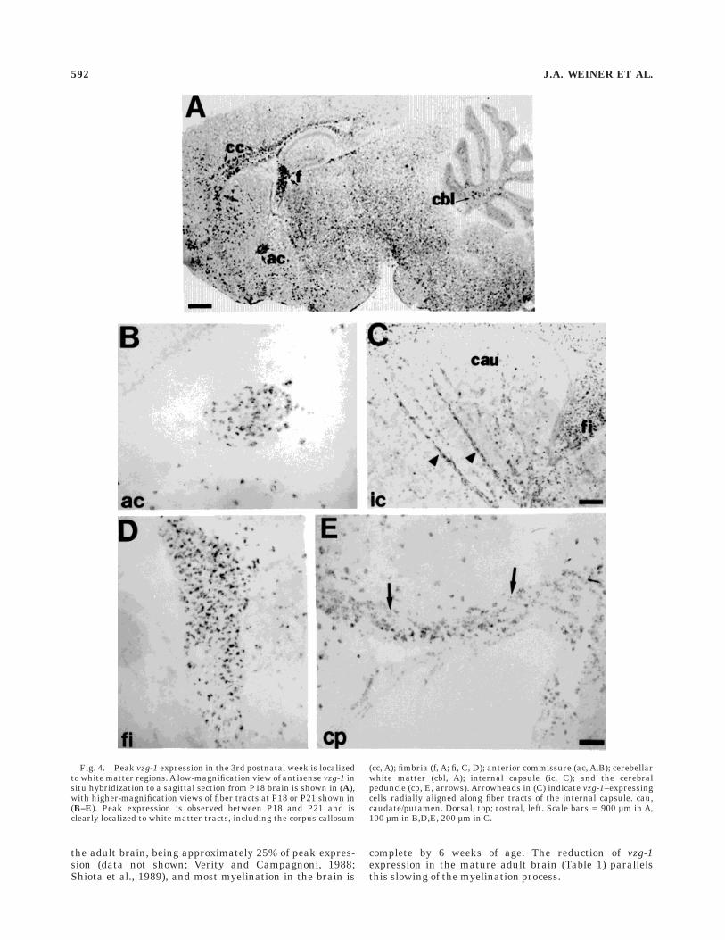
Fig. 4. Peak vzg-1 expression in the 3rd postnatal week is localizedto white matter regions. A low-magnification view of antisense vzg-1 insitu hybridization to a sagittal section from P18 brain is shown in (A),with higher-magnification views of fiber tracts at P18 or P21 shown in(B–E). Peak expression is observed between P18 and P21 and isclearly localized to white matter tracts, including the corpus callosum
(cc, A); fimbria (f, A; fi, C, D); anterior commissure (ac, A,B); cerebellarwhite matter (cbl, A); internal capsule (ic, C); and the cerebralpeduncle (cp, E, arrows). Arrowheads in (C) indicate vzg-1–expressingcells radially aligned along fiber tracts of the internal capsule. cau,caudate/putamen. Dorsal, top; rostral, left. Scale bars 5 900 µm in A,100 µm in B,D,E, 200 µm in C.
592 J.A. WEINER ET AL.
Vzg-1–expressing cells in the postnatal brainare oligodendrocytes
The temporal and spatial expression pattern describedabove suggested that the postnatal vzg-1–expressing cellswere glia, and probably oligodendrocytes associated withmyelination. Consistent with this, northern blot analysisindicated that vzg-1 was expressed by the C6 rat gliomacell line, which has oligodendrocyte-like properties (Bendaet al., 1968; Parker et al., 1980), and by RN2 (Pfeiffer andWechsler, 1972), a tumor line with properties of Schwanncells, the myelinating glia of the PNS (Fig. 5). To confirmthat vzg-1–expressing cells in the postnatal brain wereoligodendrocytes, we employed a double-label, nonradioac-tive in situ hybridization method combining a digoxigenin-labeled vzg-1 riboprobe with a fluorescein-labeled ribo-probe to the PLP gene as a marker for oligodendrocytes(see Materials and Methods). The PLP gene gives rise totwo protein isoforms: PLP, the complete protein, andDM-20, which has a 35-amino acid deletion (Griffiths et al.,1995). Although low expression of the DM-20 transcripthas been reported in some embryonic neural cell types(Timsit et al., 1992; Yu et al., 1994), the PLP gene isexpressed solely by mature, myelinating oligodendrocytesin the postnatal brain (Griffiths et al., 1995; Yan et al.,1996).
In sections from all ages examined, essentially all vzg-1–expressing cells detected by in situ hybridization co-expressed PLP mRNA. Figure 6 shows results from experi-ments performed on brain sections from P18, the peak ofvzg-1 and PLP expression. In these experiments, the vzg-1hybridization signal appears purple-brown, whereas PLPhybridization signal appears bright pink. Double-labeled
cells were clearly identifiable, appearing a brownish redcolor distinct from that produced by either label alone. Alow-magnification view of the hippocampal region, double-labeled for vzg-1 and PLP (Fig. 6A), demonstrated co-expression in the cells which make up the fimbria and thecorpus callosum; as expected, neither transcript was de-tected in the neuronal hippocampus. An adjacent section(Fig. 6B), double-labeled with riboprobes to PLP (pink) andto png-1 (purple-brown), a postmitotic neuronal marker(Weiner and Chun, 1997), demonstrated the expectedseparate populations of oligodendrocytes and neurons andserved as a control, confirming that no spurious double-labeling was produced by our technique.
Higher-magnification views of three adjacent sectionsthrough the anterior commisure, hybridized with ribo-probes to vzg-1 (Fig. 6C), PLP (Fig. 6D), or both (Fig. 6E),clearly demonstrated the different appearance of the single-and double-labeled cells, and showed that essentially all ofthe vzg-1–expressing cells co-expressed PLP (and viceversa; co-expression was near 100% in more than 1,000cells counted from various areas). In the P18 cortex, thefew vzg-1–expressing cells also expressed PLP mRNA (Fig.6F) and appeared to have small, irregularly shaped nucleidistinct from those of many of the surrounding corticalneurons (Fig. 6G). Cells expressing vzg-1 also expressedPLP mRNA in every region examined at P18, including theinternal capsule (Fig. 6H) and the corpus callosum (Fig.6I), and at every age examined (data not shown). Even insections from P3 brain, around the time the first vzg-1–(see Fig. 2) and PLP– (data not shown; Verity and Campag-noni, 1988; Shiota et al., 1989) expressing cells wereobserved, the two transcripts were co-expressed.
Astrocytes are also observed in some portions of whitematter tracts. Although the expression pattern of vzg-1 didnot appear to overlap with that reported for the astrocytemarker gene GFAP (Landry et al., 1990), it remainedpossible that a very small number of vzg-1–expressingcells were astrocytes. To examine this possibility, double-label experiments were performed using a fluorescein-labeled riboprobe to GFAP, along with the digoxigenin-labeled vzg-1 riboprobe. Vzg-1 expression did not overlapwith GFAP mRNA expression at any age examined, asshown in a section from the P18 fimbria (Fig. 7B). In thisand in other white matter tracts, the few GFAP-positivecells (pink) clearly did not co-express vzg-1 (purple-brown),and vice versa. We also used this double-labeling proce-dure on sections of the P1 brain, to determine whether thevzg-1–expressing cells observed around the meninges (seeFig. 1) were astrocytes, which form the glial limitans inthese regions (Landry et al., 1990; Jacobson, 1991). Co-expression of vzg-1 and GFAP was also not observed inthese sections (Fig. 7A), suggesting that the vzg-1–expressing cells at these early ages may be mesenchymalor neural crest-derived (Jacobson, 1991).
Vzg-1 expression is reduced in the brainsof jimpy mice
In the jimpy mutant mouse, a mutation in the X-linkedPLP gene leads to aberrant oligodendrocyte differentiationand to increased oligodendrocyte cell death in hemizygousmales (Knapp et al., 1986; Griffiths et al., 1995). Becausevzg-1 is expressed by differentiated oligodendrocytes, weasked whether vzg-1 expression was reduced in jimpybrain. Northern blot analysis using a vzg-1 probe (Fig. 8A)demonstrated that expression is in fact reduced in thebrains of 3-week-old jimpy mice, as compared to litter-
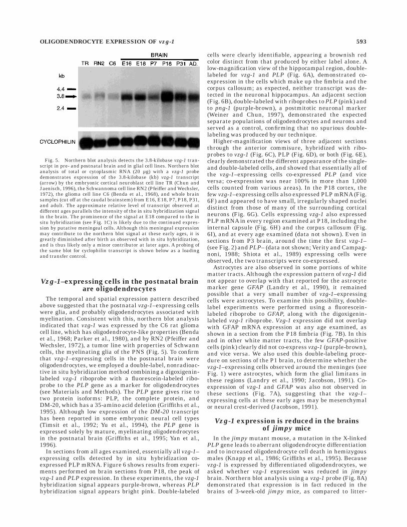
Fig. 5. Northern blot analysis detects the 3.8-kilobase vzg-1 tran-script in pre- and postnatal brain and in glial cell lines. Northern blotanalysis of total or cytoplasmic RNA (20 µg) with a vzg-1 probedemonstrates expression of the 3.8-kilobase (kb) vzg-1 transcript(arrow) by the embryonic cortical neuroblast cell line TR (Chun andJaenisch, 1996), the Schwannoma cell line RN2 (Pfeiffer and Wechsler,1972), the glioma cell line C6 (Benda et al., 1968), and whole brainsamples (cut off at the caudal brainstem) from E16, E18, P7, P18, P31,and adult. The approximate relative level of transcript observed atdifferent ages parallels the intensity of the in situ hybridization signalin the brain. The prominence of the signal at E18 compared to the insitu hybridization (see Fig. 1C) is likely due to the continued expres-sion by putative meningeal cells. Although this meningeal expressionmay contribute to the northern blot signal at these early ages, it isgreatly diminished after birth as observed with in situ hybridization,and is thus likely only a minor contributor at later ages. A probing ofthe same blot for cyclophilin transcript is shown below as a loadingand transfer control.
OLIGODENDROCYTE EXPRESSION OF vzg-1 593

Figure 6
594 J.A. WEINER ET AL.

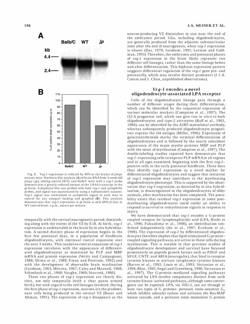
mates and to wild-type Balb/C mice. A duplicate northernblot probed for vzg-1 was quantitated radioanalytically,with signal intensity normalized to that of the ubiqui-tously expressed cyclophilin gene in each lane (Fig. 8B).The vzg-1 signal in jimpy brain was only 60% of that inwild-type littermates, consistent with the reduced numberof mature oligodendrocytes in the mutant brains. In situhybridization experiments (not shown) on sections fromP18 jimpy brains demonstrated fewer vzg-1–positive cellsand further suggested that the expression level per cellwas reduced.
DISCUSSION
We have examined the developmental expression of theLPA receptor gene vzg-1/lpA1/edg-2 in the postnatal mu-rine brain. Vzg-1 expression in the embryonic cerebralcortical VZ diminishes at the end of the neurogeneticperiod (E18) and is absent from the brain at birth. Expres-sion reappears in the hindbrain during the first fewpostnatal days and expands in a caudal-to-rostral mannerover the next 3 weeks, reaching a peak at P18. At all ages,
vzg-1 expression is concentrated in and around developingwhite matter tracts. Double-labeling experiments demon-strate that vzg-1–expressing cells co-express mRNA encod-ing PLP but not GFAP, identifying them as mature oligo-dendrocytes. Consistent with this, vzg-1 mRNA expressionis reduced by 40% in the brains of jimpy mutant mice,which are known to exhibit aberrant oligodendrocytedifferentiation and increased oligodendrocyte cell death.
Expression of vzg-1 is biphasic in theperinatal murine brain
During the embryonic development of the CNS, vzg-1expression is restricted to the cortical VZ (Hecht et al.,1996; see Fig. 1A). This first phase of expression correlates
Fig. 6. Vzg-1 is expressed by mature oligodendrocytes. Double-label in situ hybridization experiments on P18 brain sections employ-ing a digoxigenin-labeled vzg-1 riboprobe (detected with an anti-digoxigenin antibody; purple-brown precipitate) and a fluorescein-labeled PLP riboprobe (detected with an anti-fluorescein antibody;bright pink precipitate) demonstrate that vzg-1–expressing cells areoligodendrocytes. A: A low-magnification view of the P18 hippocampalarea shows double-labeled cells (reddish-brown) in the white matterregions surrounding the unlabeled hippocampal granule neurons; incontrast, double-labeling with riboprobes to PLP (pink cells) and topng-1, a neuronal marker (Weiner and Chun, 1997; purple-browncells), reveals the distinct oligodendroglial and neuronal cell popula-tions (B). Adjacent sections through the anterior commissure demon-strate the appearance of vzg-1 detection alone (C), PLP detection alone(D), and combined double-labeling (E). Double-labeled cells are clearlydistinguishable by their dark reddish-brown color. A double-labeledcell in the cortex (F, short arrow; long arrow points to pial surface) hasa small and irregularly shaped nucleus (G, 48,6-diamidino-2-phenylin-dole [DAPI] fluorescence of the same section). H–J: High-magnifica-tion views of double-labeled cells in the internal capsule (ic), corpuscallosum (cc), and anterior commissure (ac) clearly demonstrate theco-localization of the two reaction products. fi, fimbria; sp, stratumpyramidale; dg, dentate gyrus. A–G: dorsal, top, rostral, left. Scalebars 5 200 µm in A,B, 30 µm in C–E, 10 µm in F–J.
TABLE 2. Correlation of Postnatal Age of Peak vzg-1 Expressionin Various Tracts With That of Peak Myelin Gene Expression
and Observable Myelination1
Region/tract
Peakvzg-1
expression
PeakPLP/MBP
expression2
Myelinationobserved
morphologically
Cerebellum—white matter P15 P14 Peak between P13 andP163
Corpus callosum P18–P21 P20 Peak at P204
Anterior commisure P21 P20 P17 start, sharp increaseby P245
Fimbria P18 P20 P12 start, peak a few daysthereafter5
Internal capsule P15–P18 P14–P20 P10 start, sharp increaseP14–P225
Pontine fibers P18 P20 P14 start, peak thereafter5
1PLP, proteolipid protein; MBP, myelin basic protein.2Mouse: Verity and Campagnoni (1988), Shiota et al. (1989), Foran and Peterson (1992),present study.3Rat: Mitrova (1967).4Mouse: Sturrock (1980).5Rat: Jacobson (1963).
Fig. 7. Vzg-1 is not expressed by astrocytes. Double-label in situhybridization experiments on P1 (A) and P18 (B) brain sectionsemploying a digoxigenin-labeled vzg-1 riboprobe (detected with ananti-digoxigenin antibody; purple-brown precipitate) and a fluorescein-labeled glial fibrillary acidic protein (GFAP) riboprobe (detected withan anti-fluorescein antibody; bright pink precipitate) demonstratethat vzg-1 is not expressed by astrocytes. In the P1 forebrain (A), vzg-1is expressed by cells which are likely to be meningeal (white arrows;see also Fig. 1). GFAP-positive astrocytes forming the glial limitans(black arrows) are closely apposed to the vzg-1–positive cells; however,double-labeled cells are not observed. GFAP-positive astrocytes linethe outer edge of the P18 fimbria (B, black arrows), whereas vzg-1–positive cells lie within the tract itself (white arrows); again, double-labeled cells are not observed. Scale bars 5 30 µm in A, 10 µm in B.
OLIGODENDROCYTE EXPRESSION OF vzg-1 595
temporally with the cortical neurogenetic period, diminish-ing along with the extent of the VZ by E18. At birth, vzg-1expression is undetectable in the brain by in situ hybridiza-tion. A second distinct phase of expression begins in thefirst few postnatal days, in a population of hindbrainoligodendrocytes, with continual rostral expansion overthe next 3 weeks. This caudal-to-rostral expansion of vzg-1expression correlates with the appearance of differenti-ated oligodendrocytes as monitored by PLP and MBPmRNA and protein expression (Verity and Campagnoni,1988; Shiota et al., 1989; Foran and Peterson, 1992) andwith the development of observable brain myelination(Jacobson, 1963; Mitrova, 1967; Caley and Maxwell, 1968;Schonbach et al., 1968; Vaughn, 1969; Sturrock, 1980).
These two phases of vzg-1 expression are clearly dis-tinct, not only temporally (with a clear pause aroundbirth), but with regard to the cell lineages involved. Duringthe first phase of vzg-1 expression, neurons are the predomi-nant cells being produced in the cortical VZ (Bayer andAltman, 1991). The expression of vzg-1 disappears as the
neuron-producing VZ diminishes in size near the end ofthe embryonic period. Glia, including oligodendrocytes,are generally produced from the adjacent subventricularzone after the end of neurogenesis, when vzg-1 expressionis absent (Das, 1979; Jacobson, 1991; Levison and Gold-man, 1993). Therefore, the embryonic and postnatal phasesof vzg-1 expression in the brain likely represent twodifferent cell lineages, rather than the same lineage beforeand after differentiation. This biphasic expression patternsuggests differential regulation of the vzg-1 gene pre- andpostnatally, which may involve distinct promoters (J.J.A.Contos and J. Chun, unpublished observations).
Vzg-1 encodes a noveloligodendrocyte-associated LPA receptor
Cells of the oligodendrocyte lineage pass through anumber of different stages during their differentiation,which can be identified by the sequential expression ofvarious molecular markers (Compston et al., 1997). TheO2-A progenitor cell, which can give rise in vitro to botholigodendrocytes and type-2 astrocytes (Raff et al., 1983,1984), can be identified by the A2B5 monoclonal antibody,whereas subsequently produced oligodendrocyte progeni-tors express the O4 antigen (Miller, 1996). Expression ofgalactocerebroside marks the terminal differentiation ofoligodendrocytes and is followed by the nearly coincidentappearance of the major myelin proteins MBP and PLPwith the onset of myelination (Compston et al., 1997). Thedouble-labeling studies reported here demonstrate thatvzg-1–expressing cells co-express PLP mRNA in all regionsand at all ages examined, beginning with the first vzg-1–positive cells in the early postnatal hindbrain. These datathus identify vzg-1 expression as a novel marker fordifferentiated oligodendrocytes and suggest that initiationof vzg-1 expression may contribute to the myelinatingoligodendrocyte phenotype. This is supported by the obser-vation that vzg-1 expression, as detected by in situ hybrid-ization, is downregulated in the oligodendrocytes of olderanimals, after myelination has been completed. The possi-bility exists that residual vzg-1 expression in some post-myelinating oligodendrocytes could confer an ability torespond to survival or remyelination signals in response toinjury.
We have demonstrated that vzg-1 encodes a G-proteincoupled receptor for lysophosphatidic acid (LPA; Hecht etal., 1996; Fukushima et al., 1998), an identification con-firmed independently (An et al., 1997; Erickson et al.,1998). The expression of vzg-1 by differentiated oligoden-drocytes therefore implies that lipid-stimulated G-protein–coupled signaling pathways are active in these cells duringmyelination. This is notable in that previous studies ofoligodendrocyte development and survival have focussedprominently on peptide growth factors such as PDGF andbFGF, CNTF, and ARIA (neuregulin), that bind to receptortyrosine kinases or activate cytoplasmic tyrosine kinases(Barres et al., 1992; Louis et al., 1993; Vartanian et al.,1994; Bhat, 1995; Segal and Greenberg, 1996; Vartanian etal., 1997). The G-protein–mediated signaling pathwaysactivated by LPA involve components distinct from suchtyrosine kinase–activated pathways, although some conver-gence can be expected. LPA, via VZG-1, can act through atleast two types of G proteins: pertussis toxin–sensitive Gi,which inhibits adenylyl cyclase and activates the Ras-MAPkinase cascade, and a pertussis toxin–insensitive G protein
Fig. 8. Vzg-1 expression is reduced by 40% in the brains of jimpymutant mice. Northern blot analysis (A) of brain RNA from 3-week-oldjimpy ( jp), sibling control (WT), and Balb/C mice with a vzg-1 probedemonstrates a greatly reduced amount of the 3.8-kb transcript in thejp brain. A duplicate blot was probed with both vzg-1 and cyclophilinprobes, and signal was quantitated by using a radioanalytic scanner.Vzg-1 signal was normalized to cyclophilin signal in each lane tocontrol for any unequal loading and graphed (B). This analysisdemonstrates that vzg-1 expression in jp brain is only 60% of that inWT control brain. c.p.m., counts per minute.
596 J.A. WEINER ET AL.
that activates the Rho cytoskeletal pathway (Hecht et al.,1996; Fukushima et al., 1998). It will be important infuture studies to identify which of these signaling path-ways are active in oligodendrocytes and to determine howthey interact with those employed by other growth orsurvival factors.
LPA is a novel potential mediatorof myelination
LPA produces a wide variety of effects on many differentcell types, including activation of proliferation, inhibitionof gap junction communication, and morphological changessuch as stress fiber formation and neurite retraction(Jalink et al., 1994; Hecht et al., 1996; Moolenaar et al.,1997; Fukushima et al., 1998). Studies examining theeffects of LPA on oligodendrocytes or their PNS counter-parts, Schwann cells, have yet to be reported. However,our demonstration of vzg-1 expression by oligodendrocytespredicts that LPA can act on these cells. Some potentialfunctions may be inferred from the effects of serum, whichcontains high concentrations of LPA (Eichholtz et al.,1993), on oligodendrocytes in vitro. For example, serum isable to prolong the survival of oligodendrocytes derivedfrom the O2-A cell line CG-4 (Louis et al., 1992), and aserum factor, possibly a lipid, has been shown to increasegalactocerebroside levels in primary oligodendrocyte cul-tures (Bologa et al., 1988). Serum also has a variety ofeffects on cultured sciatic nerve Schwann cells; theseperipheral myelinating cells also express vzg-1 both in vivoand in vitro (not shown; Weiner and Chun, unpublishedobservations).
A potential relationship of LPA signaling to the elabora-tion of the myelin sheath is suggested by the fact thatlipids, including in large part complex phospholipids,make up approximately 75–80% of the dry weight of CNSand PNS myelin (Gould et al., 1992; Stoffel and Bosio,1997). Lysophospholipids such as LPA and the structurallyand functionally similar sphingosine-1-phosphate could beproduced during the elaboration of myelin, and could actas an autocrine ‘‘feedback’’ signal for oligodendrocytes. Notmutually exclusive is the possibility that axons could alsoproduce these signaling lysophospholipids. Future studieson receptor protein localization, in vitro and in vivofunction, and LPA production in the nervous system willclarify the role of lysophospholipid signaling in oligodendro-cytes and its relationship to known peptide growth factorsignaling mechanisms.
ACKNOWLEDGMENTS
We thank Carol Akita for her expert histological assis-tance, Dr. Steven Post for assisting with work not includedhere, Drs. Greg Lemke, Martin Gore, and Todd Zorick foradvice and the kind gift of the RN2 cell line and PLPplasmid, and Dr. Harvey Karten for the use of photo-graphic facilities. This work was supported by N.I.M.H.grants R29 MH51699 and R01 MH56158 to J.C. and grantF31 MH11480 to J.A.W.
LITERATURE CITED
An, S., M.A. Dickens, T. Bleu, O.G. Hallmark, and E.J. Goetzl (1997)Molecular cloning of the human Edg2 protein and its identification as afunctional cellular receptor for lysophosphatidic acid. Biochem. Bio-phys. Res. Commun. 231:619–622.
An, S., T. Bleu, W. Huang, O.G. Hallmark, S.R. Coughlin, and E.J. Goetzl(1998) Identification of cDNAs encoding two G protein-coupled recep-tors for lysosphingolipids. FEBS Lett. 417:279–282.
Ausubel, F.M., R. Brent, R.E. Kingston, D.D. Moore, J.G. Seidman, J.A.Smith, and K. Struhl (1994) Current Protocols in Molecular Biology.New York: John Wiley and Sons.
Barres, B.A., I.K. Hart, H.S.R. Coles, J.F. Burne, J.T. Voyvodic, W.D.Richardson, and M.C. Raff (1992) Cell death and control of cell survivalin the oligodendrocyte lineage. Cell 70:31–46.
Bayer, S.A., and J. Altman (1991) Neocortical Development. New York:Raven Press.
Benda, P., J. Lightbody, G. Sato, L. Levine, and W. Sweet (1968) Differenti-ated rat glial cell strain in tissue culture. Science 161:370.
Bhat, N.R. (1995) Signal transduction mechanisms in glial cells. Dev.Neurosci. 17:267–284.
Bologa, L., R. Cole, F. Chiappelli, R.P. Saneto, and J. de Vellis (1988) Serumcontains inducers and repressors of oligodendrocyte differentiation. J.Neurosci. Res. 20:182–188.
Boulder Committee (1970) Embryonic vertebrate central nervous system:Revised terminology. Anat. Rec. 166:257–261.
Caley, D.W., and D.S. Maxwell (1968) An electron microscopic study of theneuroglia during postnatal development of the rat cerebrum. J. Comp.Neurol. 133:45–70.
Chun, J., and R. Jaenisch (1996) Clonal cell lines produced by infection ofneocortical neuroblasts using multiple oncogenes transduced by retrovi-ruses. Mol. Cell. Neurosci. 7:304–321.
Chun, J.J.M., D.G. Schatz, M.A. Oettinger, R. Jaenisch, and D. Baltimore(1991) The recombination activating gene-1 (RAG-1) transcript ispresent in the murine central nervous system. Cell 64:189–200.
Chun, J., J.J.A., Contos, and D. Munroe (1998) A growing family of receptorgenes for lysophosphatidic acid (LPA) and other lysophospholipids(LPs). Cell Biochem. Biophys (in press).
Compston, A., J. Zajicek, J. Sussman, A. Webb, G. Hall, D. Muir, C. Shaw, A.Wood, and N. Scolding (1997) Glial lineages and myelination in thecentral nervous system. J. Anat. 190:161–200.
Das, G.D. (1979) Gliogenesis and ependymogenesis during embryonicdevelopment of the rat. An autoradiographic study. J. Neurol. Sci.43:193–204.
Dubin, A.E., T. Bahnson, N. Fukushima, and J. Chun (1997) Lysophospha-tidic acid (LPA) depolarizes embryonic cortical neuroblasts through twodistinct ionic mechanisms and alters cell morphology. Soc. Neurosci.Abstr. 23:590.
Eichholtz, T., K. Jalink, I., Fahrenfort, and W.H. Moolenaar (1993) Thebioactive phospholipid lysophosphatidic acid is released from activatedplatelets. Biochem. J. 291:677–680.
Erickson, J.P., J.J. Wu, J.G. Goddard, G. Tigyi, K. Kawanishi, D. Tomei, andM.C. Kiefer (1998) Edg-2/vzg-1 couples to the yeast pheromone re-sponse pathway selectively in response to lysophosphatidic acid. J. Biol.Chem. 273:1506–1510.
Foran, D.R., and A.C. Peterson (1992) Myelin acquisition in the centralnervous system of the mouse revealed by an MBP-Lac Z transgene. J.Neurosci. 12:4890–4897.
Fukushima, N., and J. Chun (1997) Polarized, actin ‘‘microspike cap’’formation is induced by lysophosphatidic acid (LPA) in primary culturesof embryonic cortical neuroblasts. Soc. Neurosci. Abstr. 23:1143.
Fukushima, N., Y. Kimura, and J. Chun (1998) A single receptor encoded byvzg-1/lpA1/edg-2 couples to G-proteins and mediates multiple cellularresponses to lysophosphatidic acid (LPA). Proc. Natl. Acad. Sci. USA95:6151–6156.
Gould, R.M., K.R. Jessen, R. Mirsky, and G. Tennekoon (1992) The cell ofSchwann: an update. In R.E. Martenson (ed): Myelin: Biology andChemistry. Boca Raton, FL: CRC Press, pp. 123–171.
Griffiths, I.R., P. Montague, and P. Dickinson (1995) The proteolipid proteingene. Neuropathol. Appl. Neurobiol. 21:85–96.
Hecht, J.H., J.A. Weiner, S.R. Post, and J. Chun (1996) Ventricular zonegene-1 (vzg-1) encodes a lysophosphatidic acid receptor expressed inneurogenic regions of the developing cerebral cortex. J. Cell Biol.135:1071–1083.
Jacobson, S. (1963) Sequence of myelinization in the brain of the albino rat:A. cerebral cortex, thalamus, and related structures. J. Comp. Neurol.121:5–29.
Jacobson, M. (1991) Developmental Neurobiology, 3rd Edition. New York:Plenum Press.
OLIGODENDROCYTE EXPRESSION OF vzg-1 597
Jalink, K., P.L. Hordijk, and W.H. Moolenaar (1994) Growth factor-likeeffects of lysophosphatidic acid, a novel lipid mediator. Biochim. Bio-phys. Acta 1198:185–196.
Knapp, P.E., R.P. Skoff, and D.W. Redstone (1986) Oligodendroglial celldeath in jimpy mice: an explanation for the myelin deficit. J. Neurosci.6:2813–2822.
Landry, C.F., G.O. Ivy, and I.R. Brown (1990) Developmental expression ofglial fibrillary acidic protein mRNA in the rat brain analyzed by in situhybridization. J. Neurosci. Res. 25:194–203.
Lee, M.-J., J.R. Van Brocklyn, S. Thangada, C.H. Liu, A.R. Hand, R.Menzeleev, S. Spiegel, and T. Hla (1998) Sphingosine-1-phosphate as aligand for the G protein-coupled receptor EDG-1. Science 279:1552–1555.
Levison, S.M., and J.E. Goldman (1993) Both oligodendrocytes and astro-cytes develop from progenitors in the subventricular zone of postnatalrat forebrain. Neuron 10:201–212.
Louis, J.-C., E. Magal, D. Muir, M. Manthorpe, and S. Varon (1992) CG-4, anew bipontential glial cell line from rat brain, is capable of differentiat-ing in vitro into either mature oligodendrocytes or type-2 astrocytes. J.Neurosci. Res. 31:193–204.
Louis, J.-C., E. Magal, S. Takayama, and S. Varon (1993) CNTF protectionof oligodendrocytes against natural and tumor necrosis factor-induceddeath. Science 259:689–692.
Miller, R.H. (1996) Oligodendrocyte origins. Trends Neurosci. 19:92–96.Mitrova, E. (1967) Karyometric investigation of glia cells in the cerebellum
in the course of myelination. Z. Mikrosk.-Anat. Forsch. 77:304–312.Moolenaar, W.H. (1995) Lysophosphatidic acid signalling. Curr. Opin. Cell
Biol. 7:203–210.Moolenaar, W.H., O. Kranenburg, F.R. Postma, and G.C.M. Zondag (1997)
Lysophosphatidic acid: G-protein signalling and cellular responses.Curr. Opin. Cell Biol. 9:168–173.
Parker, K.K., M.D. Norenberg, and A. Vernadakis (1980) ‘‘Transdifferentia-tion’’ of C6 glial cells in culture. Science 208:179–181.
Pfeiffer, S.E., and W. Wechsler (1972) Biochemically differentiated neoplas-tic clone of Schwann cells. Proc. Natl. Acad. Sci. USA 69:2885–2889.
Raff, M.C., R.H. Miller, and M. Noble (1983) A glial progenitor cell thatdevelops in vitro into an astrocyte or an oligodendrocyte depending onculture medium. Nature 303:390–396.
Raff, M.C., B.P. Williams, and R.H. Miller (1984) The in vitro differentiationof a bipotential glial progenitor cell. EMBO J. 3:1857–1864.
Schonbach, J., K.H. Hu, and R.L. Friede (1968) Cellular and chemicalchanges during myelination: Histologic, autoradiographic, histochemi-
cal and biochemical data on myelination in the pyramidal tract andcorpus callosum of rat. J. Comp. Neurol. 134:21–38.
Segal, R.A., and M.E. Greenberg (1996) Intracellular signaling pathwaysactivated by neurotrophic factors. Annu. Rev. Neurosci. 19:463–489.
Shiota, C., M. Miura, and K. Mikoshiba (1989) Developmental profile anddifferential localization of mRNAs of myelin proteins (MBP and PLP) inoligodendrocytes in the brain and in culture. Dev. Brain Res. 45:83–94.
Stoffel, W., and A. Bosio (1997) Myelin glycolipids and their functions. Curr.Opin. Neurobiol. 7:654–661.
Sturrock, R.R. (1980) Myelination of the mouse corpus callosum. Neuro-pathol. Appl. Neurobiol. 6:415–420.
Timsit, S.G., L. Bally-Cuif, D.R. Colman, and B. Zalc (1992) DM-20 mRNAis expressed during the embryonic development of the nervous systemof the mouse. J. Neurochem. 58:1172–1175.
Vartanian, T., G. Corfas, Y. Li, G.D. Fischbach, and K. Stefansson (1994) Arole for the acetylcholine receptor-inducing protein ARIA in oligodendro-cyte development. Proc. Natl. Acad. Sci. USA 91:11626–11630.
Vartanian, T., A. Goodearl, A. Viehover, and G. Fischbach (1997) Axonalneuregulin signals cells of the oligodendrocyte lineage through activa-tion of HER4 and Schwann cells through HER2 and HER3. J. Cell Biol.137:211–220.
Vaughn, J.E. (1969) An electron microscopic analysis of gliogenesis in ratoptic nerves. Z. Zellforsch. 94:293–324.
Verity, A.N., and A.T. Campagnoni (1988) Regional expression of myelinprotein genes in the developing mouse brain: In situ hybridizationstudies. J. Neurosci. Res. 21:238–248.
Weiner, J.A., and J. Chun (1997) Png-1, a nervous system-specific zincfinger gene, identifies regions containing postmitotic neurons duringmammalian embryonic development. J. Comp. Neurol. 381:130–142.
Weiner, J.A., J.H Hecht, S.R. Post, and J. Chun (1997) Biphasic perinatalexpression of vzg-1, a lysophosphatidic acid (LPA) receptor, suggestsdual roles for LPA in brain development. Soc. Neurosci. Abstr. 23:1689.
Yan, Y., V. Narayanan, and C. Lagenaur (1996) Expression of members ofthe proteolipid protein gene family in the developing murine centralnervous system. J. Comp. Neurol. 370:465–478.
Yu, W.-P., E.J. Collarini, N.P. Pringle, and W.D. Richardson (1994) Embry-onic expression of myelin genes: evidence for a focal source of oligoden-drocyte precursors in the ventricular zone of the neural tube. Neuron12:1353–1362.
598 J.A. WEINER ET AL.


![Computer-aided Assignment of DDC Numbers · DDC: Dewey Decimal Classification ] VZG Verbundzentrale des GBV (VZG) ... T1--028, T1—0285, T1--028563} vc_dcl: vzg colibri_ddc classifier](https://img.pdfslide.us/doc/110x75/5b2ab0057f8b9a55068b723d/computer-aided-assignment-of-ddc-numbers-ddc-dewey-decimal-classification-.jpg)