Embed Size (px)
Citation preview

L
AHVR
*NMGLP�
Bdcpptcsc(aitmmtTrcacwdcetwlnsutdraap
K
BA
SIC–LIV
ER,
PA
NCREA
S,A
ND
BILIA
RY
TRA
CT
GASTROENTEROLOGY 2010;139:1008–1018
ysophosphatidic Acid Is a Potential Mediator of Cholestatic Pruritus
NDREAS E. KREMER,* JOB J. W. W. MARTENS,* WIM KULIK,‡ FRANZISKA RUËFF,§ EDITH M. M. KUIPER,�
ENK R. VAN BUUREN,� KAREL J. VAN ERPECUM,¶ JURATE KONDRACKIENE,# JESUS PRIETO,** CHRISTIAN RUST,‡‡
ICTORIA L. GEENES,§§ CATHERINE WILLIAMSON,§§ WOUTER H. MOOLENAAR,� � ULRICH BEUERS,* andONALD P. J. OUDE ELFERINK*
Tytgat Institute for Liver and Intestinal Research and ‡Laboratory Genetic Metabolic Diseases, Academic Medical Center, University of Amsterdam, Amsterdam, Theetherlands; §Departments of Dermatology and Allergology, University of Munich, Munich, Germany; ‡‡Internal Medicine II - Grosshadern, University of Munich,unich, Germany; �Department of Gastroenterology & Hepatology, Erasmus MC University Medical Center, Rotterdam, The Netherlands; ¶Department ofastroenterology and Hepatology, University Medical Center, Utrecht, The Netherlands; #Department of Gastroenterology, Kaunas University of Medicine, Kaunas,ithuania; **Department of Medicine and Liver Unit, Clinica Universitaria, Medical School and Center for Applied Medical Research (CIMA), University of Navarra,amplona, Spain; §§Maternal and Fetal Disease Group, Institute of Reproductive and Developmental Biology, Imperial College London, London, England; and
�Division of Cell Biology, The Netherlands Cancer Institute, Amsterdam, The Netherlands
CciDpcmspsboaafltiobell
ddobfqil
fcp
ACKGROUND & AIMS: Pruritus is a common andisabling symptom in cholestatic disorders. However, itsauses remain unknown. We hypothesized that potentialruritogens accumulate in the circulation of cholestaticatients and activate sensory neurons. METHODS: Cy-osolic free calcium ([Ca2�]i) was measured in neuronalell lines by ratiometric fluorometry upon exposure toerum samples from pruritic patients with intrahepaticholestasis of pregnancy (ICP), primary biliary cirrhosisPBC), other cholestatic disorders, and pregnant, healthy,nd nonpruritic disease controls. Putative [Ca2�]i-induc-ng factors in pruritic serum were explored by analyticalechniques, including quantification by high-perfor-
ance liquid chromatography/mass spectroscopy. Inice, scratch activity after intradermal pruritogen injec-
ion was quantified using a magnetic device. RESULTS:ransient increases in neuronal [Ca2�]i induced by pru-
itic PBC and ICP sera were higher than correspondingontrols. Lysophosphatidic acid (LPA) could be identifieds a major [Ca2�]i agonist in pruritic sera, and LPAoncentrations were increased in cholestatic patientsith pruritus. LPA injected intradermally into mice in-uced scratch responses. Autotaxin, the serum enzymeonverting lysophosphatidylcholine into LPA, was mark-dly increased in patients with ICP versus pregnant con-rols (P � .0001) and cholestatic patients with versusithout pruritus (P � .0001). Autotaxin activity corre-
ated with intensity of pruritus (P � .0001), which wasot the case for serum bile salts, histamine, tryptase,ubstance P, or �-opioids. In patients with PBC whonderwent temporary nasobiliary drainage, both itch in-ensity and autotaxin activity markedly decreased duringrainage and returned to preexistent levels after drainemoval. CONCLUSIONS: We suggest that LPA andutotaxin play a critical role in cholestatic pruritusnd may serve as potential targets for future thera-eutic interventions.
eywords: Autotaxin; Bile Salts; Cholestasis; Itch.
hronic pruritus is a disabling symptom accompany-ing a broad range of systemic disorders such as
hronic liver diseases, chronic renal failure, malignancies,nfections, and endocrine and hematologic diseases.1,2
espite the recent discovery of itch-specific neuronalathways, including novel itch mediators and their re-eptors,1,3,4 the pathogenesis of pruritus remains enig-atic. Regardless of the underlying cause, various chole-
tatic disorders such as intrahepatic cholestasis ofregnancy (ICP), benign recurrent intrahepatic cholesta-is, progressive familial intrahepatic cholestasis, primaryiliary cirrhosis (PBC), and primary sclerosing cholangitisften induce pruritus. These cholestatic liver disordersre all characterized by an impairment of hepatocellularnd/or cholangiocellular secretory function and bileow.5 In these patients, pruritus may become refractoryo all medical treatments and can in severe cases be anndication for liver transplantation, even in the absencef liver failure.6 In the past, enhanced serum levels ofoth bile salts and �-opioids have been implicated in thetiology of cholestatic pruritus.7 However, neither corre-ations between itch intensity and bile salt or opioidevels nor a causative link have ever been established.
Autotaxin (ATX) was originally identified in the con-itioned medium of human A2058 melanoma cells andescribed as an autocrine cell motility factor.8 ATX isverexpressed in several other tumor entities and haseen linked to tumor cell proliferation, motility, andormation of metastasis.9 Physiologically, ATX is re-uired for angiogenesis and neuronal development, as
ndicated by ATX-deficient mice, which are embryonicethal due to vascular malformation and neuronal abnor-
Abbreviations used in this paper: ATX, autotaxin; [Ca2�]i, cytosolicree calcium; HBSS, Hank’s balanced salt solution; ICP, intrahepaticholestasis of pregnancy; LPA, lysophosphatidic acid; LPC, lysophos-hatidylcholine; PBC, primary biliary cirrhosis.
© 2010 by the AGA Institute0016-5085/$36.00
doi:10.1053/j.gastro.2010.05.009

meafbe(tlocbdLpd
erisLmt
hlDliadntcAuhc
hwgrmsMtgb
wftbtvp
t
z(otLbfw
dcL
s
slIimwH(3wsafiec
IbN0p
BA
SIC–L
IVER
,PA
NCREA
S,A
ND
BIL
IARY
TRA
CT
September 2010 LPA AND CHOLESTATIC PRURITUS 1009
alities.10,11 Recently, ATX could be identified as anxtracellular secreted enzyme with lysophospholipase Dctivity, which generates lysophosphatidic acid (LPA)rom lysophosphatidylcholine (LPC).12,13 LPA is a smallut potent bioactive phospholipid with a wide variety offfects in many cell types ranging from cytoskeletalre)organization and cell migration to cytokine produc-ion and platelet activation.9,14 Effects of ATX are be-ieved to be mainly mediated by the enzymatic formationf LPA, which activates at least 6 different G protein–oupled receptors.9,15 Most interest in ATX has so fareen directed toward its functions in cancer and earlyevelopment. However, it was recently established thatPA plays a crucial role in the induction of neuropathicain16 and causes reprogramming of gene expression inifferent types of afferent nerve fibers.17
Here we report that levels of LPA and ATX are mark-dly increased in serum of patients with cholestatic pru-itus. Moreover, serum levels closely correlate with itchntensity, and intradermal injections of LPA inducecratching behavior in mice. We therefore suggest thatPA and ATX play a critical role in cholestatic itch anday serve as potential targets for future therapeutic in-
erventions.
Materials and MethodsHuman SubjectsPeripheral venous blood was obtained from
ealthy donors, pregnant women, and patients with cho-estatic disorders after informed consent according to the
eclaration of Helsinki. The study was approved by theocal medical ethical committees. Blood samples weremmediately centrifuged at 4°C, and serum was frozen inliquots at �80°C. ICP was diagnosed, as previouslyescribed,18 in pregnant women with pruritus who hado rash in conjunction with increased serum liverransaminase and/or bile salt levels. Women were ex-luded if they had abnormal hepatitis serology (hepatitis, B, and C) or extrahepatic biliary obstruction followingltrasonographic examination. Pregnant controls had noistory of liver dysfunction or any complication in theurrent or previous pregnancies.
AnimalsA Teflon-coated magnet was implanted in each
ind paw of female C57BL/6J mice (6 – 8 weeks of age) 1eek before experiments were performed. Mice wereiven 120 minutes to acclimate to the chamber sur-ounded by a magnetic coil before they were briefly re-
oved from the chamber and intradermally injected withaline (50 �L) or LPA (8 –200 nmol in 50 �L) in the neck.
ovements of the magnets induced an electric current inhe magnetic field, which was registered by an oscillo-raph attached to a computer. The number of scratch
outs was analyzed as previously described.19 Software aas used to count scratch movements with a low cutoffrequency of 10 Hz, a high cutoff frequency of 20 Hz, ahreshold level of 300 mV, a minimum of 4 beats perout, and a maximal coefficient of variation of 40% be-ween the beats of a bout. The analytical procedure wasalidated with intradermal compound 48/80, showing aositive predictive value of 95% at a sensitivity of 50%.All mouse experiments were approved by the Institu-
ional Animal Care and Use Committee.
MaterialsCell culture media were from Lonza (Basel, Swit-
erland); stearoyl-LPA (LPA 18:1) and myristoyl-LPCLPC 14:0) from Avanti Lipids (Alabaster, AL); cholinexidase, horseradish peroxidase, homovanillic acid, per-ussis toxin, and ionomycin from Sigma-Aldrich (Stouis, MO); and Ki16245 and ATX antibody for Westernlotting from Cayman (Ann Arbor, MI). Indo-1 AM wasrom Invitrogen (Carlsbad, CA), and Microcon filtersere from Millipore (Billerica, MA).
Cell CultureSH-SY5Y cells were cultured in Ham’s F12K me-
ium containing 10% (vol/vol) fetal bovine serum, peni-illin (100 IU/mL), streptomycin (100 �g/mL), and-glutamine (0.2 mmol/L) at 37°C in a humidified atmo-phere of 5% CO2/95% air.
Fluorometric Measurement of Cytosolic FreeCalcium LevelsSH-SY5Y cells were detached, washed twice, and
uspended in HEPES-buffered Hank’s balanced salt so-ution (HBSS). Cells were incubated with 10 �mol/Lndo-1 AM for 30 minutes at 37°C, washed, resuspendedn HEPES-buffered HBSS, and incubated for another 30
inutes at 25°C to allow dye hydrolysis. After anotherash step, cells were resuspended in HEPES-bufferedBSS. Analyses were performed in a NOVOstar analyzer
BMG Labtech GmbH, Offenburg, Germany; excitation,20 nm; emission, 405 nm and 520 nm). Cell suspensionsere allowed to adapt to 37°C for 10 minutes before
erum or extracts were added. Receptor blockers weredded 10 minutes before addition of serum. Cytosolicree calcium ([Ca2�]i) was calculated after calibration withonomycin (10 �mol/L) and ethylene glycol-bis(�-amino-thyl ether)-N,N,N=,N=-tetraacetic acid (5 mmol/L) ac-ording to Grynkiewicz et al.20
ATX Activity AssayATX activity was analyzed as recently described.21
n short, serum and bile samples were incubated with auffer containing 1 mmol/L LPC 14:0, 500 mmol/LaCl, 5 mmol/L MgCl2, 100 mmol/L Tris (pH 9.0), and
.05% Triton X-100 for 60 minutes at 37°C. The phos-hodiesterase activity of ATX was determined by the
mount of liberated choline, as detected by an enzymatic
flhaempta4
tedt
Dw
bMah
bPb
d
cu
sivSw�spdi
btgcg
baSccr
wdcotndtisclishsftc(ficfia1Ttctttcaah
BA
SIC–LIV
ER,
PA
NCREA
S,A
ND
BILIA
RY
TRA
CT
1010 KREMER ET AL GASTROENTEROLOGY Vol. 139, No. 3
uorometric method using choline oxidase (2 U/mL),orseradish peroxidase (1.6 U/mL), and homovanilliccid as substrate for peroxidase. After addition of bothnzymes in a buffer (consisting of 20 mmol/L CaCl2, 2mol/L homovanillic acid, 50 mmol/L 3-(N-morpholino)
ropanesulfonic acid [pH 8.0], and 0.1% Triton X-100),he increase in fluorescence was monitored at 37°C on
NOVOstar analyzer (excitation, 320 nm; emission,05 nm).
Quantitative Determination of LPA LevelsA detailed description of the procedure is given in
he Supplementary Methods. Briefly, serum lipids werextracted after addition of myristoyl-LPA as internal stan-ard and analyzed by high-performance liquid chroma-ography/mass spectroscopy.
Determination of Bile Salt LevelsTotal serum bile salt levels were determined using
iazyme total bile salts kit (Diazyme Laboratories, Po-ay, CA) according to the manufacturer’s instructions.
Determination of Histamine LevelsSerum histamine concentrations were measured
y a competitive enzyme immunoassay (Immunotech,arseille, France) based on the competition between free
cylated histamine and alkaline phosphatase acylatedistamine conjugate.
Determination of Tryptase LevelsSerum tryptase concentrations were determined
y a fluoroenzyme immunoassay (UNICAP Tryptase;harmacia Diagnostics, Freiburg, Germany) detectingoth �- and �-tryptase.
Determination of �-Opioid ActivityTotal serum �-opioid activity was determined as
escribed by Swain et al.22
Determination of Substance P LevelsSubstance P concentrations were analyzed by a
ompetitive enzyme immunoassay according to the man-facturer’s instructions (Bachem, Torrance, CA).
Sodium Dodecyl Sulfate/Polyacrylamide GelElectrophoresis and Western BlottingTo concentrate ATX, 50 �L of serum and bile
amples was first incubated for 4 hours at 4°C withmmunoprecipitating ATX antibody 5E5 (kindly pro-ided by J. Aoki)10 bound to Sepharose. After washing,epharose beads were incubated for 10 minutes at 37°Cith sodium dodecyl sulfate–loading buffer containing-mercaptoethanol and spun down. Equal amounts ofupernatant were separated by sodium dodecyl sulfate/olyacrylamide gel electrophoresis, blotted on polyvinyli-ene difluoride membranes, blocked with 5% skim milk
n phosphate-buffered saline/0.05% Tween 20, and incu- p
ated with anti-ATX (1:1500; Cayman) overnight. Pro-eins were visualized with horseradish peroxidase– conju-ated immunoglobulins and detected by enhancedhemiluminescence (Amersham, Buckinghamshire, En-land).
Statistical AnalysisStatistical differences were evaluated for 2 groups
y Student t test and for 3 or more groups by 1-waynalysis of variance with Bonferroni correction usingPSS (version 16.0; SPSS Inc, Chicago, IL). Spearman’sorrelation coefficient and corresponding P values werealculated to assess the relationship between tested pa-ameters. All data are expressed as means � SD.
ResultsNeuron-Activating Serum Factor Identifiedas LPATo identify potential pruritogens in cholestasis,
e screened sera from pruritic patients for activation inifferent neuronal cell lines. We chose [Ca2�]i as an indi-ator of neuronal activation because it is a key mediatorf the neuronal secretory response toward diverse recep-or-dependent and -independent stimuli. In the humaneuroblastoma cell line SH-SY5Y, we observed a dose-ependent increase in intracellular calcium concentra-ions with the addition of serum (Figure 1A). Interest-ngly, sera from women with ICP showed a markedlytronger neuronal activation compared with pregnantontrols and healthy female controls (Figure 1B). Simi-arly, sera from pruritic patients with PBC tended tonduce higher transient increases in [Ca2�]i levels thanera from patients with PBC without pruritus andealthy controls (Figure 1C). We further analyzed theerum samples to identify the neuron-activating serumactor. Pretreatment of serum with 90% ethanol or pro-einase K hardly diminished the [Ca2�]i response, indi-ating that the serum factor was not a peptide or proteinFigure 2A). Serum samples were centrifuged throughlters to estimate the molecular size. The serum factorould pass a 100-kilodalton filter, but not a 10-kilodaltonlter. Interestingly, pretreatment of serum with 90% eth-nol enabled the factor to partially pass through the0-kilodalton and even a 3-kilodalton filter (Figure 2B).his observation could be explained by strong binding of
he factor to albumin (�60 kilodaltons). Thus, like un-onjugated bilirubin, the substance appeared to be par-ially forced into solution on ethanol-induced precipita-ion of albumin. Total recovery of the serum factorhrough a 10-kilodalton filter was achieved by addition ofholate above its critical micellar concentration, enablinghydrophobic substance to be completely solved in an
queous solution (Figure 2B). Because those micellesave a size of approximately 4.4 kilodaltons, they barely
ass a 3-kilodalton filter. Hence, we were dealing with a
sweaatt2cf
lc[oTcnKrt(
stp2sn
ctp�iwl
AItwpcfdsctrhsAphp
bdPottaat(tpitEs
Fh1p[c .s., n
BA
SIC–L
IVER
,PA
NCREA
S,A
ND
BIL
IARY
TRA
CT
September 2010 LPA AND CHOLESTATIC PRURITUS 1011
mall, hydrophobic substance. Its chemical propertiesere further analyzed by a 2-phase Bligh and Dyer lipid
xtraction. At neutral pH, the compound presented anmphiphilic character but dissolved better in the lowerqueous phase. Lowering the pH to 1.0 and thus poten-ially protonating the serum factor led to a recovery ofhe substance mainly in the upper lipid phase (FigureC). The increased hydrophobicity upon acidificationould be explained by protonation of phosphate or sul-ate groups in the molecule.
These observations suggested that a small phospho-ipid could be responsible for the activation of neuronalells. As pertussis toxin diminished the increase inCa2�]i, signaling of the unknown substance appeared toccur via a G-protein– coupled receptor (Figure 2D).hese observations rendered LPA a likely candidate be-ause LPA has been reported to increase calcium levels ineuronal cells.23 Pretreatment of the neuronal cells withi16425, a specific LPA receptor blocker, significantly
educed the increase in [Ca2�]i, indicating that LPA washe major serum factor in our cholestatic serum samplesFigure 2D).
Analyzing the LPA content in serum samples by masspectrometry indeed showed markedly higher concentra-ions of LPA 18:1 in sera from women with ICP com-ared with gestation-matched pregnant controls (FigureE). Similar differences were observed for other LPApecies such as LPA 16:0, 18:0, 18:2, 20:3, and 20:4 (dataot shown).
ATX Is Enhanced in Pruritus of CholestasisLPA is formed in the blood through cleavage of
holine from LPC by ATX, which has recently been iden-ified as a lysophospholipase D.12,13 Because LPC isresent in plasma at relatively high concentrations (�100mol/L), the amount of LPA (in low micromolar range)
n blood primarily depends on ATX activity.11 Therefore,e analyzed whether ATX activity in blood also corre-
igure 1. [Ca2�]i is increased in human neuroblastoma cell line SH-Sealthy controls. A shows that serum induced a transient increase in [C:320 induced an increase of [Ca2�]i. (B) Sera from women with ICP indregnancies (PC) and age-matched female controls (HC). (C) Sera from
Ca2�]i compared with age-matched healthy female controls. *P � .015alcium concentration as shown in A: maximal [Ca2�]i – basal [Ca2�]i. n
ated with the occurrence of itch. We observed higher t
TX activity in sera from women with pruritus due toCP as compared with pregnant and nonpregnant con-rols (Figure 3A). The enhanced ATX activity correlatedith increased ATX protein content in sera from theseatients (Figure 3B). We studied whether this observationould be extended to other forms of cholestasis. There-ore, sera from patients with different cholestatic disor-ers with and without pruritus were analyzed. Quitetrikingly, we found that, irrespective of the cause ofholestasis, ATX levels were markedly enhanced in pa-ients with pruritus compared with patients without pru-itus (Figure 3C–E). Recently, in patients with chronicepatitis C, liver ATX messenger RNA expression24 anderum ATX levels25 have been reported to be enhanced.TX serum levels were also increased in our group ofatients with hepatitis C virus when compared withealthy controls but were significantly lower than inruritic patients (Figure 3F).
ATX Activity Correlates With Intensity ofPruritusPruritus is a subjective perception that differs
etween individuals. Quantification of this symptom isifficult but can be achieved using visual analog scales.1
atients quantified their itch intensity at the time pointf blood drawing on a scale ranging from 0 (no pruritus)o 10 (most severe form of pruritus). Next we analyzedhe correlation between itch intensities and the ATXctivity in serum from these patients by linear regressionnalysis. We found a highly significant correlation be-ween enzyme activity and intensity of itch perceptionFigure 4A). In contrast, other agents proposed as poten-ial pruritogens in cholestasis and other diseases in theast7,26 did not show any correlation with itch intensity
n our patient cohort. This was tested for histamine,ryptase, substance P, and �-opioid activity (Figure 4B–F).ven though cholestatic patients with pruritus as a grouphowed higher serum bile salt concentrations (Table 1),
y sera from patients with cholestatic pruritus more than by sera fromhat was dose dependent. Note that even high serum dilutions of up tohigher increases of [Ca2�]i compared with gestation-matched normal
ients with PBC with and without pruritus induced a higher increase in� .005. �[Ca2�]i represents the peak of calcium transient minus basalot significant.
Y5Y ba2�]i tuced
pat, **P
here was no correlation with itch intensity (Figure 4D).

AhrshnirlsaTd
ttltitb(f
FKBoae31(
BA
SIC–LIV
ER,
PA
NCREA
S,A
ND
BILIA
RY
TRA
CT
1012 KREMER ET AL GASTROENTEROLOGY Vol. 139, No. 3
direct role of histamine and bile salts as pruritogensas already been questioned in the past.7,27 An antipru-itic effect of �-opioid antagonists has been reported inome patients.28 However, �-opioid activity was not en-anced in patients with ICP compared with regular preg-ancies and only very few pruritic patients with PBC had
ncreased �-opioid levels, questioning a major causativeole of opioids for the pathogenesis of pruritus in cho-estasis (Figure 4F).29 In some patients with PBC, exten-ive long-lasting pruritus was intractable and did notdequately respond to any recommended medication.6,26
hese patients underwent nasobiliary drainage for 2 to 7
igure 2. Identification of [Ca2�]i-enhancing serum factor as LPA. A shon [Ca2�]i in neuronal cells. U, untreated; E, supernatant of 90% ethashows the effect of filter experiments on serum. 100 kD, 10 kD, flow thr
f 90% ethanol precipitation (before filtering); CA, resuspension in 2 mmot pH values of 7.4 and 1.0. WP pH 7.4, LP pH 7.4, WP pH 1.0, and LPxtraction at pH values of 7.4 and 1.0 (n � 3). (D) Effect of pertussis tox). �405/520 represents the change in fluorescence at 405 nm (Ca2�-8:1 is enhanced in ICP cases compared with gestation-matched non
added as an internal standard before extraction). *P � .05, **P � .01.
ays as an experimental treatment of most severe pruri- d
us.27,30 In all 4 procedures, this led to dramatic reduc-ion or complete relief of pruritus within 24 hours thatasted for several days to weeks. Interestingly, concomi-ant with relief of pruritus, ATX activity decreased andncreased back to pretreatment levels when pruritus re-urned (Figure 5A). This effect was not due to directiliary clearance of ATX because neither ATX activityFigure 5B) nor protein (Figure 5C) could be found in bilerom these and other patients.
Induction of Pruritus by LPATo investigate a potential role of LPA in the in-
the effect of pretreatments of serum with 90% ethanol and proteinaserecipitation; Prot K, incubation for 24 hours with proteinase K (n � 3).of a 100-kilodalton and 10-kilodalton filter, respectively; E, supernatantolate (before filtering) (n � 3). (C) Effect of Bligh and Dyer lipid extraction
0 indicate the water phase (WP) and lipid phase (LP) of serum after lipidX) and LPA receptor blocker Ki16245 on cell activation by serum (n �
tive signal) divided by that at 520 nm (Ca2�-insensitive signal). (E) LPAstatic pregnant controls. LPA 18:1 is shown as the ratio to LPA 14:0
owsnol poughl/L ch
pH 1.in (PTsensichole
uction of pruritus in vivo, we used female C57BL/J6

Fr(ibe
BA
SIC–L
IVER
,PA
NCREA
S,A
ND
BIL
IARY
TRA
CT
September 2010 LPA AND CHOLESTATIC PRURITUS 1013
igure 3. Serum ATX is elevated in cholestatic patients with pruritus irrespective of the cause of cholestasis. (A) ATX activity (measured as cholineelease upon incubation with LPC) is highly enhanced in patients with ICP compared with pregnant controls and healthy female controls. ***P � .0001.B) Western blot for ATX protein in patient sera. Recombinant ATX (rATX) was used as a positive control. (C and D) ATX activity was highly enhancedn cholestatic women and men with pruritus compared with nonpruritic cholestatic patients and healthy controls. **P � .01, ***P � .0001. (E) Westernlot for ATX protein in serum from patients with PBC. rATX was used as positive control. (F) ATX activity in patients with chronic hepatitis C was
nhanced compared with controls but significantly lower compared with cholestatic patients with pruritus. ***P � .0001.
mpcn6mriu
c
dltucm
spcbu
Fprbf ithou
BA
SIC–LIV
ER,
PA
NCREA
S,A
ND
BILIA
RY
TRA
CT
1014 KREMER ET AL GASTROENTEROLOGY Vol. 139, No. 3
ice. Scratch movements were registered as describedreviously.19 Intradermal injections of LPA, but not thearrier, induced significant scratching behavior shown asumber of scratch bouts per 5-minute intervals (FigureA), which was in line with a previous report.31 Further-ore, we could demonstrate that the induction of pru-
itus by LPA in mice was dose dependent, showing anncreased scratching behavior from 20 nmol upward (Fig-re 6B).
DiscussionPruritus is a common and disabling symptom in
igure 4. Only ATX activity, but not histamine, tryptase, substance P, satients with cholestatic itch. (A) ATX activity showed a significant linearanging from 0 (no pruritus) to 10 (most severe form of pruritus). Speaetween histamine levels, tryptase concentrations, substance P levels,
emale and pregnant controls compared with women with ICP, PBC w
holestatic liver diseases and many other systemic disor- r
ers, including renal insufficiency; endocrine, hemato-ogic, and metabolic diseases; various infections; and cer-ain malignancies. The causal factors of pruritus arenknown in most of these diseases. Here we providelinical and experimental evidence that LPA is a potentialediator of cholestatic itch.The discovery of itch-specific sensory neurons in the
kin by Schmelz et al revolutionized the research field ofruritus.32 Thus, primary sensory neurons could be lo-alized that only responded to the pruritogen histamineut were insensitive to mechanically induced pain stim-li. Recently, another class of itch-specific sensory neu-
bile salts, or �-opioid activity, in serum correlated with itch intensity oflation with the itch intensity represented on a visual analog scale (VAS)
’s correlation coefficient: r � 0.7764, P � .0001. (B–E) No correlationtal serum bile salt levels and itch intensity. (F) Total �-opioid activity int pruritus, or PBC with pruritus. n.s., not significant.
erumcorre
rmanor to
ons has been described that mediates pruritic stimuli
ifiwcdasto
iprhamawcithvtp
ord
olLspLcoswsmtecfiiiTai
T
MADA�BSAAAC
Npc ds; M
Facc
BA
SIC–L
IVER
,PA
NCREA
S,A
ND
BIL
IARY
TRA
CT
September 2010 LPA AND CHOLESTATIC PRURITUS 1015
ndependent of histamine.33 Hence, pruritoceptive nervebers seem to consist of different subsets of neurons, asas already known for nociceptive nerve fibers. This
ould explain the varying characters of itch sensations inifferent diseases ranging from “tickling” over “burningnd painful” to agonizing pruritus.34 We are, however,till far away from an explanation for the itch sensationhat is experienced by many patients with systemic dis-rders.
Cholestatic disorders are frequently accompanied bytch sensation, and bile salts, and endogenous opioideptides have, among others, been hypothesized as pru-itogens.7 However, in line with previous reports,29,35 weave found no correlation between the severity of itchnd these 2 parameters. Furthermore, although hista-ine is a well-established mediator of pruritus during
llergic reactions, its serum levels showed no correlationith itch intensity in our patient cohort, in line with the
linical observation that antihistamines are in generalneffective in the treatment of cholestatic itch.6 Serumryptase is a marker of mast cell activation, and tryptaseas been reported to induce pruritus via protease-acti-ated receptor 2 (PAR-2).36 However, in our study,ryptase concentrations were not enhanced in cholestaticatients and did not correlate with itch intensity. We
able 1. Characteristics and Serum Chemistry of Cholestatic
Patie
ale/femalege (y)isease (PBC/primary sclerosing cholangitis/other)lkaline phosphatase (IU/L)-Glutamyltransferase (IU/L)ilirubin (mg/dL)erum bile salts (�mol/L)lbumin (g/dL)lanine aminotransferase (IU/L)spartate aminotransferase (IU/L)-reactive protein (mg/dL)
OTE. All values are expressed as mean � SD. P values are for comruritus. A majority of samples were obtained from the outpatient clinollected at the Universities of Utrecht and Rotterdam, The Netherlan
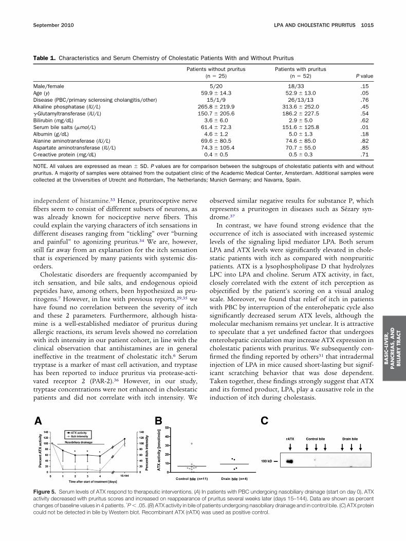
igure 5. Serum levels of ATX respond to therapeutic interventions. (Activity decreased with pruritus scores and increased on reappearancehanges of baseline values in 4 patients. *P � .05. (B) ATX activity in bile o
ould not be detected in bile by Western blot. Recombinant ATX (rATX) wasbserved similar negative results for substance P, whichepresents a pruritogen in diseases such as Sézary syn-rome.37
In contrast, we have found strong evidence that theccurrence of itch is associated with increased systemic
evels of the signaling lipid mediator LPA. Both serumPA and ATX levels were significantly elevated in chole-tatic patients with itch as compared with nonpruriticatients. ATX is a lysophospholipase D that hydrolyzesPC into LPA and choline. Serum ATX activity, in fact,losely correlated with the extent of itch perception asbjectified by the patient’s scoring on a visual analogcale. Moreover, we found that relief of itch in patientsith PBC by interruption of the enterohepatic cycle also
ignificantly decreased serum ATX levels, although theolecular mechanism remains yet unclear. It is attractive
o speculate that a yet undefined factor that undergoesnterohepatic circulation may increase ATX expression inholestatic patients with pruritus. We subsequently con-rmed the finding reported by others31 that intradermal
njection of LPA in mice caused short-lasting but signif-cant scratching behavior that was dose dependent.aken together, these findings strongly suggest that ATXnd its formed product, LPA, play a causative role in thenduction of itch during cholestasis.
ents With and Without Pruritus
ithout pruritus� 25)
Patients with pruritus(n � 52) P value
/20 18/33 .15� 14.3 52.9 � 13.0 .05
5/1/9 26/13/13 .76� 219.9 313.6 � 252.0 .45� 205.6 186.2 � 227.5 .54� 6.0 2.9 � 5.0 .62� 72.3 151.6 � 125.8 .01� 1.2 5.0 � 1.3 .18� 80.5 74.6 � 85.0 .82� 105.4 70.7 � 55.0 .85� 0.5 0.5 � 0.3 .71
on between the subgroups of cholestatic patients with and withoutthe Academic Medical Center, Amsterdam. Additional samples wereunich Germany; and Navarra, Spain.
atients with PBC undergoing nasobiliary drainage (start on day 0), ATXruritus several weeks later (days 15–144). Data are shown as percentents undergoing nasobiliary drainage and in control bile. (C) ATX protein
Pati
nts w(n
559.9
1265.8150.7
3.661.4
4.669.674.3
0.4
parisic of
) In pof p
f pati
used as positive control.
tliuLcrfilrwmamflIir
bies
tthactctthctip
peearcatcmapLorni(LntAsm
LdcmwedPlRpTfp
Fvsd.t*
BA
SIC–LIV
ER,
PA
NCREA
S,A
ND
BILIA
RY
TRA
CT
1016 KREMER ET AL GASTROENTEROLOGY Vol. 139, No. 3
Strikingly, we observed a much clearer difference be-ween pruritic and nonpruritic patients in serum ATXevels than in serum LPA levels and concurrent serum-nduced �[Ca2�]i in neuroblastoma cells (compare Fig-res 1 and 2 with Figure 3). One might argue that serumPA levels (and the concurrent Ca2� transient) shouldlosely follow the serum ATX level, because the latter isesponsible for increased LPA levels. Although we didnd a clear correlation between serum ATX and LPA
evels as well as Ca2� transients (data not shown), theange of ATX levels was much more dynamic and levelsere more consistently increased in pruritic patients. Theost likely explanation for this discrepancy is that LPA ishighly unstable lipid derivative that undergoes rapidetabolism in the circulation. In addition, LPA can be
ormed during and after blood collection, and thereforeevels depend on the procedure of processing and storage.n contrast, the enzyme ATX turns out to be highly stablen vitro and therefore represents a much more direct andeliable parameter.
The source of increased serum ATX levels remains toe determined. Enhanced levels could either be caused by
ncreased ATX expression or by reduced clearance of thenzyme. Recently, liver sinusoidal endothelial cells were
igure 6. Dose-dependent induction of scratch responses by LPA inivo. (A) Intradermal injections of LPA (100 nmol) led to increasedcratching behavior compared with vehicle injections being significanturing the first 15 minutes. Injections were performed in 7 mice. *P �
05. (B) Dose-dependent scratching behavior after intradermal injec-ions of LPA. Injections were performed in the indicated number of mice.P � .05, **P � .01, ***P � .001.
hown to play an important role in uptake and degrada- r
ion of ATX.38 This is in agreement with our observationhat ATX could not be detected in bile. Although ATXas a half-life of only several minutes in circulation, itsctivity can easily be detected in serum, suggesting aontinuous synthesis and release by peripheral cells andissues. Among these may be endothelial cells and adipo-ytes,39,40 but ATX was also reported to be expressed inhe liver.41 Interestingly, in patients with chronic hepati-is C, liver ATX messenger RNA expression was en-anced,24 which might lead to enhanced serum ATXoncentrations in these patients.25 In our group of pa-ients with hepatitis C virus, ATX levels were higher thann healthy controls but lower compared with cholestaticatients with pruritus.ATX was originally described as a motility-stimulating
rotein secreted from melanoma cells.8 Nowadays, theffects of ATX are believed to be mainly mediated by itsnzymatic product LPA.14 The bioactive lipid LPA is angonist of a family of at least 6 G protein– coupledeceptors that promote a great variety of biological pro-esses, ranging from cell motility, proliferation, survival,nd tumor progression to vascular development and cy-okine production.14 Furthermore, LPA has been impli-ated in neuronal cell functions such as brain develop-ent and neurite remodeling but also demyelination and
fter neurotrauma.42 In mice, LPA initiates neuropathicain after a single intrathecal injection.16 Mice lackingPA1 receptor do not develop any signs of demyelinationr neuropathic pain.16 It was shown that LPA causeseprogramming of signal transmission through differenterve fibers. While transmission through type 3 A� fibers
s increased, the transmission through type 1 C fibersinvolving substance P) is dramatically reduced.17 Thus,PA may induce neuropathic pain via LPA1 receptors onociceptive nerve fibers and at the same time contributeo pruritus via LPA receptors on pruritoceptive neurons.lternatively, LPA might indirectly cause pruritus by
timulating the release of a pruritogenic cytokine or lipidediator from cells located in the skin.43
It is intriguing to speculate on the role of ATX andPA in the pathogenesis of pruritus in other systemiciseases. Wound healing after injury or surgery is typi-ally accompanied by local itch perception. LPA pro-otes re-epithelialization and healing of cutaneousounds.44 High local concentrations of LPA might thus
licit itch-specific neurons, leading to the well-knownesire to scratch a wound during its healing process.ruritus is also a common symptom in patients with
ymphoma, especially in those with Hodgkin’s disease.45
ecently, Epstein–Barr virus—infected Hodgkin lym-homa cells have been shown to highly express ATX.46
hus, these cells might release high amounts of ATX,orming high local concentrations of LPA that not onlyromote tumor growth9 but may also activate the neu-
onal itch pathway. Intriguingly, patients with Hodgkin’s
dt
rdnidctr
aG1
1
1
1
1
1
1
1
1
1
1
2
2
2
2
2
2
2
2
2
2
3
3
3
3
3
3
3
3
3
BA
SIC–L
IVER
,PA
NCREA
S,A
ND
BIL
IARY
TRA
CT
September 2010 LPA AND CHOLESTATIC PRURITUS 1017
isease with intense pruritus have a shorter survival thanhose without itch.47
Unraveling the molecular mechanisms leading to pru-itus in systemic diseases will have a major impact on theevelopment of novel treatment strategies for this ago-izing symptom. At least in cholestatic disorders, ATX
nhibitors and LPA receptor blockers, which are currentlyeveloped for the treatment of patients with malignan-ies to reduce disease progression and formation of me-astasis,48 might also represent a novel class of antipru-itic drugs.
Supplementary Material
Note: To access the supplementary materialccompanying this article, visit the online version ofastroenterology at www.gastrojournal.org, and at doi:0.1053/j.gastro.2010.05.009.
References
1. Paus R, Schmelz M, Biro T, et al. Frontiers in pruritus research:scratching the brain for more effective itch therapy. J Clin Invest2006;116:1174–1186.
2. Ikoma A, Steinhoff M, Stander S, et al. The neurobiology of itch.Nat Rev Neurosci 2006;7:535–547.
3. Sun YG, Chen ZF. A gastrin-releasing peptide receptor mediatesthe itch sensation in the spinal cord. Nature 2007;448:700–703.
4. Sun YG, Zhao ZQ, Meng XL, et al. Cellular basis of itch sensation.Science 2009;325:1531–1534.
5. Beuers U. Drug insight: mechanisms and sites of action of ur-sodeoxycholic acid in cholestasis. Nat Clin Pract GastroenterolHepatol 2006;3:318–328.
6. EASL clinical practice guidelines: management of cholestatic liverdiseases. J Hepatol 2009;51:237–267.
7. Bergasa NV. The pruritus of cholestasis. J Hepatol 2005;43:1078–1088.
8. Stracke ML, Krutzsch HC, Unsworth EJ, et al. Identification, puri-fication, and partial sequence analysis of autotaxin, a novelmotility-stimulating protein. J Biol Chem 1992;267:2524–2529.
9. Mills GB, Moolenaar WH. The emerging role of lysophosphatidicacid in cancer. Nat Rev Cancer 2003;3:582–591.
0. Tanaka M, Okudaira S, Kishi Y, et al. Autotaxin stabilizes bloodvessels and is required for embryonic vasculature by producinglysophosphatidic acid. J Biol Chem 2006;281:25822–25830.
1. van Meeteren LA, Ruurs P, Stortelers C, et al. Autotaxin, asecreted lysophospholipase D, is essential for blood vessel for-mation during development. Mol Cell Biol 2006;26:5015–5022.
2. Tokumura A, Majima E, Kariya Y, et al. Identification of humanplasma lysophospholipase D, a lysophosphatidic acid-producingenzyme, as autotaxin, a multifunctional phosphodiesterase.J Biol Chem 2002;277:39436–39442.
3. Umezu-Goto M, Kishi Y, Taira A, et al. Autotaxin has lysophos-pholipase D activity leading to tumor cell growth and motility bylysophosphatidic acid production. J Cell Biol 2002;158:227–233.
4. van Meeteren LA, Moolenaar WH. Regulation and biological ac-tivities of the autotaxin-LPA axis. Prog Lipid Res 2007;46:145–160.
5. Aoki J, Inoue A, Okudaira S. Two pathways for lysophosphatidicacid production. Biochim Biophys Acta 2008;1781:513–518.
6. Inoue M, Rashid MH, Fujita R, et al. Initiation of neuropathic painrequires lysophosphatidic acid receptor signaling. Nat Med
2004;10:712–718.7. Ueda H. Molecular mechanisms of neuropathic pain-phenotypicswitch and initiation mechanisms. Pharmacol Ther 2006;109:57–77.
8. Dixon PH, van Mil SW, Chambers J, et al. Contribution of variantalleles of ABCB11 to susceptibility to intrahepatic cholestasis ofpregnancy. Gut 2009;58:537–544.
9. Inagaki N, Igeta K, Kim JF, et al. Involvement of unique mecha-nisms in the induction of scratching behavior in BALB/c mice bycompound 48/80. Eur J Pharmacol 2002;448:175–183.
0. Grynkiewicz G, Poenie M, Tsien RY. A new generation of Ca2�indicators with greatly improved fluorescence properties. J BiolChem 1985;260:3440–3450.
1. Nakamura K, Ohkawa R, Okubo S, et al. Measurement of lyso-phospholipase D/autotaxin activity in human serum samples.Clin Biochem 2007;40:274–277.
2. Swain MG, Rothman RB, Xu H, et al. Endogenous opioids accu-mulate in plasma in a rat model of acute cholestasis. Gastroen-terology 1992;103:630–635.
3. Simpson PB, Villullas IR, Schurov I, et al. Native and recombinanthuman Edg4 receptor-mediated Ca(2�) signalling. Assay DrugDev Technol 2002;1:31–40.
4. Cooper AB, Wu J, Lu D, et al. Is autotaxin (ENPP2) the linkbetween hepatitis C and hepatocellular cancer? J GastrointestSurg 2007;11:1628–1634; discussion 1634–1635.
5. Watanabe N, Ikeda H, Nakamura K, et al. Both plasma lysophos-phatidic acid and serum autotaxin levels are increased in chronichepatitis C. J Clin Gastroenterol 2007;41:616–623.
6. Kremer AE, Beuers U, Oude-Elferink RP, et al. Pathogenesis andtreatment of pruritus in cholestasis. Drugs 2008;68:2163–2182.
7. Beuers U, Gerken G, Pusl T. Biliary drainage transiently relievesintractable pruritus in primary biliary cirrhosis. Hepatology 2006;44:280–281.
8. Bergasa NV, Alling DW, Talbot TL, et al. Effects of naloxoneinfusions in patients with the pruritus of cholestasis. A double-blind, randomized, controlled trial. Ann Intern Med 1995;123:161–167.
9. Spivey JR, Jorgensen RA, Gores GJ, et al. Methionine-enkephalinconcentrations correlate with stage of disease but not pruritus inpatients with primary biliary cirrhosis. Am J Gastroenterol 1994;89:2028–2032.
0. Stapelbroek JM, van Erpecum KJ, Klomp LW, et al. Nasobiliarydrainage induces long-lasting remission in benign recurrent intra-hepatic cholestasis. Hepatology 2006;43:51–53.
1. Hashimoto T, Ohata H, Momose K. Itch-scratch responses in-duced by lysophosphatidic acid in mice. Pharmacology 2004;72:51–56.
2. Schmelz M, Schmidt R, Bickel A, et al. Specific C-receptors foritch in human skin. J Neurosci 1997;17:8003–8008.
3. Davidson S, Zhang X, Yoon CH, et al. The itch-producing agentshistamine and cowhage activate separate populations of primatespinothalamic tract neurons. J Neurosci 2007;27:10007–10014.
4. Stander S, Schmelz M. Chronic itch and pain—similarities anddifferences. Eur J Pain 2006;10:473–478.
5. Murphy GM, Ross A, Billing BH. Serum bile acids in primary biliarycirrhosis. Gut 1972;13:201–206.
6. Steinhoff M, Neisius U, Ikoma A, et al. Proteinase-activatedreceptor-2 mediates itch: a novel pathway for pruritus in humanskin. J Neurosci 2003;23:6176–6180.
7. Duval A, Dubertret L. Aprepitant as an antipruritic agent? N EnglJ Med 2009;361:1415–1416.
8. Jansen S, Andries M, Vekemans K, et al. Rapid clearance of thecirculating metastatic factor autotaxin by the scavenger recep-tors of liver sinusoidal endothelial cells. Cancer Lett 2009;284:
216–221.
3
4
4
4
4
4
4
4
4
4
R
ISN
A
AfT(psLc
C
F
BA
SIC–LIV
ER,
PA
NCREA
S,A
ND
BILIA
RY
TRA
CT
1018 KREMER ET AL GASTROENTEROLOGY Vol. 139, No. 3
9. Ferry G, Tellier E, Try A, et al. Autotaxin is released from adipo-cytes, catalyzes lysophosphatidic acid synthesis, and activatespreadipocyte proliferation. Up-regulated expression with adipo-cyte differentiation and obesity. J Biol Chem 2003;278:18162–18169.
0. Kanda H, Newton R, Klein R, et al. Autotaxin, an ectoenzyme thatproduces lysophosphatidic acid, promotes the entry of lympho-cytes into secondary lymphoid organs. Nat Immunol 2008;9:415–423.
1. Giganti A, Rodriguez M, Fould B, et al. Murine and human auto-taxin alpha, beta, and gamma isoforms: gene organization, tis-sue distribution, and biochemical characterization. J Biol Chem2008;283:7776–7789.
2. Savaskan NE, Rocha L, Kotter MR, et al. Autotaxin (NPP-2) in thebrain: cell type-specific expression and regulation during devel-opment and after neurotrauma. Cell Mol Life Sci 2007;64:230–243.
3. Zhao Y, Tong J, He D, et al. Role of lysophosphatidic acid recep-tor LPA2 in the development of allergic airway inflammation in amurine model of asthma. Respir Res 2009;10:114.
4. Balazs L, Okolicany J, Ferrebee M, et al. Topical application of thephospholipid growth factor lysophosphatidic acid promoteswound healing in vivo. Am J Physiol Regul Integr Comp Physiol2001;280:R466–R472.
5. Rubenstein M, Duvic M. Cutaneous manifestations of Hodgkin’sdisease. Int J Dermatol 2006;45:251–256.
6. Baumforth KR, Flavell JR, Reynolds GM, et al. Induction of auto-taxin by the Epstein-Barr virus promotes the growth and survivalof Hodgkin lymphoma cells. Blood 2005;106:2138–2146.
7. Gobbi PG, Attardo-Parrinello G, Lattanzio G, et al. Severe pruritusshould be a B-symptom in Hodgkin’s disease. Cancer 1983;51:
1934–1936. 18. Peyruchaud O. Novel implications for lysophospholipids,lysophosphatidic acid and sphingosine 1-phosphate, as drugtargets in cancer. Anticancer Agents Med Chem 2009;9:381–391.
Received January 24, 2010. Accepted May 11, 2010.
eprint requestsAddress requests for reprints to: Andreas E. Kremer, MD, Tytgat
nstitute for Liver and Intestinal Research, Academic Medical Center,1-164, University of Amsterdam, NL-1105 BK Amsterdam, Theetherlands. e-mail: [email protected]; fax: (31) 20-566-9190.
cknowledgmentsThe authors thank Dr H. Reesink (Academic Medical Center,
msterdam, The Netherlands) for kindly providing blood samplesrom patients with hepatitis C virus, Dr J. Aoki (University of Tokyo,okyo, Japan) for 5E5-autotaxin antibody, Dr H. van LentheAcademic Medical Center, Amsterdam, The Netherlands) forerforming high-performance liquid chromatography/masspectroscopy, and Jenny Chambers (Imperial College London,ondon, England) for her elaborate contribution to collection andharacterization of patient samples.
onflicts of interestThe authors disclose no conflicts.
undingSupported by the Deutsche Forschungsgemeinschaft (KR3618/1-
to A.E.K.).
wt11trw1sttEwopppfw
0r0m0tbwmtmwaciimqlwDt
September 2010 LPA AND CHOLESTATIC PRURITUS 1018.e1
Supplementary Materials and Methods
Quantitative LPA DeterminationAs internal standard myristoyl-LPA (LPA 14:0)
as added to serum samples reaching a final concentra-ion of 1 �M. Lipids were subsequently extracted from00 �L of serum by one-phase lipid extraction usingmL of methanol/chloroform (1:1, vol/vol). The extrac-
ion fluid was evaporated to dryness (45°C, vacuum). Theesidue was dissolved in 100 �l of chloroform/methanol/ater (50:45:5, v/v/v) containing 0.01% NH4OH, and0 �L of this solution was injected into the HPLC–MSystem. The HPLC system consisted of a Surveyor qua-ernary gradient pump, a vacuum degasser, a columnemperature controller, and an autosampler (Thermolectron, Waltham, MA, USA). The column temperatureas maintained at 25°C. The lipid extract was injectednto a LiChrospher 2 250 mm silica-60 column, 5 �marticle diameter (Merck, Darmstadt, Germany). Thehospholipids were separated from interfering com-ounds by a linear gradient between solution B (chloro-orm/methanol, 97:3, v/v) and solution A (methanol/
ater, 85:15, v/v). Solutions A and B contained 1 and s.1 mL of 25% (v/v) aqueous ammonia per liter of eluent,espectively. The gradient (0.3 mL/min) was as follows:–10 min, 20% A–100% A; 10 –12 min, 100% A; 12–12.1in, 100% A– 0% A; and 12.1–17 min, equilibration with
% A. All gradient steps were linear, and the total analysisime, including the equilibration, was 17 min. A splitteretween the HPLC column and the mass spectrometeras used, and 75 �L/min eluent was introduced into theass spectrometer. A TSQ Quantum AM (Thermo Elec-
ron) was used in the negative electrospray ionizationode. Nitrogen was used as the nebulizing gas. Argonas used as the collision gas. The skimmer offset was sett 10 V. The spray voltage used was 3600 V, and theapillary temperature was 300°C. Selected reaction mon-toring (SRM) was used to monitor precursor to producton transition of m/z 381.2 ¡ 227.2 for LPA(14:0) and
/z 435.25 ¡ 281.25 for LPA(18:1). Quadrupole 1 anduadrupole 3 were maintained at 0.3 and 0.7 unit reso-
ution (FWHM) respectively. The collision gas pressureas 0.5 mTorr and the collision energy was set at 50 V.well time was 0.150 s for both the analytes and IS. All
he parameters of LC and MS were controlled by Xcalibur
oftware version 2.0.7.