Embed Size (px)
Citation preview
Acta Biomaterialia 7 (2011) 954–958
Contents lists available at ScienceDirect
Acta Biomaterialia
journal homepage: www.elsevier .com/locate /ac tabiomat
Lysine–poly(2-hydroxyethyl methacrylate) modified polyurethane surfacewith high lysine density and fibrinolytic activity
Dan Li a,b, Hong Chen a,⇑, Shasha Wang a,b, Zhaoqiang Wu a,c, John L. Brash a,c
a College of Chemistry, Chemical Engineering and Materials Science, Soochow University, 199 Renai Rd., Suzhou 215123, Jiangsu, PR Chinab School of Materials Science and Engineering, Wuhan University of Technology, Wuhan 430070, PR Chinac School of Biomedical Engineering and Department of Chemical Engineering, McMaster University, Hamilton, Ontario, Canada
a r t i c l e i n f o a b s t r a c t
Article history:Received 21 July 2010Received in revised form 9 October 2010Accepted 20 October 2010Available online 25 October 2010
Keywords:Fibrinolytic surfacePolyurethanePoly(2-hydroxyethyl methacrylate)Protein adsorptionGraft density
1742-7061/$ - see front matter � 2010 Acta Materialdoi:10.1016/j.actbio.2010.10.021
⇑ Corresponding author. Tel./fax: +86 512 6588082E-mail address: [email protected] (H. Chen).
We have developed a potentially fibrinolytic surface in which a bioinert polymer is used as a spacer toimmobilize lysine such that the e-amino group is free to capture plasminogen when in contact with blood.Adsorbed plasminogen can be activated to plasmin and potentially dissolve nascent clots formed on thesurface. In previous work lysine was immobilized through a poly(ethylene glycol) (PEG) spacer; however,the graft density of PEG was limited and the resulting adsorbed quantity of plasminogen was insufficient todissolve clots efficiently. The aim of the present work was to optimize the surface using graft-polymerizedpoly(2-hydroxyethyl methacrylate) (poly(HEMA)) as a spacer to increase the grafting density of lysine. Sucha poly(HEMA)–lysine modified polyurethane (PU) surface is expected to have increased plasminogen bind-ing capacity and clot lysing efficiency compared with PEG–lysine modified PU. A lysine density of2.81 nmol cm�2 was measured on the PU–poly(HEMA)–Lys surface vs. 0.76 nmol cm�2 on a comparablePU–PEG–Lys surface reported previously. The poly(HEMA)–lysine-modified surface was shown to reducenon-specific (fibrinogen) adsorption while binding plasminogen from plasma with high affinity. Withincreased plasminogen binding capacity these surfaces showed more rapid clot lysis (20 min) in a standardin vitro assay than the corresponding PEG–lysine system (40 min). The data suggest that poly(HEMA) issuperior to PEG when used as a spacer in the immobilization of bioactive molecules at high density. Thismethod of modification may also provide a generic approach for preparing bioactive PU surfaces of highactivity and low non-specific adsorption of proteins.
� 2010 Acta Materialia Inc. Published by Elsevier Ltd. All rights reserved.
1. Introduction grafted polymers have abundant side-chains with active chain
Surface modification with bioactive agents capable of inhibitingenzymes in the coagulation cascade is a widely used strategy forimproving the blood compatibility of a biomaterial [1]. Polyethyl-ene glycol (PEG) has been used as a spacer to couple these bioac-tive moieties to surfaces because of its excellent proteinresistance [2,3]. We have developed the concept of a fibrinolyticsurface in which PEG is used as a spacer to immobilize lysine suchthat the e-amino group is free to capture plasminogen and tissuetype plasminogen activator (t-PA) when in contact with bloodand dissolve nascent clots formed on the surface [4–6]. However,the surface density of PEG achievable by ‘‘grafting’’ is limited dueto steric hindrance [7], and the density of terminally conjugatedbioactive molecules is correspondingly limited.
An alternative approach is to generate the spacer via surface-initiated polymerization (SIP) [7]. SIP is well known to generatemuch denser polymer layers, and, more importantly, if the surface
ia Inc. Published by Elsevier Ltd. A
7.
ends this permits the generation of a high concentration of chem-ically active sites on the surface to bind bioactive molecules. Of thevarious monomers available to form such grafts, poly(2-hydroxy-ethyl methacrylate) (poly(HEMA)) has found the most widespreaduse. Due to its excellent biocompatibility and physical propertiessimilar to those of living tissues, poly(HEMA) has been extensivelystudied for applications in tissue engineering [8], drug delivery [9],antifouling materials [10] and biosensors [11]. For the purposes ofgenerating non-fouling bioactive surfaces, HEMA has been graftpolymerized on various substrates, with attachment of bioactivemoieties via reaction with the hydroxyl groups in the side chains.For example, Xu et al. prepared poly(HEMA) modified silicon sur-faces by surface-initiated atom transfer radical polymerization(SI-ATRP) and coupled collagen to the pendant hydroxyl groupsto control the adhesion of 3T3 fibroblasts [12]. Hu et al. developeda versatile method of constructing glycosylated membranesurfaces in which poly(HEMA) was polymerized on microporouspolypropylene membranes (MPPMs) and then conjugated withglucose pentaacetate. The glycosylated membranes showed a highaffinity for concanavalin A protein while effectively preventingnon-specific protein adsorption [13,14].
ll rights reserved.
D. Li et al. / Acta Biomaterialia 7 (2011) 954–958 955
Although PEG is generally believed to be the most effective pro-tein repellent polymer and is widely used in surface modificationof various biomaterials, it has been shown to be prone to oxidationin the presence of oxygen and transition metal ions. Its long-termstability is thus in question [15]. In contrast, poly(HEMA), withhigh chemical and hydrolytic stability [16], may be more suitablefor longterm applications such as medical implants.
The aim of the present work was to optimize the properties of afibrinolytic polyurethane (PU) surface using graft-polymerizedpoly(HEMA) as a spacer to prevent non-specific protein adsorptionand increase the grafting density of lysine. It is expected that a highdensity of lysine should result in increased adsorption of plasmin-ogen from plasma and increased fibrinolytic potential. HEMA waspolymerized on a vinyl-functionalized PU surface [17] and lysinewas then coupled to the hydroxyl groups of the tethered poly(HE-MA) such that the e-amino group was free. Such a poly(HEMA)–ly-sine modified PU surface was expected to be rich in lysine and tohave increased plasminogen binding capacity and clot lysing effi-ciency compared with PEG–lysine modified PU. This method ofmodification may provide a generic approach for preparing bioac-tive PU surfaces of high activity and low non-specific adsorption ofproteins.
2. Materials and methods
2.1. Materials
N,N0-Disuccinimidyl carbonate (DSC) (anhydrous, P95% pure),trifluoroacetic acid (TFA), H-Lys(t-BOC)-OH and 4-nitrobenzalde-hyde were from Sigma–Aldrich Chemical Co. and used without fur-ther purification. 2-Hydroxyethyl methacrylate (HEMA) (Acros,98% pure) was distilled under reduced pressure to remove inhibi-tors. 2,20-Azoisobutyronitrile (AIBN) was recrystallized from meth-anol solution and dried under vacuum prior to use. Fibrinogen(plasminogen-free) was from Calbiochem (La Jolla, CA). Recombi-nant tissue plasminogen activator (t-PA) was from Genentech(San Francisco, CA). N,N-Dimethylformamide (DMF), acetonitrile,triethylamine (TEA) and methanol were from the Shanghai Chem-ical Reagent Co. and purified before use.
2.2. Preparation of surfaces
PU (Tecothane� TT-1095A, Thermedics, Wilmington, MA) wasextracted (Soxhlet) for 48 h with methanol to remove impurities.Films of this material were cast from a 5% (w/v) solution in DMF,dried in air at 75 �C for 48 h and vacuum dried at 60 �C for 48 hto remove solvent. Discs �5 mm in diameter and 0.5 mm thickwere punched from the PU elastomer films.
Methacryloyl isothiocyanate (MI) monomer was synthesized asreported previously [18]. The PU discs were immersed in 25 ml ofan acetonitrile solution containing 8 mmol MI. After stirring at65 �C for 12 h the vinyl group functionalized PU surface (VPU) ob-tained was washed with acetonitrile and dried in a vacuum oven at40 �C for 24 h.
Radical graft polymerization on the VPU surface was performedusing AIBN as the initiator. Briefly, HEMA (0.65 g, 5.0 mmol) andAIBN (0.0082 g, 0.05 mmol) were dissolved in 4 ml of anhydrousmethanol. VPU discs were immersed in the methanol solution.The solution was degassed by bubbling with nitrogen for 20 min.The grafting reaction was carried out at 65 �C for 12 h. Thepoly(HEMA) grafted PU surface was washed successively withmethanol, distilled water and methanol and dried in a vacuumoven at 40 �C for 24 h.
For the covalent conjugation of e-lysine the poly(HEMA) graftedPU surfaces were first added to an acetonitrile solution containing
DSC (0.05 mmol ml�1) and TEA (0.05 mmol ml�1) and stirred atroom temperature for 6 h. The resulting surfaces (PU–pHEMA–NHS) were incubated overnight in phosphate-buffered saline(PBS), pH 7.4 containing 5 mg ml�1 H-Lys(t-BOC)-OH. The surfaceswith e-NH-t-Boc groups were deprotected by treatment with 25%TFA for 90 min and subsequently washed with PBS. The resultingsurface, with the e-NH2 groups of lysine exposed (PU–pHEMA–Lys), is of the greatest interest in terms of its fibrinolytic potential.
2.3. Lysine density
The lysine graft densities were determined by reacting the sur-face amino groups with 4-nitrobenzaldehyde to form imines andsubsequent hydrolysis to liberate 4-nitrobenzaldehyde [19]. Typi-cally, six discs of PU–pHEMA–Lys or precursor PU–pHEMA surfacewere immersed in anhydrous ethanol (10 ml) containing 4-nitro-benzaldehyde (40 mg) and acetic acid (0.008 ml) under nitrogenat 50 �C for 3 h. The surfaces were then washed and sonicated inabsolute ethanol for 2 min. The discs were immersed in water(1 ml) containing acetic acid (0.002 ml) and the solution was keptat 40 �C for 3 h. The hydrolysis was run in triplicate (three groupsof three discs). The 4-nitrobenzaldehyde liberated, equivalent tothe surface amino content, was determined by measuring absor-bance at 268.5 nm.
2.4. Protein adsorption
Fibrinogen was labeled with 125I (ICN Pharmaceuticals, Irvine,CA) using the iodine monochloride (ICl) method. The productwas passed through an AG 1-X4 column (Bio-Rad Laboratories,Hercules, CA) to remove free iodide. Labeled fibrinogen was mixedwith unlabeled fibrinogen (1:19, labeled:unlabeled) in PBS at a to-tal concentration of 1 mg ml�1. Surfaces were incubated with theprotein solution for 3 h at room temperature, rinsed three times(10 min each time) with PBS, wicked onto filter paper and trans-ferred to clean tubes for radioactivity determination using aWizard 30 01480 Automatic Gamma Counter (PerkinElmer Life Sci-ences, Shelton, CT). Protein adsorption was expressed as molesper unit surface area.
2.5. Western blotting
Surfaces were incubated in 100% pooled normal human plasma(PNP) (from blood bank) for 3 h at room temperature. Adsorbedproteins were eluted using 2% aqueous sodium dodecyl sulfate(SDS). Polyacrylamide gel electrophoresis and immunoblottingwere performed as described previously [20]. Briefly, the eluateswere run on 12% reduced SDS–PAGE gels to separate the proteinsaccording to molecular weight. The proteins were then transferredfrom the gel onto an Immobilon PVDF membrane (Millipore, Bed-ford, MA). The membranes were blocked with 5% non-fat dry milkand incubated with primary antibodies (dilution of 1:1000) to plas-minogen or fibrinogen and then with an alkaline phosphatase-con-jugated secondary antibody (dilution of 1:1000). The substratesystem used to develop a color reaction for alkaline phosphatasewas 5-bromo-4-chloro-3-indolyl phosphate (BCIP) and nitrobluetetrazolium (NBT) (both from Bio-Rad), prepared as described bythe supplier.
2.6. Plasma clot lysis
Surfaces were incubated in PNP for 3 h at room temperature inmicrotitre plate wells. The films were removed from the wells,rinsed three times in Tris-buffered saline (TBS), pH 7.4 and thenplaced in clean wells. t-PA was added to the wells at a concentra-tion of 0.1 mg ml�1 in TBS and incubated for 30 min at room
956 D. Li et al. / Acta Biomaterialia 7 (2011) 954–958
temperature. The films were rinsed extensively with buffer to re-move any unbound proteins. To the extent that plasminogen is ad-sorbed from plasma, this procedure provides surfaces bearing alayer of bound plasmin. The clot lysing potential of the surfaceswith adsorbed plasmin was assessed using a modified plasmarecalcification assay. 100 ll of PNP was added to the wells contain-ing the surfaces. Following 5 min equilibration at 37 �C an equalvolume of 0.025 M CaCl2 was injected into the wells. Absorbanceat 405 nm was measured at 30 s intervals over a 1 h period.
3. Results and discussion
3.1. Preparation of poly(HEMA)–lysine-modified PU surfaces
The poly(HEMA)–lysine modified PU surfaces were prepared asoutlined in Scheme 1. MI was used as a functional monomer thatcould be bound covalently to the PU surface through reaction ofits NCS groups with NH groups in the PU. Poly(HEMA) was thengrown on the surface-bound vinyl groups by free radical polymer-ization. These reactions were confirmed (X-ray photoelectronspectroscopy) in a previous study [17]. The poly(HEMA) graftedsurface contains a high concentration of hydroxyl groups, whichprovide reactive sites for the attachment of lysine to give surfacesof high lysine density. DSC was chosen as a bioconjugate reagent tolink lysine to these tethered hydroxyl groups. Lysine was conju-gated through its a-amino groups using e-amino protected lysine.The protective groups were removed by hydrolysis after grafting.This procedure is required since lysine exhibits an affinity for plas-minogen and t-PA only when its e-NH2 and COOH groups are free[21]. Such a poly(HEMA)–lysine modified PU surface was expectedto be superior in plasminogen binding capacity to PEG–lysine-modified surfaces reported previously.
3.2. Lysine density
The lysine density is an important parameter that determinesthe extent of plasminogen binding. Surface immobilized lysinewas determined by reacting the e-NH2 groups of lysine on thesurface with 4-nitrobenzaldehyde to form a Schiff base. The4-nitrobenzaldehyde liberated by hydrolysis of the Schiff base ismeasured [19] by absorbance at 268.5 nm. The lysine density
Scheme 1. Schematic illustrating the
was found to be 2.81 nmol cm�2 on the PU–pHEMA–Lys surface,much higher than on a comparable PU–PEG–Lys surface(0.76 nmol cm�2) determined using the same method [6]. This ispresumably due to the higher density of hydroxyl groups availablefor reaction with lysine. The lysine density is comparable with thatreported in previous studies in which the lysine-containing sur-faces were prepared by a photochemical method; the lysine den-sity ranged from 0.2 to 3.2 nmol cm�2 [21]. The photochemicalmethod, however, is difficult to apply to complex shapes, such asthe inner surface of a small diameter vascular graft. The high den-sity of amino groups present on the PU–pHEMA–Lys surface sug-gests a correspondingly high density of lysine.
It should be noted that the values of lysine density reported forthese surfaces are based on the nominal surface area, which islikely to be less than the effective area. Densities of �3 nmol cm�2
imply areas per lysine of the order of 6 Å2, i.e. unrealistically small.However, it is expected that the pHEMA layer to which the lysine isattached has a significant thickness dimension. Thus the effectivearea will be greater than the nominal and the true lysine densitieswill be smaller than the reported ones.
3.3. Fibrinogen adsorption from buffer
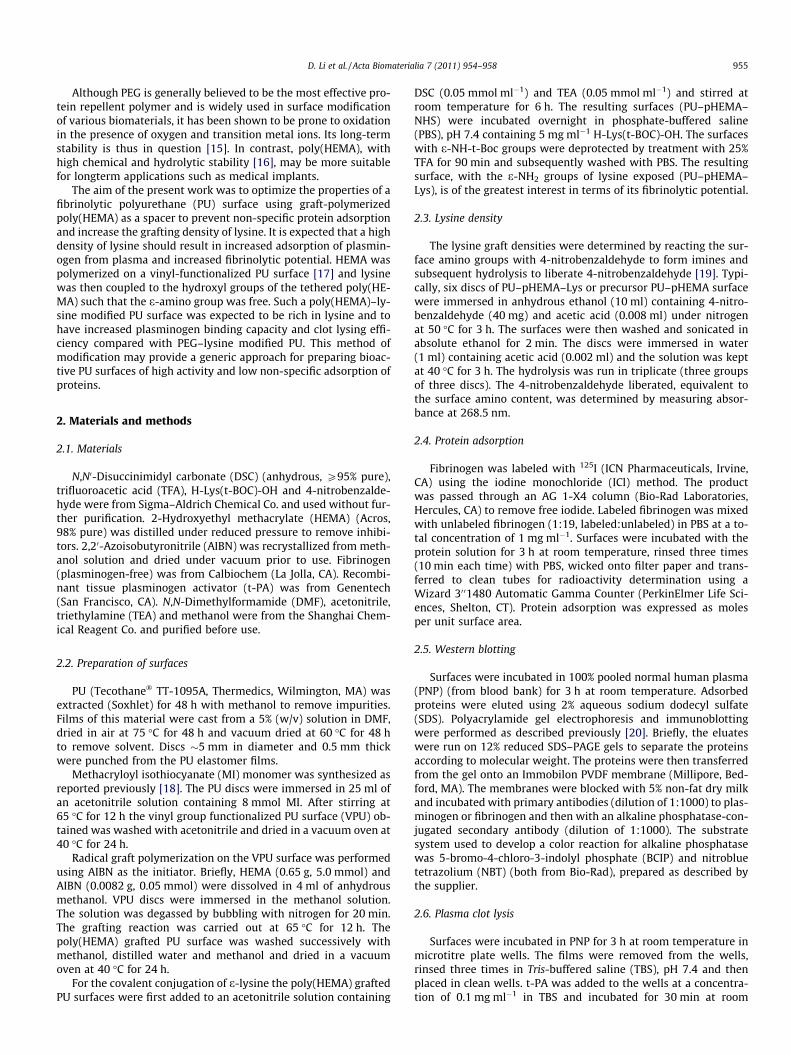
Fibrinogen is a key protein of the coagulation cascade and playsa leading role in mediating platelet adhesion to biomaterials. Inthis research fibrinogen adsorption was measured to provide anindication of the protein resistance of the modified surfaces. Asshown in Fig. 1, the unmodified PU surface showed the highest le-vel of adsorption, �0.91 lg cm�2, while only �0.1 lg cm�2 fibrino-gen was adsorbed on the poly(HEMA) modified surface, a decreaseof �90%. An optimized PU–PEG surface was shown previously todecrease fibrinogen adsorption by �95%, i.e. only slightly morethan the PU–pHEMA surface in the present work. The protein resis-tant properties of poly(HEMA) may be attributed, at least in part, toits hydrophilicity, which is generally recognized as essential forprotein resistance. Hu et al. prepared polypropylene microporousmembranes grafted with poly(HEMA) and found that with increas-ing grafting density the water contact angle decreased and the pro-tein resistance increased [10]. Furthermore, Yoshikawa et al.reported that both high (0.7 chains nm�2) and medium density(0.06 chains cm�2) poly(HEMA) brushes could resist larger proteins
processes of surface modification.
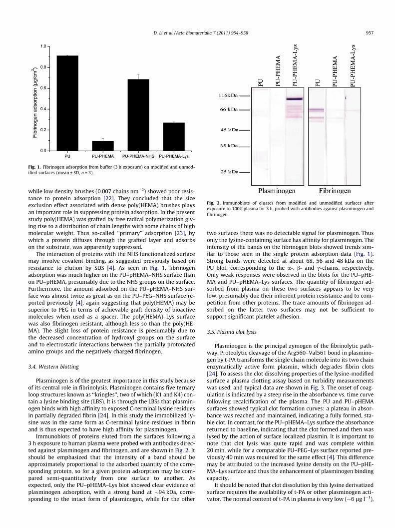
Fig. 1. Fibrinogen adsorption from buffer (3 h exposure) on modified and unmod-ified surfaces (mean ± SD, n = 3).
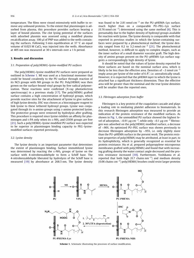
Fig. 2. Immunoblots of eluates from modified and unmodified surfaces afterexposure to 100% plasma for 3 h, probed with antibodies against plasminogen andfibrinogen.
D. Li et al. / Acta Biomaterialia 7 (2011) 954–958 957
while low density brushes (0.007 chains nm�2) showed poor resis-tance to protein adsorption [22]. They concluded that the sizeexclusion effect associated with dense poly(HEMA) brushes playsan important role in suppressing protein adsorption. In the presentstudy poly(HEMA) was grafted by free radical polymerization giv-ing rise to a distribution of chain lengths with some chains of highmolecular weight. Thus so-called ‘‘primary’’ adsorption [23], bywhich a protein diffuses through the grafted layer and adsorbson the substrate, was apparently suppressed.
The interaction of proteins with the NHS functionalized surfacemay involve covalent binding, as suggested previously based onresistance to elution by SDS [4]. As seen in Fig. 1, fibrinogenadsorption was much higher on the PU–pHEMA–NHS surface thanon PU–pHEMA, presumably due to the NHS groups on the surface.Furthermore, the amount adsorbed on the PU–pHEMA–NHS sur-face was almost twice as great as on the PU–PEG–NHS surface re-ported previously [4], again suggesting that poly(HEMA) may besuperior to PEG in terms of achievable graft density of bioactivemolecules when used as a spacer. The poly(HEMA)–Lys surfacewas also fibrinogen resistant, although less so than the poly(HE-MA). The slight loss of protein resistance is presumably due tothe decreased concentration of hydroxyl groups on the surfaceand to electrostatic interactions between the partially protonatedamino groups and the negatively charged fibrinogen.
3.4. Western blotting
Plasminogen is of the greatest importance in this study becauseof its central role in fibrinolysis. Plasminogen contains five ternaryloop structures known as ‘‘kringles’’, two of which (K1 and K4) con-tain a lysine binding site (LBS). It is through the LBSs that plasmin-ogen binds with high affinity to exposed C-terminal lysine residuesin partially degraded fibrin [24]. In this study the immobilized ly-sine was in the same form as C-terminal lysine residues in fibrinand is thus expected to have high affinity for plasminogen.
Immunoblots of proteins eluted from the surfaces following a3 h exposure to human plasma were probed with antibodies direc-ted against plasminogen and fibrinogen, and are shown in Fig. 2. Itshould be emphasized that the intensity of a band should beapproximately proportional to the adsorbed quantity of the corre-sponding protein, so for a given protein adsorption may be com-pared semi-quantitatively from one surface to another. Asexpected, only the PU–pHEMA–Lys blot showed clear evidence ofplasminogen adsorption, with a strong band at �94 kDa, corre-sponding to the intact form of plasminogen, while for the other
two surfaces there was no detectable signal for plasminogen. Thusonly the lysine-containing surface has affinity for plasminogen. Theintensity of the bands on the fibrinogen blots showed trends sim-ilar to those seen in the single protein adsorption data (Fig. 1).Strong bands were detected at about 68, 56 and 48 kDa on thePU blot, corresponding to the a-, b- and c-chains, respectively.Only weak responses were observed in the blots for the PU–pHE-MA and PU–pHEMA–Lys surfaces. The quantity of fibrinogen ad-sorbed from plasma on these two surfaces appears to be verylow, presumably due their inherent protein resistance and to com-petition from other proteins. The trace amounts of fibrinogen ad-sorbed on the latter two surfaces may not be sufficient tosupport significant platelet adhesion.
3.5. Plasma clot lysis
Plasminogen is the principal zymogen of the fibrinolytic path-way. Proteolytic cleavage of the Arg560–Val561 bond in plasmino-gen by t-PA transforms the single chain molecule into its two chainenzymatically active form plasmin, which degrades fibrin clots[24]. To assess the clot dissolving properties of the lysine-modifiedsurface a plasma clotting assay based on turbidity measurementswas used, and typical data are shown in Fig. 3. The onset of coag-ulation is indicated by a steep rise in the absorbance vs. time curvefollowing recalcification of the plasma. The PU and PU–pHEMAsurfaces showed typical clot formation curves: a plateau in absor-bance was reached and maintained, indicating a fully formed, sta-ble clot. In contrast, for the PU–pHEMA–Lys surface the absorbancereturned to baseline, indicating that the clot formed and then waslysed by the action of surface localized plasmin. It is important tonote that clot lysis was quite rapid and was complete within20 min, while for a comparable PU–PEG–Lys surface reported pre-viously 40 min was required for the same effect [4]. This differencemay be attributed to the increased lysine density on the PU–pHE-MA–Lys surface and thus the enhancement of plasminogen bindingcapacity.
It should be noted that clot dissolution by this lysine derivatizedsurface requires the availability of t-PA or other plasminogen acti-vator. The normal content of t-PA in plasma is very low (�6 lg l�1),
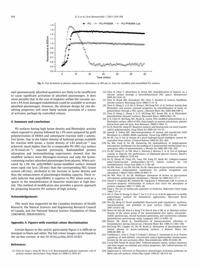
Fig. 3. Clot formation in plasma expressed as absorbance at 405 nm vs. time for modified and unmodified PU surfaces.
958 D. Li et al. / Acta Biomaterialia 7 (2011) 954–958
and spontaneously adsorbed quantities are likely to be insufficientto cause significant activation of adsorbed plasminogen. It doesseem possible that in the case of implants within the vascular sys-tem t-PA from damaged endothelium could be available to activateadsorbed plasminogen. However, the ultimate design for clot dis-solving properties will most likely include provision of a sourceof activator, perhaps by controlled release.
4. Summary and conclusions
PU surfaces having high lysine density and fibrinolytic activitywhen exposed to plasma followed by t-PA were prepared by graftpolymerization of HEMA and subsequent reaction with e-amino-free lysine. Due to the higher density of hydroxyl groups availablefor reaction with lysine, a lysine density of 2.81 nmol cm�2 wasachieved, much higher than for a comparable PU–PEG–Lys surface(0.76 nmol cm�2) reported previously. Radiolabelled proteinadsorption and immunoblotting experiments showed that themodified surfaces were fibrinogen-resistant and only the lysine-containing surface adsorbed plasminogen from plasma. When acti-vated by t-PA the poly(HEMA)–lysine-modified surface showedmore rapid clot lysis (20 min) than the corresponding PEG–lysinesystem (40 min), attributed to the increase in lysine density andthus the enhancement of plasminogen binding capacity. These re-sults indicate that poly(HEMA) is superior to PEG when used as aspacer in the immobilization of bioactive molecules at high den-sity. This method of modification also provides a generic approachfor preparing bioactive PU surfaces of high activity.
Acknowledgements
This work was supported by the Canadian Institutes of HealthResearch, the Natural Sciences and Engineering Research Councilof Canada, and the National Natural Science Foundation of China(20634030, 20920102035).
Appendix A. Figures with essential colour discrimination
Certain figures in this article, particularly Figure 2 is difficult tointerpret in black and white. The full colour images can be found inthe on-line version, at doi:10.1016/j.actbio.2010.10.021
References
[1] Chen H, Yuan L, Song W, Wu Z, Li D. Biocompatible polymer materials: role ofprotein–surface interactions. Prog Polym Sci 2008;33:1059–87.
[2] Chen H, Chen Y, Sheardown H, Brook MA. Immobilization of heparin on asilicone surface through a heterobifunctional PEG spacer. Biomaterials2005;26:7418–24.
[3] Chen H, Brook MA, Sheardown HD, Chen Y, Klenkler B. Generic bioaffinitysilicone surfaces. Bioconjug Chem 2006;17:21–8.
[4] Chen H, Zhang Y, Li D, Hu X, Wang L, McClung WG, et al. Surfaces having dualfibrinolytic and protein resistant properties by immobilization of lysine onpolyurethane through a PEG spacer. J Biomed Mater Res 2009;90A:940–6.
[5] Chen H, Wang L, Zhang Y, Li D, McClung WG, Brook MA, et al. Fibrinolyticpoly(dimethyl siloxane) surfaces. Macromol Biosci 2008;8:863–70.
[6] Li D, Chen H, McClung WG, Brash JL. Lysine–PEG-modified polyurethane as afibrinolytic surface. effect of PEG chain length on protein interactions, plateletinteractions and clot lysis. Acta Biomater. 2009;5:1864–71.
[7] Xu FJ, Neoh KG, Kang ET. Bioactive surfaces and biomaterials via atom transferradical polymerization. Prog Polym Sci 2009;34:719–61.
[8] Lahooti S, Sefton MV. Microencapsulation of normal and transfected L929fibroblasts in a HEMA–MMA copolymer. Tissue Eng 2000;6:139–49.
[9] He HY, Cao X, Lee LJ. Design of a novel hydrogel-based intelligent system forcontrolled drug release. J Control Release 2004;95:391–402.
[10] Hu MX, Yang Q, Xu ZK. Enhancing the hydrophilicity of polypropylenemicroporous membranes by the grafting of 2-hydroxyethyl methacrylate via asynergistic effect of photoinitiators. J Membr Sci 2006;285:196–205.
[11] Yu BZ, Wang CY, Ju YM, West L, Harmon J, Moussy Y, et al. Use of hydrogelcoating to improve the performance of implanted glucose sensors. BiosensBioelectron 2008;23:1278–84.
[12] Xu FJ, Zhong SP, Yung LYL, Tong YW, Kang ET, Neoh KG. Collagen-coupledpoly(2-hydroxyethyl methacrylate)–Si(111) hybrid surfaces for cellimmobilization. Tissue Eng 2005;11:1736–48.
[13] Hu MX, Wan LS, Liu ZM, Dai ZW, Xu ZK. Fabrication of glycosylated surfaces onmicroporous polypropylene membranes for protein recognition andadsorption. J Mater Chem 2008;18:4663–9.
[14] Hu MX, Wan LS, Xu ZK. Multilayer adsorption of lectins on glycosylatedmicroporous polypropylene membranes. J Membr Sci 2009;335:111–7.
[15] Ostuni E, Chapman RG, Holmlin RE, Takayama S, Whitesides GM. A survey ofstructure–property relationships of surfaces that resist the adsorption ofprotein. Langmuir 2001;17:5605–20.
[16] Kalal J. The use of methacrylic polymers in medicine. Makromol Chem Suppl1984;7:31–9.
[17] Wu Z, Chen H, Huang H, Zhao T, Liu X, Li D, et al. A facile approach to modifypolyurethane surfaces for biomaterial applications. Macromol Biosci2009;9:1165–8.
[18] Wu ZQ, Meng LZ. Novel amphiphilic fluorescent graft copolymers: synthesis,characterization, and potential as gene carriers. Polym Adv Technol2007;18:853–60.
[19] Moon LH, Kim JH, Kim K-j, Kang T-H, Kim B, Kim C-HK, et al. Absolute surfacedensity of the amine group of the aminosilylated thin layers: ultraviolet–visible spectroscopy, second harmonic generation, and synchrotron-radiationphotoelectron spectroscopy study. Langmuir 1997;13:4305–10.
[20] Mulzer SR, Brash JL. Identification of plasma-proteins adsorbed tohemodialyzers during clinical use. J Biomed Mater Res 1989;23:1483–504.
[21] McClung WG, Clapper DL, Hu SP, Brash JL. Adsorption of plasminogen fromhuman plasma to lysine-containing surfaces. J Biomed Mater Res2000;49:409–14.
[22] Yoshikawa C, Goto A, Tsujii Y, Fukuda T, Kimura T, Yamamoto K, et al. Proteinrepellency of well-defined, concentrated poly(2-hydroxyethyl methacrylate)brushes by the size-exclusion effect. Macromolecules 2006;39:2284–90.
[23] Currie EPK, Norde W, Stuart MAC. Tethered polymer chains: surface chemistryand their impact on colloidal and surface properties. Adv Colloid Interface Sci2003;100:205–65.
[24] Anglescano E. Overview on fibrinolysis–plasminogen activation pathways onfibrin and cell-surfaces. Chem Phys Lipids 1994;67–68:353–62.