Embed Size (px)
Citation preview
RESEARCH Open Access
Lysine methyltransferase G9a is not required forDNMT3A/3B anchoring to methylatednucleosomes and maintenance of DNAmethylation in somatic cellsShikhar Sharma1,2,3, Daniel S Gerke1, Han F Han2,4, Shinwu Jeong1,2, Michael R Stallcup1, Peter A Jones1,2 andGangning Liang1,2*
Abstract
Background: DNA methylation, histone modifications and nucleosome occupancy act in concert for regulation ofgene expression patterns in mammalian cells. Recently, G9a, a H3K9 methyltransferase, has been shown to play arole in establishment of DNA methylation at embryonic gene targets in ES cells through recruitment of de novoDNMT3A/3B enzymes. However, whether G9a plays a similar role in maintenance of DNA methylation in somaticcells is still unclear.
Results: Here we show that G9a is not essential for maintenance of DNA methylation in somatic cells. Knockdownof G9a has no measurable effect on DNA methylation levels at G9a-target loci. DNMT3A/3B remain stably anchoredto nucleosomes containing methylated DNA even in the absence of G9a, ensuring faithful propagation ofmethylated states in cooperation with DNMT1 through somatic divisions. Moreover, G9a also associates withnucleosomes in a DNMT3A/3B and DNA methylation-independent manner. However, G9a knockdown synergizeswith pharmacologic inhibition of DNMTs resulting in increased hypomethylation and inhibition of cell proliferation.
Conclusions: Taken together, these data suggest that G9a is not involved in maintenance of DNA methylation insomatic cells but might play a role in re-initiation of de novo methylation after treatment with hypomethylatingdrugs, thus serving as a potential target for combinatorial treatments strategies involving DNMTs inhibitors.
Keywords: G9a, DNMT3A, DNMT3B, DNA methylation, Nucleosome, Maintenance, Epigenetics
BackgroundDNA methylation is an essential epigenetic gene silen-cing mechanism which interplays with histone modifica-tions and nucleosome occupancy for regulation oftissue-specific gene expression patterns and chromatinarchitecture in mammalian cells [1]. DNA methylationpatterns are established during embryogenesis and thenfaithfully maintained in differentiated tissues, enablingpreservation of cellular identity through multiplesomatic divisions. Failure in proper maintenance of
methylation patterns can result in development of dis-ease states such as cancer [2,3]. Thus, it is essential tofaithfully maintain DNA methylation patterns in differ-entiated cells through somatic divisions.In mammals, the de novo DNA methytransferases,
DNMT3A/3B, primarily establish the methylationpatterns during embryonic development [4] and latermaintain them in differentiated tissues through coop-eration with the maintenenace DNA methyltransfer-ase, DNMT1 [5-7]. Recent studies by our group andothers have revealed distinct maintenance mechan-isms used by these enzymes for preservation ofmethylation patterns in somatic cells [8,9]. WhileDNMT1 transiently interacts with the chromatin andprimarily performs its maintenance activity in
* Correspondence: [email protected] of Biochemistry and Molecular Biology, USC/NorrisComprehensive Cancer Center, Keck School of Medicine, University ofSouthern California, Los Angeles, CA 90089-9181, USAFull list of author information is available at the end of the article
Sharma et al. Epigenetics & Chromatin 2012, 5:3http://www.epigeneticsandchromatin.com/content/5/1/3
© 2012 Sharma et al; licensee BioMed Central Ltd. This is an Open Access article distributed under the terms of the Creative CommonsAttribution License (http://creativecommons.org/licenses/by/2.0), which permits unrestricted use, distribution, and reproduction inany medium, provided the original work is properly cited.

association with the replication fork, DNMT3A/3Bremain preferentially bound to nucleosomes in chro-matin regions containing methylated repeats andCpG islands, which stabilizes these proteins andassists in faithful propagation of DNA methylationwithin the methylated domains through cooperativeactivity of DNMT3A/3B and DNMT1 enzymes [9-11].However, the mechanisms responsible for preferentialtargeting of DNMT3A/3B to such methylated regionsare still poorly understood.In embryonic stem (ES) cells, targeting of de novo
DNMT3A/3B enzymes to specific chromatin regionsinvolves interactions with auxiliary factors in addition totheir direct interactions with the nucleosomes [12,13].DNMT3A/3B interact with various chromatin-associatedproteins including heterochromatin protein 1 (HP1), his-tone deacetylase 1 (HDAC1), UHRF1 and histonemethyltransferases such as EZH2, suggested to play arole in recruitment of DNMT3A/3B to specific chroma-tin regions for de novo methylation in ES cells [9,13,14].However, recent studies suggest that these auxiliary pro-teins are not required for interaction of DNMT3A/3Bwith the nucleosomes in somatic cells suggesting exis-tence of other mechanisms involved in association ofDNMT3A/3B with chromatin [10].H3K9 methylation has also been established to inter-
play with DNA methylation for gene silencing in cells.In neurospora crassa, H3K9 methylation directs DNAmethylation to transposable elements [15,16]. A similarlink between H3K9 methylation and DNA methylationis present in mammals where heterochromatic H3K9methyltransferase, Suv39h1, has been shown to directDNA methylation to major satellite repeats at peri-centric heterochromatin [17]. Recently, G9a, a euchro-matic H3K9 methyltransferase, has also been found todirect DNA methylation to H3K9 methylated regionsin ES cells [18]. G9a, along with its partner proteinGLP, is crucial for H3K9 (mainly H3K9me andH3K9me2) methylation of euchromatin and is involvedin transcriptional silencing [19,20]. G9a binds to itsown product, H3K9me and H3K9me2 residues,through its ankyrin domain, a mechanism suggested toplay a role in propagation of H3K9 methylationthrough cell divisions [21,22]. Recent studies haveshown that G9a physically interacts with Dnmt3a/3band recruits them to G9a-target gene promoters, retro-transposons and major satellite repeats for de novomethylation in ES cells [23], independent of its histonemethyltransferase activity [24]. While an essential roleof G9a in directing de novo DNA methylation in EScells has been established, whether it plays a similarrole in maintenance of DNA methylation patterns insomatic cells remains unclear.
Here we show that G9a is not required for anchoringof DNMT3A/3B to nucleosomes in methylated chroma-tin regions and for maintenance of DNA methylation insomatic cells. Using sucrose gradient chromatin fractio-nation analysis, we show that G9a strongly binds toboth mononucleosomes and polynucleosomes, similar toDNMT3A/3B. However, knockdown of G9a in somaticcells does not decrease binding of DNMT3A/3B tonucleosomes and no discernible reduction in DNAmethylation levels of G9a-associated target genomicregions occurs even in the absence of G9a, suggestingno role of G9a in maintenance of DNA methylation.However, G9a-knockdown renders cancer cells moresensitive to 5-Aza-CdR (5-aza-2’-deoxycytidine) treat-ment resulting in increased DNA hypomethylation andcell growth inhibition, indicating that G9a might beinvolved in re-initiation of de novo methylation and thuscould serve as a promising target for combinatorial can-cer treatment strategies involving DNA hypomethylatingdrugs.
ResultsG9a strongly associates with polynucleosomes similar toDNMT3A/3BTo test the role of G9a, the euchromatic histonemethyltransferase, in the strong nucleosome anchoringmanifested by DNMT3A/3B, we examined theirnucleosomal binding patterns using sucrose densitygradient analysis. We subjected purified HCT116nuclei to partial digestion with MNase, which cuts lin-ker DNA to generate nucleosomal fragments of varioussizes, yielding a mixture of mono- and poly-nucleo-somes. The nucleosomal digests were then fractionatedon sucrose gradients containing 300 mM NaCl and thedistribution of chromatin-associated proteins analyzedthrough immunoblotting (Figure 1A). G9a associatedstrongly with polynucleosomes with substantialamounts of G9a protein sedimenting in nucleosome-containing fractions, possibly in association with itsown enzymatic products, H3K9me and H3K9me2 [21].Strikingly, the sedimentation profile of G9a was verysimilar to that of DNMT3A/3B (Figure 1B). Interest-ingly, SUV39h1, the heterochromatic histone methyl-transferase, was also found associated with thepolynucleosomes. However, its sedimentation profilewas slightly shifted towards the bottom fractions of thegradient, containing the larger chromatin fragmentsindicating association with the condensed heterochro-matin [17,25,26] (Figure 1A, B). Strong association ofDNMT3A/3B and histone methyltransferases withnucleosomes is in agreement with the inheritancemodel recently proposed for DNA methylation andhistone modifications where the enzymes remain
Sharma et al. Epigenetics & Chromatin 2012, 5:3http://www.epigeneticsandchromatin.com/content/5/1/3
Page 2 of 12

associated with their products to enable their properpropagation [8,21,26,27].
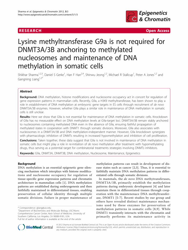
G9a tightly binds to intact mononucleosomesWe next asked whether G9a could bind to mononucleo-somes in a manner similar to that previously observedfor DNMT3A/3B. Mononucleosomal MNase digestsfrom HCT116 cells were analyzed on sucrose gradients
containing 300 mM NaCl. Mononucleosomes containingapproximately 146 bp DNA fragments and core histoneslocalized in a peak at fraction 6 (Figure 2A). DNMT1dissociated from nucleosomes, forming a peak at frac-tion 4, while DNMT3A/3B formed a peak at fraction 7suggesting that DNMT3A/3B are bound to mononu-cleosomes and that their presence altered the sedimen-tation of bound nucleosomes by one fraction relative to
Figure 1 Strong association of G9a and SUV39h1 with polynucleosomes similar to DNMT3A/3B. (A) Nucleosomes released from nucleipartially digested with MNase at low ionic strength were resolved by ultracentrifugation on a sucrose density gradient (5% to 25%) containing300 mM NaCl. Gradients were fractionated and analyzed as described in the Methods section. Ponceau S staining shows core histonestransferred onto the membrane from the SDS/PAGE gel. The control lanes (denoted as C) on the gels were loaded with unfractionated nuclearextract loaded on the gels to monitor the quality of the immunostaining of the membranes. (B) Quantitation of protein bands obtained fromthe western blot was done using Quantity One software (Bio-Rad). Plotting of levels of individual proteins in each fraction shows co-sedimentation of G9a and DNMT3A/3B while SUV39h1 shows a sedimentation profile shifted towards bottom of the gradient indicatingassociation with heavier condensed heterochromatin fragments.
Sharma et al. Epigenetics & Chromatin 2012, 5:3http://www.epigeneticsandchromatin.com/content/5/1/3
Page 3 of 12
bulk mononucleosomes. G9a also remained tightlyanchored to the mononucleosomes similar toDNMT3A/3B enzymes. SUV39h1 also displayed strongbinding to mononucleosomes (Additional file 1, FigureS1). However, under such extensive digestion of chro-matin, a substantial portion of cellular SUV39h1 proteindissociated from the nucleosomes, possibly due to dis-ruption of condensed heterochromatin structure [25].The sedimentation profiles of G9a and SUV39h1showed marked changes when the extent of MNasedigestion was altered from partial to extensive (Figure 1,2A, S1), similar to that observed previously forDNMT3A/3B [10], indicating physical association ofthese proteins with nucleosomes. Similar binding of G9a
and SUV39h1 to mononucleosomes was also observedin 293T cells indicating that the strong nucleosomalassociation of these proteins takes place in both celltypes and is not due to potential cell type-specific inter-actions (Additional file 1, Figure S1).To further explore whether these enzymes require
intact nucleosomal structures for their association withchromatin, we performed the sucrose gradient analysison the mononucleosomes treated with ethidium bro-mide (EtBr) which disrupts the nucleosomal structureby intercalating into DNA but does not interfere withthe protein-protein interactions [17,28-30]. Mononu-cleosomes were incubated with EtBr prior to loadingonto sucrose gradients containing 300 mM NaCl (Figure
Figure 2 G9a along with DNMT3A/3B binds to intact nucleosomal structures. Mononucleosomes released by extensive digestion withMNase were incubated in the absence (A) or presence (B) of 300 μg/ml EtBr for 10 min at room temperature, before loading onto the sucrosedensity gradients (5% to 25%) containing 300 mM NaCl. In (A), absorbance of each fraction, read at 260 nm, has been plotted on the top torepresent the amount of DNA sedimenting in each fraction. Gradients were fractionated and analyzed as described previously.
Sharma et al. Epigenetics & Chromatin 2012, 5:3http://www.epigeneticsandchromatin.com/content/5/1/3
Page 4 of 12
2B). The distribution of G9a changed dramatically, simi-lar to DNMT3A/3B, upon disruption of nucleosomalstructure by EtBr with the enzymes now sedimentingmainly in factions 3 to 5, which do not contain mea-sureable histone components of the nucleosome. Thesedata show that G9a and DNMT3A/3B enzymes requireintact nucleosomal structures for their association withchromatin.
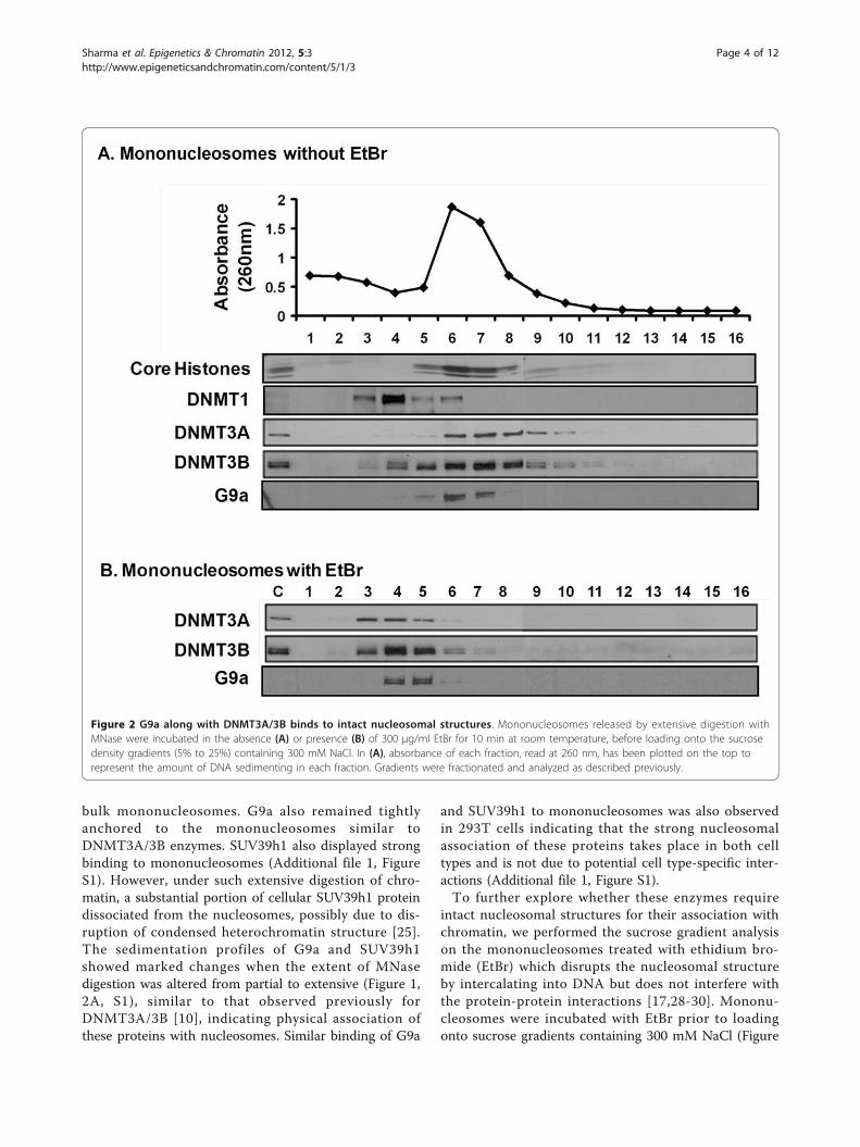
G9a is not essential for maintenance of DNA methylationin somatic cellsRecently, G9a was shown to direct DNA methylation toretrotransposons, major satellite repeats and denselymethylated CpG-rich promoters in ES cells throughrecruitment of DNMT3A/3B proteins [23]. To ascertainwhether a similar role of G9a exists in maintenance ofDNA methylation in somatic cells, we knocked downG9a in HCT116 cells using shRNA constructs. G9a pro-tein levels were severely reduced in G9a shRNA(shG9a5, shG9a7) infected cells compared to the non-specific shRNA (NS) control infected cells (Figure 3A).DNMT3A protein levels did not show any differenceamong the infected cell lines. Next we examined DNAmethylation levels at various CpG poor and CpG island
promoter regions and repeats in G9a knockdown (G9a-kd) and control infected HCT116 cells. We selected sixhighly methylated regions including some previouslyverified G9a-target regions in HCT116 cells [31] (CpGpoor promoters: RUNX3P1, MAGE-A1, SPANXA1; CpGisland promoters: ATBF1, XAGE1; Repeats: LINE1) andone unmethylated region (CpG island: RUNX3P2) forour analysis [18-20,23,32]. Cooperative activity ofDNMT3A/3B and DNMT1 has been previously shownto be required for maintenance of DNA methylation atthese methylated loci in HCT116 cells [33-35]. We didnot observe any substantial change in DNA methylationlevels at the analyzed regions in the G9a-kd cells com-pared to control infected HCT116 cells (Figure 3B).These data suggest that unlike in ES cells, G9a is notessential for maintenance of DNA methylation at theseloci in somatic cells, in agreement with some otherrecent studies [31,36].
DNMT3A/3B do not require G9a for anchoring tonucleosomesThe finding that DNA methylation is maintained in theabsence of G9a in somatic cells prompted us to examinewhether DNMT3A/3B, which are recruited andanchored to target chromatin regions by G9a for DNAmethylation in ES cells [23,24], can still strongly associ-ate with nucleosomes even in the absence of G9a insomatic cells. Since DNMT3A/3B cooperate withDNMT1 for maintenance of DNA methylation at var-ious methylated genomic loci in somatic HCT116 cells,including the G9a-target loci examined in this study[33], their continued association with such loci mightenable faithful propagation of DNA methylation even inthe absence of G9a. We analyzed mononucleosomaldigests from HCT116 cells infected with either G9ashRNA or non-specific control on sucrose gradientscontaining 300 mM NaCl. DNMT3A/3B along with G9awere tightly associated with nucleosomes in the controlcells as expected (Figure 4A). Interestingly, even in G9aknockdown cells, DNMT3A/3B remained stronglybound to the nucleosomes indicating their continuedassociation with the target loci even in the absence ofG9a. Taken together, these data indicate that in somaticcells, the strong association of DNMT3A/3B withmethylated chromatin regions is not mediated by G9aand DNMT3A/3B, in cooperation with DNMT1, canfaithfully maintain DNA methylation at their target lociindependent of G9a. We also asked whether impairedDNA methylation could feed back on H3K9 methyla-tion, as previously suggested [37], and disrupt binding ofG9a to H3K9 methylated regions. Mononucleosomaldigests from severely hypomethylated DNMT-deficientDKO1 (DNMT1ΔE2-5, DNMT3B -/- ) cells, containingseverely reduced levels of DNMT3A [11,33], were
Figure 3 Depletion of G9a does not impair maintenance ofDNA methylation in somatic cells. (A) Western blot of nuclearextracts from HCT116 cells infected with either control (NS) shRNAor shRNAs against G9a (shG9a5 or shG9a7), prepared 14 days afterinfection. (B) The levels of DNA methylation at different loci inHCT116 cells infected with either control (NS) shRNA or shRNAsagainst G9a (shG9a5 or shG9a7) were measured through Ms-SNuPE14 days after infection. Data represent mean and SEM ofmethylation levels measured at three independent CpG sites withineach locus.
Sharma et al. Epigenetics & Chromatin 2012, 5:3http://www.epigeneticsandchromatin.com/content/5/1/3
Page 5 of 12
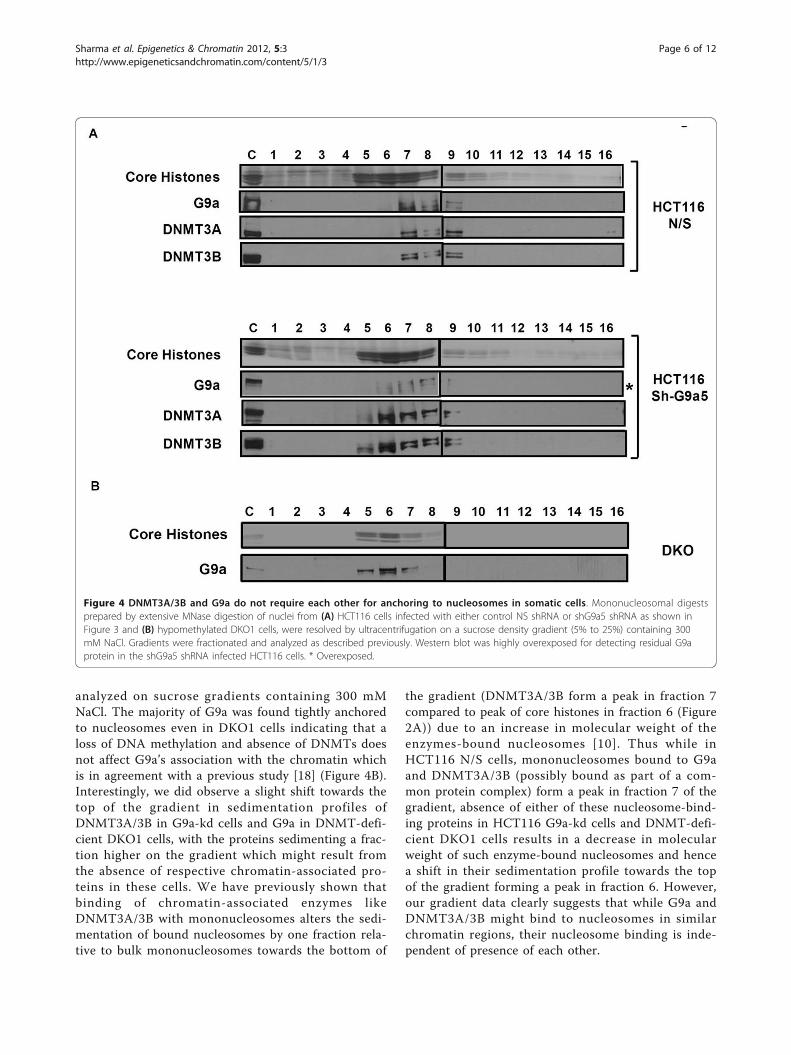
analyzed on sucrose gradients containing 300 mMNaCl. The majority of G9a was found tightly anchoredto nucleosomes even in DKO1 cells indicating that aloss of DNA methylation and absence of DNMTs doesnot affect G9a’s association with the chromatin whichis in agreement with a previous study [18] (Figure 4B).Interestingly, we did observe a slight shift towards thetop of the gradient in sedimentation profiles ofDNMT3A/3B in G9a-kd cells and G9a in DNMT-defi-cient DKO1 cells, with the proteins sedimenting a frac-tion higher on the gradient which might result fromthe absence of respective chromatin-associated pro-teins in these cells. We have previously shown thatbinding of chromatin-associated enzymes likeDNMT3A/3B with mononucleosomes alters the sedi-mentation of bound nucleosomes by one fraction rela-tive to bulk mononucleosomes towards the bottom of
the gradient (DNMT3A/3B form a peak in fraction 7compared to peak of core histones in fraction 6 (Figure2A)) due to an increase in molecular weight of theenzymes-bound nucleosomes [10]. Thus while inHCT116 N/S cells, mononucleosomes bound to G9aand DNMT3A/3B (possibly bound as part of a com-mon protein complex) form a peak in fraction 7 of thegradient, absence of either of these nucleosome-bind-ing proteins in HCT116 G9a-kd cells and DNMT-defi-cient DKO1 cells results in a decrease in molecularweight of such enzyme-bound nucleosomes and hencea shift in their sedimentation profile towards the topof the gradient forming a peak in fraction 6. However,our gradient data clearly suggests that while G9a andDNMT3A/3B might bind to nucleosomes in similarchromatin regions, their nucleosome binding is inde-pendent of presence of each other.
Figure 4 DNMT3A/3B and G9a do not require each other for anchoring to nucleosomes in somatic cells. Mononucleosomal digestsprepared by extensive MNase digestion of nuclei from (A) HCT116 cells infected with either control NS shRNA or shG9a5 shRNA as shown inFigure 3 and (B) hypomethylated DKO1 cells, were resolved by ultracentrifugation on a sucrose density gradient (5% to 25%) containing 300mM NaCl. Gradients were fractionated and analyzed as described previously. Western blot was highly overexposed for detecting residual G9aprotein in the shG9a5 shRNA infected HCT116 cells. * Overexposed.
Sharma et al. Epigenetics & Chromatin 2012, 5:3http://www.epigeneticsandchromatin.com/content/5/1/3
Page 6 of 12

Next, we asked whether G9a could target the trun-cated ΔDNMT3B variants, which lack parts of N-term-inal domain of DNMT3B including the PWWP domainbut possess a conserved active catalytic domain, tonucleosomes (Figure 5A). The truncated ΔDNMT3Bvariants have been associated with aberrant promotermethylation in non-small cell lung cancer (NSCLC) celllines, possibly arising from their aberrant targeting dueto a reduction in their chromatin binding affinities[10,38,39]. G9a associates with DNMT3A/3B throughthe interaction of its ankyrin domain with the catalyticdomains of DNMT3A/3B [23], therefore it should beable to physically interact and recruit catalytically active
ΔDNMT3B variants to the target chromatin regions.Mononucleosomal digests from 293T cells expressingMyc-tagged ΔDNMT3B variants were analyzed onsucrose gradients containing 300 mM salt. While G9aremained tightly bound to the nucleosomes, the deltaDNMT3B variants completely dissociated from thenucleosomes (Figure 5B). Taken together, these datasuggest that unlike in ES cells, where G9a is essentialfor DNA methylation through recruitment ofDNMT3A/3B enzymes to the H3K9 methylated chro-matin regions, G9a is not essential for anchoring ofDNMT3A/3B to the nucleosomes and for propagationof DNA methylation in somatic cells.
Figure 5 G9a cannot anchor ΔDNMT3B truncated variants to the nucleosomes. (A) Map of DNMT3B1 and Delta DNMT3B isoforms showingthe PWWP and PHD-like domains located in the N-terminal regions, and the catalytic methylase domains in the C-terminal region. (B)Mononucleosomal digests prepared by extensive MNase digestion of nuclei from 293T cells expressing either ΔDNMT3B2 or ΔDNMT3B4, wereresolved by ultracentrifugation on a sucrose density gradient (5% to 25%) containing 300 mM NaCl. Gradients were fractionated and analyzedthrough immunoblotting as described previously.
Sharma et al. Epigenetics & Chromatin 2012, 5:3http://www.epigeneticsandchromatin.com/content/5/1/3
Page 7 of 12

G9a knockdown synergizes with 5-Aza-CdR treatmentresulting in increased DNA hypomethylation andinhibition of cell growthWhile our results suggested no role of G9a in maintain-ing DNA methylation in somatic cells, it remained plau-sible that G9a might be involved in re-initiation of denovo DNA methylation (rebound methylation) at hypo-methylated sites after treatment with hypomethylatingdrugs, a phenomenon commonly observed after treat-ment of cancer cells with DNA methylation inhibitors[33]. Since G9a plays a role in directing de novo DNAmethylation to various G9a target regions in ES cells[23], a similar role of G9a in directing rebound methyla-tion in somatic cells is possible. Therefore, we hypothe-sized that knockdown of G9a might synergize withtreatment with DNMTs inhibitors enabling greaterreduction in DNA methylation through inhibition ofinterplay between the histone methylation and DNAmethylation machineries. To test this hypothesis, wetreated G9a shRNA (shG9a5) and non-specific control(NS) infected HCT116 cells with 5-Aza-CdR (5-aza-2’-deoxycytidine). Strikingly, G9a-kd cells treated with 5-Aza-CdR showed substantially greater inhibition of cellproliferation compared to 5-Aza-CdR treated controlinfected HCT116 cells (Figure 6A). Analysis of DNAmethylation at the MAGE-A1 promoter, a G9a-targetlocus, and p16exon2, 72 hrs after drug treatment showedgreater reduction in DNA methylation at both loci inG9a-kd cells compared to control infected cells (Figure6B). The increased hypomethylation of MAGE-A1 locusin 5-Aza-CdR treated G9a-kd cells compared to non-specific control treated cells was further confirmed bybisulfite sequencing showing a decrease in MAGE-A1promoter methylation to 45% in G9a-kd cells comparedto 55% in NS control cells after drug treatment (Addi-tional file 2, Figure S2). Since both G9a-kd and controlHCT116 cells displayed similar growth rates in mockPBS treatments, the increased hypomethylation causedby 5-Aza-CdR in G9a-kd cells compared to control cellswas not due to differential incorporation of the druginto replicating DNA in these cells (Figure 6A). How-ever, while we did see reduced methylation levels 72 hrsafter 5-Aza-CdR treatment, DNA methylation levelsrecovered to their initial high levels in both G9a-kd andcontrol HCT116 cells with increased time in culture(Day 15), possibly due to the activity of residualDNMT3A/3B enzymes still associated with such regionsand the gradual recovery in levels of cellular DNMT3A/3B and DNMT1 enzymes in absence of the hypomethy-lating drug [11,40]. After observing the pronouncedinhibition of cell proliferation by 5-Aza-CdR upondepletion of G9a, we also tried pharmacologic inhibitionof G9a by performing a double treatment using a G9a-inhibitor, BIX-01294 [41] in combination with 5-Aza-
CdR. However, BIX-01294 was very toxic to cells, aspreviously discussed [42], and thus could not be usedfor combination treatment. Taken together, these datasuggest that G9a knockdown synergizes with DNMTsinhibition leading to higher cell growth inhibition andDNA hypomethylation and G9a might serve as an effec-tive target for combinatorial cancer treatment strategiesinvolving DNMTs inhibitors.
DiscussionWe have previously shown that the majority ofDNMT3A/3B strongly anchor to nucleosomes contain-ing methylated DNA in somatic cells. Such binding,which requires the presence of DNA methylation,further stabilizes these proteins [10,11]. Based on thesedata, we proposed a revised inheritance model for DNAmethylation where DNMT3A/3B remain associated withmethylated chromatin domains i.e. in association withtheir product 5-methylcytosine enabling proper mainte-nance of DNA methylated states through somatic divi-sions in cooperation with DNMT1 [8]. This model issimilar to proposed inheritance models for histonemarks where the histone methyltransferases remain
Figure 6 Increased DNA hypomethylation and growthinhibition by 5-Aza-CdR in G9a knockdown cells. (A) Cellgrowth curve and (B) DNA methylation levels at MAGE-A1 andp16exon2 locus, of G9a-knockdown (shG9a5) and control (NS)HCT116 cells treated with either PBS or 5-Aza-CdR. Cells weretreated with the drug for 24 h. The drug containing media wasthen replaced with fresh media. The cells were kept in culture andcell counts and DNA methylation levels were assessed at respectivetime points. For 5-Aza-CdR treatment, samples examined were takenafter 3 (Day 3) and 15 (Day 15) days of drug treatment. For PBScontrol treatment, sample examined was taken after 3 days of drugtreatment. DNA methylation levels were measured using Ms-SNuPEassay as described in the Methods section. Data represent meanand range of methylation levels from two independent biologicalreplicate drug treatment experiments, in each measuringmethylation levels at three independent CpG sites within eachlocus.
Sharma et al. Epigenetics & Chromatin 2012, 5:3http://www.epigeneticsandchromatin.com/content/5/1/3
Page 8 of 12

associated with the chromatin domains containing theirown mark enabling faithful maintenance [21,26,27]. Thestrong binding of DNMT3A/3B and H3K9 methyltrans-ferases, G9a and SUV39h1, to nucleosomes observed inour sucrose density gradient experiments strongly sup-ports the existence of such proposed inheritance models.Whether a common link exists between these two simi-lar inheritance models for H3K9 methylation and DNAmethylation is still unclear. H3K9 methylation and DNAmethylation patterns are found to be highly coincidentin mammalian cells [14,43,44] suggesting the possibilityof interplay between these two epigenetic machineries.Indeed recent studies revealed such a link between thesetwo mechanisms in ES cells where G9a was shown todirect DNA methylation to certain loci in ES cells [9].However, the presence of a similar role of G9a in direct-ing maintenance of DNA methylation in somatic cells isstill inconclusive.Here we have shown that unlike in ES cells, G9a is
not essential for propagation of DNA methylation insomatic cells since depletion of G9a in somatic cellsdid not impair maintenance of DNA methylation atG9a-target loci. Our data are in agreement with someother recent studies indicating that while G9a is essen-tial for de novo methylation, it is dispensable for main-tenance of DNA methylation [31,36]. In addition, ourdata provide insights into possible mechanisms respon-sible for such G9a-independent maintenance of DNAmethylation in somatic cells. We have shown thatDNMT3A/3B remain bound to methylated chromatinregions even in the absence of G9a indicating thatthese enzymes do not require G9a for their presence atsilent methylated domains and they can faithfullymaintain DNA methylation in somatic cells indepen-dent of G9a.Based on previous studies in ES cells and our current
data, we propose that G9a primarily plays a role inestablishment of DNA methylation at target loci in EScells by recruiting DNMT3A/3B for de novo methyla-tion at such regions. G9a may act in concert withDNMT3L, a regulatory factor expressed only in EScells which stimulates DNMT3A/3B activities [45], inthe de novo methylation process. However, once themethylation patterns have been established, DNMT3A/3B remain stably associated with nucleosomes inmethylated chromatin regions in somatic cells, inde-pendent of G9a, and faithfully propagate DNA methy-lation in such domains in cooperation with DNMT1through multiple somatic divisions. One possible rea-son for such differential effects of G9a on DNA methy-lation observed in ES cells compared to somatic cellsmight be the greater level of ‘epigenetic plasticity’ inthe ES cells. ES cells require a plastic epigenome sincethey need to reprogram themselves along specific
lineages [46]. In contrast, differentiated somatic cellspossess a more restricted chromatin structure whichthey need to faithfully maintain to preserve their cellu-lar identity through somatic divisions [46]. Therefore,in somatic cells, DNMT3A/3B enzymes remain stablyassociated with the previously methylated chromatindomains ensuring faithful maintenance of methylatedstates without causing any aberrant de novo methyla-tion [11]. Interestingly, we also found that the pre-sence/absence of DNA methylation does not affectassociation of G9a, which binds to H3K9me andH3K9me2 residues, with nucleosomes. WhileDNMT3A/3B require DNA methylation for their bind-ing to nucleosomes [11], our current data suggest thatimpaired DNA methylation does not affect G9a’s asso-ciation with H3K9 methylated chromatin regions anddistinct mechanisms are involved in localization ofthese enzymes in somatic cells.Our data also show that while G9a is not required
for maintenance of DNA methylation in differentiatedcells, simultaneous targeting of G9a and DNMTsresults in increased inhibition of cell proliferation andgreater DNA hypomethylation. The striking increase ininhibition of cell proliferation observed in our experi-ments highlights the strong potential of combinatorialcancer treatment strategies targeting repressive histonemethylation and DNA methylation machineriestogether. The pronounced cell growth inhibitionobserved upon simultaneous knockdown of G9a alongwith treatment with 5-Aza-CdR might be a result ofwidespread changes in DNA methylation and accom-panying changes in histone modifications [47]. Ourdata also show that such combinatorial targetingapproach further increases the efficacy of the DNAhypomethylating drugs in reducing levels of DNAmethylation. The increase in DNA hypomethylationmight arise from a possible role of G9a in re-initiatingDNA methylation at hypomethylated sites after 5-Aza-CdR treatment. G9a might direct DNA methylation atsuch hypomethylated sites through recruitment of denovo DNMT3A/3B enzymes, similar to its role in EScells [23]. Inhibition of G9a protein observed during 5-Aza-CdR treatment further supports a role of simulta-neous inhibition of DNMTs and G9a in drug-inducedDNA hypomethylation [48]. It is also possible that theincreased DNA hypomethylation observed upon 5-Aza-CdR treatment of G9a knockdown cells might havearisen from an alteration in the cell growth rate lead-ing to differential drug incorporation in DNA. 5-Aza-CdR has been previously shown to result in severehypomethylation of rapidly dividing cells [49]. How-ever, 5-Aza-CdR treated G9a-kd cells displayed areduced growth rate compared to treated G9a WTcells ruling out such a possibility.
Sharma et al. Epigenetics & Chromatin 2012, 5:3http://www.epigeneticsandchromatin.com/content/5/1/3
Page 9 of 12

ConclusionsTaken together, our data suggest that G9a is notrequired for preferential targeting of DNMT3A/3B tosilent methylated domains and faithful maintenance ofDNA methylation in differentiated somatic cells. How-ever, G9a might serve as a potential target for combina-torial cancer treatment strategies involving DNMTsinhibitors to achieve greater drug-induced DNA hypo-methylation and anti-proliferation effects.
MethodsCell cultureHCT116 and 293T cells were maintained in McCoy’s 5Aand DMEM, respectively, containing 10% inactivatedfetal bovine serum, 100 units/ml penicillin, and 100 μg/ml streptomycin. Puromycin was included in the culturemedium at 3 μg/ml to maintain infected cells. 293Tcells expressing different ΔDNMT3B isoforms were pre-pared as described previously [10]. For 5-Aza-CdR treat-ment experiments, cells were cultured in the presence of0.3 μM 5-Aza-CdR for 24 h. After 24 h, the drug con-taining media was replaced with fresh media and cellswere kept in culture till the mentioned time points. Cellcounts were made using Coulter Counter (BeckmanCoulter, Brea, CA).
Nuclei preparationNuclei were prepared according to the proceduredescribed previously [50]. Briefly, cells were trypsinizedand washed once with PBS. The cells were then resus-pended in ice-cold RSB buffer (10 mM Tris-HCl, pH7.4, 10 mM NaCl, 3 mM MgCl2) containing proteaseinhibitors and kept on ice for 10 min before Douncehomogenization in the presence of 0.5% to 1% NP-40 tobreak up cell membranes. Nuclei were washed twicewith RSB plus the protease inhibitors (Roche) withoutthe detergent. Nuclear extracts were prepared by resus-pending nuclei in RIPA buffer (50 mM Tris-HCl, pH8.0, 150 mM NaCl, 1% NP-40, 0.5% DOC, 0.1% SDS)followed by sonication.
MNase digestion and sucrose density gradientcentrifugationSucrose density gradient experiments were performed asdescribed previously [10]. Purified nuclei (1x10⁸) resus-pended in 1 ml of RSB containing 0.25 M sucrose, 3mM CaCl2, and 100 μM PMSF, were digested with 36units of MNase (Worthington) for partial and 500 unitsof MNase (Worthington) for extensive digestion for 15min at 37°C, and then the reaction was stopped withEDTA/EGTA (up to 10 mM). After microcentrifugationat 5,000 rpm for 5 min, the nuclear pellet was resus-pended in 0.65 ml of the elution buffer (10 mM Tris-
HCl, pH 7.4, 10 mM NaCl) containing 5 mM EDTA/EGTA, gently rocked for 1 hr at 4°C followed by micro-centrifugation to obtain soluble nucleosomes. A quantityof 0.55 ml of soluble nucleosome containing buffer wasfractionated through a sucrose density gradient solution(5% to 25% sucrose, 10 mM Tris-HCl, pH 7.4, 0.25 mMEDTA) containing NaCl of indicated concentrations, bycentrifuging at 30,000 rpm for 16 h at 4°C. Fractionswere taken from the top of the centrifuge tube to 16 ali-quots. Proteins from same volume of each fraction (200to 250 μl) were concentrated by TCA precipitation andsubjected to western blot analysis. The EtBr treatmentof the mononucleosome samples was done by adding 20mg/ml EtBr to the samples (300 μg/ml at final), followedby incubation at room temperature for 10 min beforeloading onto the gradient.
Lentiviral knockdownThe lentivirus particles containing N/S, shG9a5 andshG9a7 shRNA sequences were prepared using standardprotocols. For lentivirus production, the vesicular stoma-titis virus envelope protein G expression construct pMD.G1, the packaging vector pCMV ΔR8.91 and the transfervector pHRCMVpuroSin8 were used as described pre-viously [51]. Short hairpin RNA sequences encodingnon-specific and sequences targeting G9a were as fol-lows: N/S 5’- AAAACTGCAGAAAAAGGGTAGGTTCGACTAGCAGGACTCTTCTCTTGAAAGAGTCTTGCTAGTTGAACC-
TACCCGGTGTTTCGTCCTTTCCACAAG-3’; shG9a55’- AAAACTGCAGAAAAAGACAGCAAGTCTGAAGTTGAAGCTCTCTCTTGAAGAGCTTTAACTTCATACTTGCTGTCGGT
GTTTCGTCCTTTCCACAAG -3’ and shG9a7 5’-AAAACTGCAGAAAAAGGATGAATCTGAGAATCTTGAGGGATCTCTTGAATCCCTCCAGATTCTTAGATTCA
TCCGGTGTTTCGTCCTTTCCACAAG -3’. Infected HCT116 cells were selected in the
presence of 3 μg/ml puromycin for two weeks.
Western blot analysisProteins from the same volume of each fraction (200 to250 μl) were concentrated by TCA precipitation, dis-solved in SDS/b-mercaptoethanol loading buffer, andresolved on a 4% to 15% gradient SDS/PAGE gel (Bio-Rad, Hercules, CA). Antibodies against H3 (ab1791),DNMT3A (ab2850), and SUV39h1 (ab12405) were pur-chased from Abcam Inc. (Cambridge, UK); DNMT1 (sc-20701) and DNMT3B (sc-10235) from Santa Cruz Bio-tech (Santa Cruz, CA); Myc epitope tag (05-724) fromUpstate (now Millipore, Billerica, MA) and G9a (G6919); b-Actin (A 5316) from Sigma (Saint Louis, MO).
Sharma et al. Epigenetics & Chromatin 2012, 5:3http://www.epigeneticsandchromatin.com/content/5/1/3
Page 10 of 12
The image of individual proteins was visualized usingECL detection system (Thermo Scientific, Waltham, MAand Millipore, Billerica, MA).
DNA methylation analysisMs-SNuPE assay was performed as described previously[52,53]. Genomic DNA was prepared from HCT116cells infected with either N/S, shG9a5, or shG9a7 con-structs 14 days after lentiviral infection. To analyze themethylation status of individual DNA molecules, wecloned bisulfite PCR fragments of the loci of interestinto the pCR2.1 vector using the TOPO-TA cloning kit(Invitrogen, Carlsbad, CA). Primer sequences are avail-able on request. Individual colonies were screened forthe insert and the region of interest was sequencedusing M13 primers. DNA methylation levels were esti-mated by calculating the percentage of CpGs remainingmethylated from the total number of CpGs assayedamong all individual cells.
Additional material
Additional file 1: Figure S1. G9a and SUV39h1 strongly associate withmononucleosomes in both HCT116 and 293T cells. Mononucleosomaldigests prepared by extensive MNase digestion of nuclei from (A)HCT116 cells and (B) 293T cells were resolved by ultracentrifugation on asucrose density gradient (5% to 25%) containing 300 mM NaCl. Gradientswere fractionated and analyzed as described previously. The controllanes (denoted as C) on the gels were loaded with unfractionatednuclear extract loaded on the gels to monitor the quality of theimmunostaining of the membranes.
Additional file 2: Figure S2. Increased DNA hypomethylation of MAGE-A1 promoter in G9a knockdown cells upon treatment with 5-Aza-CdR.Methylation of MAGE-A1 promoter in G9a knockdown (shG9a5) andcontrol (NS) HCT116 cells, treated with 5-Aza-CdR and PBS, was analyzed72 h after drug treatment using bisulfite sequencing. CpG sites in themap of MAGE-A1 promoter are represented by the lower tick marks (top).Each straight line, with circles representing CpG sites, represents MAGE-A1 promoter sequence from a single cell (bottom). White circles indicateunmethylated CpG sites and black circles indicate methylated CpG sites.Cross indicates methylation status could not be determined. ResidualDNA methylation levels were estimated by calculating the percentage ofCpGs remaining methylated after drug treatment from the total numberof CpGs assayed for the MAGE-A1 loci among all individual cells.
AcknowledgementsThis work was supported by National Institute of Health Grants (RO1CA124518 to GL, R37 CA082422 to PAJ). The funders had no role in studydesign, data collection and analysis, decision to publish, or preparation ofthe manuscript.
Author details1Department of Biochemistry and Molecular Biology, USC/NorrisComprehensive Cancer Center, Keck School of Medicine, University ofSouthern California, Los Angeles, CA 90089-9181, USA. 2Department ofUrology, USC/Norris Comprehensive Cancer Center, Keck School of Medicine,University of Southern California, Los Angeles, CA 90089-9181, USA.3Program in Genetic, Molecular and Cellular Biology, USC/NorrisComprehensive Cancer Center, Keck School of Medicine, University ofSouthern California, Los Angeles, CA 90089-9181, USA. 4Department of
Pharmacology and Pharmaceutical Sciences, USC/Norris ComprehensiveCancer Center, Keck School of Medicine, University of Southern California,Los Angeles, CA 90089-9181, USA.
Authors’ contributionsSS, PAJ, and GL have made substantial contributions to conception anddesign. SS and HFH have made substantial contributions to acquisition ofdata. SS, GL, and PAJ have made contributions to analysis and interpretationof data. SS, DSG, HFH, SH, MRS, PAJ, and GL have been involved in draftingthe manuscript or revising it critically for important intellectual content. SS,DSG, HFH, SH, MRS, PAJ, and GL have given final approval of the version tobe published.
Authors’ informationSS, Ph.D; DSG, graduate student; HFH, graduate student, SJ, Ph.D (ResearchAssistant Professor); MRS, Ph.D (Professor and Department Chairman); PAJ,Ph.D (Professor and Director of Norris Comprehensive Cancer Center); GL,Ph.D and MD (Associate Professor of Research).
Competing interestsThe authors declare that they have no competing interests.
Received: 25 August 2011 Accepted: 27 January 2012Published: 27 January 2012
References1. Bird A: DNA methylation patterns and epigenetic memory. Genes Dev
2002, 16:6-21.2. Sharma S, Kelly TK, Jones PA: Epigenetics in cancer. Carcinogenesis 2010,
31:27-36.3. Jones PA, Baylin SB: The fundamental role of epigenetic events in cancer.
Nat Rev Genet 2002, 3:415-428.4. Okano M, Bell DW, Haber DA, Li E: DNA methyltransferases Dnmt3a and
Dnmt3b are essential for de novo methylation and mammaliandevelopment. Cell 1999, 99:247-257.
5. Liang G, Chan MF, Tomigahara Y, Tsai YC, Gonzales FA, Li E, Laird PW,Jones PA: Cooperativity between DNA methyltransferases in themaintenance methylation of repetitive elements. Mol Cell Biol 2002,22:480-491.
6. Chen T, Ueda Y, Dodge JE, Wang Z, Li E: Establishment and maintenanceof genomic methylation patterns in mouse embryonic stem cells byDnmt3a and Dnmt3b. Mol Cell Biol 2003, 23:5594-5605.
7. Riggs AD, Xiong Z: Methylation and epigenetic fidelity. Proc Natl Acad SciUSA 2004, 101:4-5.
8. Jones PA, Liang G: Rethinking how DNA methylation patterns aremaintained. Nat Rev Genet 2009, 10:805-811.
9. Law JA, Jacobsen SE: Establishing, maintaining and modifying DNAmethylation patterns in plants and animals. Nat Rev Genet 11:204-220.
10. Jeong S, Liang G, Sharma S, Lin JC, Choi SH, Han H, Yoo CB, Egger G,Yang AS, Jones PA: Selective anchoring of DNA methyltransferases 3Aand 3B to nucleosomes containing methylated DNA. Mol Cell Biol 2009,29:5366-5376.
11. Sharma S, De Carvalho DD, Jeong S, Jones PA, Liang G: Nucleosomescontaining methylated DNA stabilize DNA methyltransferases 3A/3B andensure faithful epigenetic inheritance. PLoS Genet 2011, 7:e1001286.
12. Hermann A, Gowher H, Jeltsch A: Biochemistry and biology ofmammalian DNA methyltransferases. Cell Mol Life Sci 2004, 61:2571-2587.
13. Klose RJ, Bird AP: Genomic DNA methylation: the mark and its mediators.Trends Biochem Sci 2006, 31:89-97.
14. Cedar H, Bergman Y: Linking DNA methylation and histone modification:patterns and paradigms. Nat Rev Genet 2009, 10:295-304.
15. Tamaru H, Zhang X, McMillen D, Singh PB, Nakayama J, Grewal SI, Allis CD,Cheng X, Selker EU: Trimethylated lysine 9 of histone H3 is a mark forDNA methylation in Neurospora crassa. Nat Genet 2003, 34:75-79.
16. Margueron R, Reinberg D: Chromatin structure and the inheritance ofepigenetic information. Nat Rev Genet 2010, 11:285-296.
17. Lehnertz B, Ueda Y, Derijck AA, Braunschweig U, Perez-Burgos L, Kubicek S,Chen T, Li E, Jenuwein T, Peters AH: Suv39h-mediated histone H3 lysine 9methylation directs DNA methylation to major satellite repeats atpericentric heterochromatin. Curr Biol 2003, 13:1192-1200.
Sharma et al. Epigenetics & Chromatin 2012, 5:3http://www.epigeneticsandchromatin.com/content/5/1/3
Page 11 of 12

18. Tachibana M, Matsumura Y, Fukuda M, Kimura H, Shinkai Y: G9a/GLPcomplexes independently mediate H3K9 and DNA methylation tosilence transcription. Embo J 2008, 27:2681-2690.
19. Tachibana M, Sugimoto K, Nozaki M, Ueda J, Ohta T, Ohki M, Fukuda M,Takeda N, Niida H, Kato H, Shinkai Y: G9a histone methyltransferase playsa dominant role in euchromatic histone H3 lysine 9 methylation and isessential for early embryogenesis. Genes Dev 2002, 16:1779-1791.
20. Tachibana M, Ueda J, Fukuda M, Takeda N, Ohta T, Iwanari H, Sakihama T,Kodama T, Hamakubo T, Shinkai Y: Histone methyltransferases G9a andGLP form heteromeric complexes and are both crucial for methylationof euchromatin at H3-K9. Genes Dev 2005, 19:815-826.
21. Collins RE, Northrop JP, Horton JR, Lee DY, Zhang X, Stallcup MR, Cheng X:The ankyrin repeats of G9a and GLP histone methyltransferases aremono- and dimethyllysine binding modules. Nat Struct Mol Biol 2008,15:245-250.
22. Collins R, Cheng X: A case study in cross-talk: the histone lysinemethyltransferases G9a and GLP. Nucleic Acids Res 2010, 38:3503-3511.
23. Epsztejn-Litman S, Feldman N, Abu-Remaileh M, Shufaro Y, Gerson A,Ueda J, Deplus R, Fuks F, Shinkai Y, Cedar H, Bergman Y: De novo DNAmethylation promoted by G9a prevents reprogramming ofembryonically silenced genes. Nat Struct Mol Biol 2008, 15:1176-1183.
24. Dong KB, Maksakova IA, Mohn F, Leung D, Appanah R, Lee S, Yang HW,Lam LL, Mager DL, Schübeler D, Tachibana M, Shinkai Y, Lorincz MC: DNAmethylation in ES cells requires the lysine methyltransferase G9a butnot its catalytic activity. Embo J 2008, 27:2691-2701.
25. Peters AH, O’Carroll D, Scherthan H, Mechtler K, Sauer S, Schöfer C,Weipoltshammer K, Pagani M, Lachner M, Kohlmaier A, Opravil S, Doyle M,Sibilia M, Jenuwein T: Loss of the Suv39h histone methyltransferasesimpairs mammalian heterochromatin and genome stability. Cell 2001,107:323-337.
26. Felsenfeld G, Groudine M: Controlling the double helix. Nature 2003,421:448-453.
27. Hansen KH, Bracken AP, Pasini D, Dietrich N, Gehani SS, Monrad A,Rappsilber J, Lerdrup M, Helin K: A model for transmission of theH3K27me3 epigenetic mark. Nat Cell Biol 2008, 10:1291-1300.
28. Nielsen AL, Oulad-Abdelghani M, Ortiz JA, Remboutsika E, Chambon P,Losson R: Heterochromatin formation in mammalian cells: interactionbetween histones and HP1 proteins. Mol Cell 2001, 7:729-739.
29. McMurray CT, van Holde KE: Binding of ethidium bromide causesdissociation of the nucleosome core particle. Proc Natl Acad Sci USA 1986,83:8472-8476.
30. McMurray CT, Small EW, van Holde KE: Binding of ethidium to thenucleosome core particle. 2. Internal and external binding modes.Biochemistry 1991, 30:5644-5652.
31. Link PA, Gangisetty O, James SR, Woloszynska-Read A, Tachibana M,Shinkai Y, Karpf AR: Distinct roles for histone methyltransferases G9a andGLP in cancer germ-line antigen gene regulation in human cancer cellsand murine embryonic stem cells. Mol Cancer Res 2009, 7:851-862.
32. Esteve PO, Patnaik D, Chin HG, Benner J, Teitell MA, Pradhan S: Functionalanalysis of the N- and C-terminus of mammalian G9a histone H3methyltransferase. Nucleic Acids Res 2005, 33:3211-3223.
33. Egger G, Jeong S, Escobar SG, Cortez CC, Li TW, Saito Y, Yoo CB, Jones PA,Liang G: Identification of DNMT1 (DNA methyltransferase 1) hypomorphsin somatic knockouts suggests an essential role for DNMT1 in cellsurvival. Proc Natl Acad Sci USA 2006, 103:14080-14085.
34. James SR, Link PA, Karpf AR: Epigenetic regulation of X-linked cancer/germline antigen genes by DNMT1 and DNMT3b. Oncogene 2006,25:6975-6985.
35. Rhee I, Bachman KE, Park BH, Jair KW, Yen RW, Schuebel KE, Cui H,Feinberg AP, Lengauer C, Kinzler KW, Baylin SB, Vogelstein B: DNMT1 andDNMT3b cooperate to silence genes in human cancer cells. Nature 2002,416:552-556.
36. Leung DC, Dong KB, Maksakova IA, Goyal P, Appanah R, Lee S,Tachibana M, Shinkai Y, Lehnertz B, Mager DL, Rossi F, Lorincz MC: Lysinemethyltransferase G9a is required for de novo DNA methylation and theestablishment, but not the maintenance, of proviral silencing. Proc NatlAcad Sci USA 108:5718-5723.
37. Esteve PO, Chin HG, Smallwood A, Feehery GR, Gangisetty O, Karpf AR,Carey MF, Pradhan S: Direct interaction between DNMT1 and G9acoordinates DNA and histone methylation during replication. Genes Dev2006, 20:3089-3103.
38. Wang J, Walsh G, Liu DD, Lee JJ, Mao L: Expression of Delta DNMT3Bvariants and its association with promoter methylation of p16 andRASSF1A in primary non-small cell lung cancer. Cancer Res 2006,66:8361-8366.
39. Wang L, Wang J, Sun S, Rodriguez M, Yue P, Jang SJ, Mao L: A novelDNMT3B subfamily, DeltaDNMT3B, is the predominant form of DNMT3Bin non-small cell lung cancer. Int J Oncol 2006, 29:201-207.
40. Velicescu M, Weisenberger DJ, Gonzales FA, Tsai YC, Nguyen CT, Jones PA:Cell division is required for de novo methylation of CpG islands inbladder cancer cells. Cancer Res 2002, 62:2378-2384.
41. Chang Y, Zhang X, Horton JR, Upadhyay AK, Spannhoff A, Liu J, Snyder JP,Bedford MT, Cheng X: Structural basis for G9a-like protein lysinemethyltransferase inhibition by BIX-01294. Nat Struct Mol Biol 2009,16:312-317.
42. Chang Y, Ganesh T, Horton JR, Spannhoff A, Liu J, Sun A, Zhang X,Bedford MT, Shinkai Y, Snyder JP, Cheng X: Adding a lysine mimic in thedesign of potent inhibitors of histone lysine methyltransferases. J MolBiol 400:1-7.
43. Meissner A, Mikkelsen TS, Gu H, Wernig M, Hanna J, Sivachenko A, Zhang X,Bernstein BE, Nusbaum C, Jaffe DB, Gnirke A, Jaenisch R, Lander ES:Genome-scale DNA methylation maps of pluripotent and differentiatedcells. Nature 2008, 454:766-770.
44. Mikkelsen TS, Ku M, Jaffe DB, Issac B, Lieberman E, Giannoukos G, Alvarez P,Brockman W, Kim TK, Koche RP, Lee W, Mendenhall E, O’Donovan A,Presser A, Russ C, Xie X, Meissner A, Wernig M, Jaenisch R, Nusbaum C,Lander ES, Bernstein BE: Genome-wide maps of chromatin state inpluripotent and lineage-committed cells. Nature 2007, 448:553-560.
45. Gowher H, Liebert K, Hermann A, Xu G, Jeltsch A: Mechanism ofstimulation of catalytic activity of Dnmt3A and Dnmt3B DNA-(cytosine-C5)-methyltransferases by Dnmt3L. J Biol Chem 2005, 280:13341-13348.
46. Bernstein BE, Meissner A, Lander ES: The mammalian epigenome. Cell2007, 128:669-681.
47. Nguyen CT, Weisenberger DJ, Velicescu M, Gonzales FA, Lin JC, Liang G,Jones PA: Histone H3-lysine 9 methylation is associated with aberrantgene silencing in cancer cells and is rapidly reversed by 5-aza-2’-deoxycytidine. Cancer Res 2002, 62:6456-6461.
48. Wozniak RJ, Klimecki WT, Lau SS, Feinstein Y, Futscher BW: 5-Aza-2’-deoxycytidine-mediated reductions in G9A histone methyltransferaseand histone H3 K9 di-methylation levels are linked to tumor suppressorgene reactivation. Oncogene 2007, 26:77-90.
49. Egger G, Liang G, Aparicio A, Jones PA: Epigenetics in human disease andprospects for epigenetic therapy. Nature 2004, 429:457-463.
50. Gal-Yam EN, Jeong S, Tanay A, Egger E, Lee AS, Jones PA: ConstitutiveNucleosome Depletion and Ordered Factor Assembly at the GRP78Promoter Revealed by Single Molecule Footprinting. Plos Genetics 2006, 2:e160.
51. Ou CY, Kim JH, Yang CK, Stallcup MR: Requirement of cell cycle andapoptosis regulator 1 for target gene activation by Wnt and beta-catenin and for anchorage-independent growth of human coloncarcinoma cells. J Biol Chem 2009, 284:20629-20637.
52. Gonzalgo ML, Liang G: Methylation-sensitive single-nucleotide primerextension (Ms-SNuPE) for quantitative measurement of DNAmethylation. Nat Protoc 2007, 2:1931-1936.
53. Yoo CB, Jeong S, Egger G, Liang G, Phiasivongsa P, Tang C, Redkar S,Jones PA: Delivery of 5-aza-2’-deoxycytidine to cells usingoligodeoxynucleotides. Cancer Res 2007, 67:6400-6408.
doi:10.1186/1756-8935-5-3Cite this article as: Sharma et al.: Lysine methyltransferase G9a is notrequired for DNMT3A/3B anchoring to methylated nucleosomes andmaintenance of DNA methylation in somatic cells. Epigenetics &Chromatin 2012 5:3.
Sharma et al. Epigenetics & Chromatin 2012, 5:3http://www.epigeneticsandchromatin.com/content/5/1/3
Page 12 of 12