Embed Size (px)
Citation preview

Lymphoma
2011-2012
Prof.Dr.Rejin Kebudi, M.D

Pediatric cancerBeyin Tm
19%
Lenfoma13%
Nöroblastom8%
Yumuşak Doku Tm8%
Wilms Tm6%
Kemik Tm5%
Retinoblastom3%
Diğer Tm8%
Lösemi30%

Lymphoma: Epidemiology 3rd most common cancer in children in the US
(contrast: 2rd most common cancer in Turkey) #1 leukemias #2 brain tumors
Annual incidence of 13.2 per million children Major types include Hodgkin's and Non-
Hodgkin's lymphoma 60% are NHL 40% Hodgkin’s Lymphoma

Hodgkin’s Lymphoma

Epidemiology of Hodgkin’s 5 % of all ped.ca. Incidence by age is bimodal
In industrialized countries, peak- late 20’s and after 50’s In developing countries, early peak is before adolescence
Epidemiologic studies demonstrate 3 distinct forms: Childhood form (<14 years) Young adult form (15-34) Older form (55-74)

Epidemiology
Rarely diagnosed in kids <10 years In kids <10 years, M>F In adolescence, M=F
More common in patients with congenital and acquired immune system abnormalities Ataxia telangiectasia AIDS

Who is at higher risk
Clustering of cases in families & concordance in primary relatives ?genetic predisposition
increased association with certain HLA types ?common exposures to causal agent
Higher concordance in monozygotic twins

What causes it?
Studies have suggested several infectious agents: EBV Human Herpes Virus 6 CMV
High EBV titers and the presence of EBV genomes in Reed-Sternberg cells
Surface markers suggest T cell or B cell lineage

Reed-Sternberg Cell

How do they present?

Clinical Presentation Most common presentation in children is
asymptomatic cervical lymphadenopathy Painless, firm,not inflammatory
Extension from one lymph node group to another
2/3 of patients have mediastinal adenopathy at presentation Cough or SOB if significant compression
Infrequently presents as axillary or inguinal adenopathy

Extranodal Metastasis Hodgkin’s spreads through the lymphatic system Most frequent sites of extranodal involvement in
decreasing order of frequency bone marrow, bone,liver, lung, pericardium or pleura
Paraneoplastic syndromes More likely seen in relapsing patients with widespread
disease and NHL Hematologic, skin, nervous system, kidneys

Location at diagnosis

Diagnostic Workup Tissue is needed for definitive histologic diagnosis
Sample the node that is most accessible PE with careful attention and measurement of lymph
nodes Labs
CBC with diff ESR LFT,Renal function Alkaline phosphatase; ferritin,copper elevated (Immune response decreased, Cytokines Il 1,6,TNF- B symptoms, Il 2 elevated)

Diagnostic & Staging Workup Cervical area US/CT/MR Thoracic imaging
Chest Xray, CT scan of chest (ant/middle mediastinum) best visualization of lung parenchyma, pleura
Abdominal imaging US/CT/MRI Lymphangiogram
Most reliable method of detecting retroperitoneal lymph nodes
Rarely done in children

Diagnostic & Staging Workup Gallium Scan/ PET scan
Search the body for other involvement Staging laparotomy
Not used routinely any more Previously done routinely as part of staging
Bone marrow biopsy Recommended for stage IIB or higher Bone marrow involvement at presentation is rare
Bone scan Recommended for kids with bone pain, elevated alk phos, or
extranodal disease

CT of chest

Nuc Med & PET scans

Histologic Subtypes
Rye Classification (Classical Hodgkin Disease)
Nodular Sclerosing Most common
Mixed Cellularity Lymphocyte depletion
Least common,Least favorable
Lymphocyte Predominance Most favorable

Nodular Sclerosing Most common subtype in developed countries Accounts for 50-75% of all cases of HD Accounts for 40% of younger patients and 70% of
adolescents with HD Thickened lymph node capsule, organized
collagenous bands forming circumscribed nodules Often involves lower cervical, supraclavicular, and
mediastinal nodes

Mixed Cellularity Accounts for 15-30% of all cases of HD Common in younger children (<10 years) Most frequent subtype in HIV patients Many Reed-Sternberg cells LN has inflammatory background with
lymphocytes, plasma cells, eosinophils, histiocytes, and malignant reticular cells
Frequently presents with advanced disease and extranodal extension at diagnosis

Lymphocyte Predominance B-cell lineage Accounts for 10-15% of children with HD More common in younger patients Often presents as localized disease More common in males (2:1) LN structure partially or completely destroyed Often misdiagnosed as reactive hyperplasia
(benign appearing lymphocytes) Reed-Sternberg cells are rare

Lymphocyte Depletion
Rare in children May actually be diffuse large cell lymphoma Many bizarre, malignant reticular cells Many RS cells Few lymphocytes Diffuse fibrosis and necrosis Often presents with widespread disease with
bone and bone marrow involvement

Anatomic definition of lymph node regions
used for staging purposes

Staging: Ann Arbor Classification
Stage I Involvement of a single lymph node region (I) or of a
single extralymphatic organ or site (IE)
Stage II Involvement of two or more regions on the same side of
the diaphragm (II) or localized involvement of an extralymphatic organ or site and one or more node regions on the same side of the diaphragm (IIE)

Staging: Ann Arbor Classification
Stage III Involvement of lymph node regions on both sides of the
diaphragm (III), which may be accompanied by involvement of the spleen (IIIS) or by localized involvement of an extralymphatic organ (IIIE) or both (IIISE)
Stage IV Diffuse or disseminated involvement of one or more
extralymphatic organs or tissues with or without lymph node involvement

Staging: Ann Arbor Classification
B symptoms: Fever of 38 for 3 consecutive days Drenching night sweats Unexplained loss of 10% or more of body weight
in the 6 months preceding diagnosis
A Absence of above symptoms

Treatment Balance ensuring the best opportunity for long-
term, disease free survival and the lowest risk of severe treatment toxicity
Chemotherapy + involved field radiation therapy Multiagent chemo: ABVE-PC, ABVD,OPPA/COPP
Adria, Bleo, Vinc, Etoposide, PDN, Ctx
No. of courses of chemo according to stage 2-4 early stage, 4-6 advanced stage disease
Early relapse: Bone marrow/SC transplant
Prognosis Stratified into risk groups
Stage, bulky disease, histology Response to treatment Presence of mediastinal mass B symptoms

Prognosis Early Stage/Favorable disease
Stages I-II, IIIA 80 - 90% DFS
Advanced Stage/Unfavorable Disease Stages IIIB and IV DFS rates 60-80 %

Poor Prognosis Poor prognostic factors:
Patients who fail to achieve complete remission Patients who have a brief remission
12 months or less Patients who develop multiple relapses

Late Complications of Therapy Secondary malignancies
Leukemia/non-Hodgkin’s lymphoma Solid tumors (usually occur within field of previous
radiation) Gonadal toxicity Hypothyroidism Heart toxicity
cardiomyopathy or constrictive pericardial dz Lung toxicity
radiation pneumonitis and fibrosis

The most common presentation of Hodgkin’s…
Painless LAD
Cervical/thoracic

B symptoms include…
Fever > 38 on 3 consecutive days
Drenching night sweats
Unexplained 10% wt loss

Non Hodgkin’s Lymphoma

Contrast and compare Hodgkins
Indolent Cervical, mediastinal,
supraclavicular LAD B sx common
Non-Hodgkins Rapid (tumor lysis) Abdominal, mediastinal
masses and LAD Abdominal pain common Intussusception and appy

High Risk for NHL Familial cases
Rare reports Inherited immunodeficiencies
Wisckott-Aldrich, X-linked immunoproliferative, ataxia telangiectasia
Acquired immunodeficiencies HIV, organ transplant, post-BMT
EBV malaria
Chemicals Pesticides and solvents

NHL in general Rapidly growing
Potential doubling time of 16 hours
High metastatic potential 2/3 have widespread disease at the time of
diagnosis Bone marrow and CNS most common

NHL breakdown
50%
33%
17%
Small non-cleaved
Lymphoblastic
Large cell

Cell origins Small non-cleaved
B cell exclusively
Lymphoblastic T cell predominantly
Large cell B or T cell (most B)
Burkitt’s
ALL
Lymphoma vs Leukemia25% BM involvement
LeukemiaArbitrary cut-off

Presentations Small noncleaved (B cell)
Abdominal tumor (80%)—ileocecal region R iliac fossa mass, mistaken for appy Intussusception occasionally
Metastases common Bone, testis, breast, salivary glands, thyroid
Lymphoblastic (T cell) Mediastinal mass (50-70%)
Pleural effusions LAD, supradiaphragmatic (50-80%)
Large cell T cell: anterior mediastinal mass B cell: abdominal mass
Belly
Thorax

Burkitt’s lymphoma

C-myc oncogene
All B cell lymphomas have a translocation of the c-myc oncogene Although the exact
site differs between different types

Burkitt’s histopath
Small and uniform in shape and size
Nucleus with chromatin
Hi ratio of nuclear:cytoplasm
Basophilic cytoplasm Lipid vacuoles 2-5 nucleoli

Burkitt’s lymphomaBurkitt’s lymphoma‘starry sky’‘starry sky’
On low power, macrophages
appear as stars against the dark
background
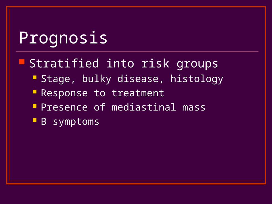
Endemic vs. Sporadic Endemic
African variety Maxilla and mandible Associated with EBV
Sporadic Seen all over
Abdominal organs 20% EBV association

The EBV connection Review of immunology
B cells are infected with EBV T cells (cytotoxic) are involved in the response to EBV
infection Theory
Malaria, and other major infections, causes immunosuppression
Host is unable to generate an adequate T cell response, to keep infection in check
The B cells then proliferate lymphoma

W/U of NHL PE CBC Chem
Electrolytes Liver, renal panels LDH, uric acid
Imaging CT chest and abd Gallium scan
FDG PET scan
Bone marrow CSF exam
Marker of tumor burden, important
determinant of outcome
Measure for tumor lysis
Metastatic w/u

CT scan vs. PET scan

Gallium vs. FDG-PETFDG is tagged glucose
Therapy Chemo only
Surgery only for abdominal emergency Radiation for SVC obstruction, or paraspinal
compression B cell
High dose intensive therapy T cell
Similar to ALL therapy

Complications Tumor related
SVC syndrome Spinal cord compression Pleural and pericardial
effusions Pulmonary embolism Obstructive uropathy Pharyngeal/ airway obs
Metabolic Tumor lysis SIADH Hypo/Hyperglycemia
GI Bleeding, fistulae,
obstruction
Cytokine mediated Cachexia, fever malaise
Hematologic BM infiltration Pancytopenia

Tumor Lysis!!! Evaluate
Phosphorus Uric acid Calcium Potassium
Life threatening emergency Hydrate Alkalinize

EFS for lo and hi stage Burkitts

Quiz Time

What is the genetic problem? NHL B cell?
C-myc
Neuroblastoma? N-myc

Hodgkins vs. Non-Hodgkins Indolent
Hodgkins B symptoms
Hodgkins Abdominal mass
presentation NHL
60% of lymphomas NHL
EBV association BOTH
Reed Sternberg cells Hodgkin
Starry Sky NHL
Painless cervical adenopathy presentation Hodgkin’s
Associated with immune dysfunction BOTH