Embed Size (px)
Citation preview
THE EUROPEAN JOURNALOF
lymphologyand related problems
VOLUME 16 • No. 49 • 2006
INDEXED IN EXCERPTA MEDICA
OFFICIAL ORGANOF THE
EUROPEAN GROUP OF LYMPHOLOGYLATIN-MEDITERRANEAN CHAPTER
OF ISLSOCIETÀ ITALIANA DI LINFANGIOLOGIA
CZECH SOCIETY OF LYMPHOLOGYROMANIAN SOCIETY OF LYMPHOLOGY
SUMMARY
CLINICAL SCIENCES
Original articles
– Translational lymphology and the FöldiklinikMarlys H. Witte, M.D. p. 1
– Imaging findings in pulmonary lymphangiectasiaCarlo Bellini, M.D., P.H.D.; Francesco Boccardo, M.D., P.H.D.;Corradino Campisi, M.D.; Eugenio Bonioli, M.D. p. 10
– Demonstration of Flowave’s effectiveness through lymphoscintigraphyMaurizio Ricci, Simona Paladini. p. 14
– Early or late diagnosis of Lymphedema in our Lymphedema unitIsabel Forner-Cordero, Raquel Navarro-Monsoliu, Jose Muñoz-Langa, Pilar Rel-Monzó p. 19
– Role of lymphoscintigraphy in the indications to microsurgical treatment of peripheral lymphedemasFrancesco Boccardo, Carlo Bellini, Corrado Campisi, Costantino Eretta, D. Pertile,Emanuela Benatti, Mirko Campisi, Giuseppina Talamo, Alberto Macciò, Corradino Campisi p. 24
Calendar
PRI-MED MID-ATLANTIC - NOVEMBER 30 / DECEMBER 2, 2006 - BALTIMORE (MARYLAND)
SOCIETÀ ITALIANA DI FLEBOLOGIA - 5th MEDITERRANEAN CONGRESS OF PHLEBOLOGY - 28 APRIL / 1 MAY, 2007PORTO HELY (GREECE)
XXXIII CONGRESS OF EUROPEAN GROUP OF LYMPHOLOGY - MAY 12-13, 2007, PRAGUE (CZECH REPUBLIC)
CONGRESO ARGENTINO DE FLEBOLOGIA - 23 A 25 DE MAYO DEL 2007 - TUCUMÁN (ARGENTINA)
INTERNATIONAL UNION OF PHLEBOLOGY (UIP) - WORLD CONGRESS CHAPTER MEETING - 18 AL 20 DE JUNIO DE 2007KYOTO (JAPAN)
SOCIETÀ ITALIANA DI FLEBOLINFOLOGIA (SIFL) - V CONGRESSO INTERNAZIONALE DI PHLEBOLOGY - 1-4 LUGLIO 2007CORFÙ
21st INTERNATIONAL CONGRESS OF LYMPHOLOGY - SEPTEMBER 26-30, 2007, SHANGHAI (CHINA)
UNION INTERNATIONALE DE PHLEBOLOGIE (UIP 50) - 16° CONGRESSO MONDIALE - AUGUST 31 / SEPTEMBER 2, 2009GRIMALDI FORUM - PRINCIPATO DI MONACO p. 29
ISSN 0778-5569
00_COVER_VOL_XVI_N_49_2006 23-11-2006 9:47 Pagina 1
1THE EUROPEAN JOURNAL OF LYMPHOLOGY - Vol. XVI - Nr. 49 - 2006
ABSTRACT
The opening of the new wing of the FöldiKlinik in Hinterzarten,Germany, in October 2004, afforded the opportunity to reflect onthe recent advances in molecular lymphology and their potentialimpact on basic research and clinical lymphology. We are now ina position both to expand our delineation of the lymphatic“phenotype” (particularly through non-invasive, multimodalimaging) as well as pinpoint the lymphvascular genotype and itsstructural and functional expression in patients with lymphedema,lymphangiodysplasias, and other lymphatic system disease. Newtherapeutic approaches, in addition to current non-operative andoperative options, are on the horizon, including gene therapy,lymphangioinhibitory and stimulatory drugs, stem cell therapy,and tissue engineering. As we translate these advances from benchto bedside and clinic, more than ever global collaborations andadvanced communication technologies (e.g.,TeleLymphologybased at the University of Arizona Health Sciences Center) areneeded to carry out optimal testing and evaluation of thesealternatives for therapeutic benefit vs. risk and also to assure thewidespread availability of effective modalities throughout theworld community. The newly expanded FöldiKlinik will be apremier hub in this effort.
KEY WORDS: lymphedema, lymphology, lymphvascular phenotype,lymphvascular genomics, translational medicine,telemedicine, FöldiKlinik.
BACKGROUND
What an exciting moment and great honor it was for me to bepresent for the celebration of the opening of the new wing of theFöldiKlink! In the United States, there’s a movement afoot called“translational medicine,” encouraging physicians and basicscientists to come together to expedite the movement of basicscience advances from the “bench to the trench,” i.e., from thelaboratory to the bedside/clinic and community. Whereastranslation may be a new movement for some specialties, it’snothing new for lymphology and lymphologists. Since before theofficial founding of the International Society of Lymphology in1966 and the introduction of the new word “lymphology” for thisdiscipline by visionary Swiss radiologist Alois Rüttimann and hisco-founders, we have recognized that translational biologists need
THE EUROPEAN JOURNALOF
lymphologyand related problems
VOLUME 16 • No. 49 • 2006
INDEXED IN EXCERPTA MEDICA
TRANSLATIONAL LYMPHOLOGY AND THE FÖLDIKLINIK
MARLYS H. WITTE, M.D.
Department of Surgery, University of Arizona, Tucson, Arizona USA
Corresponding author: Marlys H. Witte, MDProfessor of SurgeryUniversity of Arizona1501 N. Campbell Ave., Room 4406P.O. Box 245200Tucson, Arizona 85724-5200e-mail: [email protected]
to work alongside translational physicians and transcend thebarriers of language, geography, and specialization. There’s hardlya lymphologist, exemplified by pioneers such as Ernest Starlingexploring the physiologic principles governing lymph formationand edema or Florence Sabin meticulously dissecting thelymphatic development of the human embryo, who wasn’tlooking, either as basic scientists to implications of their findingsfor clinical practice, or, on the other hand, astute clinicians, likeBritish surgeon John Kinmonth seeking out basic scientists forbetter explanations of the inheritance patterns and dysfunctionallymphographic images he was seeing for the first time in patientswith lymphedema-angiodysplasia syndromes including thefamiliar hereditary forms.
Since the 1960's, the Földis, Michael or Ethel, one or the other orboth together, have framed the boundaries of lymphology withtheir ideas, experiments, teachings, and clinical practice.Publication of the landmark tome by the Hungarian team ofRusznyak, Földi and Szabo, in 1960 (Rusznyák et al., 1967)defined the field, describing lymphostatic disorders, andpresenting some commonalities, themes, and theories of theirpathophysiology based on experimental study and clinicalobservation. This was the moment, in essence, lymphology wasintroduced to the global scientific community. As lymphographersbrought the hidden lymphatic vasculature and lymph nodes intoview, displaying vivid living images of lymphatic disorders, themultidisciplinary International Society of Lymphology took shapeand purpose to provide an international forum and dispel thenotion that the lymphatic system was simply “lymph nodes heldtogether by strings” (Lymphology, 1967-2006; Progress inLymphology, 1967-2006). Again, Michael Földi was there for thecelebration and interchange and soon, Ethel joined him, first as abasic scientist exploring the pathophysiology and potentialtreatments of experimental lymphostatic diseases, and then shemoved from the laboratory into the clinic to join Michael andapply, for the first time, these pathophysiologic concepts todiseases which had previously been approached in a disorderly,haphazard fashion. Their clinic and then a full-fledged hospitalgradually evolved into the world’s premier medical institutiondevoted exclusively to lymphology with associated educationaland certification programs to ensure a coordinated clinical careteam of multidisciplinary experts for these patients. Then in 1994in Hinterzarten, Germany, the Földis, in consultation with the ISL
01-9_WITTE 23-11-2006 9:49 Pagina 1
Executive Committee, created the framework for the firstinternational consensus statement on lymphedema (ISL ExecutiveCommittee, 1995). The publication and dissemination of thisdocument stimulated a vigorous dialogue about the optimalapproach to diagnosis and treatment of lymphedema andestablished international guidelines albeit as a work in progress. Afew years later, in the mid- and late 1990's, molecular lymphologyexploded on the scene with major discoveries in genomics,proteomics, lymphangiogenesis, immunohistochemistry, andsystems biology, bringing with them the potential forrevolutionary advances in clinical lymphology (reviewed in Witteet al., 2003).So, what about lymphology’s “next future” (to borrow thisquaintly English expression from my Italian lymphologiccolleagues)? The expression implies that there is a future now, andthen there is a “next future”, one that I’d like to highlight for thenext few minutes. The opening of the new wing of the FöldiKlinikprovided an opportune moment to reflect and also to set the stagefor a prototype collaborative network of lymphology centers inthis next future of clinical care, research, and education, i.e.,translational lymphology, so that advances at the laboratory benchcan move rapidly yet judiciously to the bedside and spreadthroughout the global community.
LYMPHVASCULAR PHENOTYPES
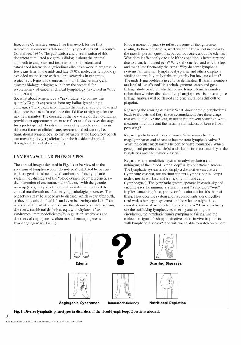
The clinical images depicted in Fig. 1 can be viewed as thespectrum of lymphvascular “phenotypes” exhibited by patientswith congenital and acquired disturbances of the lymphaticsystem, i.e., disorders of the “blood-lymph loop.” Epigenetics –the interaction of environmental influences with the geneticmakeup (the genotype) of these individuals has produced theclinical manifestations of underlying pathologic processes. Thephenotypes may be secondary to diseases which occur after birth,or they may arise in fetal life and even be “embryonic lethal” andnever seen. But what we do see are the edematous states, scarringdisorders, nutritional depletion, e.g., with chylous refluxsyndromes, immunodeficiency/dysregulation syndromes anddisorders of angiogenesis, often mixed hemangiogenesis-lymphangiogenesis (Fig. 1).
First, a moment’s pause to reflect on some of the ignorancerelating to these conditions, what we don’t know, not necessarilythe most important questions, but curious ones, about the edemas:Why does it affect only one side if the condition is hereditary anddue to a single mutated gene? Why only one leg, and why the leg,and much less frequently the arms? Why do some lymphaticsystems fail with this lymphatic dysplasia, and others display asimilar abnormality on lymphscintigraphy but have no edema?The underlying problems need to be delineated. If family membersare labeled “unaffected” in a whole genome search and genelinkage study based on whether or not lymphedema is manifestrather than whether disordered lymphangiogenesis is present, genelinkage analysis will be flawed and gene mutations difficult topinpoint.
Regarding the scarring diseases: What about chronic lymphedemaleads to fibrosis and fatty tissue accumulation? Are there drugsthat would dissolve the scar, or better yet, prevent scarring? Whatmeasures might prevent the initial lymphostasis, or keep it frompersisting?
Regarding chylous reflux syndromes: What events lead tomegalymphatics and absent or incompetent lymphatic valves?What molecular mechanisms lie behind valve formation? Whichgene(s) and protein cascade(s) underlie intrinsic contractility of thelymphatics and pacemaker activity?
Regarding immunodeficiency/immunodysregulation andunhinging of the “blood-lymph loop” in lymphostatic disorders:The lymphatic system is not simply a distinctive vasculature(lymphatic vessels), nor its fluid content (lymph), nor its lymphnodes, nor its working and trafficking immune cells(lymphocytes). The lymphatic system operates in continuity andencompasses the immune system. It is not “lymphoid”; “-oid”implies something false, phony, or faux about it but it’s the realthing. How does the system and its components work together(and with other organ systems), and how better might thesecomplex system dynamics be observed in vivo? Can we actuallysee the trafficking lymphocytes entering and exiting thecirculation, the lymphatic trunks pumping or failing, and themolecular signals flashing distinctive colors in vivo in patientswith lymphatic diseases? And will we be able to watch on remote
2THE EUROPEAN JOURNAL OF LYMPHOLOGY - Vol. XVI - Nr. 49 - 2006
Fig. 1. Diverse lymphatic phenotypes in disorders of the blood-lymph loop. Questions abound.
01-9_WITTE 23-11-2006 9:49 Pagina 2
microsensor printouts as voluminous data is generated on theeffects of designer drugs or as robotic surgeons normalize thepathologic process?
Regarding the angiogenic syndromes: What about lymphostasispromotes overgrowth of multiple cell types leading to fibrous scar,fat deposits, and also proliferation of blood vessels and lymphaticsand how does this pathologic process relate to angiomas andvascular birthmark syndromes? When and how doeslymphangiogenesis escape control and become frankly neoplastic?And how soon, as Cristobal Papendieck implores us, will anti-angiotumorigenesis agents be available to prevent or reverse theprocess and involute those disfiguring lymphangiomas andhemangiomas with a non-toxic pill rather than an extensiveendangering operation?
Lymphologists tend to be a pure breed but often with otherpastimes. Though respectful of the venous system and its links tothe lymphatic system, they are usually neither“lymphophlebologists” nor “phlebolymphologists,” thoughoccasionally lymphologist- something else (e.g., -radiologist,surgeon, dermatologist, or internist, etc.). Lymphedema is anexternal or internal manifestation of excess tissue fluidaccumulation resulting from low-output lymph circulatory failure.But it is not a disease entity. The disease is the underlyinglymphvascular system abnormality which often but not alwaysculminates in lymphedema. Fifty years ago the expression“cardiogenic edema” was still commonly used signifying that wedidn’t know the underlying heart diseases well enough on apathophysiological or molecular level to define the cause. Now wecan often pinpoint the origin of the heart disease and the specificcause of the heart failure and thereby the pathomechanism of“cardiogenic edema,” and so the latter term has been abandoned.In the same way, we are beginning to talk about the underlyinglymphatic disturbance, even the causative gene mutations forlymphatic maldevelopment and “lymphologenic edema.” Forexample, the transcription factor FOXC2 is mutated in
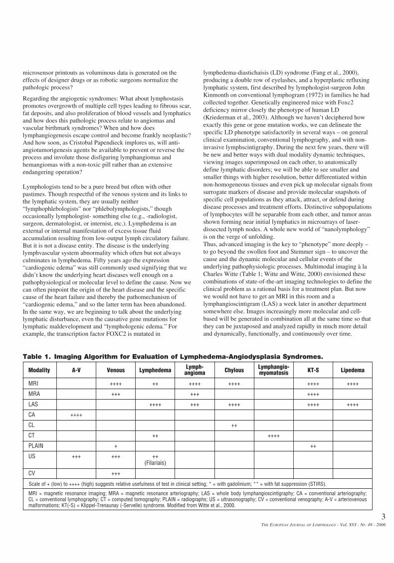
lymphedema-diastichaisis (LD) syndrome (Fang et al., 2000),producing a double row of eyelashes, and a hyperplastic refluxinglymphatic system, first described by lymphologist-surgeon JohnKinmonth on conventional lymphogram (1972) in families he hadcollected together. Genetically engineered mice with Foxc2deficiency mirror closely the phenotype of human LD(Kriederman et al., 2003). Although we haven’t deciphered howexactly this gene or gene mutation works, we can delineate thespecific LD phenotype satisfactorily in several ways – on generalclinical examination, conventional lymphography, and with non-invasive lymphscintigraphy. During the next few years, there willbe new and better ways with dual modality dynamic techniques,viewing images superimposed on each other, to anatomicallydefine lymphatic disorders; we will be able to see smaller andsmaller things with higher resolution, better differentiated withinnon-homogeneous tissues and even pick up molecular signals fromsurrogate markers of disease and provide molecular snapshots ofspecific cell populations as they attack, attract, or defend duringdisease processes and treatment efforts. Distinctive subpopulationsof lymphocytes will be separable from each other, and tumor areasshown forming near initial lymphatics in microarrays of laser-dissected lymph nodes. A whole new world of “nanolymphology”is on the verge of unfolding.Thus, advanced imaging is the key to “phenotype” more deeply –to go beyond the swollen foot and Stemmer sign – to uncover thecause and the dynamic molecular and cellular events of theunderlying pathophysiologic processes. Multimodal imaging à laCharles Witte (Table 1; Witte and Witte, 2000) envisioned thesecombinations of state-of-the-art imaging technologies to define theclinical problem as a rational basis for a treatment plan. But nowwe would not have to get an MRI in this room and alymphangioscintigram (LAS) a week later in another departmentsomewhere else. Images increasingly more molecular and cell-based will be generated in combination all at the same time so thatthey can be juxtaposed and analyzed rapidly in much more detailand dynamically, functionally, and continuously over time.
3THE EUROPEAN JOURNAL OF LYMPHOLOGY - Vol. XVI - Nr. 49 - 2006
Table 1. Imaging Algorithm for Evaluation of Lymphedema-Angiodysplasia Syndromes.
Modality A-V Venous Lymphedema Lymph- Chylous Lymphangio- KT-S Lipedemaangioma myomatosis
MRI ++++ ++ ++++ ++++ ++++ ++++
MRA +++ +++ ++++
LAS ++++ +++ ++++ ++++ ++++
CA ++++
CL ++
CT ++ ++++
PLAIN + ++
US +++ +++ ++(Filariais)
CV +++
Scale of + (low) to ++++ (high) suggests relative usefulness of test in clinical setting; * = with gadolinium; ** = with fat suppression (STIRS).
MRI = magnetic resonance imaging; MRA = magnetic resonance arteriography; LAS = whole body lymphangioscintigraphy; CA = conventional arteriography; CL = conventional lymphography; CT = computed tomography; PLAIN = radiographs; US = ultrasonography; CV = conventional venography; A-V = arteriovenousmalformations; KT(-S) = Klippel-Trenaunay (-Servelle) syndrome. Modified from Witte et al., 2000.
01-9_WITTE 23-11-2006 9:49 Pagina 3
LYMPHVASCULAR GENOTYPES
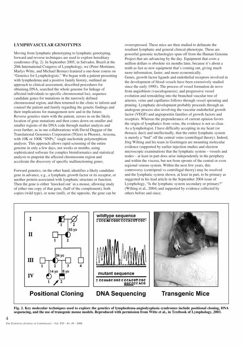
Moving from lymphatic phenotyping to lymphatic genotyping,forward and reverse techniques are used to explore hereditarysyndromes (Fig. 2). In September 2005, in Salvador, Brazil at the20th International Congress of Lymphology, we (Peter Mortimer,Marlys Witte, and Michael Bernas) featured a one-hour course on“Genetics for Lymphologists.” We began with a patient presentingwith lymphedema and a positive family history, outlined anapproach to clinical assessment, described procedures forobtaining DNA, searched the whole genome for linkage ofaffected individuals to specific chromosomal loci, sequencecandidate genes for mutations in the narrowly definedchromosomal region, and then returned to the clinic to inform andcounsel the patient and family regarding the genetic findings andtheir implications for management now and in the future. Reverse genetics starts with the patient, zeroes in on the likelylocation of gene mutations and then cones down on smaller andsmaller regions of the DNA code through marker analysis andeven further, as in our collaborations with David Duggan of theTranslational Genomics Corporation (TGen) in Phoenix, Arizona,with 10K or 100K “SNPs,” single nucleotide polymorphismanalysis. This approach allows rapid screening of the entiregenome in only a few days, not weeks or months, usingsophisticated software for complex bioinformatics and statisticalanalysis to pinpoint the affected chromosome region andaccelerate the discovery of specific malfunctioning genes.
Forward genetics, on the other hand, identifies a likely candidategene in advance, e.g., a lymphatic growth factor or its receptor, oranother protein associated with lymphatic structure or function.Then the gene is either ‘knocked out’ in a mouse, allowing studyof either one copy of that gene, (half of the complement), bothcopies (wild type), or none (null), or the opposite, the gene can be
overexpressed. These mice are then studied to delineate theresultant lymphatic and general clinical phenotype. These arepowerful genomic technologies spun off from the Human GenomeProject that are advancing by the day. Equipment that costs amillion dollars is obsolete six months later, because it’s about atenth as fast as new equipment that’s coming out, giving muchmore information, faster, and more economically. Genes, growth factor ligands and endothelial receptors involved inthe development of blood vessels have been extensively studiedsince the early 1990's. The process of vessel formation de novofrom angioblasts (vasculogenesis), and progressive vesselevolution and remodeling into the branched vascular tree ofarteries, veins and capillaries follows through vessel sprouting andpruning. Lymphatic development probably proceeds through ananalogous process also involving the vascular endothelial growthfactor (VEGF) and angiopoeitin families of growth factors andreceptors. Whereas the preponderance of current opinion favorsthe origin of lymphatics from veins, the evidence is not so clear.As a lymphologist, I have difficulty accepting in my heart (orthoracic duct) and intellectually, that the entire lymphatic systemis merely a “bud” off the central veins (centrifugal theory). Indeed,Jörg Wilting and his team in Goettingen are mounting molecularevidence (supported by earlier injection studies and electronmicroscopic examination) that the lymphatic system – vessels andnodes – at least in part does arise independently in the peripheryand within the viscera, but not from sprouts of the central or evenregional venous system. Within the next few years, thiscontroversy (centripetal vs centrifugal theory) may be resolvedand the lymphatic system shown, at least in part, to be primary assuggested in his lead article in the September 2004 issue ofLymphology, “Is the lymphatic system secondary or primary?”(Wilting et al., 2004) and supported by evidence collected byothers before and since.
4THE EUROPEAN JOURNAL OF LYMPHOLOGY - Vol. XVI - Nr. 49 - 2006
Fig. 2. Key molecular techniques used to explore the genetics of lymphedema-angiodysplasia syndromes include positional cloning, DNAsequencing, and the use of transgenic mouse models. Reproduced with permission from Witte et al., in Textbook of Lymphology, 2003.
01-9_WITTE 23-11-2006 9:49 Pagina 4
The specific growth factors, receptors, and transcription factorsthat have been described to influence lymphatic growth anddevelopment are shown in Table 2 (modified from Witte et al.,2003). Some are not unexpected, like the members of the VEGFand angiopoeitin families of growth factors and theircorresponding endothelial receptors. But there are alsotranscription factors, such as FOXC2, SOX18, and PROX1. Whowould have imagined that the fruit fly “forkhead” gene Foxc2would have anything to do with the human lymphatic system andfurthermore cause, when defective, the very specific clinicalsyndrome of lymphedema-diastichaisis? There will be moresurprises, and any all-encompassing scheme of lymphaticdevelopment is likely to be extensively revisited over the next fewyears as more discoveries are made and the interacting pathwaysand feedback loops prove to be more complex than firstenvisioned. The Foxc2 haploinsufficient (+/-) mouse (Kriederman et al., 2003)with just one allele knocked out phenotypically resembles thepatient I encountered in my first visit in the early days of theFöldiKlinik several decades ago. She had a double row ofeyelashes and pubertal onset lymphedema and a family history ofthe condition. The mice, like the lymphedema-distichiasis patientswith FOXC2 mutations (Fang et al., 2000), invariably show adouble row of eyelashes and often exhibit a hyperplastic refluxinglymphatic system resembling Kinmonth’s distinctivelymphograms (1972) with occasional limb edema along with avariety of other ocular abnormalities like their human counterparts.A smaller number of affected humans exhibit cleft palate andtetralogy of Fallot. When Foxc2 is completely knocked out –homozygous null mouse (-/-) – they die before birth displayingboth cleft palate and combined aortic arch and cardiacinterventricular septal defects, closely resembling tetralogy ofFallot (Iida et al., 1997); they also exhibit defective lymphaticvalvular formation (Petrova et al., 2004) as documented earlier byextensive visceral lymph reflux seen in the Foxc2haploinsufficient mouse (Kriederman et al., 2003). Curiously, theFoxc2 overexpressing mouse (with an adipocyte promoter) alsoexhibits lymphatic system hyperplasia like the haploinsufficientmouse but without distichiasis or other ocular abnormalities (Noonet al., 2006) suggesting that Foxc2 gene dose imbalance may beinvolved in the distinctive lymphatic phenotype.
On the other hand, the angiopoietin-2 transgenic mousephenotypically resembles a variety of human lymphedemasyndromes. When the gene for vascular growth factor angiopoeitin2, known to be involved in the modeling and remodeling of bloodvessels, has been totally knocked out (-/-), we have found nolymphatics or lymph nodes (or only very rudimentary ones) in thelower limbs, and the upper limb and cervical lymphatic system isalso hypoplastic or aplastic (Gale et al., 2002). Lymphedema ofthe lower limbs and chylous ascites and chylothorax are typicallypresent. But no reported human lymphedema syndromes havebeen associated with angiopoietin-2 mutations, i.e., this is agenetically engineered “mouse syndrome” looking for a humangenetic counterpart.
When the human lymphvascular genome map (Fig. 3), as we knowit today, is laid out with the chromosomes lined up in pairs as akaryotype, potential genes or loci pertaining to lymphaticdevelopment are found on most chromosomes. Beginning in 1998,three publications (Ferrell et al., 1998; Witte et al., 1998; Evans etal., 1999) including one in Lymphology, linked a form of Milroysyndrome to the distal arm (q34-35) of chromosome 5, andmultiple mutations in VEGFR3, the receptor for VEGF-C, havebeen reported (Karkkainen, 2000). Subsequently, two otherlymphangiogenesis genes have been identified in other hereditarylymphedema syndromes. On chromosome 16, is the causativeFOXC2 gene for lymphedema distichiasis discovered in aUniversity of Arizona-University of Michigan collaboration (Fanget al., 2000). On chromosome 20 is the gene for transcriptionfactor SOX18 associated with lymphedema-hypotrichosis-telangiectasia syndrome (Irrthum et al., 2003). In addition, otherchromosome locations but not the specific genes have been linked,that is, we know where the genes are, but not which ones and howthey are altered.Chromosomal aneuploidy – extra or fewer chromosomes than thenormal diploid complement of 46 + X,X or X,Y – are commonlyassociated with lymphatic maldevelopment (Northup et al., 2003).Aneuploid embryos often die early in fetal life even beforelymphatic development takes place, but some such as trisomy 21and Klinefelter syndrome can even reach adulthood.Rearrangements or “chromosomal mutations,” where a piece ofchromosome is deleted, added, or translocated, involve groups of
5THE EUROPEAN JOURNAL OF LYMPHOLOGY - Vol. XVI - Nr. 49 - 2006
Table 2. Vascular Growth Factors, Receptors, and Transcription Factors in Vascular, IncludingLymphatic, Development
Vascular Growth Factors
PIGF VEGF-A VEGF-B VEGF-C* VEGF-D* VEGF-E Ang-1* Ang-2* Ephrin-B2*
VEGFR-1/Flt-1 ◆ ◆ ◆
VEGFR-2/Flk-1 ◆ ◆ ◆
VEGFR-3/Flt-4* ◆ ◆
Neuropilin-1 ◆ ◆ ◆ ◆
Neuropilin-2* ◆ ◆ ◆
Tie-1 ◆
Tie-2* ◆ ◆
Eph-B4 ◆
◆ Corresponding growth factor-receptor combinations. *Implicated specifically in lymphatic development (bold and italic text).
01-9_WITTE 23-11-2006 9:49 Pagina 5
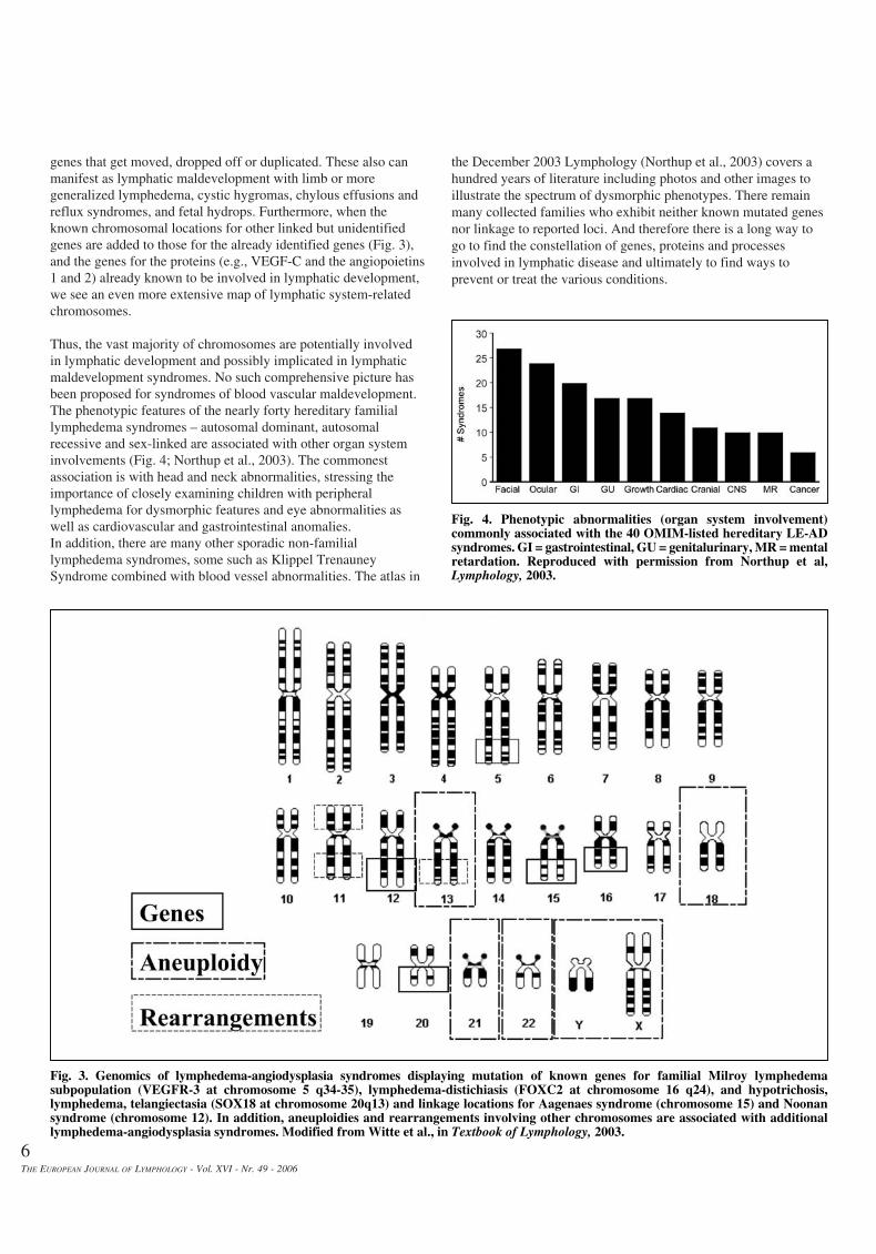
genes that get moved, dropped off or duplicated. These also canmanifest as lymphatic maldevelopment with limb or moregeneralized lymphedema, cystic hygromas, chylous effusions andreflux syndromes, and fetal hydrops. Furthermore, when theknown chromosomal locations for other linked but unidentifiedgenes are added to those for the already identified genes (Fig. 3),and the genes for the proteins (e.g., VEGF-C and the angiopoietins1 and 2) already known to be involved in lymphatic development,we see an even more extensive map of lymphatic system-relatedchromosomes.
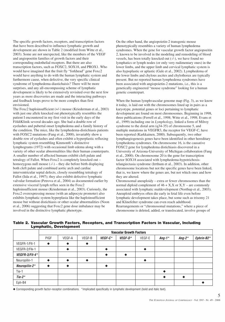
Thus, the vast majority of chromosomes are potentially involvedin lymphatic development and possibly implicated in lymphaticmaldevelopment syndromes. No such comprehensive picture hasbeen proposed for syndromes of blood vascular maldevelopment.The phenotypic features of the nearly forty hereditary familiallymphedema syndromes – autosomal dominant, autosomalrecessive and sex-linked are associated with other organ systeminvolvements (Fig. 4; Northup et al., 2003). The commonestassociation is with head and neck abnormalities, stressing theimportance of closely examining children with peripherallymphedema for dysmorphic features and eye abnormalities aswell as cardiovascular and gastrointestinal anomalies. In addition, there are many other sporadic non-familiallymphedema syndromes, some such as Klippel TrenauneySyndrome combined with blood vessel abnormalities. The atlas in
the December 2003 Lymphology (Northup et al., 2003) covers ahundred years of literature including photos and other images toillustrate the spectrum of dysmorphic phenotypes. There remainmany collected families who exhibit neither known mutated genesnor linkage to reported loci. And therefore there is a long way togo to find the constellation of genes, proteins and processesinvolved in lymphatic disease and ultimately to find ways toprevent or treat the various conditions.
6THE EUROPEAN JOURNAL OF LYMPHOLOGY - Vol. XVI - Nr. 49 - 2006
Fig. 4. Phenotypic abnormalities (organ system involvement)commonly associated with the 40 OMIM-listed hereditary LE-ADsyndromes. GI = gastrointestinal, GU = genitalurinary, MR = mentalretardation. Reproduced with permission from Northup et al,Lymphology, 2003.
Fig. 3. Genomics of lymphedema-angiodysplasia syndromes displaying mutation of known genes for familial Milroy lymphedemasubpopulation (VEGFR-3 at chromosome 5 q34-35), lymphedema-distichiasis (FOXC2 at chromosome 16 q24), and hypotrichosis,lymphedema, telangiectasia (SOX18 at chromosome 20q13) and linkage locations for Aagenaes syndrome (chromosome 15) and Noonansyndrome (chromosome 12). In addition, aneuploidies and rearrangements involving other chromosomes are associated with additionallymphedema-angiodysplasia syndromes. Modified from Witte et al., in Textbook of Lymphology, 2003.
01-9_WITTE 23-11-2006 9:49 Pagina 6
TRANSLATION TO CLINICAL LYMPHOLOGY
Regarding treatments: How can or do we approach swellingdiseases, scarring diseases, immunodeficiencies, nutritionaldepletion, and angiogenic syndromes resulting from disruption ofthe blood lymph loop? Such disorders often have no currenttreatments, operative or non-operative. Others, such as the edemas,immunodeficiencies, nutritional depletion can be handled but notoptimally. Prevention is a distant goal. Right now, the cornerstoneremains the “Földi Method”, although Michael Földi assures methere is no such thing. It’s just tailoring the treatment, byunderstanding the patient, evaluating through multimodal imaging,and diagnosing and working through each problem, whether achylous reflux syndrome or Cristobal Papendieck’s specialty – themassive, disfiguring lymphangioma-hemangioma syndromes.
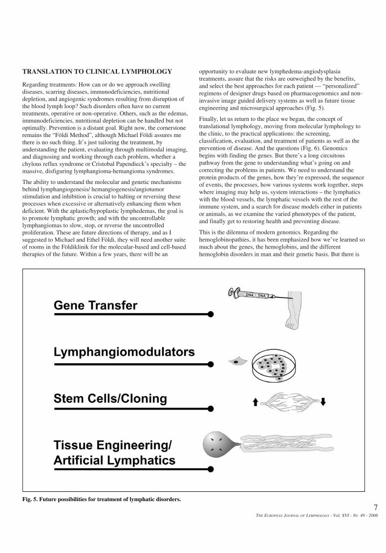
The ability to understand the molecular and genetic mechanismsbehind lymphangiogenesis/ hemangiogenesis/angiotumorstimulation and inhibition is crucial to halting or reversing theseprocesses when excessive or alternatively enhancing them whendeficient. With the aplastic/hypoplastic lymphedemas, the goal isto promote lymphatic growth; and with the uncontrollablelymphangiomas to slow, stop, or reverse the uncontrolledproliferation. These are future directions of therapy, and as Isuggested to Michael and Ethel Földi, they will need another suiteof rooms in the Földiklinik for the molecular-based and cell-basedtherapies of the future. Within a few years, there will be an
opportunity to evaluate new lymphedema-angiodysplasiatreatments, assure that the risks are outweighed by the benefits,and select the best approaches for each patient — “personalized”regimens of designer drugs based on pharmacogenomics and non-invasive image guided delivery systems as well as future tissueengineering and microsurgical approaches (Fig. 5).
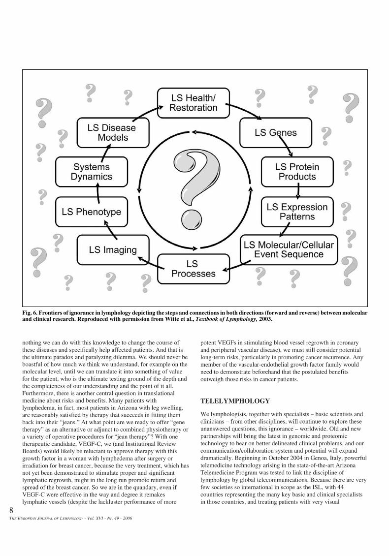
Finally, let us return to the place we began, the concept oftranslational lymphology, moving from molecular lymphology tothe clinic, to the practical applications: the screening,classification, evaluation, and treatment of patients as well as theprevention of disease. And the questions (Fig. 6). Genomicsbegins with finding the genes. But there’s a long circuitouspathway from the gene to understanding what’s going on andcorrecting the problems in patients. We need to understand theprotein products of the genes, how they’re expressed, the sequenceof events, the processes, how various systems work together, stepswhere imaging may help us, system interactions – the lymphaticswith the blood vessels, the lymphatic vessels with the rest of theimmune system, and a search for disease models either in patientsor animals, as we examine the varied phenotypes of the patient,and finally get to restoring health and preventing disease.
This is the dilemma of modern genomics. Regarding thehemoglobinopathies, it has been emphasized how we’ve learned somuch about the genes, the hemoglobins, and the differenthemoglobin disorders in man and their genetic basis. But there is
7THE EUROPEAN JOURNAL OF LYMPHOLOGY - Vol. XVI - Nr. 49 - 2006
Fig. 5. Future possibilities for treatment of lymphatic disorders.
01-9_WITTE 23-11-2006 9:49 Pagina 7
nothing we can do with this knowledge to change the course ofthese diseases and specifically help affected patients. And that isthe ultimate paradox and paralyzing dilemma. We should never beboastful of how much we think we understand, for example on themolecular level, until we can translate it into something of valuefor the patient, who is the ultimate testing ground of the depth andthe completeness of our understanding and the point of it all. Furthermore, there is another central question in translationalmedicine about risks and benefits. Many patients withlymphedema, in fact, most patients in Arizona with leg swelling,are reasonably satisfied by therapy that succeeds in fitting themback into their “jeans.” At what point are we ready to offer “genetherapy” as an alternative or adjunct to combined physiotherapy ora variety of operative procedures for “jean therapy”? With onetherapeutic candidate, VEGF-C, we (and Institutional ReviewBoards) would likely be reluctant to approve therapy with thisgrowth factor in a woman with lymphedema after surgery orirradiation for breast cancer, because the very treatment, which hasnot yet been demonstrated to stimulate proper and significantlymphatic regrowth, might in the long run promote return andspread of the breast cancer. So we are in the quandary, even ifVEGF-C were effective in the way and degree it remakeslymphatic vessels (despite the lackluster performance of more
potent VEGFs in stimulating blood vessel regrowth in coronaryand peripheral vascular disease), we must still consider potentiallong-term risks, particularly in promoting cancer recurrence. Anymember of the vascular-endothelial growth factor family wouldneed to demonstrate beforehand that the postulated benefitsoutweigh those risks in cancer patients.
TELELYMPHOLOGY
We lymphologists, together with specialists – basic scientists andclinicians – from other disciplines, will continue to explore theseunanswered questions, this ignorance – worldwide. Old and newpartnerships will bring the latest in genomic and proteomictechnology to bear on better delineated clinical problems, and ourcommunication/collaboration system and potential will expanddramatically. Beginning in October 2004 in Genoa, Italy, powerfultelemedicine technology arising in the state-of-the-art ArizonaTelemedicine Program was tested to link the discipline oflymphology by global telecommunications. Because there are veryfew societies so international in scope as the ISL, with 44countries representing the many key basic and clinical specialistsin those countries, and treating patients with very visual
8THE EUROPEAN JOURNAL OF LYMPHOLOGY - Vol. XVI - Nr. 49 - 2006
Fig. 6. Frontiers of ignorance in lymphology depicting the steps and connections in both directions (forward and reverse) between molecularand clinical research. Reproduced with permission from Witte et al., Textbook of Lymphology, 2003.
01-9_WITTE 23-11-2006 9:49 Pagina 8
abnormalities that can be transmitted on camera, we could actuallybe sharing our questions and expertise simultaneously all over theworld. We could reach remote areas, on Native Americanreservations, in villages in rural India and on tropical islands.Homes, operating rooms, and laboratories can also be as accessiblefrom the Tucson Telemedicine hub as was Corradino Campisi’sand Francesco Boccardo’s operating room in Genoa, where thesurgeons were viewing a lymphatic-venous shunt procedure liveand asking questions in an interactive fashion. So, the use ofTelemedicine technology for TeleLymphology, for clinical care,for teleconsultation, for educational purposes, researchcollaboration and planning clearly is an exciting path of the “nextfuture.” The newly expanded Földiklinik will be a premier hub inthis new TeleLymphology global alliance within the InternationalSociety of Lymphology.
REFERENCES
Evans A.L., Brice G., Sotirova V., Mortimer P., Beninson J.,Burnand K., Rosbotham J., Child A., Sarfarazi M. (1999):Mapping of primary congenital lymphedema to the 5q35.3region. Am. J. Hum. Genet., 64: 547-555.
Fang J.M., Dagenais, S.L., Erickson, R.P., Arlt, M.F., Glynn, M.W.,Gorski, J.L., Seaver, L.H., and Glover, T.W. (2000): Mutationsin FOXC2 (MFH-1), a forkhead family transcription factor, areresponsible for the hereditary lymphedema-distichiasissyndrome. Am. J. Hum. Genet., 67: 1382-1388.
Ferrell R.E., Levinson K.L., Esman J.H., Kimak M.A., LawrenceE.C., Barmada M.M., Finegold D.N. (1998): Hereditarylymphedema: Evidence for linkage and genetic heterogeneity.Hum. Mol. Genet., 7: 2073-2078.
Gale N.W., Thurston G., Hackett S.F., Renard R., Wang Q.,McClain J., Martin C., Witte C., Witte M.H., Suri C.,Campochiaro P.A., Wiegand S.J., Yancopoulos, G.D. (2002):Angiopoietin-2 is required for postnatal angiogenesis andlymphatic patterning, and only the latter role is rescued byangiopoietin-1. Developmental Cell, 3: 411-423.
Iida K., Koseki H., Kakinuma H., Kato N., Mizutani-Koseki Y.,Ohuchi H., Yoshioka H., Noji S., Kawamura K., Kataoka Y.,Ueno F., Taniguchi M., Yoshida N., Sugiyama T., Miura N.(1997): Essential roles of the winged helix transcription factorMFH-1 in aortic arch patterning and skeletogenesis.Development, 124: 4627-4638.
International Society of Lymphology Executive Committee (1995):The Diagnosis and Treatment of Peripheral Lymphedema.Lymphology, 28: 113-117.
Irrthum A., Devriend K., Chitayat D., Matthijs G., Glade C.,Steijlen P.M., Fryns J.-P., Van Steensel A.M., Vikkula M.(2003): Mutations in the transcription factor gene SOX18underlie recessive and dominant forms of hypotrichosis-lymphedema-telangiectasis. Am. J. Hum. Genet., 72: 1470-1478.
Karkkainen M.J., Ferrell R.E., Lawrence E.C., Kimak M.A.,Levinson K.L., McTigue M.A., Alitalo K., Finegold D.N.(2000): Missense mutations interfere with vascular endothelialgrowth factor receptor-3 signaling in primary lymphedema.Nature Genet., 25: 153-159.
Kinmonth, J.B. (Ed.), (1972): The Lymphatics: Diseases,Lymphography and Surgery. Edward Arnold, London.
Kriederman B.M., Myloyde T.L., Witte M.H., Dagenais S.L., WitteC.L., Rennels M., Bernas M.J., Lynch M.T., Erickson R.P.,Caulder M.S., Miura N., Jackson D., Brooks B.P., Glover T.W.(2003): Foxc2 haploinsufficient mice are a model for humanautosomal dominant lymphedema-distichiasis syndrome.Human Molecular Genetics, 12: 1179-1185.
Lymphology (1967-2006) Vols. 1-39.
Noon, A., Hunter R.J., Witte M.H., Kriederman B, Bernas M,Rennels M, Percy D, Enerback S, Erickson R.P. (2006):Comparative lymphatic ocular and metabolic phenotypes ofFoxc2 haploinsufficient and aP2-FOXC2 transgenic mice.Lymphology, 39: 84-94.
Northup K.A., Witte M.H., Witte C.L. (2003): Syndromicclassification of hereditary lymphedema. Lymphology, 36: 162-189.
Petrova T.V., Karpanen T., Norrmén C., Mellor R., Tamakoshi T.,Finegold D., Ferrell R., Kerjaschki D., Mortimer P., Ylä-Herttuala S., Miura N., Alitalo K. (2004): Defective valves andabnormal mural cell recruitment underlie lymphatic vascularfailure in lymphedema distichiasis. Nature Med, 10: 974-981.
Progress in Lymphology I-XX (1967-2006).
Rusznyák I., Földi M., Szabo G. (1960): Lymphatics and LymphCirculation. Pergamon Press, New York: Oxford, 853 p.
Wilting J., Papoutsi M., Becker J. (2004): The lymphatic vascularsystem: Secondary or primary? Lymphology, 37: 98-106.
Witte C.L., Witte M.H. (2000): An imaging evaluation ofangiodysplasia syndromes. Lymphology, 33: 158-166.
Witte M.H., Bernas M.J., Northup K.A., Witte C.L. (2003):Molecular lymphology and genetics of lymphedema-angiodysplasia syndromes. In: Földi M., Földi E., Kubik S.(Eds). “Textbook of Lymphology”. Urban & Fischer Verlag,München, Germany (English text revised by Biotext, LLC, SanFrancisco), 6th edition, Chapter 16, pp. 471-493.
Witte M.H., Erickson R., Bernas M., Andrade M., Reiser F., ConlonW., Hoyme H.E., Witte C.L. (1998): Phenotypic and genotypicheterogeneity in familial Milroy lymphedema. Lymphology, 31:145-155.
9THE EUROPEAN JOURNAL OF LYMPHOLOGY - Vol. XVI - Nr. 49 - 2006
01-9_WITTE 23-11-2006 9:49 Pagina 9
10THE EUROPEAN JOURNAL OF LYMPHOLOGY - Vol. XVI - Nr. 49 - 2006
ABSTRACT
We present the imaging findings of 4 newborns and 1 childaffected by pulmonary lymphangiectasis (PL). Congenital PL is arare developmental disorder involving the lung. Pulmonarylymphangiectasis is characterized by pulmonary subpleural,interlobar, perivascular, and peribronchial lymphatic dilatation.Both frequency and etiology are unknown. At birth, PL presentswith severe respiratory distress, tachypnea, and cyanosis, with avery high mortality rate at or within a few hours of birth. Bilaterallung reticular appearance, peribronchial cuffing, and bilateralpleural effusions on radiographic chest evaluation are verysuggestive of PL. Bilateral septal and peribronchial interstitialthickening are well evidentiated by high-resolution CT.Radiological studies, together with history and clinical data maylead to a diagnosis of PL in most cases. Lymphoscintigraphy,bronchoscopic and pleural effusion evaluation, and if necessary,lung biopsy are useful tools for confirming PL diagnosis.
KEY WORDS: Pulmonary Lymphangiectasia; Imaging findings;Newborn; Child.
INTRODUCTION
Pulmonary lymphangiectasis (PL) is a rare developmental disorderinvolving the lung, and is characterized by pulmonary subpleural,interlobar, perivascular, and peribronchial lymphatic dilatation. On the basis of an improved characterization of the clinicalpresentation, and on recent, notable advances in neonatal intensivecare, it has been suggested that PL can be divided into two majorcategories, defined as primary and secondary PL (1, 2). When presenting as a primary pulmonary developmental defect,PL may be caused by a congenital defect in the primary
THE EUROPEAN JOURNALOF
lymphologyand related problems
VOLUME 16 • No. 49 • 2006
INDEXED IN EXCERPTA MEDICA
IMAGING FINDINGS IN PULMONARY LYMPHANGIECTASIA
CARLO BELLINI1, MD, PHD; FRANCESCO BOCCARDO2, MD, PHDCORRADINO CAMPISI2, MD; EUGENIO BONIOLI3, MD1 Service of Neonatal Pathology, Department of Pediatrics,
University of Genoa, Institute G. Gaslini, Genoa, Italy2 Department of Surgery, Unit of Lymphatic Surgery,
S. Martino Hospital, University of Genoa, Italy3 Department of Pediatrics, University of Genoa, Institute G. Gaslini, Genoa, Italy
Correspondence to: Dott. Carlo BelliniDipartimento di PediatriaUniversità di GenovaIstituto G. Gaslini,Largo G. Gaslini, 516147 GenovaItalyTel.: +39 10 5636762Fax: +39 10 3770675E-mail: [email protected]
development of the lung, or may represent a localized expressionof more generalized lymphatic involvement. When it is part of generalized lymphatic dysplasia, PL presentswith dilated pulmonary lymphatics truncal lymphangiectasia,which is usually associated with peripheral lymphedema.Hemihypertrophy (which is rare in infants and young children) anddiffuse angiomatosis, in which the bone represents the mostcommon site of involvement, can also be observed. Both of theseforms may be encompassed by the definition of primary PL.Cardiovascular and lymphatic obstructive forms make up thesecondary PL group. Hypoplastic left heart syndrome, pulmonaryvein atresia, congenital mitral stenosis, cor triatum, and thoracicduct agenesis are the most likely causes of secondary PL.Although PL is nearly always fatal during the neonatal period, therecent improvements in mechanical ventilation of severelyaffected newborns have made survival more frequent. We reportour experience in the radiological diagnosis of PL, both innewborns and in older children.
PATIENTS AND RESULTS
Four newborns and 1 child affected by PL were evaluated byimaging between January 2000, and December 2003, at ourinstitute. Newborns who presented at least one sign among thefollowing at birth were examined for the possible presence ofpulmonary lymphangiectasia: non-immune hydrops fetalis,hydrothorax, hydropericardium, ascites, lymphedema of the limbs,lymphedema of the genitalia.Children who presented respiratory difficulties of varying degreewith a relapsing course, associated with recurrent cough, wheeze,or inspiratory crackle were examined for the possible presence ofpulmonary lymphangiectasia. Diagnostic investigation included
10-13_BELLINI 23-11-2006 9:52 Pagina 10
11THE EUROPEAN JOURNAL OF LYMPHOLOGY - Vol. XVI - Nr. 49 - 2006
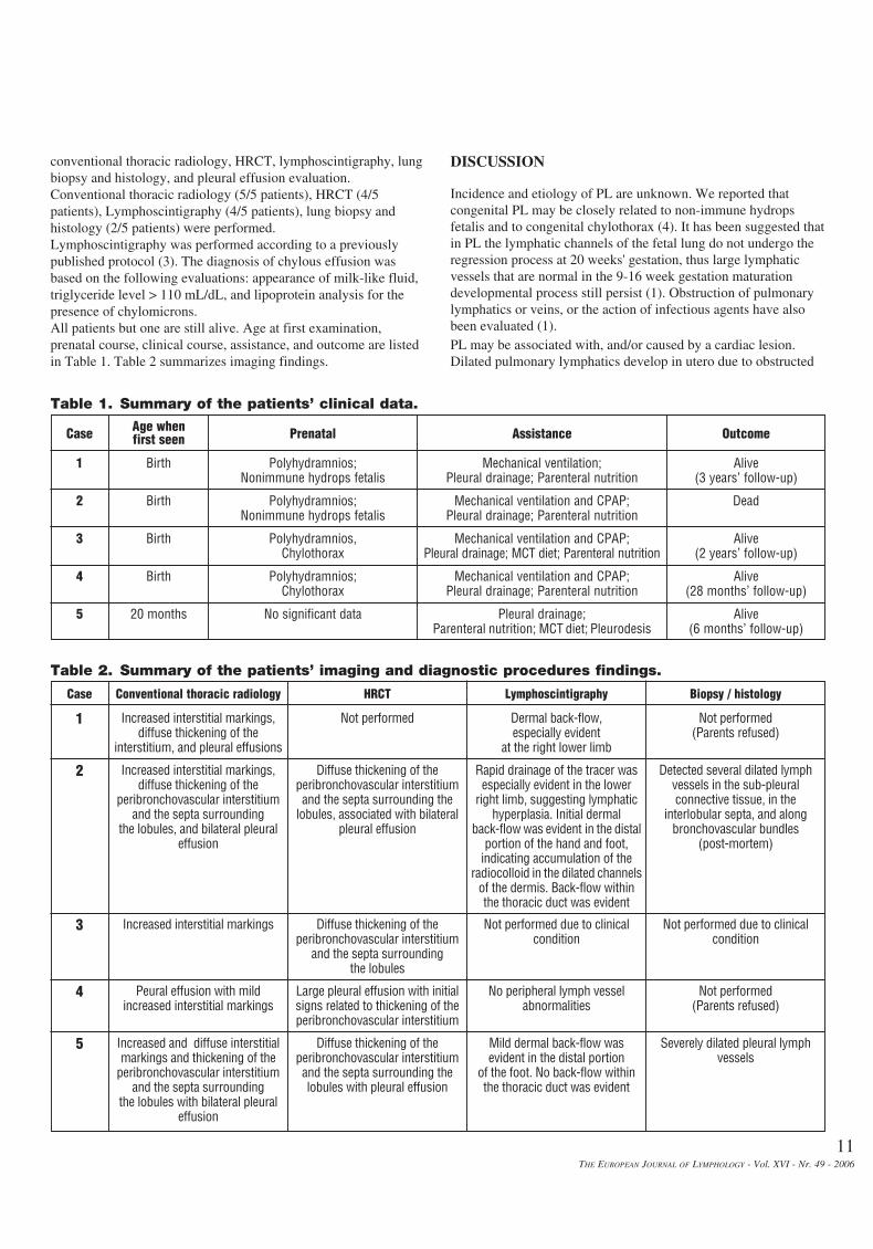
conventional thoracic radiology, HRCT, lymphoscintigraphy, lungbiopsy and histology, and pleural effusion evaluation.Conventional thoracic radiology (5/5 patients), HRCT (4/5patients), Lymphoscintigraphy (4/5 patients), lung biopsy andhistology (2/5 patients) were performed.Lymphoscintigraphy was performed according to a previouslypublished protocol (3). The diagnosis of chylous effusion wasbased on the following evaluations: appearance of milk-like fluid,triglyceride level > 110 mL/dL, and lipoprotein analysis for thepresence of chylomicrons.All patients but one are still alive. Age at first examination,prenatal course, clinical course, assistance, and outcome are listedin Table 1. Table 2 summarizes imaging findings.
DISCUSSION
Incidence and etiology of PL are unknown. We reported thatcongenital PL may be closely related to non-immune hydropsfetalis and to congenital chylothorax (4). It has been suggested thatin PL the lymphatic channels of the fetal lung do not undergo theregression process at 20 weeks' gestation, thus large lymphaticvessels that are normal in the 9-16 week gestation maturationdevelopmental process still persist (1). Obstruction of pulmonarylymphatics or veins, or the action of infectious agents have alsobeen evaluated (1).PL may be associated with, and/or caused by a cardiac lesion.Dilated pulmonary lymphatics develop in utero due to obstructed
Table 1. Summary of the patients’ clinical data.
Case Age when Prenatal Assistance Outcomefirst seen
1 Birth Polyhydramnios; Mechanical ventilation; AliveNonimmune hydrops fetalis Pleural drainage; Parenteral nutrition (3 years’ follow-up)
2 Birth Polyhydramnios; Mechanical ventilation and CPAP; DeadNonimmune hydrops fetalis Pleural drainage; Parenteral nutrition
3 Birth Polyhydramnios, Mechanical ventilation and CPAP; AliveChylothorax Pleural drainage; MCT diet; Parenteral nutrition (2 years’ follow-up)
4 Birth Polyhydramnios; Mechanical ventilation and CPAP; AliveChylothorax Pleural drainage; Parenteral nutrition (28 months’ follow-up)
5 20 months No significant data Pleural drainage; AliveParenteral nutrition; MCT diet; Pleurodesis (6 months’ follow-up)
Table 2. Summary of the patients’ imaging and diagnostic procedures findings.
Case Conventional thoracic radiology HRCT Lymphoscintigraphy Biopsy / histology
1
2 Increased interstitial markings,diffuse thickening of the
peribronchovascular interstitiumand the septa surrounding
the lobules, and bilateral pleuraleffusion
Diffuse thickening of theperibronchovascular interstitiumand the septa surrounding the
lobules, associated with bilateralpleural effusion
Rapid drainage of the tracer wasespecially evident in the lower
right limb, suggesting lymphatichyperplasia. Initial dermal
back-flow was evident in the distalportion of the hand and foot,
indicating accumulation of theradiocolloid in the dilated channels
of the dermis. Back-flow within the thoracic duct was evident
Detected several dilated lymphvessels in the sub-pleuralconnective tissue, in the
interlobular septa, and alongbronchovascular bundles
(post-mortem)
3 Increased interstitial markings Diffuse thickening of theperibronchovascular interstitium
and the septa surrounding the lobules
Not performed due to clinicalcondition
Not performed due to clinicalcondition
4 Peural effusion with mild increased interstitial markings
Large pleural effusion with initialsigns related to thickening of theperibronchovascular interstitium
No peripheral lymph vesselabnormalities
Not performed(Parents refused)
5 Increased and diffuse interstitialmarkings and thickening of the
peribronchovascular interstitiumand the septa surrounding
the lobules with bilateral pleuraleffusion
Diffuse thickening of theperibronchovascular interstitiumand the septa surrounding thelobules with pleural effusion
Mild dermal back-flow was evident in the distal portion
of the foot. No back-flow within the thoracic duct was evident
Severely dilated pleural lymphvessels
Increased interstitial markings,diffuse thickening of the
interstitium, and pleural effusions
Not performed Dermal back-flow, especially evident
at the right lower limb
Not performed(Parents refused)
10-13_BELLINI 23-11-2006 9:52 Pagina 11
12THE EUROPEAN JOURNAL OF LYMPHOLOGY - Vol. XVI - Nr. 49 - 2006
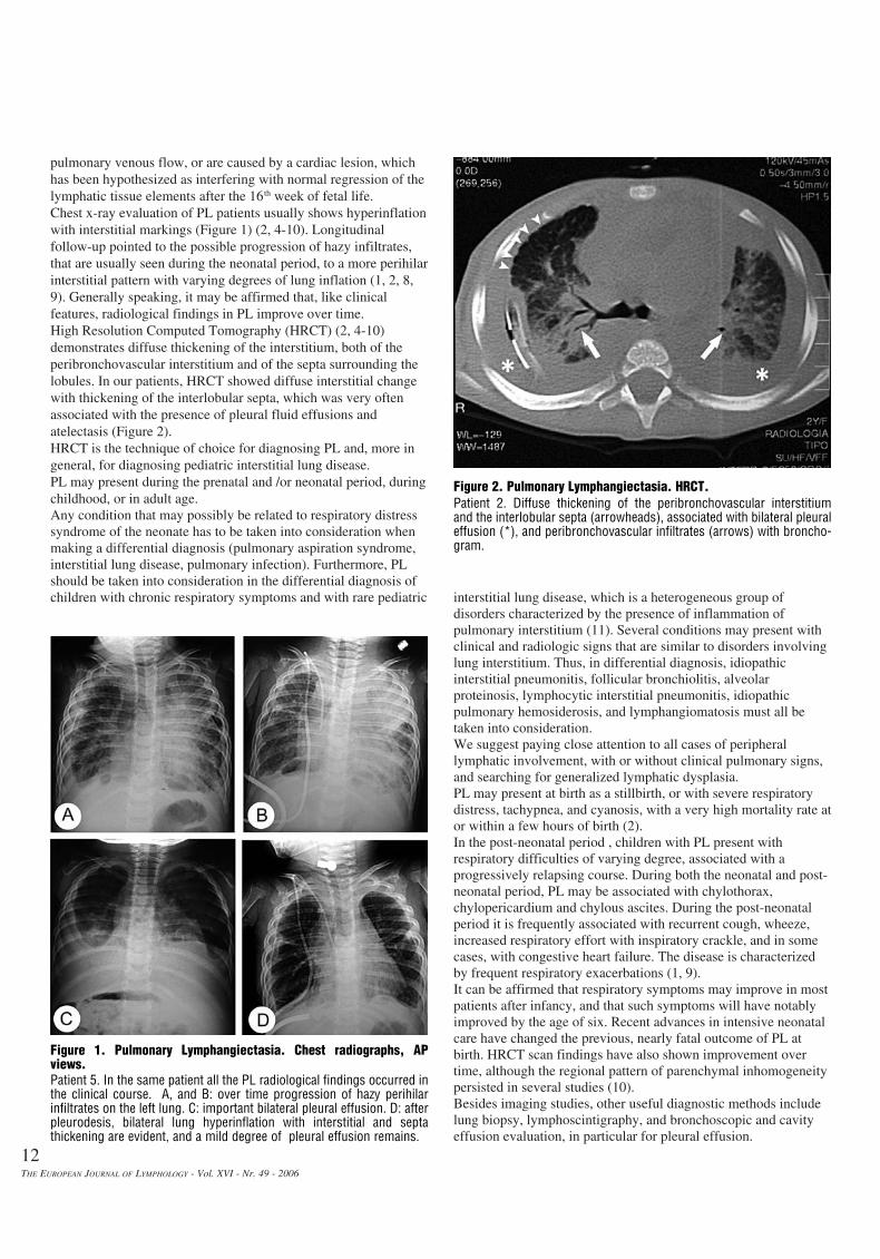
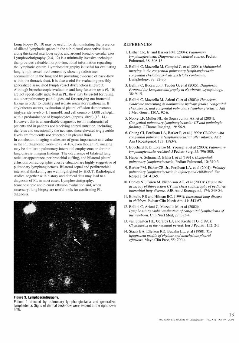
pulmonary venous flow, or are caused by a cardiac lesion, whichhas been hypothesized as interfering with normal regression of thelymphatic tissue elements after the 16th week of fetal life. Chest x-ray evaluation of PL patients usually shows hyperinflationwith interstitial markings (Figure 1) (2, 4-10). Longitudinalfollow-up pointed to the possible progression of hazy infiltrates,that are usually seen during the neonatal period, to a more perihilarinterstitial pattern with varying degrees of lung inflation (1, 2, 8,9). Generally speaking, it may be affirmed that, like clinicalfeatures, radiological findings in PL improve over time. High Resolution Computed Tomography (HRCT) (2, 4-10)demonstrates diffuse thickening of the interstitium, both of theperibronchovascular interstitium and of the septa surrounding thelobules. In our patients, HRCT showed diffuse interstitial changewith thickening of the interlobular septa, which was very oftenassociated with the presence of pleural fluid effusions andatelectasis (Figure 2).HRCT is the technique of choice for diagnosing PL and, more ingeneral, for diagnosing pediatric interstitial lung disease.PL may present during the prenatal and /or neonatal period, duringchildhood, or in adult age. Any condition that may possibly be related to respiratory distresssyndrome of the neonate has to be taken into consideration whenmaking a differential diagnosis (pulmonary aspiration syndrome,interstitial lung disease, pulmonary infection). Furthermore, PLshould be taken into consideration in the differential diagnosis ofchildren with chronic respiratory symptoms and with rare pediatric interstitial lung disease, which is a heterogeneous group of
disorders characterized by the presence of inflammation ofpulmonary interstitium (11). Several conditions may present withclinical and radiologic signs that are similar to disorders involvinglung interstitium. Thus, in differential diagnosis, idiopathicinterstitial pneumonitis, follicular bronchiolitis, alveolarproteinosis, lymphocytic interstitial pneumonitis, idiopathicpulmonary hemosiderosis, and lymphangiomatosis must all betaken into consideration.We suggest paying close attention to all cases of peripherallymphatic involvement, with or without clinical pulmonary signs,and searching for generalized lymphatic dysplasia.PL may present at birth as a stillbirth, or with severe respiratorydistress, tachypnea, and cyanosis, with a very high mortality rate ator within a few hours of birth (2). In the post-neonatal period , children with PL present withrespiratory difficulties of varying degree, associated with aprogressively relapsing course. During both the neonatal and post-neonatal period, PL may be associated with chylothorax,chylopericardium and chylous ascites. During the post-neonatalperiod it is frequently associated with recurrent cough, wheeze,increased respiratory effort with inspiratory crackle, and in somecases, with congestive heart failure. The disease is characterizedby frequent respiratory exacerbations (1, 9).It can be affirmed that respiratory symptoms may improve in mostpatients after infancy, and that such symptoms will have notablyimproved by the age of six. Recent advances in intensive neonatalcare have changed the previous, nearly fatal outcome of PL atbirth. HRCT scan findings have also shown improvement overtime, although the regional pattern of parenchymal inhomogeneitypersisted in several studies (10).Besides imaging studies, other useful diagnostic methods includelung biopsy, lymphoscintigraphy, and bronchoscopic and cavityeffusion evaluation, in particular for pleural effusion.
Figure 1. Pulmonary Lymphangiectasia. Chest radiographs, APviews.Patient 5. In the same patient all the PL radiological findings occurred inthe clinical course. A, and B: over time progression of hazy perihilarinfiltrates on the left lung. C: important bilateral pleural effusion. D: afterpleurodesis, bilateral lung hyperinflation with interstitial and septathickening are evident, and a mild degree of pleural effusion remains.
Figure 2. Pulmonary Lymphangiectasia. HRCT.Patient 2. Diffuse thickening of the peribronchovascular interstitium and the interlobular septa (arrowheads), associated with bilateral pleuraleffusion (*), and peribronchovascular infiltrates (arrows) with broncho-gram.
10-13_BELLINI 23-11-2006 9:52 Pagina 12
13THE EUROPEAN JOURNAL OF LYMPHOLOGY - Vol. XVI - Nr. 49 - 2006
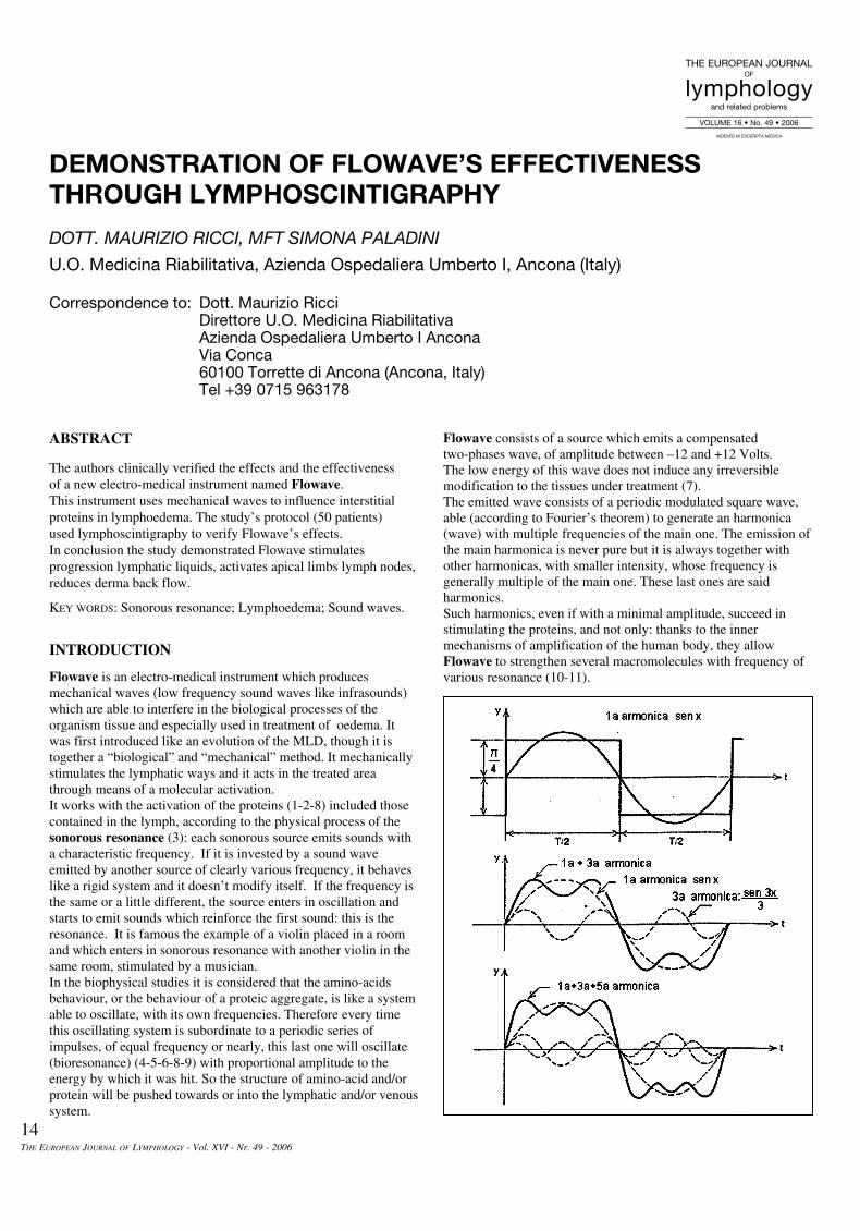
Lung biopsy (9, 10) may be useful for demonstrating the presenceof dilated lymphatic spaces in the sub-pleural connective tissue,along thickened interlobar septa, and around bronchovascular axes.Lymphoscintigraphy (2-4, 12) is a minimally invasive techniquethat provides valuable morpho-functional information regardingthe lymphatic system. Lymphoscintigraphy is useful for evaluatinglung lymph vessel involvement by showing radiotraceraccumulation in the lung and by providing evidence of back-flowwithin the thoracic duct. It is also useful for evaluating possiblygeneralized associated lymph vessel dysfunction (Figure 3).Although bronchoscopic evaluation and lung function tests (9, 10)are not specifically indicated in PL, they may be useful for rulingout other pulmonary pathologies and for carrying out bronchiallavage in order to identify and isolate respiratory pathogens. Ifchylothorax occurs, evaluation of pleural effusion demonstratestriglyceride levels > 1.1 mmol/L and cell counts > 1,000 cells/µL,with a predominance of lymphocytes (approx. 80%) (13, 14).However, this is an unreliable diagnostic test in malnourishedpatients and in patients not receiving enteral nutrition, includingthe fetus and occasionally the neonate, since elevated triglyceridelevels are frequently not detectable in pleural fluid. In conclusion, imaging studies are of great importance and valuein the PL diagnostic work-up (2, 4-10), even though PL imagingmay be similar to pulmonary interstitial emphysema or chroniclung disease imaging findings. The occurrence of bilateral lungreticular appearance, peribronchial cuffing, and bilateral pleuraleffusions on radiographic chest evaluation are highly suggestive ofpulmonary lymphangiectasis. Bilateral septal and peribronchialinterstitial thickening are well highlighted by HRCT. Radiologicalstudies, together with history and clinical data may lead to adiagnosis of PL in most cases. Lymphoscintigraphy,bronchoscopic and pleural effusion evaluation and, whennecessary, lung biopsy are useful tools for confirming PLdiagnosis.
REFERENCES
11. Esther CR, Jr. and Barker PM. (2004): Pulmonarylymphangiectasia: Diagnosis and clinical course. PediatrPulmonol, 38: 308-13.
12. Bellini C, Mazzella M, Campisi C, et al (2004): Multimodalimaging in the congenital pulmonary lymphangiectasia-congenital chylothorax-hydrops fetalis continuum.Lymphology, 37: 22-30.
13. Bellini C, Boccardo F, Taddei G, et al (2005): DiagnosticProtocol for Lymphoscintigraphy in Newborns. Lymphology,38: 9-15.
14. Bellini C, Mazzella M, Arioni C, et al (2003): Hennekamsyndrome presenting as nonimmune hydrops fetalis, congenitalchylothorax, and congenital pulmonary lymphangiectasia. AmJ Med Genet, 120A: 92-6.
15. Nobre LF, Muller NL, de Souza Junior AS, et al (2004):Congenital pulmonary lymphangiectasia: CT and pathologicfindings. J Thorac Imaging, 19: 56-9.
16. Chung CJ, Fordham LA, Barker P, et al (1999): Children withcongenital pulmonary lymphangiectasia: after infancy. AJRAm J Roentgenol, 173: 1583-8.
17. Bouchard S, Di Lorenzo M, Youssef S, et al (2000): Pulmonarylymphangiectasia revisited. J Pediatr Surg, 35: 796-800.
18. Huber A, Schranz D, Blaha I, et al (1991): Congenitalpulmonary lymphangiectasia. Pediatr Pulmonol, 10: 310-3.
19. Barker PM, Esther CR, Jr., Fordham LA, et al (2004): Primarypulmonary lymphangiectasia in infancy and childhood. EurRespir J, 24: 413-9.
10. Copley SJ, Coren M, Nicholson AG, et al (2000): Diagnosticaccuracy of thin-section CT and chest radiography of pediatricinterstitial lung disease. AJR Am J Roentgenol, 174: 549-54.
11. Bokulic RE and Hilman BC. (1994): Interstitial lung diseasein children. Pediatr Clin North Am, 41: 543-67.
12. Bellini C, Arioni C, Mazzella M, et al (2002):Lymphoscintigraphic evaluation of congenital lymphedema ofthe newborn. Clin Nucl Med, 27: 383-4.
13. van Straaten HL, Gerards LJ, and Krediet TG. (1993):Chylothorax in the neonatal period. Eur J Pediatr, 152: 2-5.
14. Staats BA, Ellefson RD, Budahn LL, et al (1980): Thelipoprotein profile of chylous and nonchylous pleuraleffusions. Mayo Clin Proc, 55: 700-4.
Figure 3. Lymphoscintigraphy.Patient 1 affected by pulmonary lymphangiectasia and generalizedlymphedema. Signs of dermal back-flow were evident at the right lowerlimb.
10-13_BELLINI 23-11-2006 9:52 Pagina 13
ABSTRACT
The authors clinically verified the effects and the effectiveness of a new electro-medical instrument named Flowave. This instrument uses mechanical waves to influence interstitialproteins in lymphoedema. The study’s protocol (50 patients) used lymphoscintigraphy to verify Flowave’s effects. In conclusion the study demonstrated Flowave stimulatesprogression lymphatic liquids, activates apical limbs lymph nodes,reduces derma back flow.
KEY WORDS: Sonorous resonance; Lymphoedema; Sound waves.
INTRODUCTION
Flowave is an electro-medical instrument which producesmechanical waves (low frequency sound waves like infrasounds)which are able to interfere in the biological processes of theorganism tissue and especially used in treatment of oedema. Itwas first introduced like an evolution of the MLD, though it istogether a “biological” and “mechanical” method. It mechanicallystimulates the lymphatic ways and it acts in the treated areathrough means of a molecular activation.It works with the activation of the proteins (1-2-8) included thosecontained in the lymph, according to the physical process of thesonorous resonance (3): each sonorous source emits sounds witha characteristic frequency. If it is invested by a sound waveemitted by another source of clearly various frequency, it behaveslike a rigid system and it doesn’t modify itself. If the frequency isthe same or a little different, the source enters in oscillation andstarts to emit sounds which reinforce the first sound: this is theresonance. It is famous the example of a violin placed in a roomand which enters in sonorous resonance with another violin in thesame room, stimulated by a musician.In the biophysical studies it is considered that the amino-acidsbehaviour, or the behaviour of a proteic aggregate, is like a systemable to oscillate, with its own frequencies. Therefore every timethis oscillating system is subordinate to a periodic series ofimpulses, of equal frequency or nearly, this last one will oscillate(bioresonance) (4-5-6-8-9) with proportional amplitude to theenergy by which it was hit. So the structure of amino-acid and/orprotein will be pushed towards or into the lymphatic and/or venoussystem.
Flowave consists of a source which emits a compensated two-phases wave, of amplitude between –12 and +12 Volts. The low energy of this wave does not induce any irreversiblemodification to the tissues under treatment (7).The emitted wave consists of a periodic modulated square wave,able (according to Fourier’s theorem) to generate an harmonica(wave) with multiple frequencies of the main one. The emission ofthe main harmonica is never pure but it is always together withother harmonicas, with smaller intensity, whose frequency isgenerally multiple of the main one. These last ones are saidharmonics.Such harmonics, even if with a minimal amplitude, succeed instimulating the proteins, and not only: thanks to the innermechanisms of amplification of the human body, they allowFlowave to strengthen several macromolecules with frequency ofvarious resonance (10-11).
14THE EUROPEAN JOURNAL OF LYMPHOLOGY - Vol. XVI - Nr. 49 - 2006
THE EUROPEAN JOURNALOF
lymphologyand related problems
VOLUME 16 • No. 49 • 2006
INDEXED IN EXCERPTA MEDICA
DEMONSTRATION OF FLOWAVE’S EFFECTIVENESSTHROUGH LYMPHOSCINTIGRAPHY
DOTT. MAURIZIO RICCI, MFT SIMONA PALADINI
U.O. Medicina Riabilitativa, Azienda Ospedaliera Umberto I, Ancona (Italy)
Correspondence to: Dott. Maurizio RicciDirettore U.O. Medicina RiabilitativaAzienda Ospedaliera Umberto I AnconaVia Conca60100 Torrette di Ancona (Ancona, Italy)Tel +39 0715 963178
14-18_RICCI 23-11-2006 9:54 Pagina 14
15THE EUROPEAN JOURNAL OF LYMPHOLOGY - Vol. XVI - Nr. 49 - 2006
A lymphatic oedema is a pathology characterized by an highinterstitial proteinous concentration which recalls and keeps watermolecules in the interstice and so it favourites fibrosis. For anefficient and long-lasting therapeutic effect it is necessary toremove these substances from the interstice in order to be followedby the water molecules.Limphoscintigraphical and ultrasonographic studies with highresolution have demonstrated that by using sound waves it ispossible to activate interstitial proteinous molecules, allowing theirremoval. Sound waves also stimulate some intracellular proteins,activating metabolic processes revealed by the expulsion ofcellular products (3-5-6-7).These data caught our attention and induced us to clinically verifythe effects and the effectiveness of Flowave with lymphoscintigraphy.So, we set up a protocol with the following criteria of inclusion:
• Patients with lymphedema, primary or secondary, mono orbilateral, of the superior or inferior limb;
• Initial stage (at least) 2 months;
• stage of the lymphedema between 2° and 4°;
• they were not receiving to other treatments and the chronicpatients observed a therapeutic wash out for at least 6 months.
Criteria of exclusion:
• presence of pacemakers;
• presence of metallic devices in the limb to treat;
• systemic disease;
• pregnancy.
The study method was represented by:
• clinical control made by the same operator;
• centimetric measurement (7 points on the superior and inferiorlimb) made at the beginning and at the end of the cycletreatment with Flowave;
• picture of the limbs in the two controls;
• lymphoscintigraphy before and after the treatment always madeby the same operator;
• daily therapy only with Flowave, all made by the same operatorusing a standardized program.
The cases report
From 18.12.2002 to 29.11.2004 50 patients have been found withthe aforesaid characteristics. Medium age 55 years (22 - 83).Medium insurgence 25 months. 18 of them were treated withradiotherapy and 32 without it.The aetiology divided them in:
• 10 cases of venous lymphoedema of the inferior limbs;
• 3 primary lymphoedema of inferior limbs;
• 8 secondary lymphoedema of the inferior limbs;
• 28 secondary lymphoedema of the upper limbs post-mastectomy;
• 1 lipolymphoedema of the inferior limbs.
The number of treatments, was variable from 9 to 14; the periodbetween the execution of the first lymphoscintigraphy and thebeginning of the therapy never exceeded 2 days while the timebetween the end of the sitting and the second lymphoscintigraphywas variable from 1 to 7 days.
Verification of results
In all cases the patients have reported a good adaptation to thereceived treatment. The feeling of gravity and hardening of thelimbs has been eliminated in all cases. Chronic patients stopped any other therapies for about 6 monthsbefore starting this treatment..In the meanwhile the operator positively estimated the methodicalexecution of the performance.
CLINICAL RESULTS
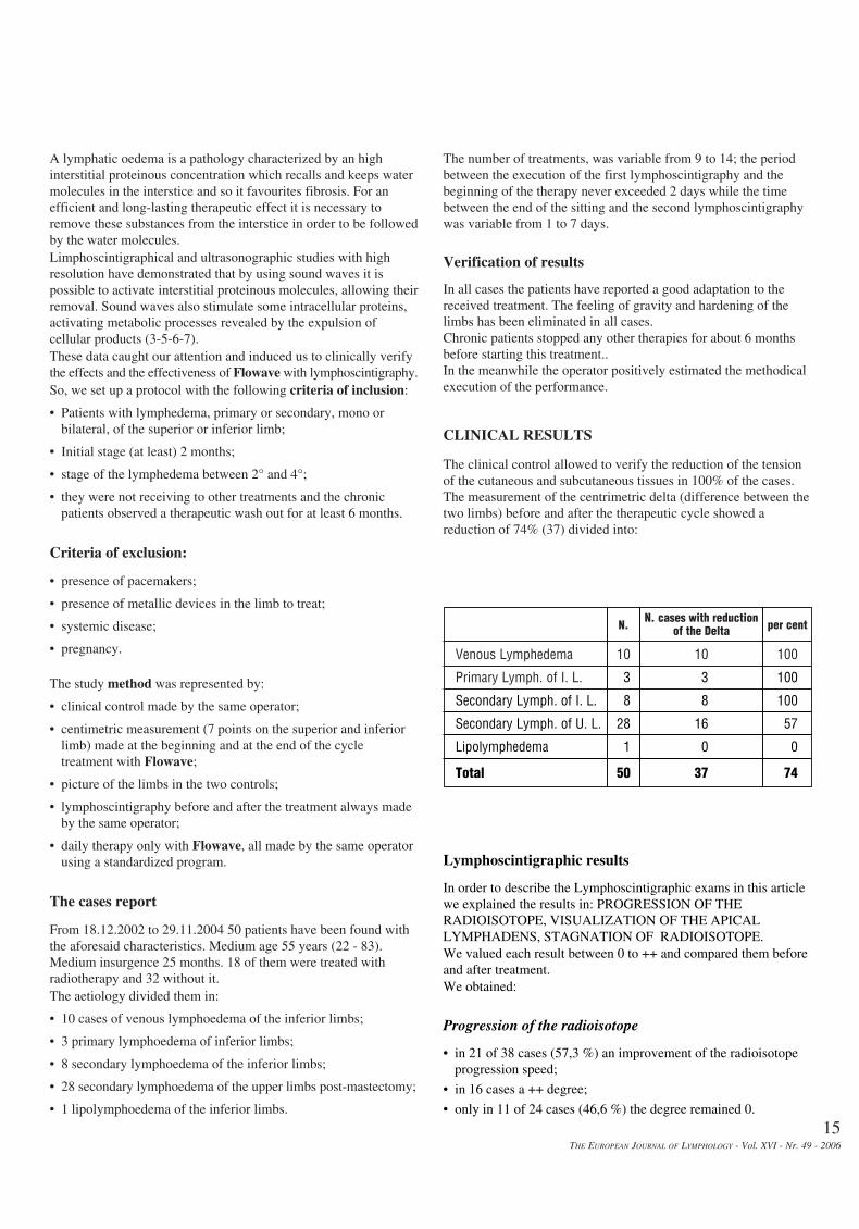
The clinical control allowed to verify the reduction of the tensionof the cutaneous and subcutaneous tissues in 100% of the cases.The measurement of the centrimetric delta (difference between thetwo limbs) before and after the therapeutic cycle showed areduction of 74% (37) divided into:
N. N. cases with reduction per centof the Delta
Venous Lymphedema 10 10 100
Primary Lymph. of I. L. 13 13 100
Secondary Lymph. of I. L. 18 18 100
Secondary Lymph. of U. L. 28 16 157
Lipolymphedema 11 10 110
Total 50 37 174
Lymphoscintigraphic results
In order to describe the Lymphoscintigraphic exams in this articlewe explained the results in: PROGRESSION OF THERADIOISOTOPE, VISUALIZATION OF THE APICALLYMPHADENS, STAGNATION OF RADIOISOTOPE.We valued each result between 0 to ++ and compared them beforeand after treatment.We obtained:
Progression of the radioisotope
• in 21 of 38 cases (57,3 %) an improvement of the radioisotopeprogression speed;
• in 16 cases a ++ degree;
• only in 11 of 24 cases (46,6 %) the degree remained 0.
14-18_RICCI 23-11-2006 9:54 Pagina 15
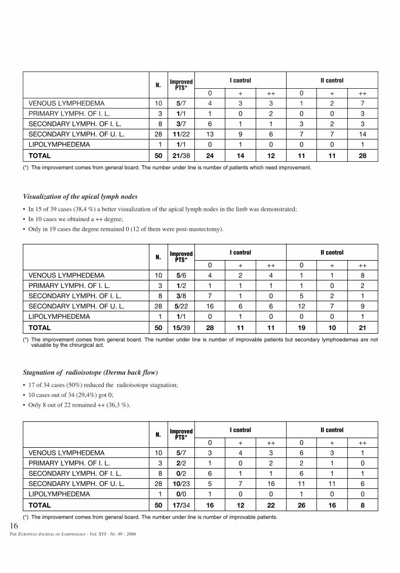
Visualization of the apical lymph nodes
• In 15 of 39 cases (38,4 %) a better visualization of the apical lymph nodes in the limb was demonstrated;
• In 10 cases we obtained a ++ degree;
• Only in 19 cases the degree remained 0 (12 of them were post-mastectomy).
Stagnation of radioisotope (Derma back flow)
• 17 of 34 cases (50%) reduced the radioisotope stagnation;
• 10 cases out of 34 (29,4%) got 0;
• Only 8 out of 22 remained ++ (36,3 %).
16THE EUROPEAN JOURNAL OF LYMPHOLOGY - Vol. XVI - Nr. 49 - 2006
N. Improved I control II controlPTS*
0 + ++ 0 + ++
VENOUS LYMPHEDEMA 10 5/7 4 3 3 1 2 7
PRIMARY LYMPH. OF I. L. 13 1/1 1 0 2 0 0 3
SECONDARY LYMPH. OF I. L. 18 3/7 6 1 1 3 2 3
SECONDARY LYMPH. OF U. L. 28 11/22 13 9 6 7 7 14
LIPOLYMPHEDEMA 11 1/1 0 1 0 0 0 1
TOTAL 50 21/38 24 14 12 11 11 28
(*) The improvement comes from general board. The number under line is number of patients which need improvement.
N. Improved I control II controlPTS*
0 + ++ 0 + ++
VENOUS LYMPHEDEMA 10 5/6 4 2 4 1 1 8
PRIMARY LYMPH. OF I. L. 13 1/2 1 1 1 1 0 2
SECONDARY LYMPH. OF I. L. 18 3/8 7 1 0 5 2 1
SECONDARY LYMPH. OF U. L. 28 5/22 16 6 6 12 7 9
LIPOLYMPHEDEMA 11 1/1 0 1 0 0 0 1
TOTAL 50 15/39 28 11 11 19 10 21
(*) The improvement comes from general board. The number under line is number of improvable patients but secondary lymphoedemas are notvaluable by the chirurgical act.
N. Improved I control II controlPTS*
0 + ++ 0 + ++
VENOUS LYMPHEDEMA 10 5/7 3 4 3 6 3 1
PRIMARY LYMPH. OF I. L. 13 2/2 1 0 2 2 1 0
SECONDARY LYMPH. OF I. L. 18 0/2 6 1 1 6 1 1
SECONDARY LYMPH. OF U. L. 28 10/23 5 7 16 11 11 6
LIPOLYMPHEDEMA 11 0/0 1 0 0 1 0 0
TOTAL 50 17/34 16 12 22 26 16 8
(*) The improvement comes from general board. The number under line is number of improvable patients.
14-18_RICCI 23-11-2006 9:54 Pagina 16
17THE EUROPEAN JOURNAL OF LYMPHOLOGY - Vol. XVI - Nr. 49 - 2006
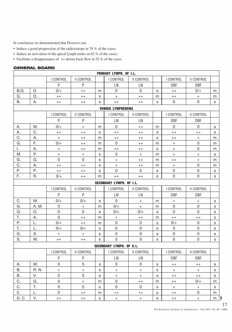
In conclusion we demonstrated that Flowave can:
• Induce a good progression of the radioisotope in 78 % of the cases;
• Induce an activation of the apical lymph nodes in 62 % of the cases;
• Facilitate a disappearance of ++ derma back flow in 52 % of the cases.
PRIMARY LYMPH. OF I.L.
I CONTROL II CONTROL I CONTROL II CONTROL I CONTROL II CONTROL
P P LN LN DBF DBFB.G. D. 0/+ ++ m 0 0 s ++ 0/+ mQ. D. ++ ++ s + ++ m ++ + mB. A. ++ ++ s ++ ++ s 0 0 s
VENOUS LYMPHEDEMA
I CONTROL II CONTROL I CONTROL II CONTROL I CONTROL II CONTROL
P P LN LN DBF DBFA. M. 0/+ + m 0 ++ m 0 0 sA. C. ++ ++ s ++ ++ s ++ ++ sC. A. + ++ m ++ ++ s ++ + mG. F. 0/+ ++ m 0 ++ m + 0 mI. A. + ++ m ++ ++ s + 0 mM. P. + + s 0 + m + + sG. G. 0 0 s + ++ m ++ + mC. A. ++ ++ s + ++ m + 0 mP. P. ++ ++ s 0 0 s 0 0 sF. R. 0/+ ++ m ++ ++ s 0 0 s
SECONDARY LYMPH. OF I.L.
I CONTROL II CONTROL I CONTROL II CONTROL I CONTROL II CONTROL
P P LN LN DBF DBFC. M. 0/+ 0/+ s 0 + m + + sG. A. M. 0 + m 0/+ + m 0 0 sQ. O. 0 0 s 0/+ 0/+ s 0 0 sT. A. 0 ++ m + ++ m ++ ++ sP. L. 0/+ ++ m 0 0 s 0/+ 0 sT. L. 0/+ 0/+ s 0 0 s 0 0 sG. S. + + s 0 0 s 0 0 sS. M. ++ ++ s 0 0 s 0 0 s
SECONDARY LYMPH. OF U.L.
I CONTROL II CONTROL I CONTROL II CONTROL I CONTROL II CONTROL
P P LN LN DBF DBFA. M. 0 0 s 0 0 s ++ ++ sB. R. N. + + s + + s + + sB. V. 0 0 s + + s ++ ++ sC. G. 0 + m 0 ++ m ++ 0/+ mC. T. 0 0 s 0 0 s + + sC. L. 0 ++ m ++ ++ s ++ 0 mD. C. V. ++ ++ s + + s ++ + m
GENERAL BOARD
14-18_RICCI 23-11-2006 9:54 Pagina 17
18THE EUROPEAN JOURNAL OF LYMPHOLOGY - Vol. XVI - Nr. 49 - 2006
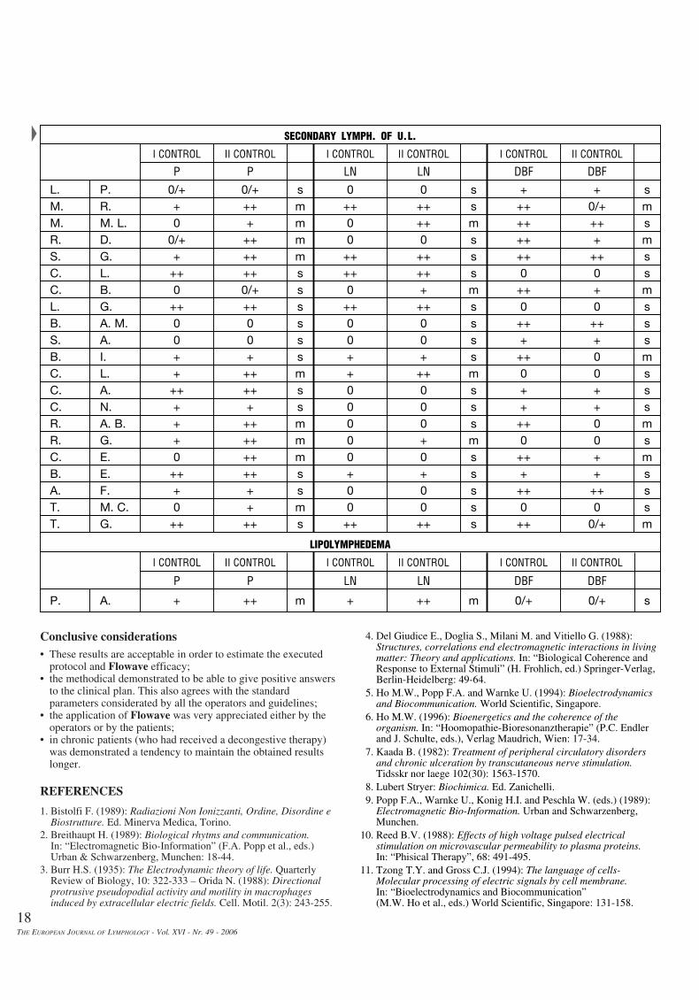
Conclusive considerations• These results are acceptable in order to estimate the executed
protocol and Flowave efficacy;• the methodical demonstrated to be able to give positive answers
to the clinical plan. This also agrees with the standardparameters considerated by all the operators and guidelines;
• the application of Flowave was very appreciated either by theoperators or by the patients;
• in chronic patients (who had received a decongestive therapy)was demonstrated a tendency to maintain the obtained resultslonger.
REFERENCES
1. Bistolfi F. (1989): Radiazioni Non Ionizzanti, Ordine, Disordine eBiostrutture. Ed. Minerva Medica, Torino.
2. Breithaupt H. (1989): Biological rhytms and communication.In: “Electromagnetic Bio-Information” (F.A. Popp et al., eds.)Urban & Schwarzenberg, Munchen: 18-44.
3. Burr H.S. (1935): The Electrodynamic theory of life. QuarterlyReview of Biology, 10: 322-333 – Orida N. (1988): Directionalprotrusive pseudopodial activity and motility in macrophagesinduced by extracellular electric fields. Cell. Motil. 2(3): 243-255.
14. Del Giudice E., Doglia S., Milani M. and Vitiello G. (1988):Structures, correlations end electromagnetic interactions in livingmatter: Theory and applications. In: “Biological Coherence andResponse to External Stimuli” (H. Frohlich, ed.) Springer-Verlag,Berlin-Heidelberg: 49-64.
15. Ho M.W., Popp F.A. and Warnke U. (1994): Bioelectrodynamicsand Biocommunication. World Scientific, Singapore.
16. Ho M.W. (1996): Bioenergetics and the coherence of theorganism. In: “Hoomopathie-Bioresonanztherapie” (P.C. Endlerand J. Schulte, eds.), Verlag Maudrich, Wien: 17-34.
17. Kaada B. (1982): Treatment of peripheral circulatory disordersand chronic ulceration by transcutaneous nerve stimulation.Tidsskr nor laege 102(30): 1563-1570.
18. Lubert Stryer: Biochimica. Ed. Zanichelli.19. Popp F.A., Warnke U., Konig H.I. and Peschla W. (eds.) (1989):
Electromagnetic Bio-Information. Urban and Schwarzenberg,Munchen.
10. Reed B.V. (1988): Effects of high voltage pulsed electricalstimulation on microvascular permeability to plasma proteins.In: “Phisical Therapy”, 68: 491-495.
11. Tzong T.Y. and Gross C.J. (1994): The language of cells-Molecular processing of electric signals by cell membrane.In: “Bioelectrodynamics and Biocommunication” (M.W. Ho et al., eds.) World Scientific, Singapore: 131-158.
SECONDARY LYMPH. OF U.L.
I CONTROL II CONTROL I CONTROL II CONTROL I CONTROL II CONTROL
P P LN LN DBF DBFL. P. 0/+ 0/+ s 0 0 s + + sM. R. + ++ m ++ ++ s ++ 0/+ mM. M. L. 0 + m 0 ++ m ++ ++ sR. D. 0/+ ++ m 0 0 s ++ + mS. G. + ++ m ++ ++ s ++ ++ sC. L. ++ ++ s ++ ++ s 0 0 sC. B. 0 0/+ s 0 + m ++ + mL. G. ++ ++ s ++ ++ s 0 0 sB. A. M. 0 0 s 0 0 s ++ ++ sS. A. 0 0 s 0 0 s + + sB. I. + + s + + s ++ 0 mC. L. + ++ m + ++ m 0 0 sC. A. ++ ++ s 0 0 s + + sC. N. + + s 0 0 s + + sR. A. B. + ++ m 0 0 s ++ 0 mR. G. + ++ m 0 + m 0 0 sC. E. 0 ++ m 0 0 s ++ + mB. E. ++ ++ s + + s + + sA. F. + + s 0 0 s ++ ++ sT. M. C. 0 + m 0 0 s 0 0 sT. G. ++ ++ s ++ ++ s ++ 0/+ m
LIPOLYMPHEDEMA
I CONTROL II CONTROL I CONTROL II CONTROL I CONTROL II CONTROL
P P LN LN DBF DBF
P. A. + ++ m + ++ m 0/+ 0/+ s
14-18_RICCI 23-11-2006 9:54 Pagina 18
19THE EUROPEAN JOURNAL OF LYMPHOLOGY - Vol. XVI - Nr. 49 - 2006
ABSTRACT
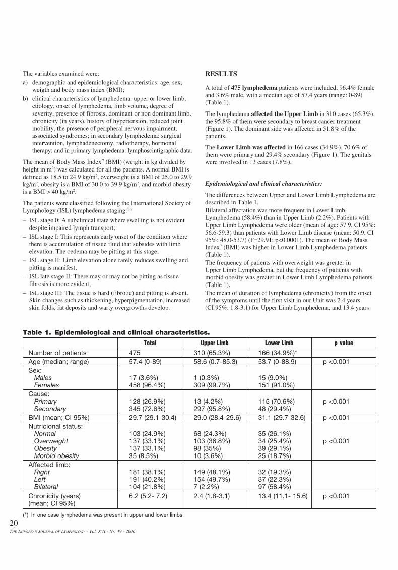
Aim: to describe the characteristics of the patients admitted to theLymphedema Unit of Hospital Universitario La Fe of Valencia(Spain).Material and method: A descriptive study of patients seen in ourUnit from September 2005 to March 2006 was performed. Theepidemiological and clinical features were recorded.Results: A total of 475 lymphedema patients were included, 310cases (65.3%) with Upper Limb Lymphedema (95.8% of themwere secondary to breast cancer treatment), and 166 cases (34.9%)with Lower Limb Lymphedema (70.6% of them were primary).The degree of the disease was more severe in Lower LimbLymphedema than in Upper Limb (p<0.0001). Dermalcomplications were present in 27.6% of the patients with UpperLimb Lymphedema and in 55.5% of the patients with Lower LimbLymphedema (p<0.0001). The most common cause forlymphedema in our country is postmastectomy lymphedema, andthe patients are referred to us a mean of 2.4 years after thebeginning of the symptoms, and in early stages. For the LowerLimb Lymphedema, the referral to our Unit was a mean of 13.4years after the beginning of the symptoms, and usually inadvanced and complicated stages.Conclusions: These data suggest that Upper Limb Lymphedema isa well-known and expected sequel of breast cancer treatment, andin the case of the Lower Limb Lymphedema it is ignored andusually not properly diagnosed by medical community. Someefforts have to be made to improve medical knowledge in thisfield, in our country.
KEY WORDS: Lymphedema, epidemiology, staging, complications.
INTRODUCTION
Lymphoedema is a chronic and progressive condition that resultsfrom any reduction in the capacity of the lymphatic system todrain fluid from the interstitium and return it to the bloodcirculation.1 Lymphedema can be Primary, when produced by
THE EUROPEAN JOURNALOF
lymphologyand related problems
VOLUME 16 • No. 49 • 2006
INDEXED IN EXCERPTA MEDICA
EARLY OR LATE DIAGNOSIS OF LYMPHEDEMA IN OUR LYMPHEDEMA UNIT
ISABEL FORNER-CORDERO*, RAQUEL NAVARRO-MONSOLIU*, JOSE MUÑOZ-LANGA**, PILAR REL-MONZÓ*
** Lymphedema Unit. Rehabilitation Department. Hospital Universitario La Fe** Medical Oncology Unit. Hospital Universitario Dr. Peset. Valencia. Spain
Correspondence to: Isabel Forner-CorderoC/ Andrés Mancebo 36, 1246023 Valencia SpainPhone: 00-34-649179852E-mail: [email protected]
congenital abnormalities of the lymphatic system, or Secondary toCancer and its treatment, traumatism, Filarial infection or ChronicVenous Disease as the main causes. Lymphedema followingtreatment for breast cancer is the most frequent type oflymphedema in our country, being its incidence from 6 to 30%2
depending from the sources.
Lymphoedema leads to aesthetic problems, disfunction due topain, weight and limited mobility, and psychological troubles.Most frequent complications1 are: infections as erysipela-lymphangitis, fibrosis, elephantiasis, lymph fistulae and lymphaticulcers.In the Hospital La Fe of Valencia, the Lymphedema Unit belongsto the Rehabilitation Department and its task is to study and treatthe patients affected of lymphedema. This Unit receives patientsfrom Valencia city and the Region of Valencia and often patientsfrom other Spanish regions.
Since the start of the Breast Cancer Unit, that is a multidisciplinaryworking group for the best management of the breast cancerpatients, the referral of the patients from oncologist specialist,radiotherapists and surgeons has improved.The aim of the study was to describe the characteristics of thepatients admitted to the Lymphedema Unit of Hospital La Fe ofValencia (Spain) and the differences between Upper and lowerlimb Lymphedema patients.
MATERIAL AND METHODS
A descriptive study of patients seen in our Unit from September2005 to March 2006 was performed. The epidemiological andclinical features were recorded.
The diagnosis of lymphedema was done by the natural history, theclinical exam and the complementary exams as blood biochemistryparameters, echo-dupplex scan, Computed Tomography scan andlymphoscintigraphy findings when possible.3,4,5,6
19-23_FORNER_CORDERO 23-11-2006 9:56 Pagina 19
The variables examined were: a) demographic and epidemiological characteristics: age, sex,
weigth and body mass index (BMI);b) clinical characteristics of lymphedema: upper or lower limb,
etiology, onset of lymphedema, limb volume, degree ofseverity, presence of fibrosis, dominant or non dominant limb,chronicity (in years), history of hypertension, reduced jointmobility, the presence of peripheral nervous impairment,associated syndromes; in secondary lymphedema: surgicalintervention, lymphadenectomy, radiotherapy, hormonaltherapy; and in primary lymphedema: lymphoscintigraphic data.
The mean of Body Mass Index 7 (BMI) (weight in kg divided byheight in m2) was calculated for all the patients. A normal BMI isdefined as 18.5 to 24.9 kg/m2, overweight is a BMI of 25.0 to 29.9kg/m2, obesity is a BMI of 30.0 to 39.9 kg/m2, and morbid obesityis a BMI > 40 kg/m2.
The patients were classified following the International Society ofLymphology (ISL) lymphedema staging: 8,9
– ISL stage 0: A subclinical state where swelling is not evidentdespite impaired lymph transport;
– ISL stage I: This represents early onset of the condition wherethere is accumulation of tissue fluid that subsides with limbelevation. The oedema may be pitting at this stage;
– ISL stage II: Limb elevation alone rarely reduces swelling andpitting is manifest;
– ISL late stage II: There may or may not be pitting as tissuefibrosis is more evident;
– ISL stage III: The tissue is hard (fibrotic) and pitting is absent.Skin changes such as thickening, hyperpigmentation, increasedskin folds, fat deposits and warty overgrowths develop.
RESULTS
A total of 475 lymphedema patients were included, 96.4% femaleand 3.6% male, with a median age of 57.4 years (range: 0-89)(Table 1).
The lymphedema affected the Upper Limb in 310 cases (65.3%);the 95.8% of them were secondary to breast cancer treatment(Figure 1). The dominant side was affected in 51.8% of thepatients.
The Lower Limb was affected in 166 cases (34.9%), 70.6% ofthem were primary and 29.4% secondary (Figure 1). The genitalswere involved in 13 cases (7.8%).
Epidemiological and clinical characteristics:
The differences between Upper and Lower Limb Lymphedema aredescribed in Table 1.Bilateral affectation was more frequent in Lower LimbLymphedema (58.4%) than in Upper Limb (2.2%). Patients withUpper Limb Lymphedema were older (mean of age: 57.9, CI 95%:56.6-59.3) than patients with Lower Limb disease (mean: 50.9, CI95%: 48.0-53.7) (F=29.91; p<0.0001). The mean of Body MassIndex7 (BMI) was higher in Lower Limb Lymphedema patients(Table 1).The frequency of patients with overweight was greater in Upper Limb Lymphedema, but the frequency of patients withmorbid obesity was greater in Lower Limb Lymphedema patients (Table 1).The mean of duration of lymphedema (chronicity) from the onsetof the symptoms until the first visit in our Unit was 2.4 years (CI 95%: 1.8-3.1) for Upper Limb Lymphedema, and 13.4 years
20THE EUROPEAN JOURNAL OF LYMPHOLOGY - Vol. XVI - Nr. 49 - 2006
Table 1. Epidemiological and clinical characteristics.
Total Upper Limb Lower Limb p value
Number of patients 475 310 (65.3%) 166 (34.9%)*Age (median; range) 57.4 (0-89) 58.6 (0.7-85.3) 53.7 (0-88.9) p <0.001Sex:
Males 17 (3.6%) 1 (0.3%) 15 (9.0%)Females 458 (96.4%) 309 (99.7%) 151 (91.0%)
Cause:Primary 128 (26.9%) 13 (4.2%) 115 (70.6%) p <0.001Secondary 345 (72.6%) 297 (95.8%) 48 (29.4%)
BMI (mean; CI 95%) 29.7 (29.1-30.4) 29.0 (28.4-29.6) 31.1 (29.7-32.6) p <0.001Nutricional status:
Normal 103 (24.9%) 68 (24.3%) 35 (26.1%)Overweight 137 (33.1%) 103 (36.8%) 34 (25.4%) p <0.001Obesity 137 (33.1%) 98 (35%) 39 (29.1%)Morbid obesity 35 (8.5%) 10 (3.6%) 25 (18.7%)
Affected limb:Right 181 (38.1%) 149 (48.1%) 32 (19.3%)Left 191 (40.2%) 154 (49.7%) 37 (22.3%)Bilateral 104 (21.8%) 7 (2.2%) 97 (58.4%)
Chronicity (years) 6.2 (5.2- 7.2) 2.4 (1.8-3.1) 13.4 (11.1- 15.6) p <0.001(mean; CI 95%)
(*) In one case lymphedema was present in upper and lower limbs.
19-23_FORNER_CORDERO 23-11-2006 9:56 Pagina 20
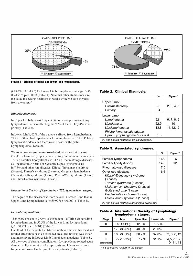
(CI 95%: 11.1-15.6) for Lower Limb Lymphedema (range: 0-55)(F=136.9; p<0.0001) (Table 1). Note that other studies measurethe delay in seeking treatment in weeks while we do it in yearsfrom the onset.10
Etiologic diagnosis:
In Upper Limb the most frequent etiology was postmastectomylymphedema that was affecting the 96% of them. Only 4% wereprimary (Table 2).
In Lower Limb, 62% of the patients suffered from Lymphedema,22.9% of them had Lipedema or Lipolymphedema, 13.8% Phlebo-lymphostatic edema and there were 2 cases with CysticLymphangioma (Table 2).
We found some syndromes associated with the clinical case(Table 3): Familiar lymphedema affecting one or more members in16.9%; Familiar lipodystrophy in 14.5%; Rheumatologic diseasesas Rheumatoid Arthritis or Systemic Lupus Erythematosus in 7.5%; and other rare diseases: Klippel-Trenaunay syndrome (3 cases); Turner’s syndrome (3 cases); Malignant lymphedema (2 cases); Goltz syndrome (1 case); Prader-Willi syndrome (1 case)and Ehler-Danlos syndrome (1 case).
International Society of Lymphology (ISL) lymphedema staging:
The degree of the disease was more severe in Lower Limb than inUpper Limb Lymphedema (χ2 = 59.927; p < 0.0001) (Table 4).
Dermal complications:
They were present in 27.6% of the patients suffering Upper LimbLymphedema and in 55.5% of the Lower Limb Lymphedema (χ2 = 30.721; p < 0.0001) (Table 5). One third of the patients had fibrosis in their limbs with a local andlimited affectation or in an extended area. The fibrosis was widerand more severe in Lower Limb Lymphedema patients (Table 5).All the types of dermal complications: Lymphedema-related acutedermatitis, Hyperkeratosis, Lymph cysts and Ulcers were morefrequent in Lower Limb Lymphedema patients (Table 5).
21THE EUROPEAN JOURNAL OF LYMPHOLOGY - Vol. XVI - Nr. 49 - 2006
Figure 1 - Etiology of upper and lower limb lymphedema.
Table 2. Clinical Diagnosis.% Figures*
Upper Limb:Postmastectomy 96 2, 3, 4, 5Primary 94
Lower Limb:Lymphedema 62.9 6, 7, 8, 9Lipedema or 22.9 10Lipolymphedema 13.8 11,12,13Phlebo-lymphostatic edemaCystic Lymphangioma (2 cases) 11.3
(*) See figures related to clinical diagnosis.
Table 3. Associated syndromes.% Figures*
Familiar lymphedema 16.9 16Familiar lipodystrophy 14.5 12Rheumatologic diseases 17.5Other rare diseases: 16.6
Klippel-Trenaunay syndrome(3 cases)Turner’s syndrome (3 cases)Malignant lymphedema (2 cases)Goltz syndrome (1 case)Prader-Willi syndrome (1 case)Ehler-Danlos syndrome (1 case)
(*) See figures related to associated syndromes.
Table 4. International Society of Lymphologylymphedema stages.
Stage Total Upper Limb Lower Limb Figures*
0 143 (9.1%) 12.9% 11.8 %
I 173 (36.6%) 40.6% 28.0%
II 180 (38.1%) 38.7% 37.8% 2, 3, 6, 12
III 177 (16.3%) 17.7% 31.1% 4,5,7,8,9, elephantiasis 10, 11, 13
(*) See figures related to the stages.
19-23_FORNER_CORDERO 23-11-2006 9:56 Pagina 21
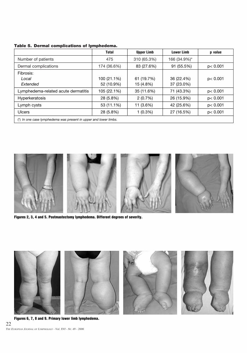
22THE EUROPEAN JOURNAL OF LYMPHOLOGY - Vol. XVI - Nr. 49 - 2006
Table 5. Dermal complications of lymphedema.
Total Upper Limb Lower Limb p value
Number of patients 475 310 (65.3%) 166 (34.9%)*
Dermal complications 174 (36.6%) 183 (27.6%) 191 (55.5%) p< 0.001
Fibrosis:Local 100 (21.1%) 61 (19.7%) 36 (22.4%) p< 0.001Extended 152 (10.9%) 15 (4.8%)1 37 (23.0%)
Lymphedema-related acute dermatitis 105 (22.1%) 35 (11.6%) 71 (43.3%) p< 0.001
Hyperkeratosis 128 (5.8%)1 2 (0.7%) 26 (15.9%) p< 0.001
Lymph cysts 153 (11.1%) 11 (3.6%)1 42 (25.6%) p< 0.001
Ulcers 128 (5.8%)1 11 (0.3%)1 27 (16.5%) p< 0.001
(*) In one case lymphedema was present in upper and lower limbs.
Figures 2, 3, 4 and 5. Postmastectomy lymphedema. Different degrees of severity.
Figures 6, 7, 8 and 9. Primary lower limb lymphedema.
19-23_FORNER_CORDERO 23-11-2006 9:56 Pagina 22
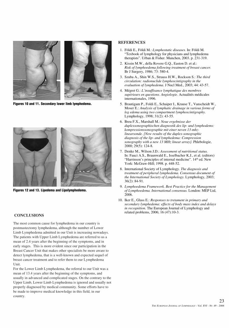
CONCLUSIONS
The most common cause for lymphedema in our country ispostmastectomy lymphedema, although the number of LowerLimb Lymphedema admitted in our Unit is increasing nowadays.The patients with Upper Limb Lymphedema are referred to us amean of 2.4 years after the beginning of the symptoms, and inearly stages. This is more evident since our participation in theBreast Cancer Unit that makes other specialists be more aware todetect lymphedema, that is a well-known and expected sequel ofbreast cancer treatment and to refer them to our LymphedemaUnit.For the Lower Limb Lymphedema, the referral to our Unit was amean of 13.4 years after the beginning of the symptoms, andusually in advanced and complicated stages. On the contrary to theUpper Limb, Lower Limb Lymphedema is ignored and usually notproperly diagnosed by medical community. Some efforts have tobe made to improve medical knowledge in this field, in ourcountry.
REFERENCES
11. Földi E., Földi M.: Lymphostatic diseases. In: Földi M.“Texbook of lymphology for physicians and lymphedematherapists”. Urban & Fisher. Munchen, 2003. p. 231-319.
12. Kissin M.W., della Rovere G.Q., Easton D. et al.: Risk of lymphoedema following treatment of breast cancer.Br J Surgery, 1986; 73: 580-4.
13. Szuba A., Shin W.S., Strauss H.W., Rockson S.: The thirdcirculation: radionuclide lymphoscintigraphy in theevaluation of lymphedema. J Nucl Med., 2003; 44: 43-57.
14. Mégret G.: L’insuffisance lymphatique des membressupérieurs en questions. Angiologie. Actualités médicalesinternationales, 1996.
15. Brautigam P., Foldi E., Schaiper I., Krause T., Vanscheidt W.,Moser E.: Analysis of lymphatic drainage in various forms ofleg edema using two compartment lymphoscintigraphy.Lymphology, 1998; 31(2): 43-55.
16. Breu F.X., Marshall M.: Neue ergebnisse derduplexsonographischen diagnostik des lip- und lymphodems:kompressionssonographie mit einer neven 13-mhz-linearsonde. [New results of the duplex-sonographicdiagnosis of the lip- and lymphedema: Compressionsonography with a new 13 MHz linear array]. Phlebologie,2000; 29(5): 124-8.
17. Denke M., Wilson J.D.: Assessment of nutritional status.In: Fauci A.S., Braunwald E., Isselbacher K.J., et al. (editors)“Harrinson’s principles of internal medicine”. 14th ed. NewYork: McGraw-Hill; 1998. p. 448-52.
18. International Society of Lymphology. The diagnosis andtreatment of peripheral lymphedema. Consensus document ofthe International Society of Lymphology. Lymphology, 2003;36(2): 84-91.
19. Lymphoedema Framework. Best Practice for the Managementof Lymphoedema. International consensus. London: MEP Ltd,2006.
10. Iker E., Glass E.: Responses to tretament in primary andsecondary lymphedema: effects of body mass index and delaysin recognition. The European Journal of Lymphology andrelated problems, 2006; 16 (47):10-3.
23THE EUROPEAN JOURNAL OF LYMPHOLOGY - Vol. XVI - Nr. 49 - 2006
Figures 10 and 11. Secondary lower limb lymphedema.
Figures 12 and 13. Lipedema and Lipolymphedema.
19-23_FORNER_CORDERO 23-11-2006 9:56 Pagina 23
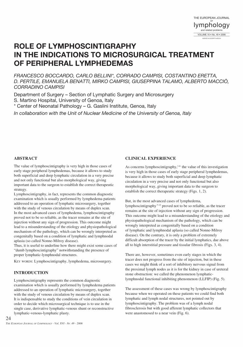
ABSTRACT
The value of lymphoscintigraphy is very high in those cases ofearly stage peripheral lymphedemas, because it allows to studyboth superficial and deep lymphatic circulation in a very preciseand not only functional but also morphological way, givingimportant data to the surgeon to establish the correct therapeuticstrategy.Lymphoscintigraphy, in fact, represents the common diagnosticexamination which is usually performed by lymphedema patientsaddressed to an operation of lymphatic microsurgery, togetherwith the study of venous circulation by means of duplex scan.In the most advanced cases of lymphedema, lymphoscintigraphyproved not to be so reliable, as the tracer remains at the site ofinjection without any sign of progression. This outcome mightlead to a misunderstanding of the etiology and physiopathologicalmechanism of the pathology, which can be wrongly interpreted ascongenitally based on a condition of lymphatic and lymphnodalaplasia (so called Nonne-Milroy disease).Thus, it is useful to underline how there might exist some cases of“dumb lymphoscintigraphy” notwithstanding the presence ofproper lymphatic-lymphnodal structures.
KEY WORDS: Lymphoscintigraphy, lymphedema, microsurgery.
INTRODUCTION
Lymphoscintigraphy represents the common diagnosticexamination which is usually performed by lymphedema patientsaddressed to an operation of lymphatic microsurgery, togetherwith the study of venous circulation by means of duplex scan.It is indispensable to study the conditions of vein circulation inorder to decide which microsurgical technique is to use in thesingle case, derivative lymphatic-venous shunt or reconstructivelymphatic-venous-lymphatic plasty.
CLINICAL EXPERIENCE
As concerns lymphoscintigraphy,1-6 the value of this investigationis very high in those cases of early stage peripheral lymphedemas,because it allows to study both superficial and deep lymphaticcirculation in a very precise and not only functional but alsomorphological way, giving important data to the surgeon toestablish the correct therapeutic strategy (Figs. 1, 2).
But, in the most advanced cases of lymphedema,lymphoscintigraphy7-12 proved not to be so reliable, as the tracerremains at the site of injection without any sign of progression.This outcome might lead to a misunderstanding of the etiology andphysiopathological mechanism of the pathology, which can bewrongly interpreted as congenitally based on a condition of lymphatic and lymphnodal aplasia (so called Nonne-Milroydisease). On the contrary, it is only a problem of extremelydifficult absorption of the tracer by the initial lymphatics, due aboveall to high interstitial pressure and tissular fibrosis (Figs. 3, 4).
There are, however, sometimes even early stages in which thetracer does not progress from the site of injection, but in thesecases we might think of a sort of inhibitory nervous signal fromthe proximal lymph nodes as it is for the kidney in case of ureteralstone obstruction: we called the phenomenon lymphatic-lymphnodal functional inhibiting phenomenon (LLFIP) (Fig. 5).
The assessment of these cases was wrong by lymphoscintigraphybecause when we operated on these patients we could find bothlymphatic and lymph nodal structures, not pointed out bylymphoscintigraphy. The problem was of a lymph nodalfibrosclerosis but with good afferent lymphatic collectors thatwere anastomosed to a near vein (Fig. 6).
24THE EUROPEAN JOURNAL OF LYMPHOLOGY - Vol. XVI - Nr. 49 - 2006
THE EUROPEAN JOURNALOF
lymphologyand related problems
VOLUME 16 • No. 49 • 2006
INDEXED IN EXCERPTA MEDICA
ROLE OF LYMPHOSCINTIGRAPHY IN THE INDICATIONS TO MICROSURGICAL TREATMENT OF PERIPHERAL LYMPHEDEMAS
FRANCESCO BOCCARDO, CARLO BELLINI*, CORRADO CAMPISI, COSTANTINO ERETTA, D. PERTILE, EMANUELA BENATTI, MIRKO CAMPISI, GIUSEPPINA TALAMO, ALBERTO MACCIÒ,CORRADINO CAMPISI
Department of Surgery – Section of Lymphatic Surgery and MicrosurgeryS. Martino Hospital, University of Genoa, Italy* Center of Neonatal Pathology – G. Gaslini Institute, Genoa, Italy
In collaboration with the Unit of Nuclear Medicine of the University of Genoa, Italy
24-28_BOCCARDO 23-11-2006 10:42 Pagina 24
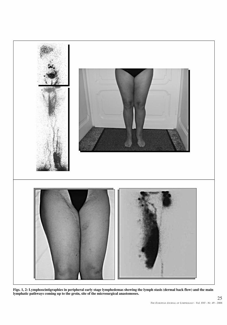
25THE EUROPEAN JOURNAL OF LYMPHOLOGY - Vol. XVI - Nr. 49 - 2006
Figs. 1, 2: Lymphoscintigraphies in peripheral early stage lymphedemas showing the lymph stasis (dermal back flow) and the mainlymphatic pathways coming up to the groin, site of the microsurgical anastomoses.
24-28_BOCCARDO 23-11-2006 10:42 Pagina 25
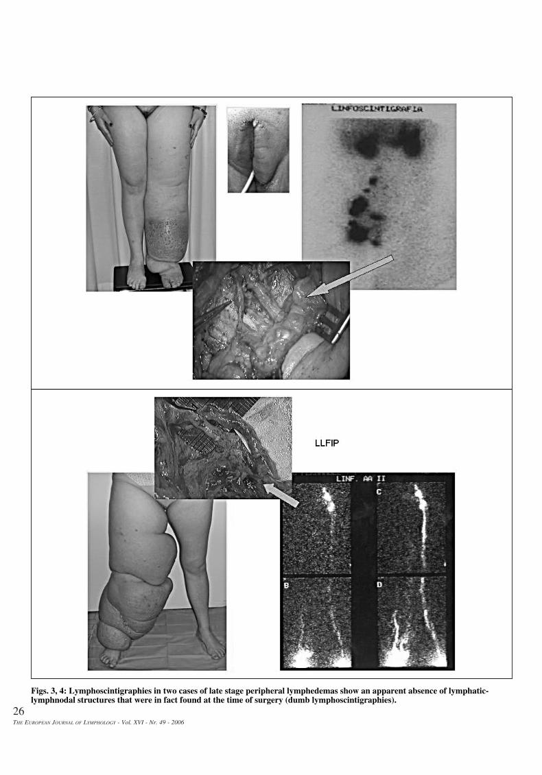
26THE EUROPEAN JOURNAL OF LYMPHOLOGY - Vol. XVI - Nr. 49 - 2006
Figs. 3, 4: Lymphoscintigraphies in two cases of late stage peripheral lymphedemas show an apparent absence of lymphatic-lymphnodal structures that were in fact found at the time of surgery (dumb lymphoscintigraphies).
24-28_BOCCARDO 23-11-2006 10:42 Pagina 26
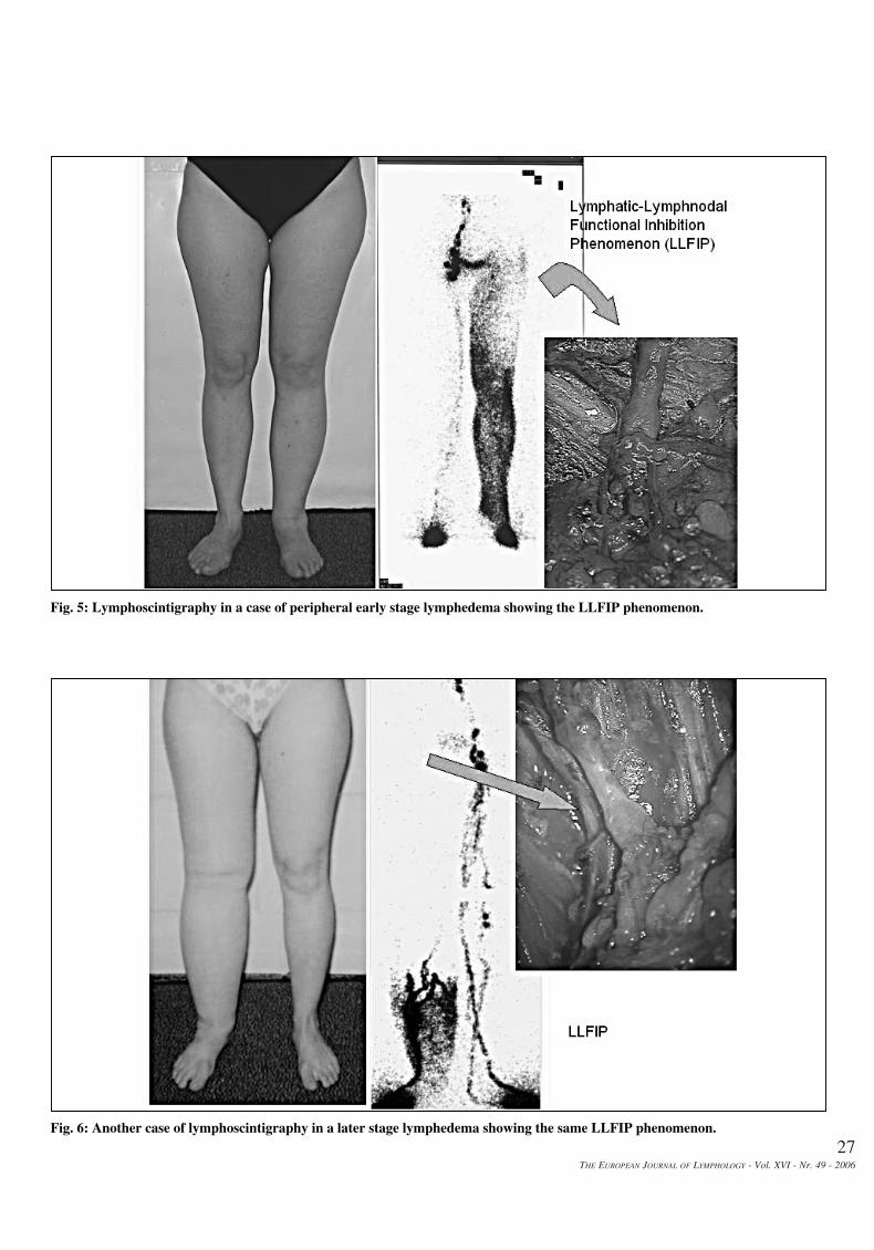
27THE EUROPEAN JOURNAL OF LYMPHOLOGY - Vol. XVI - Nr. 49 - 2006
Fig. 5: Lymphoscintigraphy in a case of peripheral early stage lymphedema showing the LLFIP phenomenon.
Fig. 6: Another case of lymphoscintigraphy in a later stage lymphedema showing the same LLFIP phenomenon.
24-28_BOCCARDO 23-11-2006 10:42 Pagina 27
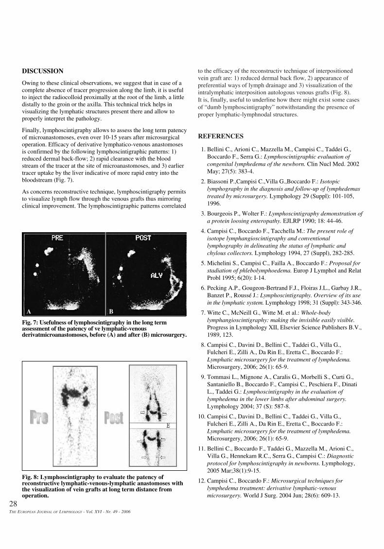
28THE EUROPEAN JOURNAL OF LYMPHOLOGY - Vol. XVI - Nr. 49 - 2006
to the efficacy of the reconstructiv technique of interpositionedvein graft are: 1) reduced dermal back flow, 2) appearance ofpreferential ways of lymph drainage and 3) visualization of theintralymphatic interposition autologous venous grafts (Fig. 8).It is, finally, useful to underline how there might exist some casesof “dumb lymphoscintigraphy” notwithstanding the presence ofproper lymphatic-lymphnodal structures.
REFERENCES
11. Bellini C., Arioni C., Mazzella M., Campisi C., Taddei G.,Boccardo F., Serra G.: Lymphoscintigraphic evaluation ofcongenital lymphedema of the newborn. Clin Nucl Med. 2002May; 27(5): 383-4.
12. Biassoni P.,Campisi C.,Villa G.,Boccardo F.: Isotopiclymphography in the diagnosis and follow-up of lymphedemastreated by microsurgery. Lymphology 29 (Suppl): 101-105,1996.
13. Bourgeois P., Wolter F.: Lymphoscintigraphy demonstration ofa protein loosing enteropathy. EJLRP 1990; 18: 44-46.
14. Campisi C., Boccardo F., Tacchella M.: The present role ofisotope lymphangioscintigraphy and conventionallymphography in delineating the status of lymphatic andchylous collectors. Lymphology 1994, 27 (Suppl), 282-285.
15. Michelini S., Campisi C., Failla A., Boccardo F.: Proposal forstadiation of phlebolymphoedema. Europ J Lymphol and RelatProbl 1995; 6(20): I-14.
16. Pecking A.P., Gougeon-Bertrand F.J., Floiras J.L., Garbay J.R.,Banzet P., Roussé J.: Lymphoscintigraphy. Overview of its usein the lymphatic system. Lymphology 1998; 31 (Suppl): 343-346.
17. Witte C., McNeill G., Witte M. et al.: Whole-bodylymphangioscintigraphy: making the invisible easily visible.Progress in Lymphology XII, Elsevier Science Publishers B.V.,1989, 123.
18. Campisi C., Davini D., Bellini C., Taddei G., Villa G.,Fulcheri E., Zilli A., Da Rin E., Eretta C., Boccardo F.:Lymphatic microsurgery for the treatment of lymphedema.Microsurgery, 2006; 26(1): 65-9.
19. Tommasi L., Mignone A., Caralis G., Morbelli S., Curti G.,Santaniello B., Boccardo F., Campisi C., Peschiera F., DinatiL., Taddei G.: Lymphoscintigraphy in the evaluation oflymphedema in the lower limbs after abdominal surgery.Lymphology 2004; 37 (S): 587-8.
10. Campisi C., Davini D., Bellini C., Taddei G., Villa G.,Fulcheri E., Zilli A., Da Rin E., Eretta C., Boccardo F.:Lymphatic microsurgery for the treatment of lymphedema.Microsurgery, 2006; 26(1): 65-9.
11. Bellini C., Boccardo F., Taddei G., Mazzella M., Arioni C.,Villa G., Hennekam R.C., Serra G., Campisi C.: Diagnosticprotocol for lymphoscintigraphy in newborns. Lymphology,2005 Mar;38(1):9-15.
12. Campisi C., Boccardo F.: Microsurgical techniques forlymphedema treatment: derivative lymphatic-venousmicrosurgery. World J Surg. 2004 Jun; 28(6): 609-13.
DISCUSSION
Owing to these clinical observations, we suggest that in case of acomplete absence of tracer progression along the limb, it is usefulto inject the radiocolloid proximally at the root of the limb, a littledistally to the groin or the axilla. This technical trick helps invisualizing the lymphatic structures present there and allow toproperly interpret the pathology.
Finally, lymphoscintigraphy allows to assess the long term patencyof microanastomoses, even over 10-15 years after microsurgicaloperation. Efficacy of derivative lymphatico-venous anastomosesis confirmed by the following lymphoscintigraphic patterns: 1)reduced dermal back-flow; 2) rapid clearance with the bloodstream of the tracer at the site of microanastomoses, and 3) earliertracer uptake by the liver indicative of more rapid entry into thebloodstream (Fig. 7).
As concerns reconstructive technique, lymphoscintigraphy permitsto visualize lymph flow through the venous grafts thus mirroringclinical improvement. The lymphoscintigraphic patterns correlated
Fig. 8: Lymphoscintigraphy to evaluate the patency ofreconstructive lymphatic-venous-lymphatic anastomoses withthe visualization of vein grafts at long term distance fromoperation.
Fig. 7: Usefulness of lymphoscintigraphy in the long termassessment of the patency of ve lymphatic-venousderivatmicroanastomoses, before (A) and after (B) microsurgery.
A B
24-28_BOCCARDO 23-11-2006 10:42 Pagina 28