Embed Size (px)
Citation preview
Lymphocyte Effector Functions:Killing
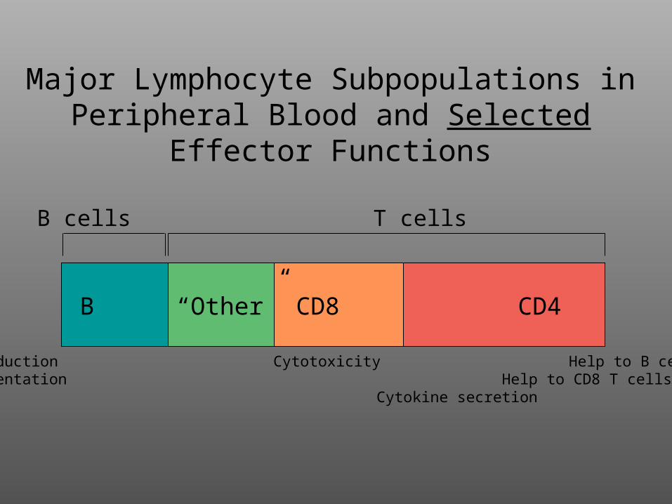
Major Lymphocyte Subpopulations in Peripheral Blood and Selected Effector
Functions
B “Other” CD8 CD4
Ab production Cytotoxicity Help to B cellsAg presentation Help to CD8 T cells
Cytokine secretion
B cells T cells
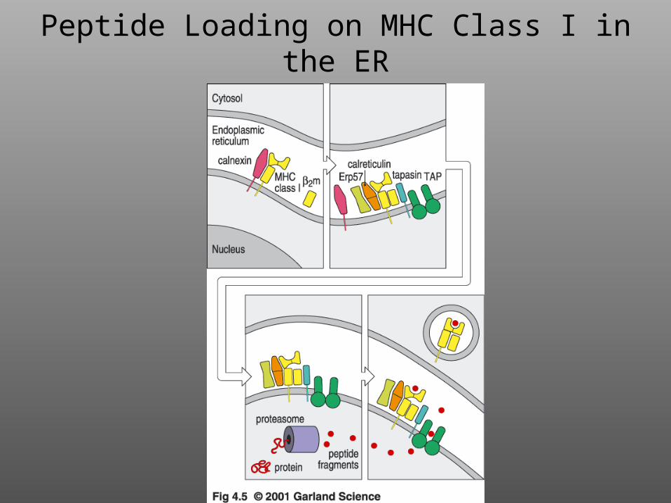
Peptide Loading on MHC Class I in the ER
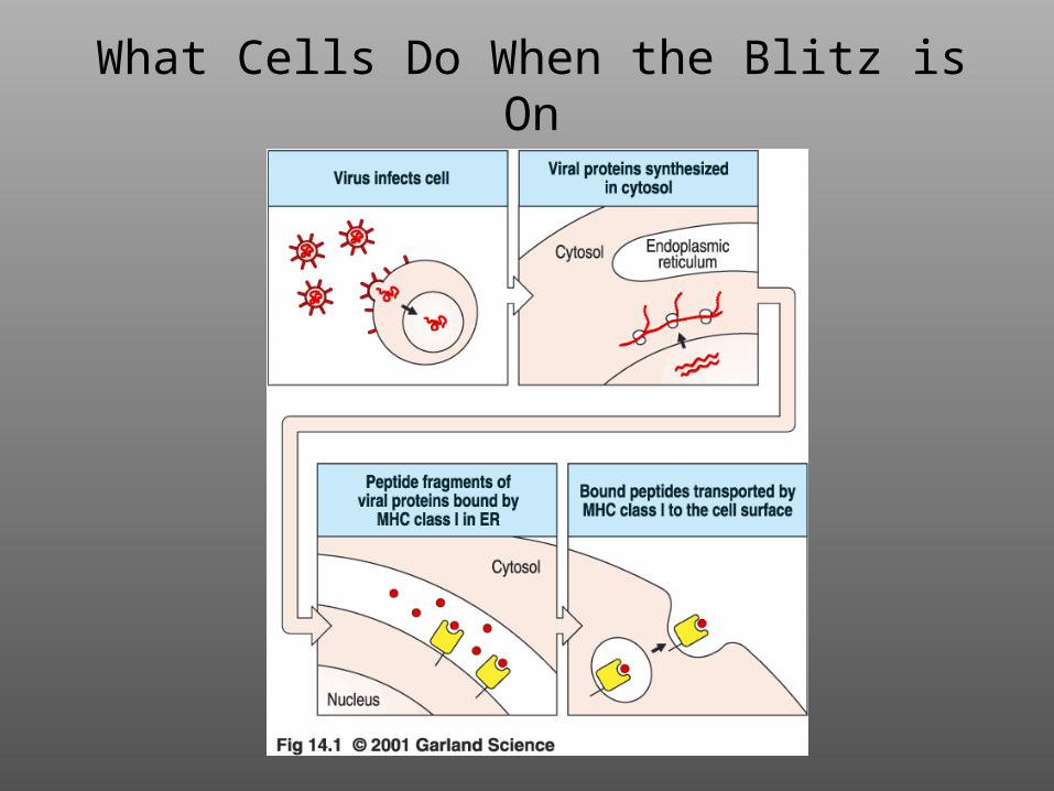
What Cells Do When the Blitz is On
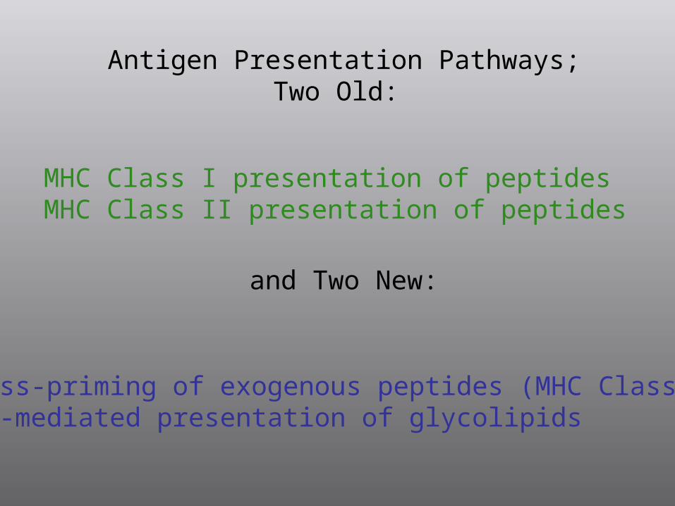
Antigen Presentation Pathways;Two Old:
and Two New:
MHC Class I presentation of peptidesMHC Class II presentation of peptides
Cross-priming of exogenous peptides (MHC Class I)CD1-mediated presentation of glycolipids
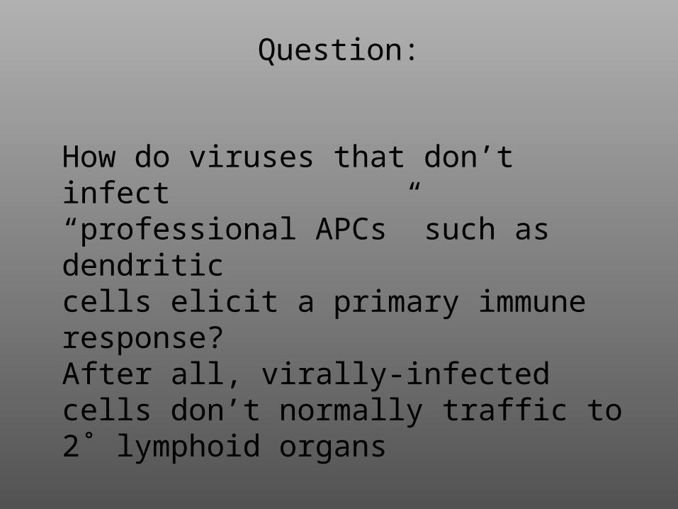
Question:
How do viruses that don’t infect “professional APCs” such as dendriticcells elicit a primary immune response?After all, virally-infected cells don’t normally traffic to 2˚ lymphoid organs
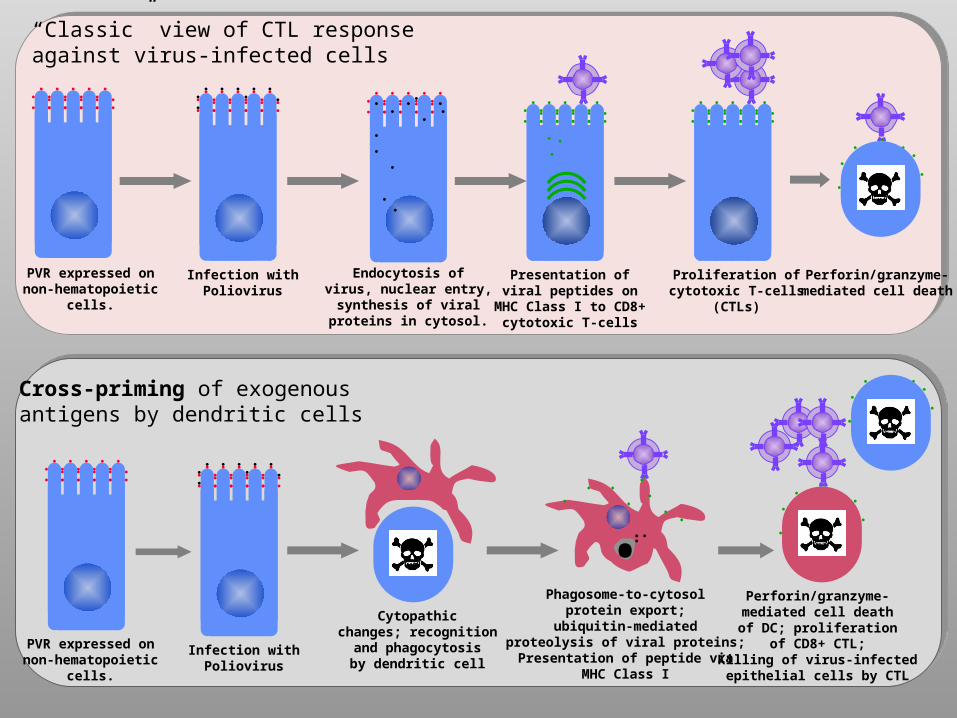
PVR expressed onnon-hematopoietic
cells.
Infection withPoliovirus
Endocytosis ofvirus, nuclear entry,
synthesis of viralproteins in cytosol.
Presentation ofviral peptides on
MHC Class I to CD8+cytotoxic T-cells
Proliferation ofcytotoxic T-cells
(CTLs)
Perforin/granzyme-mediated cell death
“Classic” view of CTL responseagainst virus-infected cells
Cross-priming of exogenousantigens by dendritic cells
PVR expressed onnon-hematopoietic
cells.
Infection withPoliovirus
Cytopathicchanges; recognition
and phagocytosisby dendritic cell
Phagosome-to-cytosolprotein export;
ubiquitin-mediatedproteolysis of viral proteins;Presentation of peptide via
MHC Class I
•Perforin/granzyme-mediated cell deathof DC; proliferation
of CD8+ CTL;Killing of virus-infectedepithelial cells by CTL
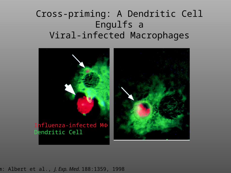
Cross-priming: A Dendritic Cell Engulfs aViral-infected Macrophages
Influenza-infected MDendritic Cell
From: Albert et al., J. Exp. Med. 188:1359, 1998
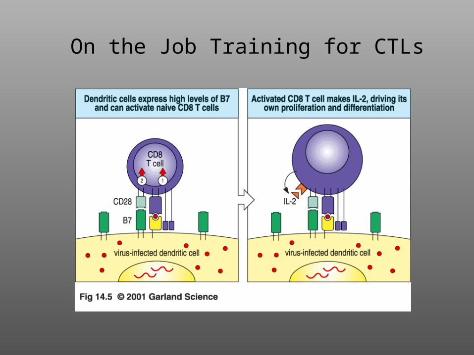
On the Job Training for CTLs
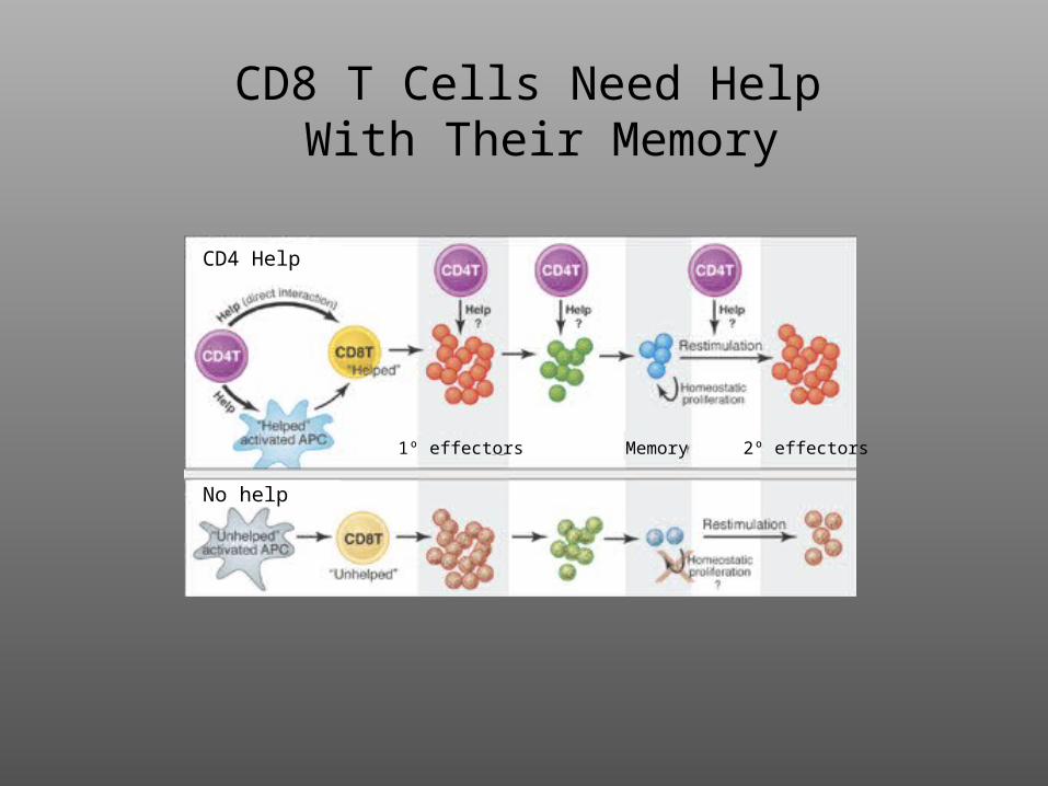
CD8 T Cells Need Help With Their Memory
CD4 Help
No help
1º effectors Memory 2º effectors

QuickTime™ and aCinepak decompressor
are needed to see this picture.
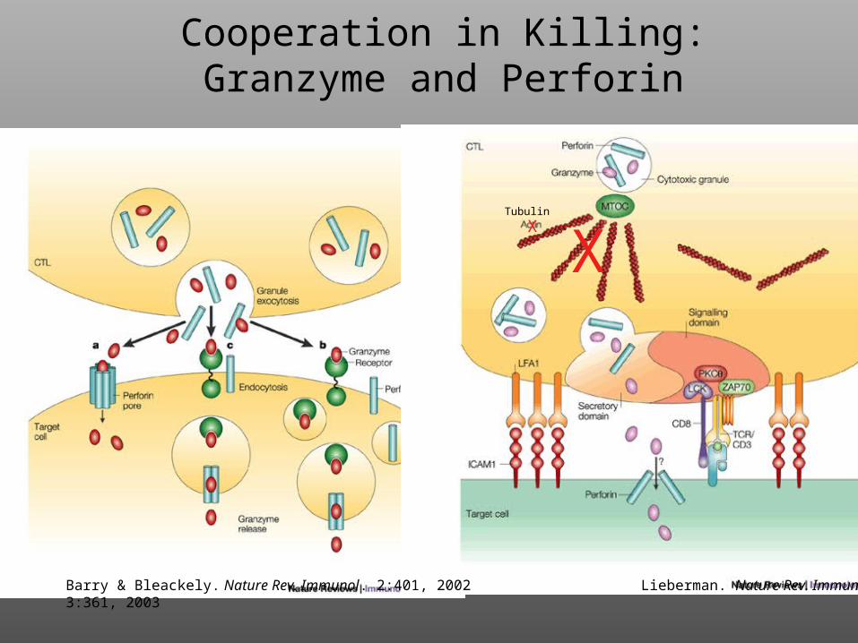
Cooperation in Killing:Granzyme and Perforin
Barry & Bleackely. Nature Rev. Immunol. 2:401, 2002 Lieberman. Nature Rev. Immunol. 3:361, 2003
XX
Tubulin
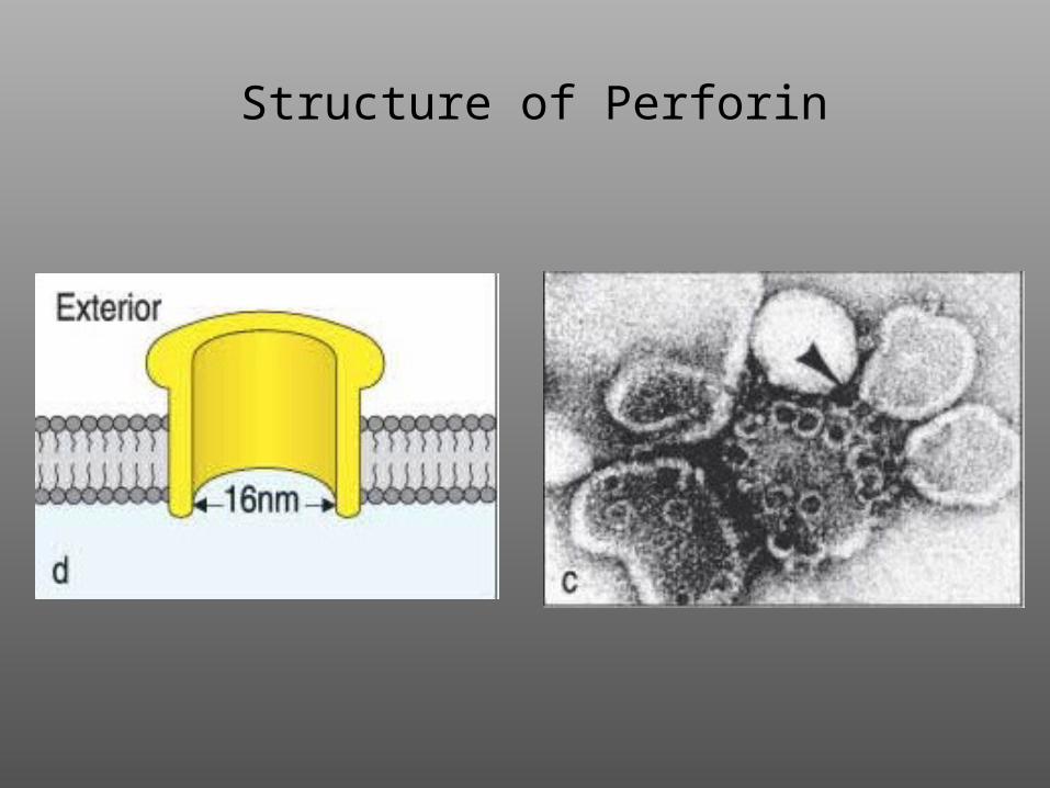
Structure of Perforin
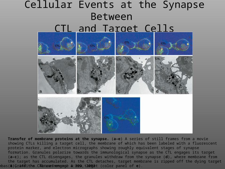
Cellular Events at the Synapse Between
CTL and Target Cells
Transfer of membrane proteins at the synapse. (a–e) A series of still frames from a movie showing CTLs killing a target cell, the membrane of which has been labeled with a fluorescent protein marker, and electron micrographs showing roughly equivalent stages of synapse formation. Granules polarize towards the immunological synapse as the CTL engages its target (a–c); as the CTL disengages, the granules withdraw from the synapse (d), where membrane from the target has accumulated. As the CTL detaches, target membrane is ripped off the dying target (e), and the CTL can engage a new target (color panel of e).
Trambas & Griffiths. Nature Immunol. 4:399, 2003
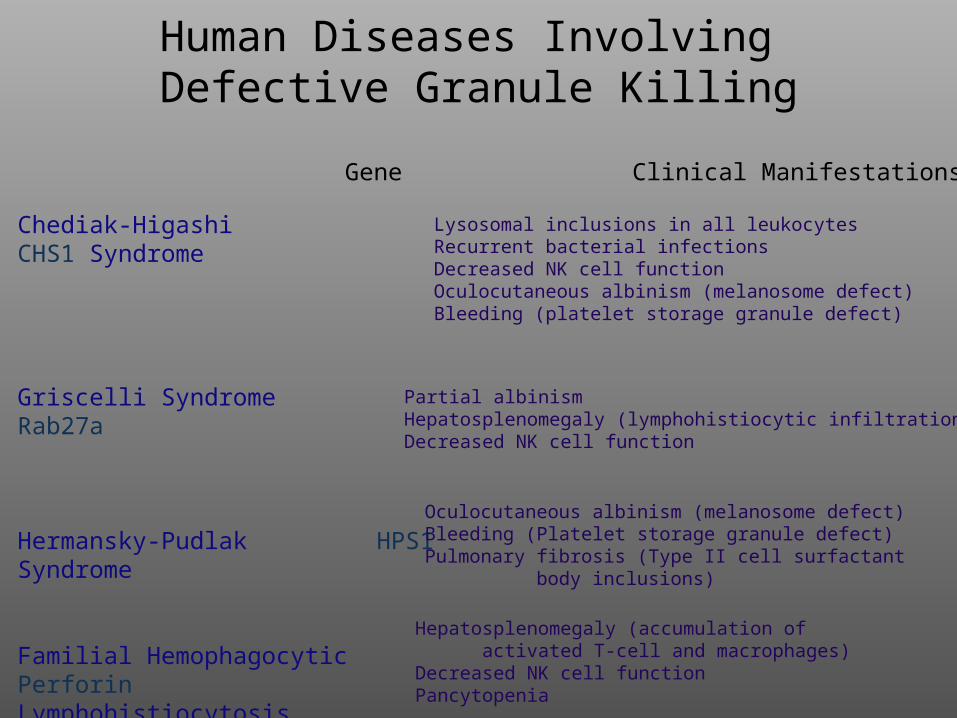
Human Diseases Involving Defective Granule Killing
Chediak-Higashi CHS1 Syndrome
Griscelli Syndrome Rab27a
Hermansky-Pudlak HPS1 Syndrome
Familial Hemophagocytic PerforinLymphohistiocytosis (30% of cases)
Lysosomal inclusions in all leukocytesRecurrent bacterial infectionsDecreased NK cell functionOculocutaneous albinism (melanosome defect)Bleeding (platelet storage granule defect)
Oculocutaneous albinism (melanosome defect)Bleeding (Platelet storage granule defect)Pulmonary fibrosis (Type II cell surfactant body inclusions)
Partial albinismHepatosplenomegaly (lymphohistiocytic infiltration)Decreased NK cell function
Hepatosplenomegaly (accumulation of activated T-cell and macrophages)Decreased NK cell functionPancytopenia
Disease Gene Clinical Manifestations
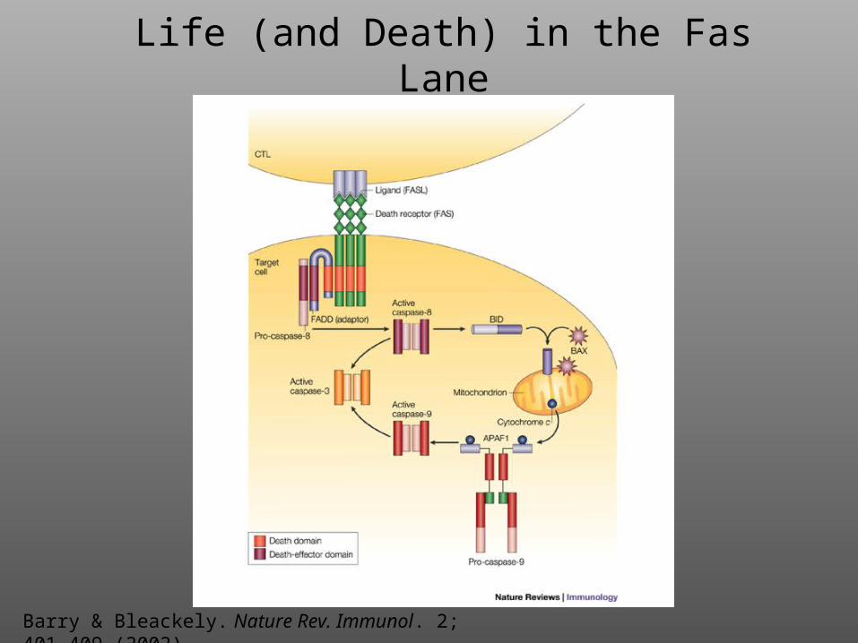
Life (and Death) in the Fas Lane
Barry & Bleackely. Nature Rev. Immunol. 2; 401-409 (2002)
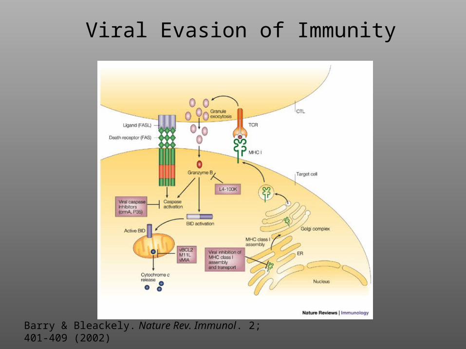
Viral Evasion of Immunity
Barry & Bleackely. Nature Rev. Immunol. 2; 401-409 (2002)
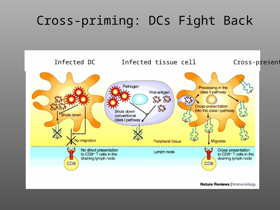
Cross-priming: DCs Fight Back
Infected DC Infected tissue cell Cross-presenting DC
B cells T cells
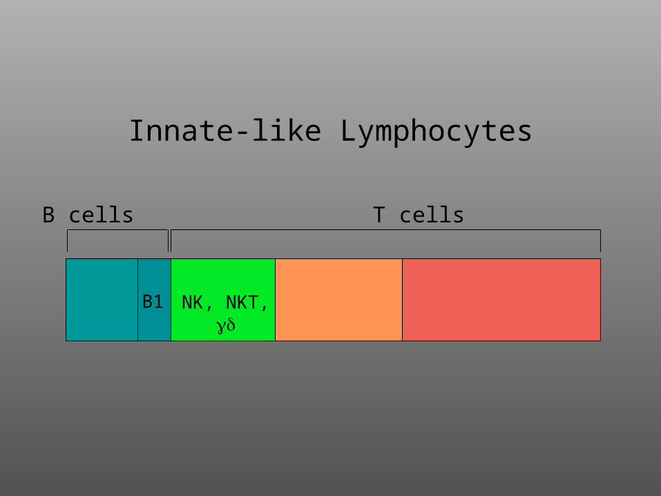
Innate-like Lymphocytes
B1 NK, NKT,
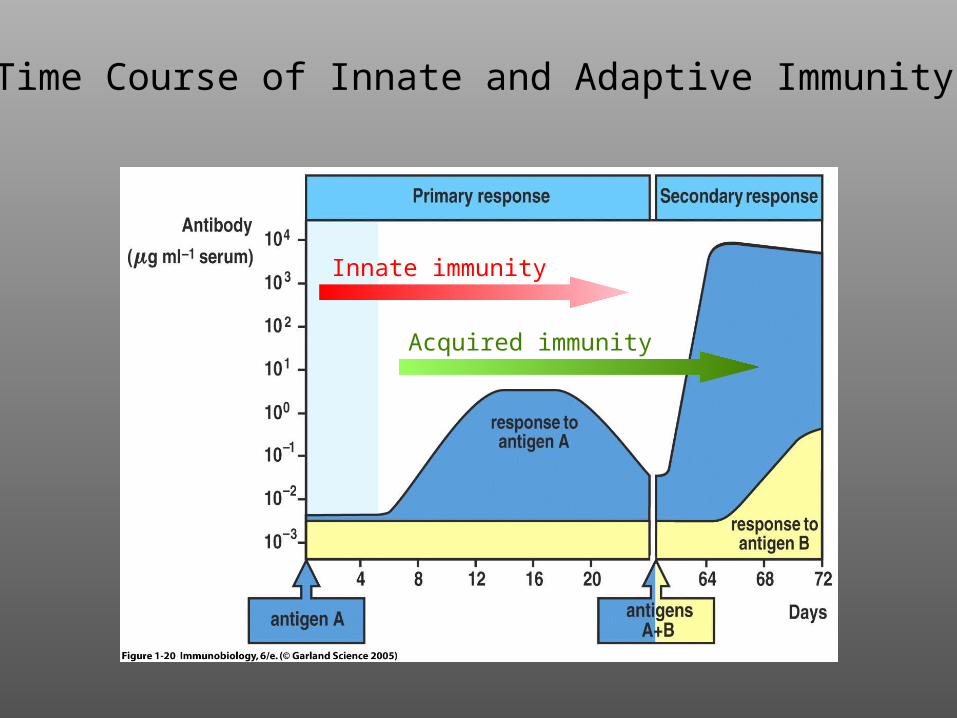
Time Course of thePrimary Immune Response
Innate immunity
Acquired immunity
Time Course of Innate and Adaptive Immunity
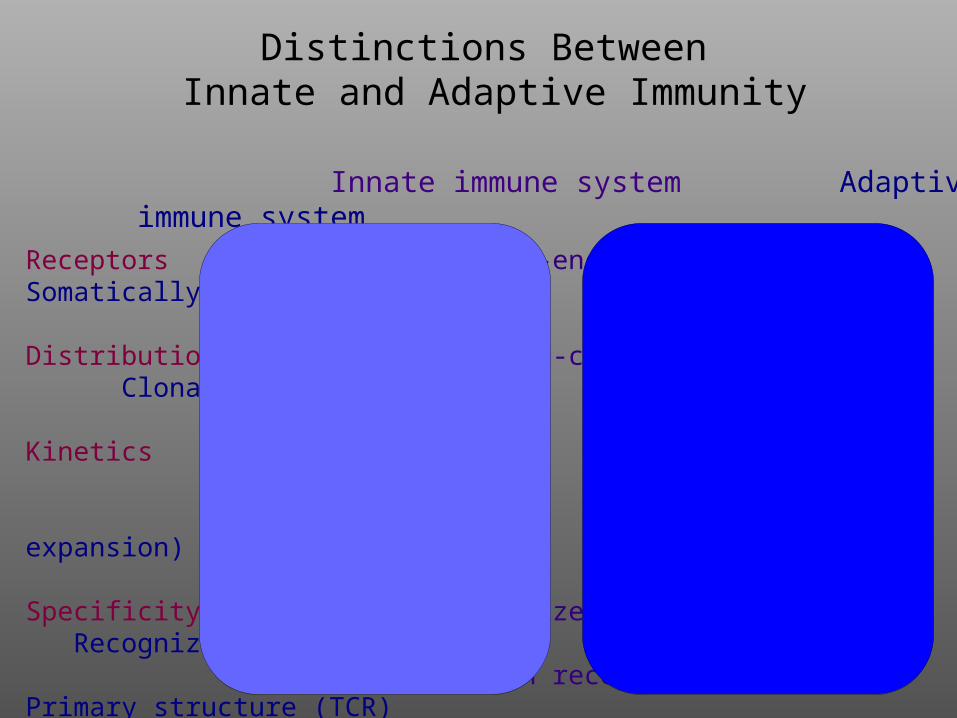
Distinctions Between
Innate and Adaptive Immunity
Innate immune system Adaptive immune system
Receptors Germline-encoded Somatically engineered
Distribution Non-clonal Clonal
Kinetics Rapid Slow (requires clonal expansion)
Specificity Recognizes non-self Recognizes “altered self”
“pattern recognition” Primary structure (TCR) Higher order structure
(Immunoglobulin; BCR)
Effector Cells All Primarily lymphocytes, DCs, M
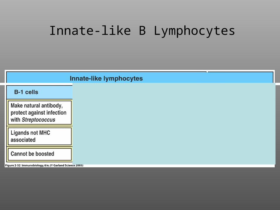
Innate-like B Lymphocytes
NK cells
not MHCassociated
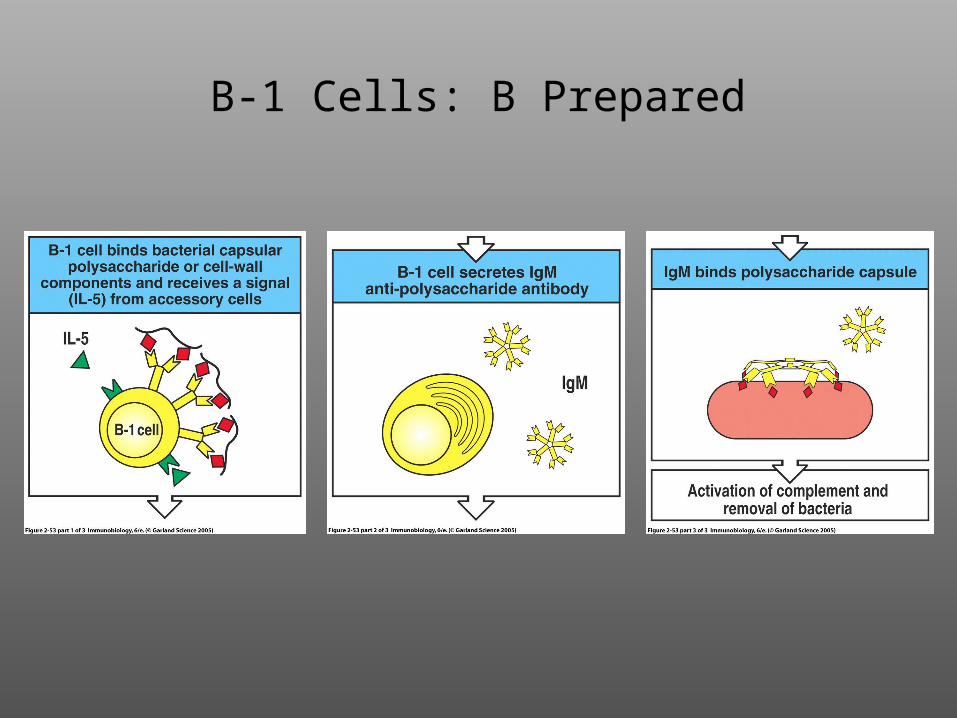
B-1 Cells: B Prepared
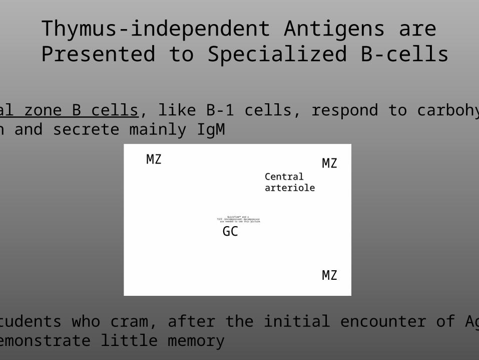
Marginal zone B cells, like B-1 cells, respond to carbohydrate antigen and secrete mainly IgM
Like students who cram, after the initial encounter of Ag, they demonstrate little memory
Thymus-independent Antigens arePresented to Specialized B-cells
QuickTime™ and aTIFF (Uncompressed) decompressor
are needed to see this picture.
MZMZ
MZ
GC
CentralCentralarteriolearteriole
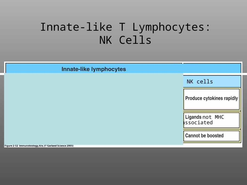
Innate-like T Lymphocytes:NK Cells
NK cells
not MHCassociated
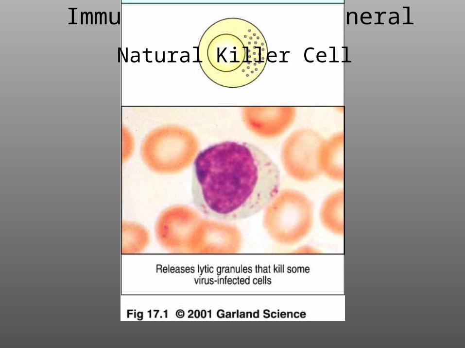
Immunology Course-General PrinciplesNatural Killer Cell
How do NK Cells Recognize Their Targets?
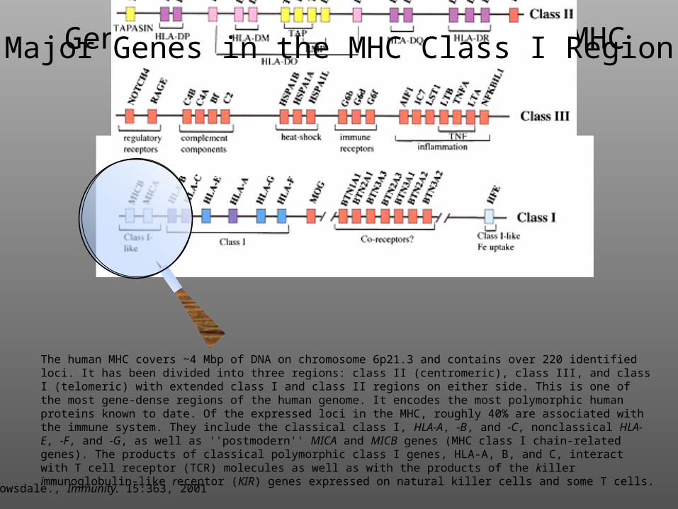
Gene Structure of the Human MHC
Trowsdale., Immunity. 15:363, 2001
The human MHC covers ~4 Mbp of DNA on chromosome 6p21.3 and contains over 220 identified loci. It has been divided into three regions: class II (centromeric), class III, and class I (telomeric) with extended class I and class II regions on either side. This is one of the most gene-dense regions of the human genome. It encodes the most polymorphic human proteins known to date. Of the expressed loci in the MHC, roughly 40% are associated with the immune system. They include the classical class I, HLA-A, -B, and -C, nonclassical HLA-E, -F, and -G, as well as ''postmodern'' MICA and MICB genes (MHC class I chain-related genes). The products of classical polymorphic class I genes, HLA-A, B, and C, interact with T cell receptor (TCR) molecules as well as with the products of the killer immunoglobulin-like receptor (KIR) genes expressed on natural killer cells and some T cells.
Major Genes in the MHC Class I Region
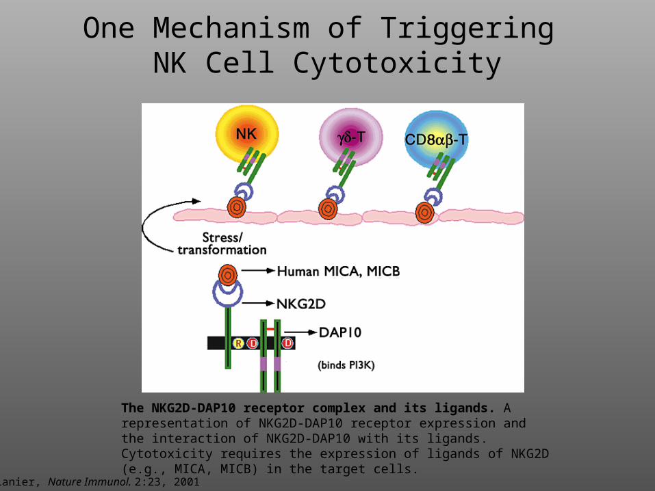
Lanier, Nature Immunol. 2:23, 2001
The NKG2D-DAP10 receptor complex and its ligands. A representation of NKG2D-DAP10 receptor expression and the interaction of NKG2D-DAP10 with its ligands. Cytotoxicity requires the expression of ligands of NKG2D (e.g., MICA, MICB) in the target cells.
One Mechanism of Triggering NK Cell Cytotoxicity
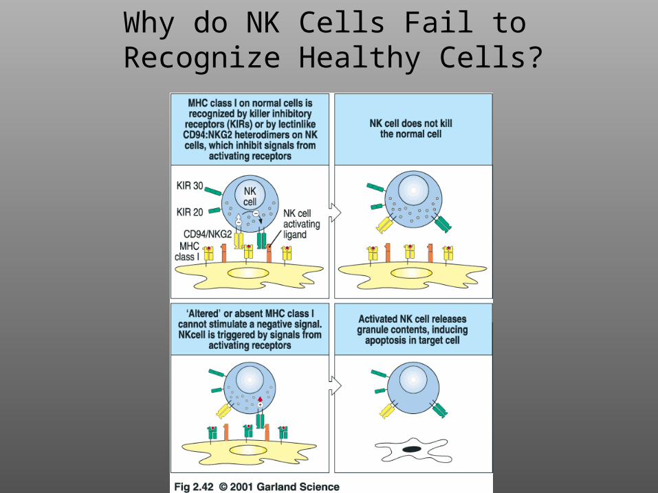
Why do NK Cells Fail to
Recognize Healthy Cells?
Y
Y
Y
Y
Y
Y
YPYP
YPYP
YPYP XX XX MM
YPYP xx xx LL
DAP12DAP12 DAP10DAP10
KIR2DSNKG2D
KIR2DL
SykSyk
PI 3-kinasePI 3-kinase
SHP-2SHP-2
++++--
CytotoxicityCytokine Secretion
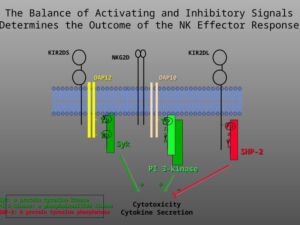
The Balance of Activating and Inhibitory SignalsDetermines the Outcome of the NK Effector Response
Syk: a protein tyrosine kinaseSyk: a protein tyrosine kinasePI 3-kinase: a phosphoinositide kinasePI 3-kinase: a phosphoinositide kinaseSHP-2: A protein tyrosine phosphataseSHP-2: A protein tyrosine phosphatase
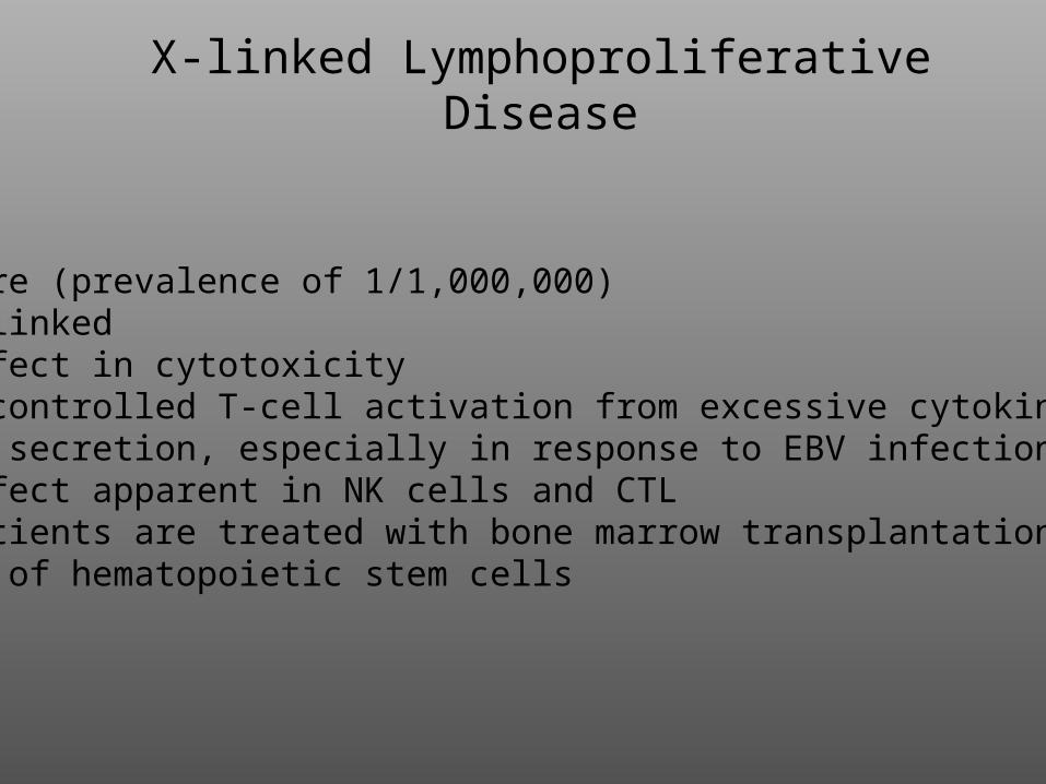
X-linked Lymphoproliferative Disease
Rare (prevalence of 1/1,000,000)X-linkedDefect in cytotoxicityUncontrolled T-cell activation from excessive cytokine secretion, especially in response to EBV infectionDefect apparent in NK cells and CTLPatients are treated with bone marrow transplantations of hematopoietic stem cells
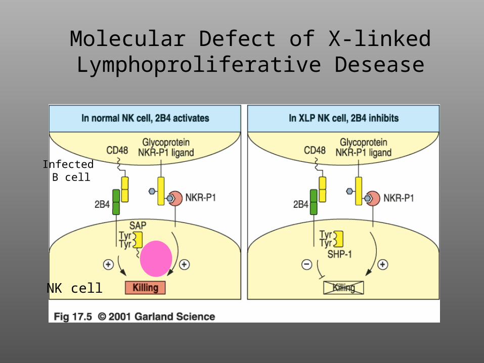
Molecular Defect of X-linked Lymphoproliferative Desease
NK cell
Infected B cell
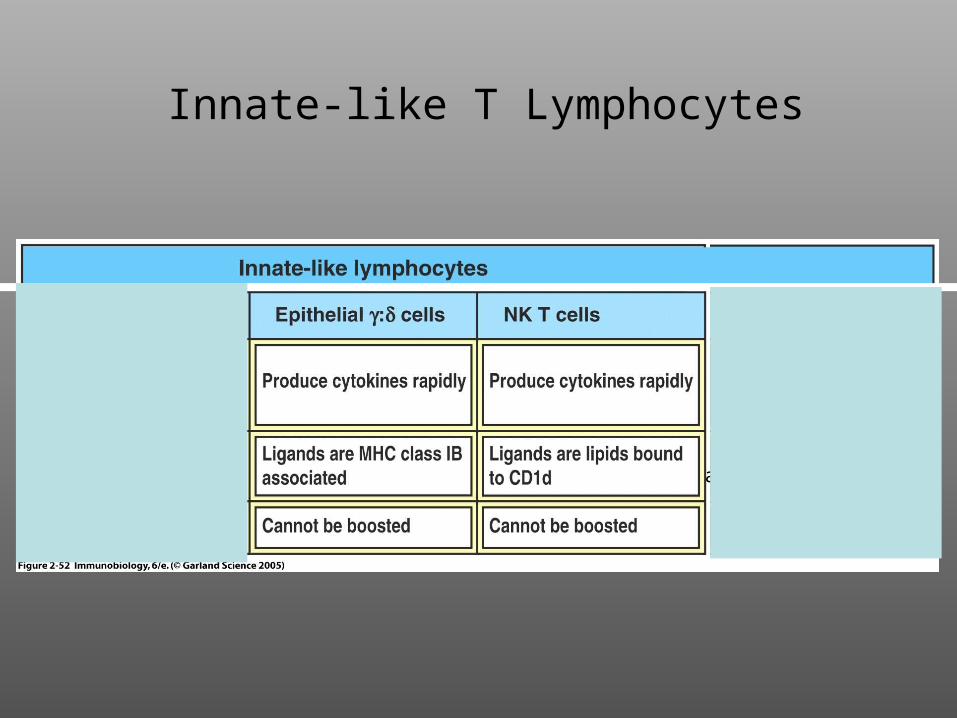
Innate-like T Lymphocytes
NK cells
not MHCassociated
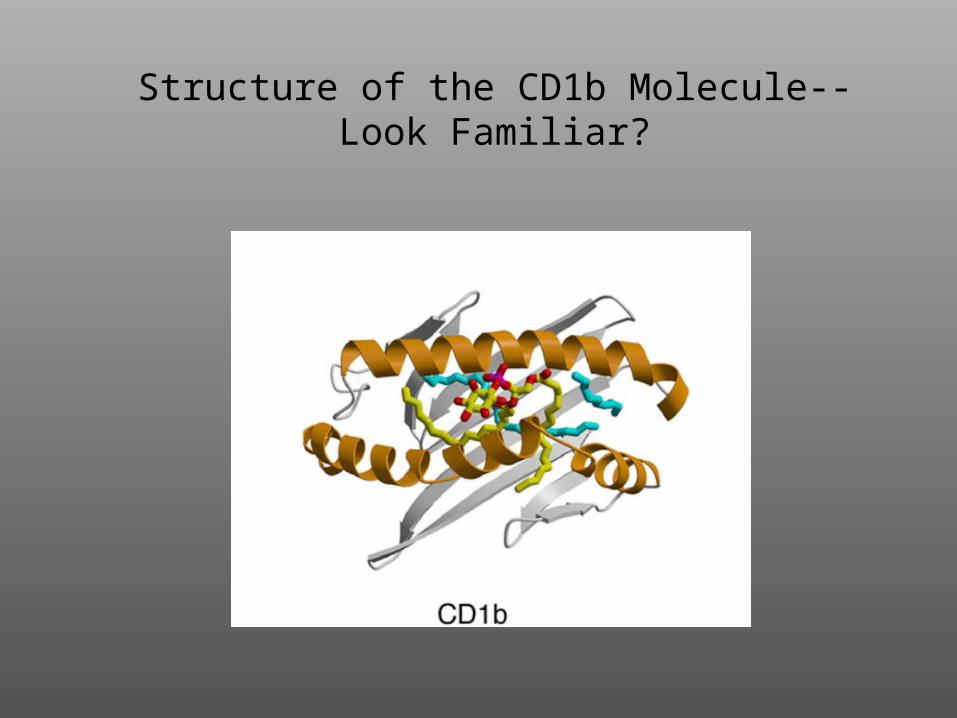
Structure of the CD1b Molecule--Look Familiar?
The NKT Cell Recognizes Glycolipid Antigen
Presented by CD1 on the APC
QuickTime™ and aTIFF (Uncompressed) decompressor
are needed to see this picture.
DCs that are infected with intracellular bacteria present foreign bacterial lipid antigens on the cell surfacbound to CD1 molecules. CD1-restricted T cells that are specific for the foreign microbial lipids are stimulatedto carry out effector functions, including the secretion of cytolytic granules containing perforin and granulysin, which lyse the infected cells and have direct antimicrobial effects, respectively, and the production ofIFN- and TNF-, which activate the microbicidal functions of macrophages.
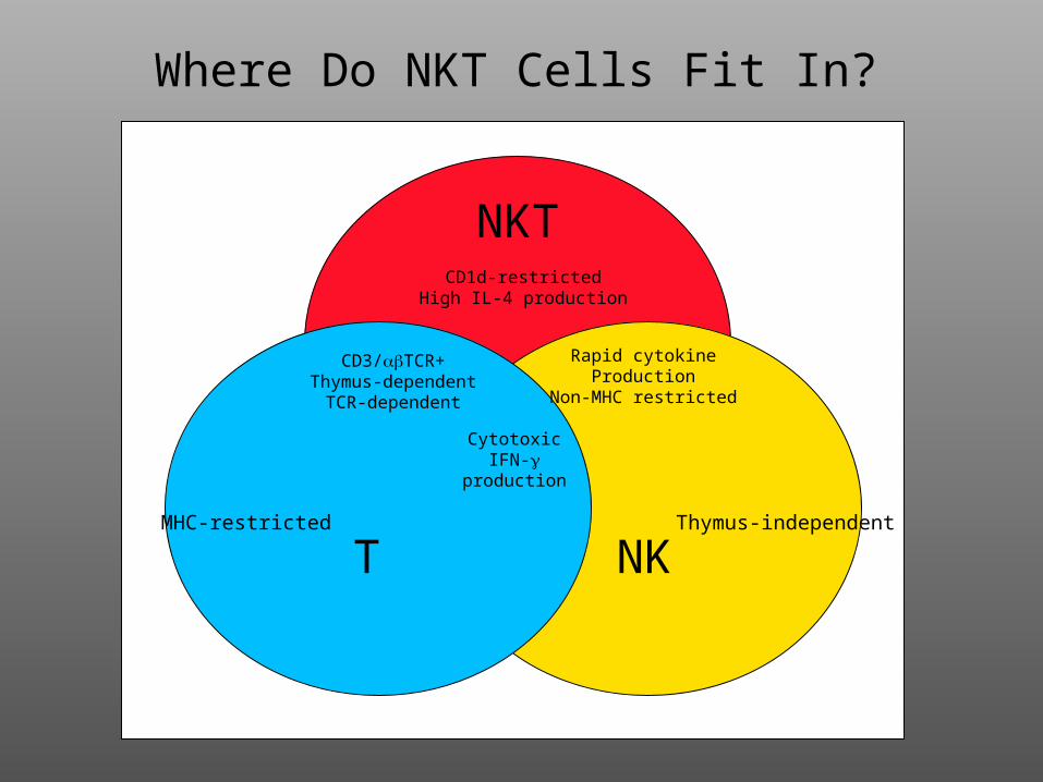
Where Do NKT Cells Fit In?
NKT
NKTMHC-restricted Thymus-independent
Rapid cytokineProduction
Non-MHC restricted
CD1d-restrictedHigh IL-4 production
CD3/TCR+Thymus-dependent
TCR-dependent
CytotoxicIFN-
production
1. For cytotoxic CD8 T-cells, ligation of the TCR by MHC I/peptide + co-stimulation results in release of granzymes and perforin and/or FasL, leading to apoptosisof the target cells.
2. Viruses evade host defense, in part, by down-regulating MHC Class I. Uninfected dendritic cells circumvent this by “cross-priming”: phagocytosis of virus-infected cell and presentation of “exogenous” viral antigens on MHC Class I.
3. The innate immune system has a rapid onset and recognizes molecular patterns in a non-clonal fashion.
4. NK cells lack TCRs, but instead express both activating and inhibitory (e.g., KIRs) receptors at their surfaces. The relative expression and ligation of these receptors determines the outcome (i.e., killing or not) of the NK effector response.
5. Innate immune B-cells (e.g., B-1 cells and marginal zone B cells) recognizecarbohydrate antigens, secrete IgM, and are not long-lived.
6. Innate immune T-cells ( T-cells, and NK T cells) recognize non-peptide antigens innon-classical MHC-like molecules. They mediate cytotoxicity & rapid cytokine secretion.
Summary