Embed Size (px)
Citation preview

Lymphatics of the Heart
By ROBERT A. JOHNSON, M.D., AND THOMAS M. BLAKE, M.D.
LYMPHATIC DRAINAGE of the heart hasreceived little attention in the past but
its potential importance has been recognizedrecently by several investigators.1-5 Review ofstudies of cardiac lymphatics4' 6 shows thatmany details of the anatomy of the system are
still unknown, largely because of limitationsof the technics of investigation. The small sizeand delicate structure of peripheral lymphat-ics and the presence of valves preclude theirdemonstration adequately by direct injection,and visualization in most instances has beenby injection into living tissue of vital dyeswhich are removed by lymphatics. Usingthese technics, the most recent investigationshave been those of Patek,9 Miller and associ-ates,3 and Golab.10 An effective method fordemonstrating lymphatics by direct applica-tion of hydrogen peroxide was described in1922 by Magnus and Stubel" and in 1963 byParke and Michels,12 and the purpose of thisreport is to present observations of the car-
diac lymphatic system obtained by thismeans. Injection and clearing technics havebeen utilized in some instances also.
MethodsHydrogen peroxide initiates an oxidoreduction
reaction with catalase and peroxidase in the tis-sue or lymph, or both, producing oxygen andwater.13-15 The released oxygen causes distentionof lymphatics and sometimes blood vessels; thetwo may be differentiated on the basis of theirmorphology. Lymphatics are superficial, trans-parent, and generally have a bulbous, irregularcontour due to the presence of valves;7' 9, 16, 17blood capillaries are of smaller diameter and con-
stant size with their general arrangement more
orderly than lymphatics. Artifacts produced byextravascular dissection of released oxygen be-
From the Department of Medicine, University ofMississippi School of Medicine, Jackson, Mississippi.
Supported in part by the Mississippi Heart As-sociation, and Grants HE-07159 and 2-F2-HE-19,626-02 from the National Heart Institute, U. S.Public Health Service.
Circulation, Volume XXXIII, January 1966
neath the epicardium or endocardium form in-consistent patterns unlike lymphatics or bloodvessels and are recognized easily.
Hearts of 25 pigs, 13 dogs, and 20 humanswere examined. Observation of subepicardial andsubendocardial lymphatics was facilitated bycutting open the hearts according to a methoddescribed by Schlesinger,18 modified by leavingthe ventricular septum intact. A 1 per cent solu-tion of hydrogen peroxide applied topically witha cotton-tipped applicator resulted in distentionof lymphatics in the region of application. Thereaction was more effective in some hearts thanin others but seemed to be improved in speci-mens refrigerated 24 hours or longer before studyand still occurred in those refrigerated for morethan a week. Frothing produced by the peroxidereaction was a complication that obscured somevessels but could be diminished by placingthe specimens in 10 per cent formalin for 30 to60 minutes prior to the use of peroxide. Wholespecimens were immersed effectively in peroxidein some instances but, in general, results werebest in unfixed tissue with local application.
Specimens were observed through a stereo-microscope or with the naked eye. As they be-came distended, lymphatics extended beyond thefrothy area where peroxide was applied and wereseen clearly. They remained distended for sev-eral minutes allowing time for photography andmeasurement with an ocular micrometer. Thechannels could be distended repeatedly by re-application of peroxide.
Photographs were made with Kodachrome IIProfessional Type A film and a 35-mm. Exactacamera back attached through one ocular of thestereomicroscope with magnification ranging from7 to 30 times. Exposure times were determinedby means of a photometer inserted into the otherocular of the stereomicroscope. Highlights of thevessels were brought out by varying the angleand intensity of the light from a standard 35-mm.slide projector equipped with a 500-watt bulb.
Injection of india ink and clearing by theSpalteholz method were used in some instances.
ResultsObservations in Dog and Pig Hearts
A dense network of subepicardial lymphaticcapillaries measuring around 15 to 20 micronsin diameter covers the outer surface of theheart. These join channels of intermediate
137
by guest on July 5, 2018http://circ.ahajournals.org/
Dow
nloaded from
JOHNSON, BLAKE
size, some of which anastomose with largerchannels accompanying the anterior and pos-terior coronary blood vessels. Others courselongitudinally and join a large channel in theatrioventricular (AV) sulcus. The duct of theAV sulcus, from which the main cardiaclymph duct arises, is formed by continuationof the anterior and posterior paracoronarylympathic ducts.8' 19 The subepicardial capil-lary network over the atria is similar to thatover the ventricles, and ducts of similar mag-nitude join the duct in the AV sulcus.Beneath the endocardium of both ventricles,
including the septum, small lymphatic vesselsof relatively uniform caliber (15 to 20 micronsin diameter) form a dense network orientedtransversely to the subjacent muscle fibers.These vessels are regular in contour and donot appear to contain valves (fig. 1). Re-approximating the cut edges of the heart andviewing the ventricular chambers from above,the network appears in a spiral arrangement.Over the apices of papillary muscles the
channels become larger, with a bulbous ap-pearance suggesting the presence of valves,and in dog hearts these channels form a dens-er network than in the pig heart. In both
Figure 1
Subendocardial lymphatic capillaries (15 to 20 mi-
crons in diameter) overlying a portion of the anteriorpapillary muscle and surrounding endocardium in theleft ventricle of a pig heart. The longitudinal axis ofthe papillary muscle extends from the left lowertoward the right upper portion of the photograph. Inthe right upper field the apex has been cut andthere is a fragment of severed chorda tendinea. Thefluffy areas are artifact produced by the peroxide re-
action. Magnification 8 X.
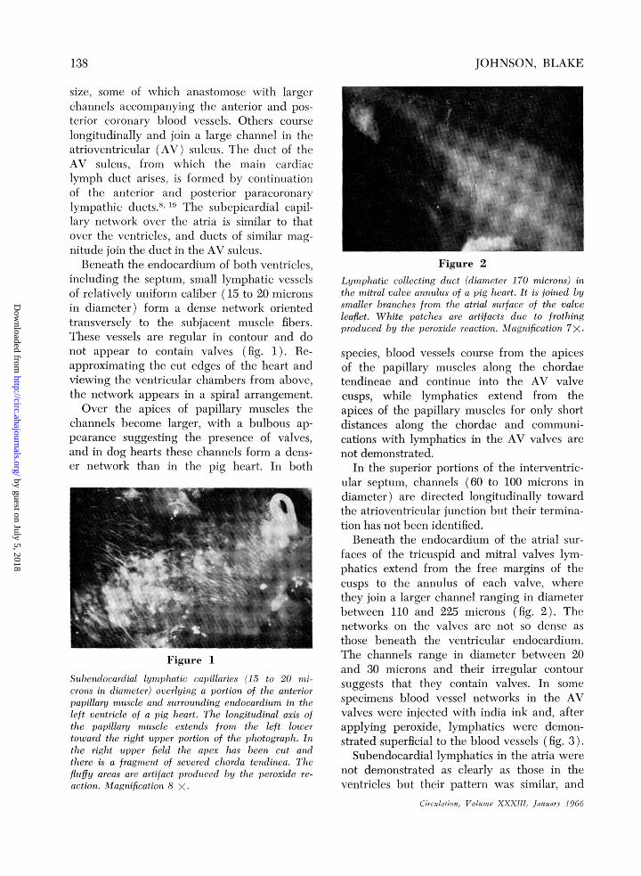
Figure 2Lymphatic collecting duct (diameter 170 microns) inthe mitral valve annulus of a pig heart. It is joined bysmaller branches from the atrial surface of the valveleaflet. White patches are artifacts due to frothingproduced by the peroxide reaction. Magnification 7x.
species, blood vessels course from the apicesof the papillary muscles along the chordaetendineae and continue into the AV valvecusps, while lymphatics extend from theapices of the papillary muscles for only shortdistances along the chordae and communi-cations with lymphatics in the AV valves arenot demonstrated.
In the superior portions of the interventric-ular septum, channels (60 to 100 microns indiameter) are directed longitudinally towardthe atrioventricular junction but their termina-tion has not been identified.Beneath the endocardium of the atrial sur-
faces of the tricuspid and mitral valves lym-phatics extend from the free margins of thecusps to the annulus of each valve, wherethey join a larger channel ranging in diameterbetween 110 and 225 microns (fig. 2). Thenetworks on the valves are not so dense asthose beneath the ventricular endocardium.The channels range in diameter between 20and 30 microns and their irregular contoursuggests that they contain valves. In somespecimens blood vessel networks in the AVvalves were injected with india ink and, afterapplying peroxide, lymphatics were demon-strated superficial to the blood vessels (fig. 3).
Subendocardial lymphatics in the atria werenot demonstrated as clearly as those in theventricles but their pattern was similar, and
Circulation, Volume XXXll, January 1966
138
by guest on July 5, 2018http://circ.ahajournals.org/
Dow
nloaded from
LYMPHATICS OF THE HEART
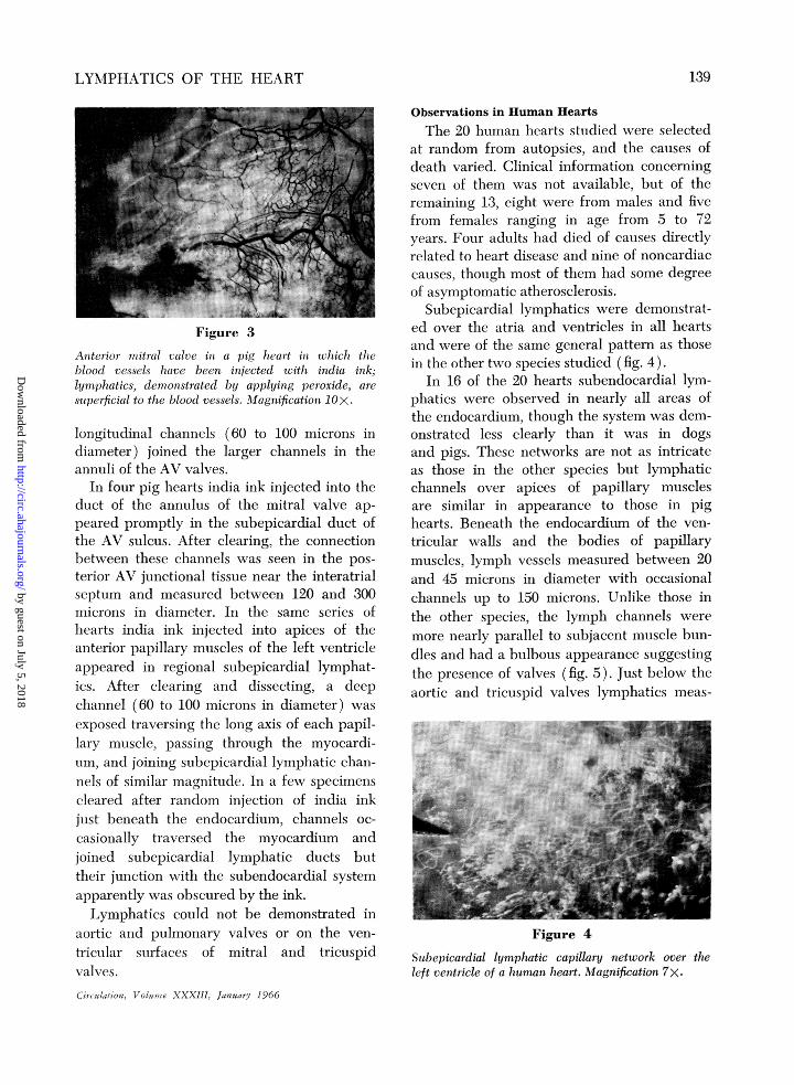
Figure 3
Anterior mitral valve in a pig heart in which theblood vessels have been injected with india ink;lymphatics, demonstrated by applying peroxide, aresuperficial to the blood vessels. Magnification lOX.
longitudinal channels (60 to 100 microns indiameter) joined the larger channels in theannuli of the AV valves.
In four pig hearts india ink injected into theduct of the annulus of the mitral valve ap-peared promptly in the subepicardial duct ofthe AV sulcus. After clearing, the connectionbetween these channels was seen in the pos-terior AV junctional tissue near the interatrialseptum and measured between 120 and 300microns in diameter. In the same series ofhearts india ink injected into apices of theanterior papillary muscles of the left ventricleappeared in regional subepicardial lymphat-ics. After clearing and dissecting, a deepchannel (60 to 100 microns in diameter) wasexposed traversing the long axis of each papil-lary muscle, passing through the myocardi-um, and joining subepicardial lymphatic chan-nels of similar magnitude. In a few specimenscleared after random injection of india inkjust beneath the endocardium, channels oc-casionally traversed the myocardium andjoined subepicardial lymphatic ducts buttheir junction with the subendocardial systemapparently was obscured by the ink.Lymphatics could not be demonstrated in
aortic and pulmonary valves or on the ven-tricular surfaces of mitral and tricuspidvalves.Circlulation, Volumze XXXIII, January 1966
Observations in Human HeartsThe 20 human hearts studied were selected
at random from autopsies, and the causes ofdeath varied. Clinical information concerningseven of them was not available, but of theremaining 13, eight were from males and fivefrom females ranging in age from 5 to 72years. Four adults had died of causes directlyrelated to heart disease and nine of noncardiaccauses, though most of them had some degreeof asymptomatic atherosclerosis.
Subepicardial lymphatics were demonstrat-ed over the atria and ventricles in all heartsand were of the same general pattern as thosein the other two species studied (fig. 4).
In 16 of the 20 hearts subendocardial lym-phatics were observed in nearly all areas ofthe endocardium, though the system was dem-onstrated less clearly than it was in dogsand pigs. These networks are not as intricateas those in the other species but lymphaticchannels over apices of papillary musclesare similar in appearance to those in pighearts. Beneath the endocardium of the ven-tricular walls and the bodies of papillarymuscles, lymph vessels measured between 20and 45 microns in diameter with occasionalchannels up to 150 microns. Unlike those inthe other species, the lymph channels weremore nearly parallel to subjacent muscle bun-dles and had a bulbous appearance suggestingthe presence of valves (fig. 5). Just below theaortic and tricuspid valves lymphatics meas-
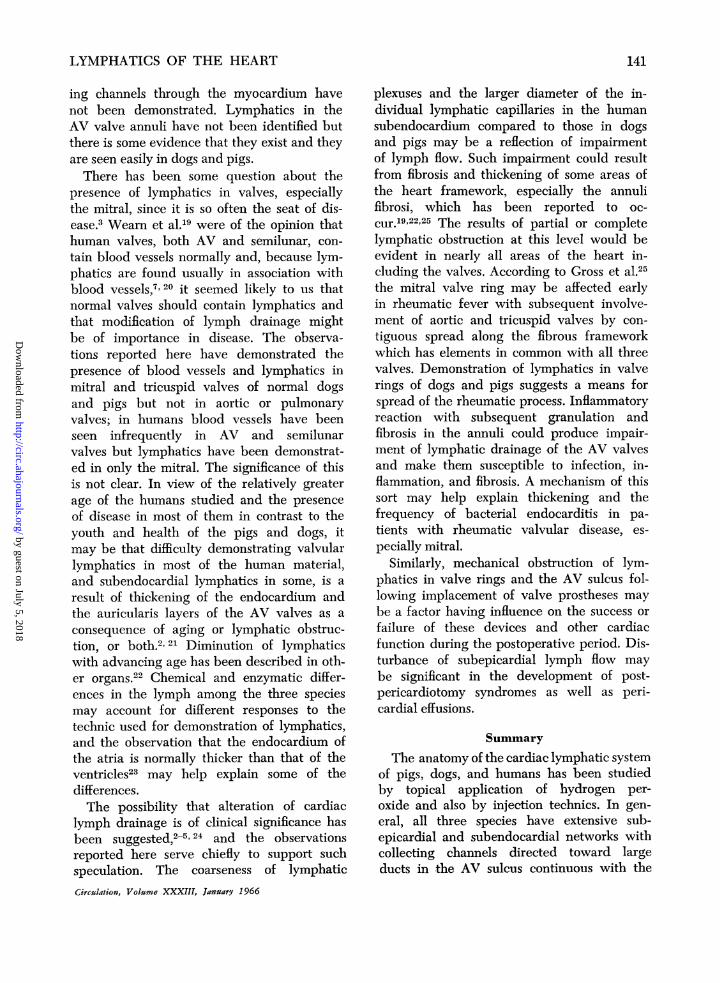
Figure 4
Subepicardial lymphatic capillary network over theleft ventricle of a human heart. Magnification 7X.
139
by guest on July 5, 2018http://circ.ahajournals.org/
Dow
nloaded from

JOHNSON, BLAKE
Figure 5 Figure bSubendocardial lymphatic capillaries (20 to 40 mi- Lymphatic capillaries (20 to 30 microns in diameter)crons in diameter) on a papillary muscle in the left on the atrial surface of the posterior leaflet of theventricle of a human heart. The longitudinal axis of nmitral valve in a human heart (see text). The cuspthe papillary mnuscle is directed from the right lower edge is just beyond the lower right margin of theto the left upper portion of the photograph. Magnifi- photograph. Frothing and air bubble artifacts are incation 25X. the left lower field. Magnification 30X.
uring up to 250 microns in diameter coursedlongitudinally deep to the valve rings towardthe AV junction but their termination has notbeen identified.
In the 13 patients for whom clinical in-formation was available, subendocardial lym-phatics were seen in hearts of three of the fourdying from causes directly related to heartdisease and in six of nine dying of noncar-diac causes. Lymphatics were not demonstrat-ed in tricuspid or semilunar valves and wereobserved in mitral valves in only two in-stances. In one, a 65-year-old man who diedwith chronic congestive heart failure, thevalves did not appear grossly to be involvedby disease. Beneath the endocardium on theatrial surface of the posterior cusp of themitral valve, lymphatics were traced fromnear the free margin to approximately halfthe distance to the valve ring. They appearedslightly larger than those in pigs and dogswith diameters in the range of 30 to 40 mi-crons (fig. 6). In the other instance, a 44-year-old woman with mitral stenosis who haddied following commissurotomy, there was asmall plexus of lymphatics on the atrial sur-face of the posterior cusp of the mitral valveabout halfway between its free edge and thevalve ring.
Transmyocardial ducts and large channels
in the AV annuli like those seen in dogs andpigs were not identified in humans butthere were channels approaching the annulifrom the atria.
In a 5-year-old boy with disseminated blas-tomycosis, subendocardial lymphatics wereseen only on the apices of the papillary mus-cles. In another child, a 6-year-old girl whohad died with acute leukemia, no subendo-cardial lymphatics were demonstrated.
In t-his series subendocardial lymphaticswere demonstraed in six of eight males andthree of five females.
DiscussionSmall differences in cardiac lymphatics of
the three species studied are described. Indogs and pigs lymphatic capillary nets be-neath the epicardium and endocardium joincollecting ducts that course longitudinally andjoin a large subepicardial duct in the AV sul-cus from which the main cardiac lymph ductarises. The subendocardial and subepicardialsystems communicate through transmyocar-dial channels in the AV valve annuli, papil-lary muscles, and at random throughout themyocardium.
In humans the arrangement of the suben-docardial and subepicardial systems is similarto that in the other species but communicat-
Circulation, Volume XXXIII, January 1966
140
by guest on July 5, 2018http://circ.ahajournals.org/
Dow
nloaded from
LYMPHATICS OF THE HEART
ing channels through the myocardium havenot been demonstrated. Lymphatics in theAV valve annuli have not been identified butthere is some evidence that they exist and theyare seen easily in dogs and pigs.There has been some question about the
presence of lymphatics in valves, especiallythe mitral, since it is so often the seat of dis-ease.3 Wearn et al.19 were of the opinion thathuman valves, both AV and semilunar, con-tain blood vessels normally and, because lym-phatics are found usually in association withblood vessels,7' 20 it seemed likely to us thatnormal valves should contain lymphatics andthat modification of lymph drainage mightbe of importance in disease. The observa-tions reported here have demonstrated thepresence of blood vessels and lymphatics inmitral and tricuspid valves of normal dogsand pigs but not in aortic or pulmonaryvalves; in humans blood vessels have beenseen infrequently in AV and semilunarvalves but lymphatics have been demonstrat-ed in only the mitral. The significance of thisis not clear. In view of the relatively greaterage of the humans studied and the presenceof disease in most of them in contrast to theyouth and health of the pigs and dogs, itmay be that difficulty demonstrating valvularlymphatics in most of the human material,and subendocardial lymphatics in some, is aresult of thickening of the endocardium andthe auricularis layers of the AV valves as aconsequence of aging or lymphatic obstruc-tion, or both.2' 21 Diminution of lymphaticswith advancing age has been described in oth-er organs.22 Chemical and enzymatic differ-ences in the lymph among the three speciesmay account for different responses to thetechnic used for demonstration of lymphatics,and the observation that the endocardium ofthe atria is normally thicker than that of theventricles23 may help explain some of thedifferences.The possibility that alteration of cardiac
lymph drainage is of clinical significance hasbeen suggested,2 5,24 and the observationsreported here serve chiefly to support suchspeculation. The coarseness of lymphaticCirculation, Volume XXXIII, January 1966
plexuses and the larger diameter of the in-dividual lymphatic capillaries in the humansubendocardium compared to those in dogsand pigs may be a reflection of impairmentof lymph flow. Such impairment could resultfrom fibrosis and thickening of some areas ofthe heart framework, especially the annulifibrosi, which has been reported to oc-cur.19'22'25 The results of partial or completelymphatic obstruction at this level would beevident in nearly all areas of the heart in-cluding the valves. According to Gross et al.25the mitral valve ring may be affected earlyin rheumatic fever with subsequent involve-ment of aortic and tricuspid valves by con-tiguous spread along the fibrous frameworkwhich has elements in common with all threevalves. Demonstration of lymphatics in valverings of dogs and pigs suggests a means forspread of the rheumatic process. Inflammatoryreaction with subsequent granulation andfibrosis in the annuli could produce impair-ment of lymphatic drainage of the AV valvesand make them susceptible to infection, in-flammation, and fibrosis. A mechanism of thissort may help explain thickening and thefrequency of bacterial endocarditis in pa-tients with rheumatic valvular disease, es-pecially mitral.
Similarly, mechanical obstruction of lym-phatics in valve rings and the AV sulcus fol-lowing implacement of valve prostheses maybe a factor having influence on the success orfailure of these devices and other cardiacfunction during the postoperative period. Dis-turbance of subepicardial lymph flow maybe significant in the development of post-pericardiotomy syndromes as well as peri-cardial effusions.
Summary
The anatomy of the cardiac lymphatic systemof pigs, dogs, and humans has been studiedby topical application of hydrogen per-oxide and also by injection technics. In gen-eral, all three species have extensive sub-epicardial and subendocardial networks withcollecting channels directed toward largeducts in the AV sulcus continuous with the
141
by guest on July 5, 2018http://circ.ahajournals.org/
Dow
nloaded from

JOHNSON, BLAKE
main cardiac lymph duct. The subepicardialand subendocardial systems in dogs and pigscommunicate via transmyocardial channelsand channels in the supporting structures ofthe AV valves; in humans these communica-tions have not been identified with certainty.Lymphatics are seen in both AV valves ofdogs and pigs but have been seen only inthe mitral valves of humans. The possiblesignificance of these observations has beendiscussed.
AcknowledgmentThe authors express appreciation for the interest
of Dr. W. Lane Williams and for the technical as-sistance of J. R. Dumas and J. T. Russell.
References1. PHILLIPS, J. H., JR.: The lymphatic system with
particular reference to cardiac edema. Bull.Tulane Med. Faculty 14: 187, 1955.
2. MILLER, A. J., PICK, R., ANI) KArZ, L. N.: Ven-tricular endomyocardial pathology producedby chronic cardiac lymphatic obstruction inthe dog. Circulation Research 8: 941, 1960.
3. MILLER, A. J., PICK, R., AND KATZ, L. N.: Lym-phatics of the mitral valve of the dog. Circula-tion Research 9: 1005, 1961.
4. MILLER, A. J.: The lymphatics of the heart.Arch. Int. Med. 112: 501, 1963.
5. KLINE, I. K., MILLER, A. J., PICK, R., AND KATZ,L. N.: The relationship between human en-
docardial fibroelastosis and obstruction of thecardiac lymphatics. Circulation 30: 728, 1964.
6. RAMSEY, E. M.: Studies in the pathology ofvascular disease. I. Nutrition of the bloodvessel wall: Review of the literature. YaleJ. Biol. & Med. 9: 14, 1936.
7. YOFFEY, J. M., AND COURTICE, F. C.: Lymphatics,Lymph, and Lymphoid Tissue. Cambridge,Harvard University Press, 1956.
8. RUSZYNYAK, I., FOLDT, M., AND SZABO, G.: Lym-phatics and Lymph Circulation. New York,Pergamon Press, Ltd., 1960.
9. PATEK, P. R.: The morphology of the lymphat-ics of the mammalian heart. Am. J. Anat.64: 203, 1939.
10. GOLAB, B.: The lymphatic vessels of the heart.The subendocardial and muscle networks. Fo-lia morphol. 12: 47, 1961.
11. MAGNUS, G., AND STUBEL, A.: Zur Kenntnis derLymphgefiisse des Auges. Bericht (iiber diedreiundvierzigste Versammlung) der Deutsch-en Ophthalmologischen Gesellschaft (Jena)Miinchen, Verlag von J. F. Bergmann, 1922,p. 36.
12. PARKE, W. P., AND MICHELS, N. A.: A methodfor demonstrating subserous lymphatics withhydrogen peroxide. Anat. Rec. 146: 165,1963.
13. HAUROWITZ, F.: Chemistry and Biology of Pro-teins. New York, Academic Press, Inc., 1950.
14. MCELROY, W. D., AND GLASS, B.: A symposiumon the mechanism of enzyme action. Balti-more, The Johns Hopkins Press, 1954.
15. DIXON, M., AND WEBB, E. C.: Enzymes. NewYork, Academic Press, Inc., 1958.
16. BORIsov, A. V.: Lymphatic capillaries andblood vessels of milky spots in the humanomentum. Arkhiv Anatomii, Gistologii, i Em-briologii, 44: 115, 1963.
17. PAPP, M., ROHLICH, P., RUSZNYAK, I., ToRo,I.: Central chyliferous vessel of intestinalvillus. Arkhiv Anatomii, Gistologii i Embriol-ogii 42: 24, 1962.
18. SCHLESINGER, M. J.: An injection plus dissectionstudy of coronary artery occlusions and anas-
tomoses. Am. Heart J. 15: 528, 1938.19. WEARN, J. T., BROMER, A. WV., AND ZSCHIESCHE,
L. J.: Incidence of blood vessels in humanheart valves. Am. Heart J. 11: 22, 1936.
20. ABRAMSON, D. I.: Blood Vessels and Lymphatics.New York, Academic Press, Inc., 1962, p. 701.
21. GROSS, L., AND KUGEL, M. A.: Topographicanatomy and histology of the valves in thehuman heart. Am. J. Path. 7: 445, 1931.
22. SPIRIN, B. A.: Internal lymphatic system of thepenis. Arkhiv Anatomii, Gistologii, i Embriologii,44: 68, 1963.
23. GOULD, S. E., ED: Pathology of the Heart.Springfield, Illinois, Charles C. Thomas, 1960.
24. MAYERSON, H. S.: Editorial: On lymph andlymphatics. Circulation 28: 839, 1963.
25. GRoss, L., AND FRIEDBERG, C. K.: Lesions ofthe cardiac valve rings in rheumatic fever.Am. J. Path. 12: 469, 1936.
Circulation, Volume XXXIII, January 1966
142
by guest on July 5, 2018http://circ.ahajournals.org/
Dow
nloaded from

ROBERT A. JOHNSON and THOMAS M. BLAKELymphatics of the Heart
Print ISSN: 0009-7322. Online ISSN: 1524-4539 Copyright © 1966 American Heart Association, Inc. All rights reserved.
is published by the American Heart Association, 7272 Greenville Avenue, Dallas, TX 75231Circulation doi: 10.1161/01.CIR.33.1.137
1966;33:137-142Circulation.
http://circ.ahajournals.org/content/33/1/137located on the World Wide Web at:
The online version of this article, along with updated information and services, is
http://circ.ahajournals.org//subscriptions/
is online at: Circulation Information about subscribing to Subscriptions:
http://www.lww.com/reprints Information about reprints can be found online at: Reprints:
document. and Rights Question and Answer
Permissionsthe Web page under Services. Further information about this process is available in thewhich permission is being requested is located, click Request Permissions in the middle column ofClearance Center, not the Editorial Office. Once the online version of the published article for
can be obtained via RightsLink, a service of the CopyrightCirculationoriginally published in Requests for permissions to reproduce figures, tables, or portions of articlesPermissions:
by guest on July 5, 2018http://circ.ahajournals.org/
Dow
nloaded from