Embed Size (px)
Citation preview

Department of Histology and Embryology, P.J. Šafárik University, Medical Faculty, Košice
Lymphatic system: Sylabus for foreign students from microscopic anatomy
Authors: prof. MUDr. Eva MECHÍROVÁ, CSc; MVDr. Štefan TÓTH, PhD.
Lymphatic system

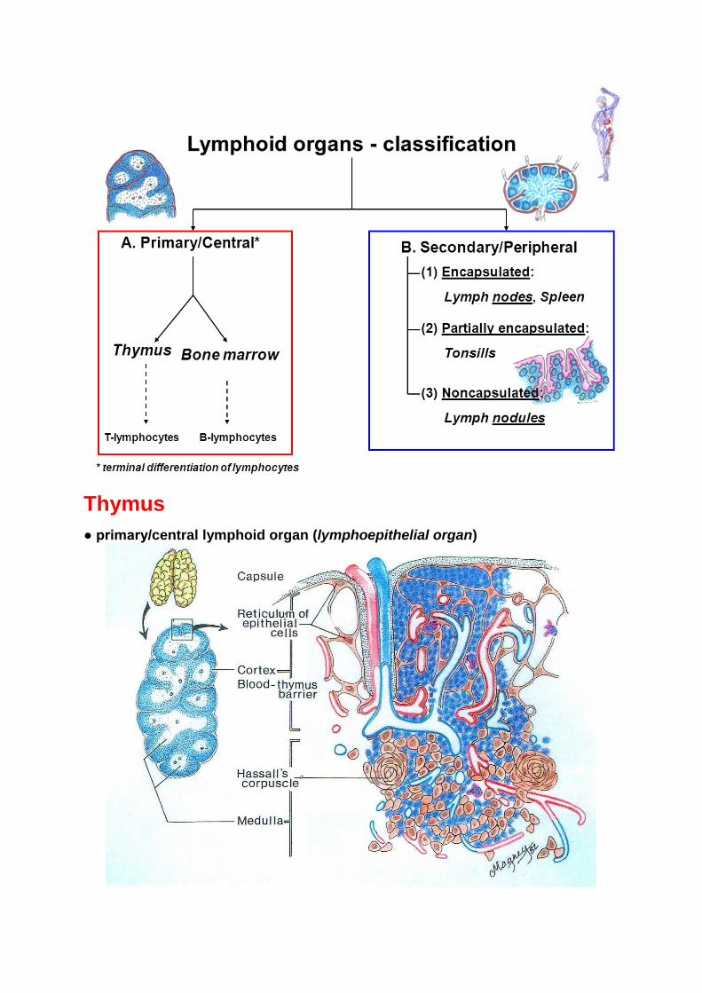
Thymus
● primary/central lymphoid organ (lymphoepithelial organ)

Functions of the thymus 1. Differentiation of immunocompetent T-lymphocytes from precursor cells of
bone marrow into T- helper and T-cytotoxic cels. 2. Development of self - tolerance reaction. 3. Secretion of polypeptides with hormonal activity by reticular epithelial cells, that regulate proliferation, maturation and function of T- lymphocytes in thymus and other lymphoid organs: - thymulin - thymopoetin - thymosin - thymic humoral factor.

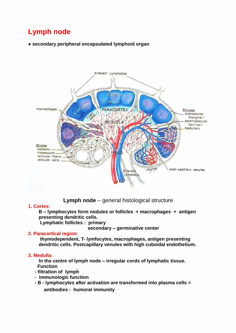
Lymph node ● secondary peripheral encapsulated lymphoid organ
Lymph node – general histological structure 1. Cortex: B – lymphocytes form nodules or follicles + macrophages + antigen presenting dendritic cells. Lymphatic follicles : primary
secondary – germinative center
2. Paracortical region: thymodependent, T- lymfocytes, macrophages, antigen presenting dendritic cells. Postcapillary venules with high cuboidal endothelium.
3. Medulla: In the centre of lymph node – irregular cords of lymphatic tissue. Function
- filtration of lymph
- immunologic function - B - lymphocytes after activation are transformed into plasma cells =
antibodies - humoral immunity.

RC – reticular cell; RF – reticular fibers; T-Ly – T lymphocyte
Lymph node – detail of subcapsular lymphatic sinus

Spleen Peripheral lymphoid encapsulated organ. Spleen is the largest lymphoid organ in the human body. Function of the spleen: production of lymphocytes immunologic destruction of defective erythrocytes Microscopic structure: The connective tissue capsule is composed of connective tissue, covered by simple squamous epithelium – mesothelium. Trabeculae arise from capsule, and there are branches of splenic artery and efferent lymphatic vessels (no afferent lymphatic vessels) Stroma is composed of reticular connective tissue. Parenchyma –> or splenic pulp can be divided into white and red.
White pulp – typical lymphoid tissue – T and B- lymphocytes, their activated forms, plasma cells, macrophages and antigen presenting cells. White pulp is in relation with arterial blood system. Branches of trabecular arteries enter the white pulp and are here called as central arteriols. They are surrounded by periarterial lymphatic sheath (PALS), that is thymodependent and contains T- lymfocytes. Sometime the sheath is surrounded by lymphoid
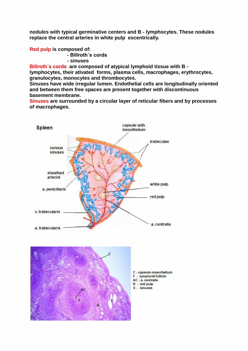
nodules with typical germinative centers and B - lymphocytes. These nodules replace the central arteries in white pulp excentrically. Red pulp is composed of: - Billroth´s cords - sinuses Billroth´s cords are composed of atypical lymphoid tissue with B - lymphocytes, their ativated forms, plasma cells, macrophages, erythrocytes, granulocytes, monocytes and thrombocytes. Sinuses have wide irregular lumen. Endothelial cells are longitudinally oriented and between them free spaces are present together with discontinuous basement membrane. Sinuses are surrounded by a circular layer of reticular fibers and by processes of macrophages.

Theory of open and closed blood circulation 1. Closed circulation - trabecular arteries are branched in central arteriols and then in penicillar arteriols. Near the termination penicillar arterioles are surrounded by a sheath of reticular cells and macrophages. Penicillar arteriols directly continue into sinuses of red pulp.
2. Open circulation - in this case the blood from end part of penicillar arteriols flows outside of circulation in the red pulp. After that is collected again in the sinuses

The open and closed blood circulation in spleen
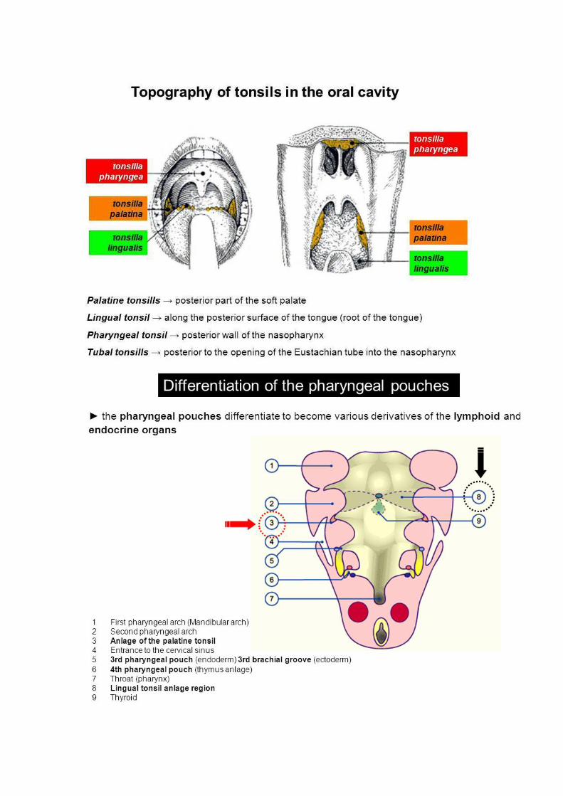
Tonsils
● partially (incompletely) encapsulated peripheral lymphoid organs ● histologically, composed of aggregates of lymphatic tissue covered by epithelium ● according to their location in the mouth and pharynx are called: 1. palatine 2. lingual 3. pharyngeal 4. tubal
A ring of lymphatic tissue of tonsils at the entrance of the oropharynx (lat. anulus Waldeyeri)

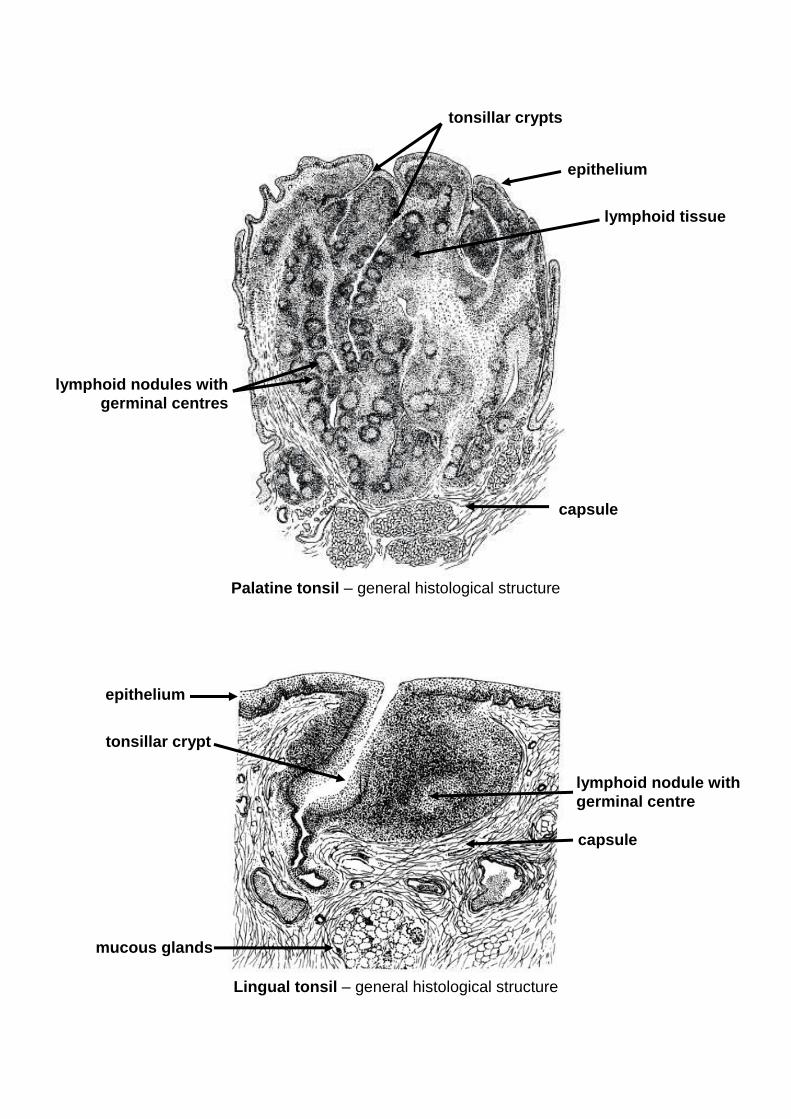
Palatine tonsil – general histological structure
Lingual tonsil – general histological structure
epithelium
lymphoid tissue
tonsillar crypts
capsule
lymphoid nodules with
germinal centres
tonsillar crypt
epithelium
mucous glands
capsule
lymphoid nodule with
germinal centre

Table: Prenatal development of lymphoid organs - overview