Embed Size (px)
Citation preview
193
Lymphology 38 (2005) 193-196
COMMENTARY
LYMPHATIC FILARIASIS AND THE ISL CONSENSUS DOCUMENT
S. Jamal
Department of Plastic Surgery, Thanjavur Medical College Hospital, Thanjavur, India
Keywords: filariasis, lymphedema staging,treatment, consensus
At the general body meeting during the19th International Congress of Lymphology(ICL) in Freiburg, Germany, I requested that the International Society of Lymphology(ISL) offer comments in the ConsensusDocument on the treatment of filarial edema.To date, this has not yet been accomplished.Therefore, I am providing my ownreflections.
Since 1979, I have been presenting myexperience on the management of filarialedema in the International Congresses ofLymphology (ICL) and treating patients withlymphatic filariasis for the past 4 decades. I have seen over 15,000 cases and operated on approximately 3,000 patients.
As per the ISL classification oflymphedema, there is no pitting on pressure,and there is no reduction of edema afterelevation in Grade II and III (1). But infilarial edema of Grade II, III and IV (in ourclassification), there is a reduction in the size,a finding I reported at the 11th ICL (2). Thisdifference relates to the fact that post-surgeryand radiation to regional nodes, there is often extensive anatomical blockade to lymphflow whereas in filariasis, there is no suchblockage, and the edema is due to over-
dilation and/or paralysis of the lymphaticvessels with valvular incompetence. Becauseof the reduction in edema after bed rest,many patients do not enter early treatment as they believe their swelling will be reducedto near normal status after simple bed rest.
For filarial edema cases, liposuction isnot advisable as the mechanism of lymphe-dema is different due to the presence of theworm in the lymphatic vessels resulting inincompetence of valves leading to backflow oflymph and more fibrosis around lymphaticsand blood vessels. The experience of thosewho attempted liposuction in these cases hasbeen disastrous with development of chroniculcers in the leg.
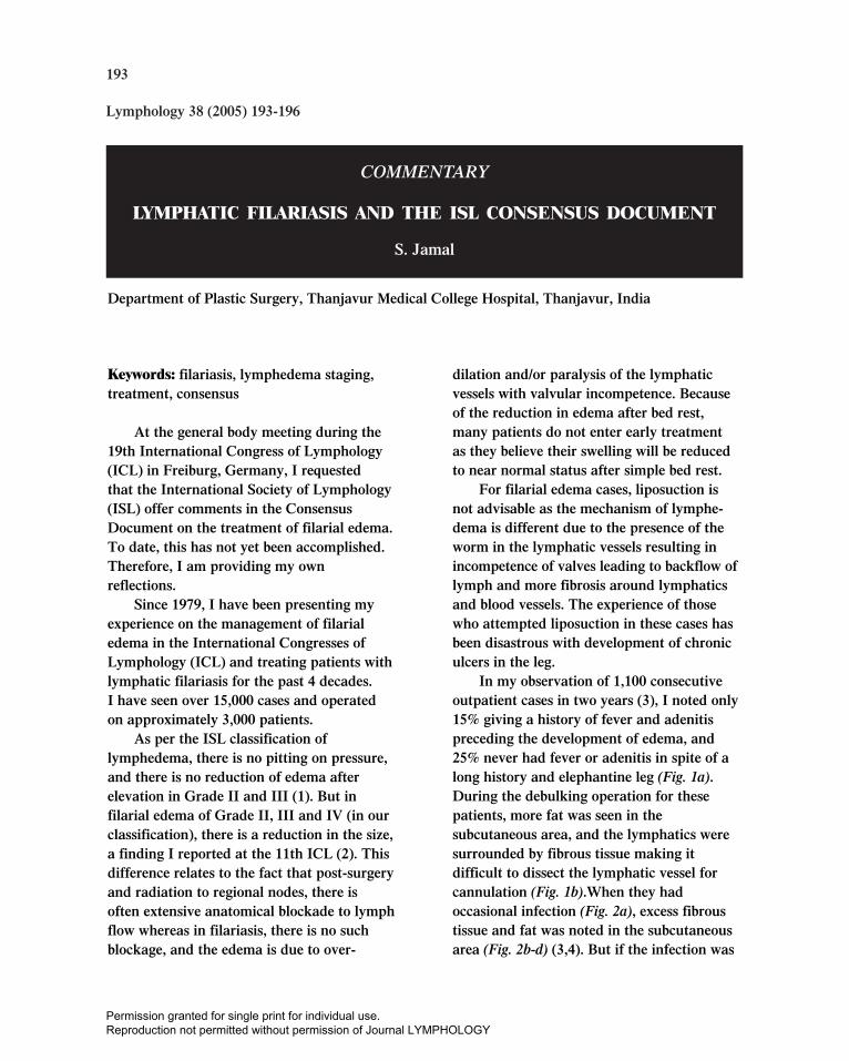
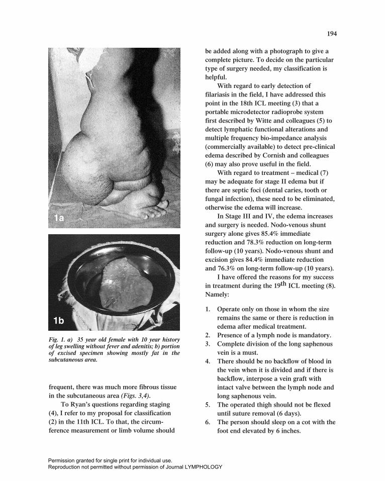
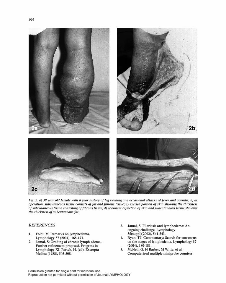
In my observation of 1,100 consecutiveoutpatient cases in two years (3), I noted only15% giving a history of fever and adenitispreceding the development of edema, and25% never had fever or adenitis in spite of along history and elephantine leg (Fig. 1a).During the debulking operation for thesepatients, more fat was seen in thesubcutaneous area, and the lymphatics weresurrounded by fibrous tissue making itdifficult to dissect the lymphatic vessel forcannulation (Fig. 1b).When they hadoccasional infection (Fig. 2a), excess fibroustissue and fat was noted in the subcutaneousarea (Fig. 2b-d) (3,4). But if the infection was
Permission granted for single print for individual use. Reproduction not permitted without permission of Journal LYMPHOLOGY
194
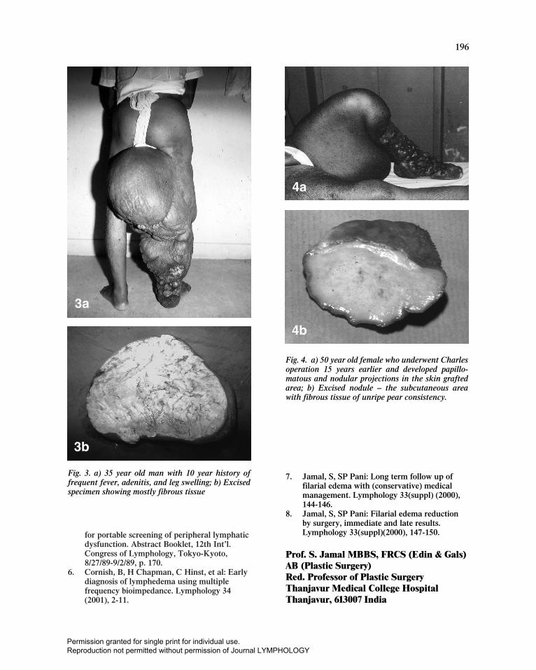
frequent, there was much more fibrous tissuein the subcutaneous area (Figs. 3,4).
To Ryan’s questions regarding staging(4), I refer to my proposal for classification(2) in the 11th ICL. To that, the circum-ference measurement or limb volume should
be added along with a photograph to give acomplete picture. To decide on the particulartype of surgery needed, my classification ishelpful.
With regard to early detection offilariasis in the field, I have addressed thispoint in the 18th ICL meeting (3) that aportable microdetector radioprobe systemfirst described by Witte and colleagues (5) todetect lymphatic functional alterations andmultiple frequency bio-impedance analysis(commercially available) to detect pre-clinicaledema described by Cornish and colleagues(6) may also prove useful in the field.
With regard to treatment – medical (7)may be adequate for stage II edema but ifthere are septic foci (dental caries, tooth orfungal infection), these need to be eliminated,otherwise the edema will increase.
In Stage III and IV, the edema increasesand surgery is needed. Nodo-venous shuntsurgery alone gives 85.4% immediatereduction and 78.3% reduction on long-termfollow-up (10 years). Nodo-venous shunt andexcision gives 84.4% immediate reductionand 76.3% on long-term follow-up (10 years).
I have offered the reasons for my successin treatment during the 19th ICL meeting (8).Namely:
1. Operate only on those in whom the sizeremains the same or there is reduction inedema after medical treatment.
2. Presence of a lymph node is mandatory.3. Complete division of the long saphenous
vein is a must.4. There should be no backflow of blood in
the vein when it is divided and if there isbackflow, interpose a vein graft withintact valve between the lymph node andlong saphenous vein.
5. The operated thigh should not be flexeduntil suture removal (6 days).
6. The person should sleep on a cot with thefoot end elevated by 6 inches.
Fig. 1. a) 35 year old female with 10 year history of leg swelling without fever and adenitis; b) portionof excised specimen showing mostly fat in thesubcutaneous area.
1a
1b
Permission granted for single print for individual use. Reproduction not permitted without permission of Journal LYMPHOLOGY
195
2a
2c
Fig. 2. a) 38 year old female with 8 year history of leg swelling and occasional attacks of fever and adenitis; b) atoperation, subcutaneous tissue consists of fat and fibrous tissue; c) excised portion of skin showing the thicknessof subcutaneous tissue consisting of fibrous tissue; d) operative reflection of skin and subcutaneous tissue showingthe thickness of subcutaneous fat.
2b
2d
REFERENCES
1. Földi, M: Remarks on lymphedema.Lymphology 37 (2004), 168-173.
2. Jamal, S: Grading of chronic lymph edema-Further refinement proposed. Progress inLymphology XI. Partch, H. (ed), ExcerptaMedica (1988), 505-508.
3. Jamal, S: Filariasis and lymphedema: Anongoing challenge. Lymphology35(suppl)(2002), 541-543.
4. Ryan, TJ: Commentary: Search for consensuson the stages of lymphedema. Lymphology 37(2004), 180-181.
5. McNeill G, H Barber, M Witte, et al:Computerized multiple miniprobe counters
Permission granted for single print for individual use. Reproduction not permitted without permission of Journal LYMPHOLOGY
196
for portable screening of peripheral lymphaticdysfunction. Abstract Booklet, 12th Int’l.Congress of Lymphology, Tokyo-Kyoto,8/27/89-9/2/89, p. 170.
6. Cornish, B, H Chapman, C Hinst, et al: Earlydiagnosis of lymphedema using multiplefrequency bioimpedance. Lymphology 34(2001), 2-11.
3a
Fig. 3. a) 35 year old man with 10 year history offrequent fever, adenitis, and leg swelling; b) Excisedspecimen showing mostly fibrous tissue
3b
Fig. 4. a) 50 year old female who underwent Charlesoperation 15 years earlier and developed papillo-matous and nodular projections in the skin graftedarea; b) Excised nodule – the subcutaneous areawith fibrous tissue of unripe pear consistency.
4b
4a
7. Jamal, S, SP Pani: Long term follow up offilarial edema with (conservative) medicalmanagement. Lymphology 33(suppl) (2000),144-146.
8. Jamal, S, SP Pani: Filarial edema reductionby surgery, immediate and late results.Lymphology 33(suppl)(2000), 147-150.
Prof. S. Jamal MBBS, FRCS (Edin & Gals)AB (Plastic Surgery)Red. Professor of Plastic SurgeryThanjavur Medical College HospitalThanjavur, 613007 India
Permission granted for single print for individual use. Reproduction not permitted without permission of Journal LYMPHOLOGY