Embed Size (px)
Citation preview

P A R T
Drugs Acting on the Central
and Peripheral Nervous Systems
4
19 Introduction to Nerves and the NervousSystem 299
20 Anxiolytic and Hypnotic Agents 310
21 Antidepressant Agents 324
22 Psychotherapeutic Agents 341
23 Antiseizure Agents 359
24 Antiparkinsonism Agents 379
25 Muscle Relaxants 392
26 Narcotics, Narcotic Antagonists, andAntimigraine Agents 405
27 General and Local Anesthetic Agents 425
28 Neuromuscular Junction Blocking Agents 441
Drugs Acting on the Central
and PeripheralNervous Systems
LWBK374_c19_p297-309.qxd 26/08/2009 12:56 PM Page 297 Aptara

LWBK374_c19_p297-309.qxd 26/08/2009 12:56 PM Page 298 Aptara

Introduction to Nervesand the Nervous System 19
G l o s s a r y o f K e y T e r m s
action potential: sudden change in electrical charge of a
nerve cell membrane; the electrical signal by which neu-
rons send information
afferent: neurons or groups of neurons that bring information
to the central nervous system; sensory nerve
axon: long projection from a neuron that carries information
from one nerve to another nerve or effector
dendrite: short projection on a neuron that transmits informa-
tion
depolarization: opening of the sodium channels in a nerve
membrane to allow the influx of positive sodium ions,
reversing the membrane charge from negative to positive
effector cell: cell stimulated by a nerve; may be a muscle, a
gland, or another nerve cell
efferent: neurons or groups of neurons that carry information
from the central nervous system to an effector; motor neu-
rons are efferent
engram: short-term memory made up of a reverberating
electrical circuit of action potentials
forebrain: upper level of the brain; consists of the two cere-
bral hemispheres, where thinking and coordination of sen-
sory and motor activity occur
ganglia: a group of nerve bodies
hindbrain: most primitive area of the brain, the brainstem;
consists of the pons and medulla, which control basic, vital
L e a r n i n g O b j e c t i v e s
Upon completion of this chapter, you will be able to:
1. Label the parts of a neuron and describe the functions of each part.
2. Describe an action potential, including the roles of the various electrolytes involved in the action
potential.
3. Explain what a neurotransmitter is, including its origins and functions at the synapse.
4. Describe the function of the cerebral cortex, cerebellum, hypothalamus, thalamus, midbrain,
pituitary gland, medulla, spinal cord, and reticular activating system.
5. Discuss what is known about learning and the impact of emotion on the learning process.
functions and arousal, and the cerebellum, which controls
motor functions that regulate balance
limbic system: area in the midbrain that is rich in epineph-
rine, norepinephrine, and serotonin and seems to control
emotions
midbrain: the middle area of the brain; it consists of the
hypothalamus and thalamus and includes the limbic
system
neuron: structural unit of the nervous system
neurotransmitter: chemical produced by a nerve and
released when the nerve is stimulated; reacts with a spe-
cific receptor site to cause a reaction
repolarization: return of a membrane to a resting state, with
more sodium ions outside the membrane and a relatively
negative charge inside the membrane
Schwann cell: insulating cell found on nerve axons; allows
“leaping” electrical conduction to speed the transmission
of information and prevent tiring of the neuron
soma: cell body of a neuron; contains the nucleus, cyto-
plasm, and various granules
synapse: junction between a nerve and an effector; consists
of the presynaptic nerve ending, a space called the
synaptic cleft, and the postsynaptic cell
299
LWBK374_c19_p297-309.qxd 26/08/2009 12:57 PM Page 299 Aptara

he nervous system is responsible for controlling the func-tions of the human body, analyzing incoming stimuli, and
integrating internal and external responses. The nervous sys-tem is composed of the central nervous system (CNS; thebrain and spinal cord) and the peripheral nervous system(PNS). The PNS is composed of sensory receptors that bringinformation into the CNS and motor nerves that carry infor-mation away from the CNS to facilitate response to stimuli.The autonomic nervous system, which is discussed inChapter 29, uses components of the CNS and PNS to regulateautomatic or unconscious responses to stimuli.
The structural unit of the nervous system is the nerve cell,or neuron. The billions of nerve cells that make up the ner-vous system are organized to allow movement realization ofvarious sensations; response to internal and external stimuli;and learning, thinking, and emotion. The mechanisms thatare involved in all of these processes are not clearly under-stood. The actions of drugs that are used to affect the func-tioning of the nerves and the responses that these drugs causethroughout the nervous system provide some of the currenttheories about the workings of the nervous system.
PHYSIOLOGY OF THE NERVOUS SYSTEM
The nervous system operates through the use of electricalimpulses and chemical messengers to transmit informationthroughout the body and to respond to internal and externalstimuli. The properties and functions of the neuron providethe basis for all nervous system function.
Neurons
As noted previously, the neuron is the structural unit of thenervous system. The human body contains about 14 billionneurons. About 10 billion of these are located in the brain,and the remainder make up the spinal cord and PNS.
Neurons have several distinctive cellular features(Figure 19.1). Each neuron is made up of a cell body, orsoma, which contains the cell nucleus, cytoplasm, and vari-ous granules and other particles. Short, branch-like projec-tions that cover most of the surface of a neuron are known asdendrites. These structures, which provide increased surface
area for the neuron, bring information into the neuron fromother neurons.
One end of the nerve body extends into a long process thatdoes not branch out until the very end of the process. Thiselongated process is called the nerve axon, and it emergesfrom the soma at the axon hillock, a slightly enlarged area ofthe soma from which the axon emerges. The axon of a nervecan be extremely tiny, or it can extend for several feet. Theaxon carries information from a nerve to be transmitted toeffector cells—cells stimulated by a nerve, which mayinclude a muscle, gland, or another nerve. This transmissionoccurs at the end of the axon, where the axon branches out inwhat is called the axon terminal.
The axons of many nerves are packed closely together inthe nervous system and look like cable or fiber tracts.Afferent fibers are nerve axons that run from peripheralreceptors into the CNS. In contrast, efferent fibers are nerveaxons that carry nerve impulses from the CNS to the periph-ery to stimulate muscles or glands. (An easy way to remem-ber the difference between afferent and efferent is to recallthat efferent fibers exit from the CNS.)
It is currently thought that neurons are unable to repro-duce; so, if nerves are destroyed, they are lost. If dendritesand axons are lost, nerves regenerate those structures; how-ever, for this regeneration to occur, the soma and the axonhillock must remain intact. For a clinical example, consider aperson who has closed a car door on his or her finger.Sensation and movement may be lost or limited for a certainperiod, but because the nerve bodies for most of the nerves inthe hand are located in ganglia (groups of nerve bodies) inthe wrist, they are able to regenerate the damaged axon ordendrites. Over time, sensation and full movement shouldreturn.
Research on possible ways to stimulate the reproductionof nerves is under way. Although scientists have used nervegrowth factor with fetal cell implants to stimulate some nervegrowth, it is currently assumed that nerves are unable toreproduce.
Action Potential
Nerves send messages by conducting electrical impulsescalled action potentials.
PART 4 Drugs Acting on the Central and Peripheral Nervous Systems300
T
DendritesNucleus
Axon
Cellbody
Neurilemma
Myelinsheath
Node ofRanvier
Synapticterminals
● FIGURE 19.1 The neuron, functional unitof the nervous system.
LWBK374_c19_p297-309.qxd 26/08/2009 12:57 PM Page 300 Aptara

Nerve membranes, which are capable of conductingaction potentials along the entire membrane, send messagesto nearby neurons or to effector cells that may be locatedinches to feet away via this electrical communication system.Like all cell membranes, nerve membranes have variouschannels or pores that control the movement of substancesinto and out of the cell. Some of these channels allow themovement of sodium, potassium, and calcium. When cellsare at rest, their membranes are impermeable to sodium.However, the membranes are permeable to potassium ions.
The sodium–potassium pump that is active in the mem-branes of neurons is responsible for this property of the mem-brane. This system pumps sodium ions out of the cell andpotassium ions into the cell. At rest, more sodium ions areoutside the cell membrane, and more potassium ions areinside. Electrically, the inside of the cell is relatively negativecompared with the outside of the membrane, which estab-lishes an electrical potential along the nerve membrane.When nerves are at rest, this is referred to as the resting mem-brane potential of the nerve.
Stimulation of a neuron causes depolarization of thenerve, which means that the sodium channels open inresponse to the stimulus, and sodium ions rush into the cell,following the established concentration gradient. If an electri-cal monitoring device is attached to the nerve at this point, apositive rush of ions is recorded. The electrical charge on theinside of the membrane changes from relatively negative torelatively positive. This sudden reversal of membrane poten-tial, called the action potential (Figure 19.2), lasts less than amicrosecond. Using the sodium–potassium pump, the cellthen returns that section of membrane to the resting mem-brane potential, a process called repolarization. The actionpotential generated at one point along a nerve membranestimulates the generation of an action potential in adjacentportions of the cell membrane, and the stimulus travels thelength of the cell membrane.
Nerves can respond to stimuli several hundred times persecond, but for a given stimulus to cause an action potential,it must have sufficient strength and must occur when thenerve membrane is able to respond—that is, when it has repo-larized. A nerve cannot be stimulated again while it is depo-larized. The balance of sodium and potassium across the cellmembrane must be re-established.
Nerves require energy (i.e., oxygen and glucose) and thecorrect balance of the electrolytes sodium and potassium tomaintain normal action potentials and transmit informationinto and out of the nervous system. If an individual hasanoxia or hypoglycemia, the nerves might not be able tomaintain the sodium–potassium pump, and that individualmay become severely irritable or too stable (not responsive tostimuli).
Long nerves are myelinated: they have a myelin sheaththat speeds electrical conduction and protects the nerves fromthe fatigue that results from frequent formation of actionpotentials. Even though many of the tightly packed nerves inthe brain do not need to travel far to stimulate another nerve,
they are myelinated. The effect of this myelination is notunderstood.
Myelinated nerves have Schwann cells, which are locatedat specific intervals along nerve axons and are very resistantto electrical stimulation (Figure 19.1). The Schwann cellswrap themselves around the axon in jellyroll fashion (Figure19.3). Between the Schwann cells are areas of uncoverednerve membrane called the nodes of Ranvier. So-called“leaping” nerve conduction occurs along these exposed nervefibers. An action potential excites one section of nerve mem-brane, and the electrical impulse then “skips” from one nodeto the next, generating an action potential. Because the mem-brane is forming fewer action potentials, the speed of con-duction is much faster and the nerve is protected from beingexhausted or using up energy to form multiple action poten-tials. This node-to-node mode of conduction is termed salta-tory or leaping conduction (Figure 19.1).
CHAPTER 19 Introduction to Nerves and the Nervous System 301
+ + + + + + + + + + + + + + + + + +
+ + + + + + + + + + + + + + + + + + +– – – – – – – – – – – – – – – – – – –
– – – – – – – – – – – – – – – – – –
Meter
A
B
potassium Sodium
potassium
Depolarization(sodium rushes into the cell)
Repolarization
(sodium pumped out of the cell)
Resting membrane potential
1 msec
sodium
–70mV
–80
60
40
20
0
20
40
60
+80
● FIGURE 19.2 The action potential. A. A segment of an axonshowing that, at rest, the inside of the membrane is relatively nega-tively charged and the outside is positively charged. A pair of elec-trodes placed as shown would record a potential difference of about–70 mV; this is the resting membrane potential. B. An action poten-tial of about 1 msec that would be recorded if the axon shown inpanel A were brought to threshold. At the peak of the action poten-tial, the charge on the membrane reverses polarity.
LWBK374_c19_p297-309.qxd 26/08/2009 12:57 PM Page 301 Aptara
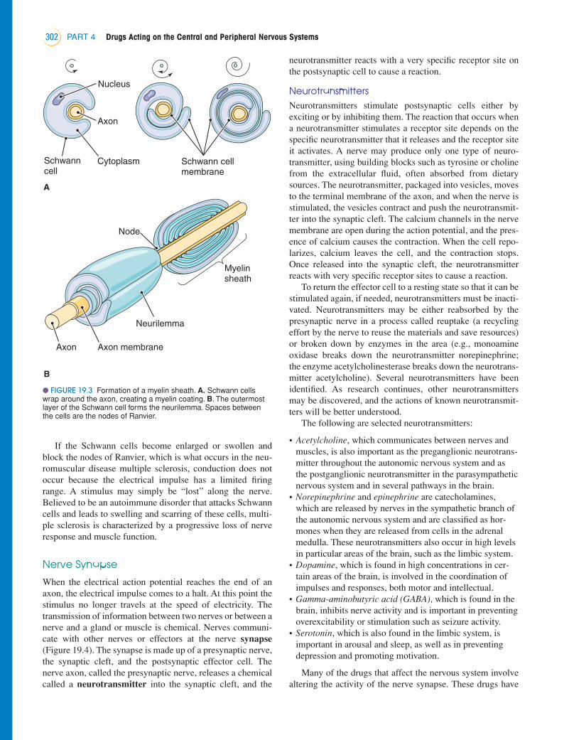
If the Schwann cells become enlarged or swollen andblock the nodes of Ranvier, which is what occurs in the neu-romuscular disease multiple sclerosis, conduction does notoccur because the electrical impulse has a limited firingrange. A stimulus may simply be “lost” along the nerve.Believed to be an autoimmune disorder that attacks Schwanncells and leads to swelling and scarring of these cells, multi-ple sclerosis is characterized by a progressive loss of nerveresponse and muscle function.
Nerve Synapse
When the electrical action potential reaches the end of anaxon, the electrical impulse comes to a halt. At this point thestimulus no longer travels at the speed of electricity. Thetransmission of information between two nerves or between anerve and a gland or muscle is chemical. Nerves communi-cate with other nerves or effectors at the nerve synapse
(Figure 19.4). The synapse is made up of a presynaptic nerve,the synaptic cleft, and the postsynaptic effector cell. Thenerve axon, called the presynaptic nerve, releases a chemicalcalled a neurotransmitter into the synaptic cleft, and the
neurotransmitter reacts with a very specific receptor site onthe postsynaptic cell to cause a reaction.
Neurotransmitters Neurotransmitters stimulate postsynaptic cells either byexciting or by inhibiting them. The reaction that occurs whena neurotransmitter stimulates a receptor site depends on thespecific neurotransmitter that it releases and the receptor siteit activates. A nerve may produce only one type of neuro-transmitter, using building blocks such as tyrosine or cholinefrom the extracellular fluid, often absorbed from dietarysources. The neurotransmitter, packaged into vesicles, movesto the terminal membrane of the axon, and when the nerve isstimulated, the vesicles contract and push the neurotransmit-ter into the synaptic cleft. The calcium channels in the nervemembrane are open during the action potential, and the pres-ence of calcium causes the contraction. When the cell repo-larizes, calcium leaves the cell, and the contraction stops.Once released into the synaptic cleft, the neurotransmitterreacts with very specific receptor sites to cause a reaction.
To return the effector cell to a resting state so that it can bestimulated again, if needed, neurotransmitters must be inacti-vated. Neurotransmitters may be either reabsorbed by thepresynaptic nerve in a process called reuptake (a recyclingeffort by the nerve to reuse the materials and save resources)or broken down by enzymes in the area (e.g., monoamineoxidase breaks down the neurotransmitter norepinephrine;the enzyme acetylcholinesterase breaks down the neurotrans-mitter acetylcholine). Several neurotransmitters have beenidentified. As research continues, other neurotransmittersmay be discovered, and the actions of known neurotransmit-ters will be better understood.
The following are selected neurotransmitters:
• Acetylcholine, which communicates between nerves andmuscles, is also important as the preganglionic neurotrans-mitter throughout the autonomic nervous system and asthe postganglionic neurotransmitter in the parasympatheticnervous system and in several pathways in the brain.
• Norepinephrine and epinephrine are catecholamines,which are released by nerves in the sympathetic branch ofthe autonomic nervous system and are classified as hor-mones when they are released from cells in the adrenalmedulla. These neurotransmitters also occur in high levelsin particular areas of the brain, such as the limbic system.
• Dopamine, which is found in high concentrations in cer-tain areas of the brain, is involved in the coordination ofimpulses and responses, both motor and intellectual.
• Gamma-aminobutyric acid (GABA), which is found in thebrain, inhibits nerve activity and is important in preventingoverexcitability or stimulation such as seizure activity.
• Serotonin, which is also found in the limbic system, isimportant in arousal and sleep, as well as in preventingdepression and promoting motivation.
Many of the drugs that affect the nervous system involvealtering the activity of the nerve synapse. These drugs have
PART 4 Drugs Acting on the Central and Peripheral Nervous Systems302
Schwanncell
Nucleus
Axon membraneAxon
Schwann cellmembrane
Axon
Cytoplasm
Neurilemma
Myelinsheath
Node
A
B
● FIGURE 19.3 Formation of a myelin sheath. A. Schwann cellswrap around the axon, creating a myelin coating. B. The outermostlayer of the Schwann cell forms the neurilemma. Spaces betweenthe cells are the nodes of Ranvier.
LWBK374_c19_p297-309.qxd 26/08/2009 12:57 PM Page 302 Aptara

CHAPTER 19 Introduction to Nerves and the Nervous System 303
DendritesNucleus
Axon
Cellbody
Neurilemma
Myelinsheath
Node ofRanvier
Synapticterminals
Presynapticnerve
terminal
Causes of neurotransmitterinactivation
A. Inactivation by enzyme
B. Diffusion
C. Reuptake
Synaptic TransmissionSequence of events
Synaptic cleft
NA+, K+, Cl-
Neuron oreffector cell
Inactive productto blood vessel
Into blood vessel
IPSP
EPSP
Bloodvessel
Contraction
Secretion
Return to presynaptic cell
Synaptic vessel
Axonterminal
Storage vesicle
Ca+
Enzymes
Neurotransmitter
Inactive product
Receptor
Enzymes
orAP
1
2
3
4
5
6
8a
8b
10
A
B
C
C
7
C-AMP
C-GMP
9a9b
Synaptic
term
inals
● FIGURE 19.4 The sequence of events in synaptic transmission: (1) Synthesis of the neurotransmit-ter; (2) uptake of the neurotransmitter into storage vesicles; (3) release of the neurotransmitter by anaction potential in the presynaptic nerve; (4) diffusion of the neurotransmitter across the synaptic cleft;(5) combination of the neurotransmitter with a receptor; (6) a sequence of events leading to activationof second messengers within the postsynaptic nerve; (7) change in permeability of the postsynapticmembrane to one or more ions, causing (8a) an inhibitory postsynaptic potential or (8b) an excitatorypostsynaptic potential. Characteristic responses of the postsynaptic cell are as follows: (9a) The glandsecretes hormones; (9b) the muscle cells have an action potential; and (10) the muscle contracts. Theaction of the neurotransmitter is terminated by one or more of the following processes. (A) inactivationby an enzyme; (B) diffusion out of the synaptic cleft and removal by the vascular system; and (C)reuptake into the presynaptic nerve followed by storage in a synaptic vesicle or deactivation by anenzyme.
LWBK374_c19_p297-309.qxd 26/08/2009 12:57 PM Page 303 Aptara

are carried bound to plasma proteins and are unable to crossinto the brain. When a patient is suffering from a braininfection, antibiotics cannot cross into the brain until theinfection is so severe that the blood–brain barrier can nolonger function.
The brain has a unique blood supply to protect the neuronsfrom lack of oxygen and glucose. Two arteries—thecarotids—branch off the aortic arch and go up into each sideof the brain at the front of the head, and two other arteries—the vertebrals—enter the back of the brain to become thebasilar arteries. These arteries all deliver blood to a commonvessel at the bottom of the brain called the circle of Willis,which distributes the blood to the brain as it is needed(Figure 19.6). The role of the circle of Willis becomes appar-ent when an individual has an occluded carotid artery.Although the passage of blood through one of the carotidarteries may be negligible, the areas of the brain on that sidewill still have a full blood supply because of the blood sent tothose areas via the circle of Willis.
Anatomy of the Brain
The brain has three major divisions: the hindbrain, the mid-brain, and the forebrain (Figure 19.7).
The hindbrain, which runs from the top of the spinal cordinto the midbrain, is the most primitive area of the brain andcontains the brainstem, where the pons and medulla oblon-gata are located. These areas of the brain control basic, vitalfunctions, such as the respiratory centers, which controlbreathing; the cardiovascular centers, which regulate blood
PART 4 Drugs Acting on the Central and Peripheral Nervous Systems304
Arachnoidvillus
Venous(dural) sinus
Skin
Sagittalsuture
Skull
Dura mater
Meninges
ArachnoidPia mater
Gray matter
Brain tissue
White matter
● FIGURE 19.5 Bony and membranous protection of the brain.
several functions, including blocking the reuptake of neuro-transmitters so that they are present in the synapse in greaterquantities and cause more stimulation of receptor sites;blocking receptor sites so that the neurotransmitter cannotstimulate the receptor site; blocking the enzymes that breakdown neurotransmitters to cause an increase in neurotrans-mitter concentration in the synapse; stimulating specificreceptor sites when the neurotransmitter is not available; andcausing the presynaptic nerve to release greater amounts ofthe neurotransmitter.
KEY POINTS
➧ The nervous system controls the body, analyzes externalstimuli, and integrates internal and external responses tostimuli.
➧ The neuron, comprising a cell body, dendrites and anaxon, is the functional unit of the nervous system.Dendrites route information to the nerve, and axons takethe information away.
➧ Nerves transmit information by way of action potentials.An action potential is a sudden change in membranecharge from negative to positive that is triggered whenstimulation of a nerve opens sodium channels and allowspositive sodium ions to flow into the cell.
➧ When sodium ions flow into a nerve, the nerve membranedepolarizes. Mechanically, this is recorded as a flow ofpositive electrical charges. Repolarization immediatelyfollows, with the sodium–potassium pump in the cellmembrane pumping sodium and potassium ions out ofthe cell, leaving the inside of the membrane relativelynegative to the outside.
➧ At the end of the axon, neurons communicate withchemicals called neurotransmitters, which are producedby the nerve. Neurotransmitters are released into thesynapse when the nerve is stimulated; they react with avery specific receptor site to cause a reaction and areimmediately broken down or removed from the synapse.
CENTRAL NERVOUS SYSTEM
The CNS consists of the brain and the spinal cord, the twoparts of the body that contain the vast majority of nerves. Thebones of the vertebrae protect the spinal cord; and the bonesof the skull, which are corrugated much like an egg cartonand serve to absorb impact, protect the brain (Figure 19.5). Inaddition, the meninges, which are membranes that cover thenerves in the brain and spine, furnish further protection.
The blood–brain barrier, a functioning boundary, alsoplays a defensive role. It keeps toxins, proteins, and otherlarge structures out of the brain and prevents their contactwith the sensitive and fragile neurons. The blood–brain bar-rier represents a therapeutic challenge to drug treatment ofbrain-related disorders because a large percentage of drugs
LWBK374_c19_p297-309.qxd 26/08/2009 12:57 PM Page 304 Aptara
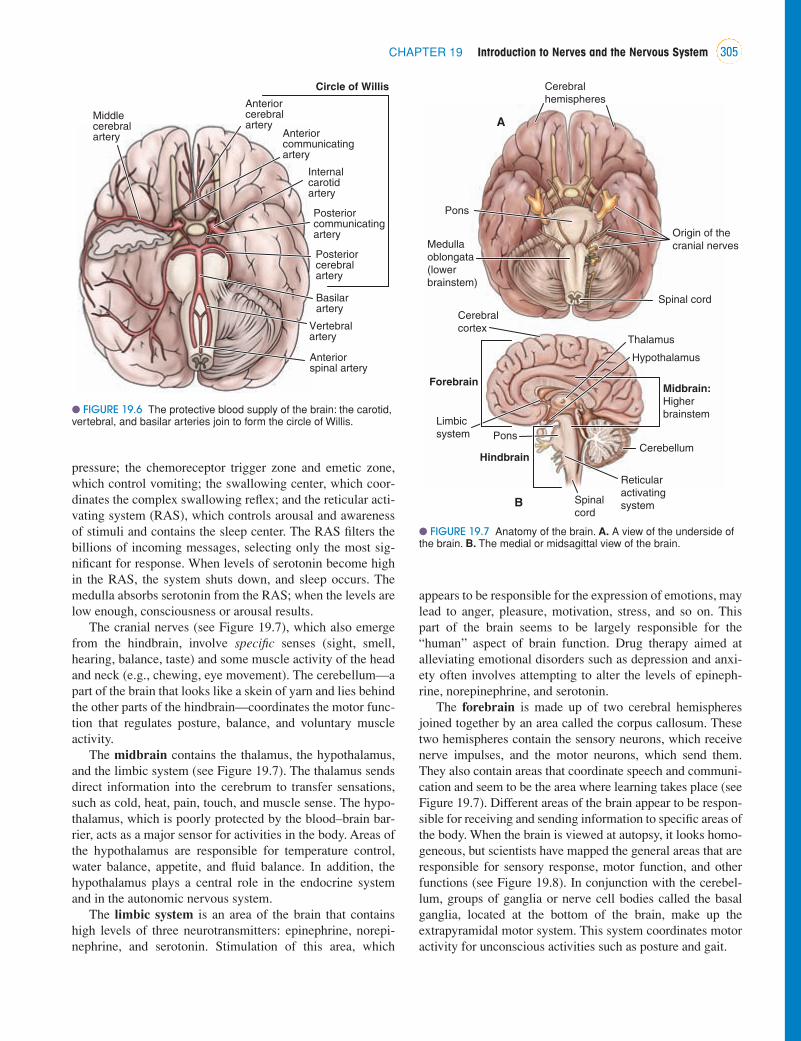
pressure; the chemoreceptor trigger zone and emetic zone,which control vomiting; the swallowing center, which coor-dinates the complex swallowing reflex; and the reticular acti-vating system (RAS), which controls arousal and awarenessof stimuli and contains the sleep center. The RAS filters thebillions of incoming messages, selecting only the most sig-nificant for response. When levels of serotonin become highin the RAS, the system shuts down, and sleep occurs. Themedulla absorbs serotonin from the RAS; when the levels arelow enough, consciousness or arousal results.
The cranial nerves (see Figure 19.7), which also emergefrom the hindbrain, involve specific senses (sight, smell,hearing, balance, taste) and some muscle activity of the headand neck (e.g., chewing, eye movement). The cerebellum—apart of the brain that looks like a skein of yarn and lies behindthe other parts of the hindbrain—coordinates the motor func-tion that regulates posture, balance, and voluntary muscleactivity.
The midbrain contains the thalamus, the hypothalamus,and the limbic system (see Figure 19.7). The thalamus sendsdirect information into the cerebrum to transfer sensations,such as cold, heat, pain, touch, and muscle sense. The hypo-thalamus, which is poorly protected by the blood–brain bar-rier, acts as a major sensor for activities in the body. Areas ofthe hypothalamus are responsible for temperature control,water balance, appetite, and fluid balance. In addition, thehypothalamus plays a central role in the endocrine systemand in the autonomic nervous system.
The limbic system is an area of the brain that containshigh levels of three neurotransmitters: epinephrine, norepi-nephrine, and serotonin. Stimulation of this area, which
appears to be responsible for the expression of emotions, maylead to anger, pleasure, motivation, stress, and so on. Thispart of the brain seems to be largely responsible for the“human” aspect of brain function. Drug therapy aimed atalleviating emotional disorders such as depression and anxi-ety often involves attempting to alter the levels of epineph-rine, norepinephrine, and serotonin.
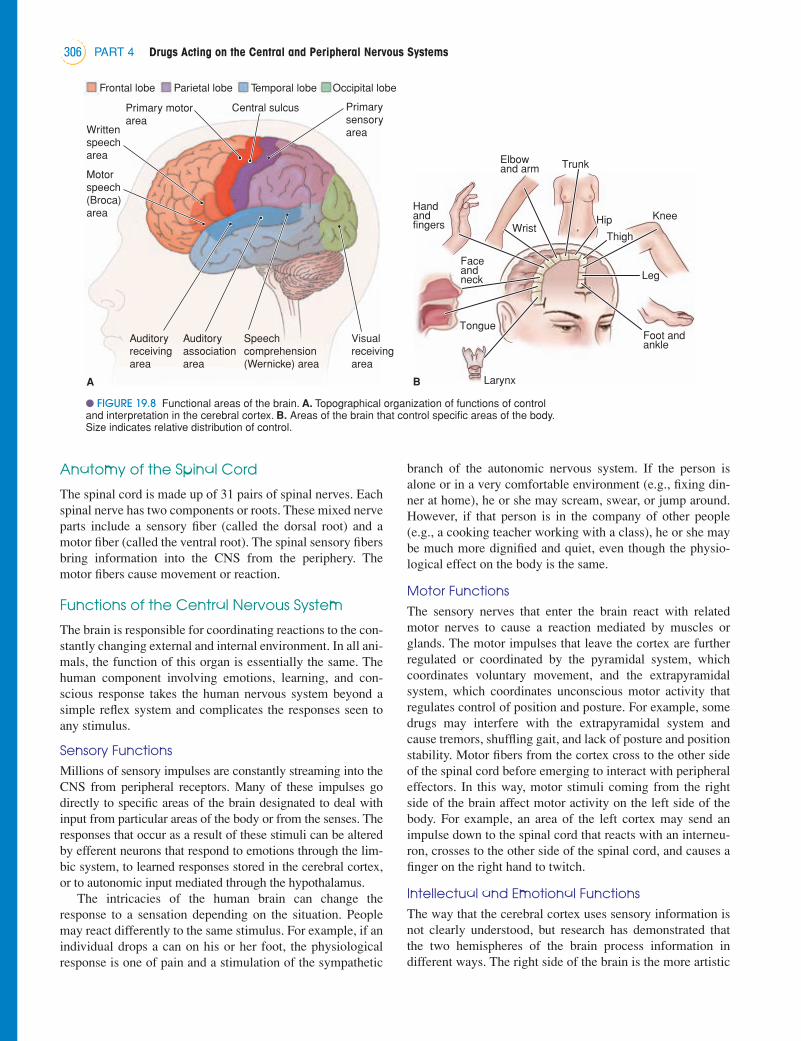
The forebrain is made up of two cerebral hemispheresjoined together by an area called the corpus callosum. Thesetwo hemispheres contain the sensory neurons, which receivenerve impulses, and the motor neurons, which send them.They also contain areas that coordinate speech and communi-cation and seem to be the area where learning takes place (seeFigure 19.7). Different areas of the brain appear to be respon-sible for receiving and sending information to specific areas ofthe body. When the brain is viewed at autopsy, it looks homo-geneous, but scientists have mapped the general areas that areresponsible for sensory response, motor function, and otherfunctions (see Figure 19.8). In conjunction with the cerebel-lum, groups of ganglia or nerve cell bodies called the basalganglia, located at the bottom of the brain, make up theextrapyramidal motor system. This system coordinates motoractivity for unconscious activities such as posture and gait.
CHAPTER 19 Introduction to Nerves and the Nervous System 305
Middlecerebralartery
Anteriorcerebralartery
Anteriorcommunicatingartery
Circle of Willis
Internalcarotidartery
Posteriorcommunicatingartery
Posteriorcerebralartery
Vertebralartery
Anteriorspinal artery
Basilarartery
● FIGURE 19.6 The protective blood supply of the brain: the carotid,vertebral, and basilar arteries join to form the circle of Willis.
Origin of thecranial nerves
Cerebralcortex
Thalamus
Limbic
system
Hypothalamus
Midbrain:Higher
brainstem
Reticular
activating
system
Cerebellum
Spinal
cord
Pons
Hindbrain
Forebrain
A
B
Pons
Cerebralhemispheres
Medulla oblongata(lower brainstem)
Spinal cord
● FIGURE 19.7 Anatomy of the brain. A. A view of the underside ofthe brain. B. The medial or midsagittal view of the brain.
LWBK374_c19_p297-309.qxd 26/08/2009 12:57 PM Page 305 Aptara
Anatomy of the Spinal Cord
The spinal cord is made up of 31 pairs of spinal nerves. Eachspinal nerve has two components or roots. These mixed nerveparts include a sensory fiber (called the dorsal root) and amotor fiber (called the ventral root). The spinal sensory fibersbring information into the CNS from the periphery. Themotor fibers cause movement or reaction.
Functions of the Central Nervous System
The brain is responsible for coordinating reactions to the con-stantly changing external and internal environment. In all ani-mals, the function of this organ is essentially the same. Thehuman component involving emotions, learning, and con-scious response takes the human nervous system beyond asimple reflex system and complicates the responses seen toany stimulus.
Sensory FunctionsMillions of sensory impulses are constantly streaming into theCNS from peripheral receptors. Many of these impulses godirectly to specific areas of the brain designated to deal withinput from particular areas of the body or from the senses. Theresponses that occur as a result of these stimuli can be alteredby efferent neurons that respond to emotions through the lim-bic system, to learned responses stored in the cerebral cortex,or to autonomic input mediated through the hypothalamus.
The intricacies of the human brain can change theresponse to a sensation depending on the situation. Peoplemay react differently to the same stimulus. For example, if anindividual drops a can on his or her foot, the physiologicalresponse is one of pain and a stimulation of the sympathetic
branch of the autonomic nervous system. If the person isalone or in a very comfortable environment (e.g., fixing din-ner at home), he or she may scream, swear, or jump around.However, if that person is in the company of other people(e.g., a cooking teacher working with a class), he or she maybe much more dignified and quiet, even though the physio-logical effect on the body is the same.
Motor FunctionsThe sensory nerves that enter the brain react with relatedmotor nerves to cause a reaction mediated by muscles orglands. The motor impulses that leave the cortex are furtherregulated or coordinated by the pyramidal system, whichcoordinates voluntary movement, and the extrapyramidalsystem, which coordinates unconscious motor activity thatregulates control of position and posture. For example, somedrugs may interfere with the extrapyramidal system andcause tremors, shuffling gait, and lack of posture and positionstability. Motor fibers from the cortex cross to the other sideof the spinal cord before emerging to interact with peripheraleffectors. In this way, motor stimuli coming from the rightside of the brain affect motor activity on the left side of thebody. For example, an area of the left cortex may send animpulse down to the spinal cord that reacts with an interneu-ron, crosses to the other side of the spinal cord, and causes afinger on the right hand to twitch.
Intellectual and Emotional FunctionsThe way that the cerebral cortex uses sensory information isnot clearly understood, but research has demonstrated thatthe two hemispheres of the brain process information indifferent ways. The right side of the brain is the more artistic
PART 4 Drugs Acting on the Central and Peripheral Nervous Systems306
Visualreceivingarea
Primary motorarea
Auditoryreceivingarea
Auditoryassociationarea
Writtenspeecharea
Motorspeech(Broca)area
Primarysensoryarea
Speechcomprehension(Wernicke) area
Central sulcus
Frontal lobe Temporal lobeParietal lobe Occipital lobe
A B
Tongue
Faceandneck
Handandfingers Wrist
Hip
Elbowand arm Trunk
Thigh
Knee
Leg
Foot andankle
Larynx
● FIGURE 19.8 Functional areas of the brain. A. Topographical organization of functions of controland interpretation in the cerebral cortex. B. Areas of the brain that control specific areas of the body.Size indicates relative distribution of control.
LWBK374_c19_p297-309.qxd 26/08/2009 12:57 PM Page 306 Aptara

side, concerned with forms and shapes, and the left side is moreanalytical, concerned with names, numbers, and processes.Why the two hemispheres are different and how they developdifferently is not known.
When learning takes place, distinct layers of the cerebralcortex are affected, and an actual membrane change occurs in aneuron to store information in the brain permanently. Learningbegins as an electrical circuit called an engram, a reverberatingcircuit of action potentials that eventually becomes a long-term,permanent memory in the presence of the proper neurotrans-mitters and hormones. Scientists do not understand exactly howthis happens, but it is known that the nerve requires oxygen,glucose, and sleep to process an engram into a permanent mem-ory, and during that processing structural changes occur to thecells involved in the engram. This reverberating circuit isresponsible for short-term memory. When patients have adecreased blood supply to the brain, short-term memory may belost, and they are not able to remember new things. Becausethey are unable to remember new things, the brain falls back onlong-term, permanent memory for daily functioning. For exam-ple, a patient may be introduced to a nurse and have no recol-lection of the nurse 2 hours later and yet be able to recall theevents of several years ago vividly.
Several substances appear to affect learning. Antidiuretichormone (ADH), which is released during reactions to stress, isone such substance. Although too much stress prevents learn-ing, feeling slightly stressed may increase a person’s ability tolearn. A patient who is a little nervous about upcoming surgery,for example, seems to display a better mastery of facts about thesurgery and postoperative procedures than a patient who is verystressed and scared or one who appears to show no interest orconcern. Oxytocin is another substance that seems to increaseactual learning. Because childbirth is the only known time thatoxytocin levels increase, the significance of this is not under-stood. Nurses who work with maternity patients should knowthat women in labor will very likely remember the smallestdetails about the whole experience and should use whateveropportunity is made available to do teaching.
In addition, the limbic system appears to play an importantrole in how a person learns and reacts to stimuli. The emotionsassociated with a memory as well as with the present have animpact on stimulus response. The placebo effect is a docu-mented effect of the mind on drug therapy: If a person per-ceives that a drug will be effective, it is much more likely toactually be effective. This effect, which uses the actions of thecerebrum and the limbic system, can have a tremendousimpact on drug response. Events that are perceived as stressfulby some patients may be seen as positive by other patients.
➧ The hindbrain, the most primitive area of the brain, con-tains the centers that control basic, vital functions. Thepons, the medulla, and the reticular activating system(RAS), which regulates arousal and awareness, are alllocated in the hindbrain. The cerebellum, which helps tocoordinate motor activity, is found at the back of thehindbrain.
➧ The midbrain consists of the hypothalamus, the thalamus,and the limbic system. The limbic system is responsiblefor the expression of emotion, and the thalamus andhypothalamus coordinate internal and external responsesand direct information into the cerebral cortex.
➧ The cerebral cortex consists of two hemispheres, whichregulate the communication between sensory and motorneurons and are the sites of thinking and learning.
CLINICAL SIGNIFICANCE OF DRUGSTHAT ACT ON THE NERVOUS SYSTEM
The features of the human nervous system, including thecomplexities of the human brain, sometimes make it difficultto predict the exact reaction of a particular patient to a givendrug. When a drug is used to affect the nervous system, theoccurrence of many systemic effects is always a possibilitybecause the nervous system affects the entire body. The chap-ters in this section address the individual classes of drugsused to treat disorders of the nervous system, including theiradverse effects. An understanding of the actions of specificdrugs makes it easier to anticipate what therapeutic andadverse effects might occur. In addition, nurses should con-sider all of the learned, cultural, and emotional aspects of thepatient’s situation in an attempt to provide optimal therapeu-tic benefit and minimal adverse effects.
C H A P T E R S U M M A R Y
• Although nerves do not reproduce, they can regenerateinjured parts if the soma and axon hillock remain intact.
• Efferent nerves take information out of the CNS to effec-tor sites; afferent nerves are sensory nerves that take infor-mation into the CNS.
• When the transmission of action potentials reaches the axonterminal, it causes the release of chemicals called neurotrans-mitters, which cross the synaptic cleft to stimulate an effec-tor cell, which can be another nerve, a muscle, or a gland.
• A neurotransmitter must be produced by a nerve (eachnerve can produce only one kind); it must be released intothe synapse when the nerve is stimulated; it must react witha very specific receptor site to cause a reaction; and it mustbe immediately broken down or removed from the synapseso that the cell can be ready to be stimulated again.
• Much of the drug therapy in the nervous system involvesreceptor sites and the release or reuptake and breakdownof neurotransmitters.
CHAPTER 19 Introduction to Nerves and the Nervous System 307
KEY POINTS
➧ The CNS consists of the brain and spinal cord, which areprotected by bone and meninges. To ensure blood flowto the brain if a vessel should become damaged, thebrain also has a protective blood supply moderated bythe circle of Willis.
LWBK374_c19_p297-309.qxd 26/08/2009 12:57 PM Page 307 Aptara

• The CNS consists of the brain and spinal cord, which areprotected by bone and meninges. To ensure blood flow to thebrain if a vessel should become damaged, the brain also hasa protective blood supply moderated by the circle of Willis.
• The hindbrain, the most primitive area of the brain, containsthe centers that control basic, vital functions. The pons, themedulla, and the reticular activating system (RAS), whichregulates arousal and awareness, are all located in the hind-brain. The cerebellum, which helps to coordinate motoractivity, is found at the back of the hindbrain.
• The midbrain consists of the hypothalamus, the thalamus,and the limbic system. The limbic system is responsiblefor the expression of emotion, and the thalamus and hypo-thalamus coordinate internal and external responses anddirect information into the cerebral cortex.
• The cerebral cortex consists of two hemispheres, whichregulate the communication between sensory and motorneurons and are the sites of thinking and learning.
• The mechanisms of learning and processing learned infor-mation are not understood. Emotion-related factors influ-ence the human brain, which handles stimuli andresponses in complex ways.
• Much remains to be learned about the human brain andhow drugs influence it. The actions of many drugs thathave known effects on human behavior are not understood.
W E B L I N K S
Health care providers and patients may want to consult thefollowing internet source:
http://www.InnerBody.com
Review of the organization and actions of the nervoussystem.
http://www.NationalGeographic.com
Interactive view of the nervous system at work.
PART 4 Drugs Acting on the Central and Peripheral Nervous Systems308
4. Which of the following could result in the initiation ofan action potential?a. Depolarizing the membraneb. Decreasing the extracellular potassium concentrationc. Increasing the activity of the sodium–potassium
active transport systemd. Stimulating the nerve with a threshold electrical
stimulus during the absolute refractory period of themembrane
5. Neurotransmitters area. produced in the muscle to communicate with nerves.b. the chemicals used to stimulate or suppress effectors
at the nerve synapse.c. usually found in the diet.d. nonspecific in their action on various nerves.
6. The limbic system is an area of the brain thata. is responsible for coordination of movement.b. is responsible for the special senses.c. is responsible for the expression of emotions.d. controls sleep.
7. The most primitive area of the brain, the brainstem,contains areas responsible fora. vomiting, swallowing, respiration, arousal, and sleep.b. learning.c. motivation and memory.d. taste, sight, hearing, and balance.
C H E C K Y O U R U N D E R S T A N D I N G
Answers to the questions in this chapter can be found inAnswers to Check Your Understanding Questions on theCD-Rom in the front of the book.
MULTIPLE CHOICE
Select the best answer to the following.
1. The cerebelluma. initiates voluntary muscle movement.b. helps regulate the tone of skeletal muscles.c. if destroyed, would result in the loss of all voluntary
skeletal activity.d. contains the centers responsible for the regulation of
body temperature.
2. At those regions of the nerve membrane where myelin ispresent, there isa. low resistance to electrical current.b. high resistance to electrical current.c. high conductance of electrical current.d. energy loss for the cell.
3. The nerve synapsea. is not resistant to electrical current.b. cannot become exhausted.c. has a synaptic cleft.d. transfers information at the speed of electricity.
LWBK374_c19_p297-309.qxd 26/08/2009 12:57 PM Page 308 Aptara

B I B L I O G R A P H Y A N DR E F E R E N C E S
Fox, S. (1991). Perspectives on human biology. Dubuque, IA: Wm.C. Brown.
Ganong, W. (2003). Review of medical physiology (21st ed.).Norwalk, CT: Appleton & Lange.
Gilman, A., Hardman, J. G., & Limbird, L. E. (Eds.). (2006).Goodman and Gilman’s the pharmacological basis of therapeu-tics (11th ed.). New York: McGraw-Hill.
Guyton, A., & Hall, J. (2007). Textbook of medical physiology(11th ed.). Philadelphia: W. B. Saunders.
Karch, A. M. (2009). 2010 Lippincott’s nursing drug guide.Philadelphia: Lippincott Williams & Wilkins.
Noback, C., Strominger, N. L., Demarest, R. J., Ruggiero, D. A.(2005). The human nervous system: Structure and function (6th ed.). Totowa, NJ: Humana Press.
Parpura, V., & Hayden, P. (2008). Astrocytes in the physiology ofthe nervous system. New York: Springer.
Porth, C. M. (2008). Pathophysiology: Concepts of altered healthstates (8th ed.). Philadelphia: Lippincott Williams & Wilkins.
Thibodeau, G., & Patton, K (2006). Anthony’s textbook of anatomyand physiology (18th ed.). St. Louis, MO: Mosby.
CHAPTER 19 Introduction to Nerves and the Nervous System 309
c. Little glucose is stored in nerve cells, so a constantsupply is needed.
d. The brain needs a constant supply of insulin and thy-roid hormone.
e. The brain swells easily and needs the blood supply toreduce swelling.
f. Circulating aldosterone levels maintain the fluid bal-ance in the brain.
2. The blood–brain barrier could be described by which ofthe following?a. It is produced by the cells that make up the
meninges.b. It is regulated by the microglia in the CNS.c. It is weaker in certain parts of the brain.d. It is uniform in its permeability throughout the CNS.e. It is an anatomical structure that can be punctured.f. It is more likely to block the entry of proteins into
the CNS.
8. A clinical indication of poor blood supply to the brain,particularly to the higher levels where learning takesplace, would bea. loss of long-term memory.b. loss of short-term memory.c. loss of coordinated movement.d. insomnia.
MULTIPLE RESPONSE
Select all that apply.
1. In explaining the importance of a constant blood sup-ply to the brain, the nurse would tell the student whichof the following?a. Energy is needed to maintain nerve membranes and
cannot be produced without oxygen.b. Carbon dioxide must constantly be removed to main-
tain the proper pH.
LWBK374_c19_p297-309.qxd 26/08/2009 12:57 PM Page 309 Aptara