Embed Size (px)
Citation preview
1
Lung Lung PatternsPatterns
VMB 960
3/7/2011
Abnormal Lung Pattern Abnormal Lung Pattern ClassificationClassification
Alveolar
Interstitial
BronchialBronchial
Vascular (covered with heart lecture)
Mixed
NormalNormalMay be the MOST DIFFICULT pattern
to diagnose!
The ability to see vessels, bronchi and some interstitial markings is NORMALg
Although “technically” not a pattern, determination of ‘normal’ is obviously critical

2
NormalNormal
Broad range of normal depending on:Age of the animal
Conformation of the animal
Phase of respiration
Be clear if you mean “radiographically normal” or “clinically normal”
Does a normal lung pattern mean an absence of lung pathology? NO!!
3
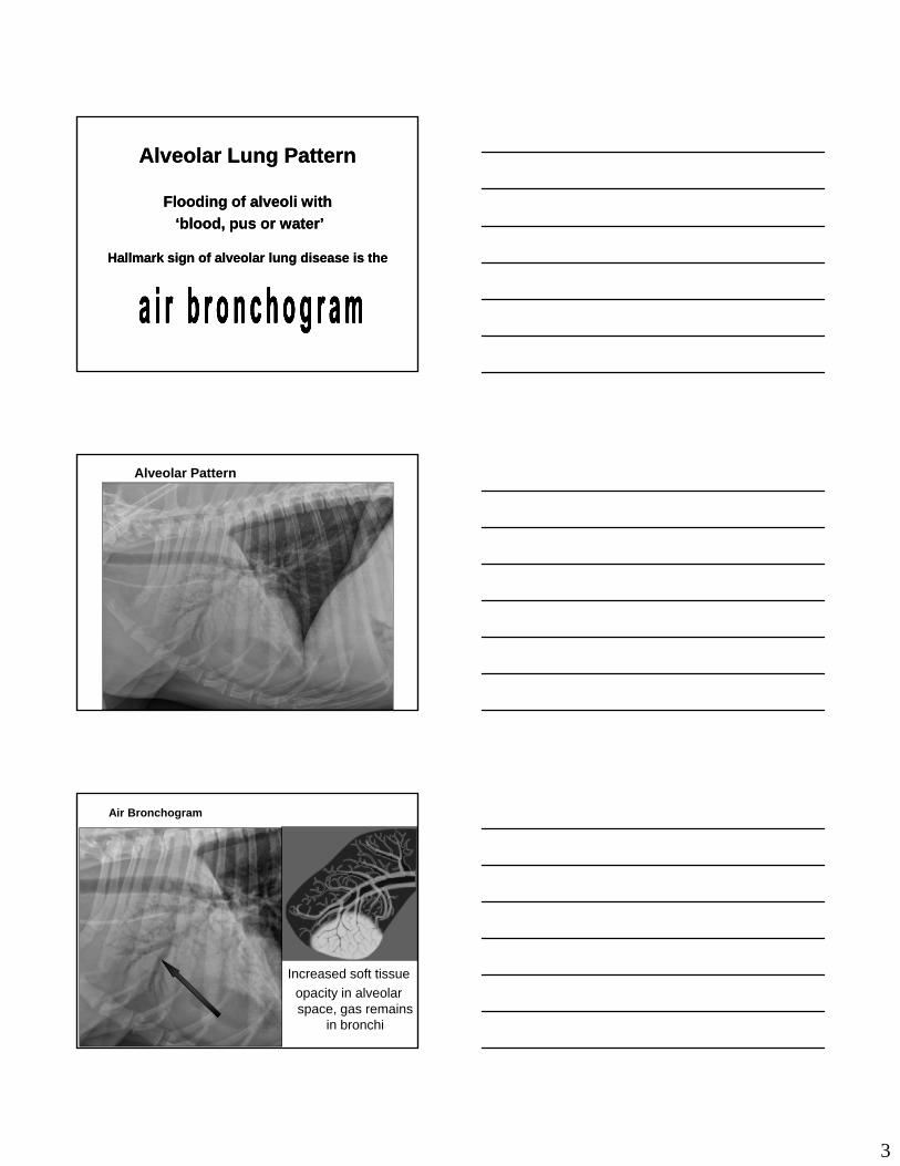
Alveolar Lung PatternAlveolar Lung Pattern
Flooding of alveoli withFlooding of alveoli with
‘blood, pus or water’ ‘blood, pus or water’
Hallmark sign of alveolar lung disease is theHallmark sign of alveolar lung disease is the
Alveolar Pattern
Air Bronchogram
Increased soft tissue
opacity in alveolar space, gas remains
in bronchi
4
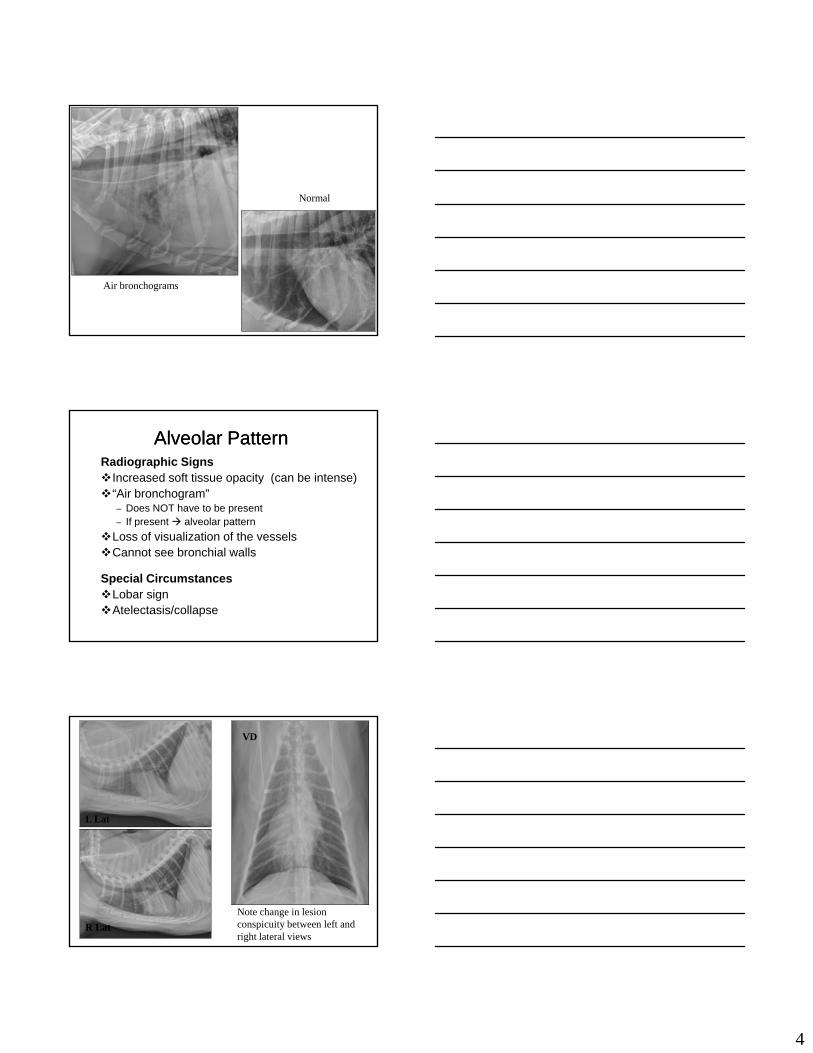
Normal
Air bronchograms
Alveolar PatternAlveolar PatternRadiographic SignsIncreased soft tissue opacity (can be intense)“Air bronchogram”
– Does NOT have to be present– If present alveolar patternp p
Loss of visualization of the vesselsCannot see bronchial walls
Special CircumstancesLobar signAtelectasis/collapse
L Lat VD
L Lat
VD
R lat
Note change in lesion conspicuity between left and right lateral views
R Lat

5
Lobar sign
R lat
L lat
Alveolar PatternAlveolar Pattern
There are two DIFFERENT mechanisms that can result in an alveolar pattern
CONSOLIDATION– Fluid or cells in the alveoli (blood, pus, water)
ATELECTASIS– Collapse of the alveoli
Both result in an increase in soft tissue opacity in the alveolar space
Alveolar PatternAlveolar Pattern
CONSOLIDATION Fluid and/or cells in the
alveoli No mediastinal shift
ATELECTASIS Collapse of the alveoli
(loss of air in alveoli)
M di ti l hift No mediastinal shift Lung lobe “normal” size Not necessarily
associated with pleural disease
Mediastinal shift
Lung lobe decreased in size
Often associated with pleural disease– Pneumothorax
– Pleural effusion
6
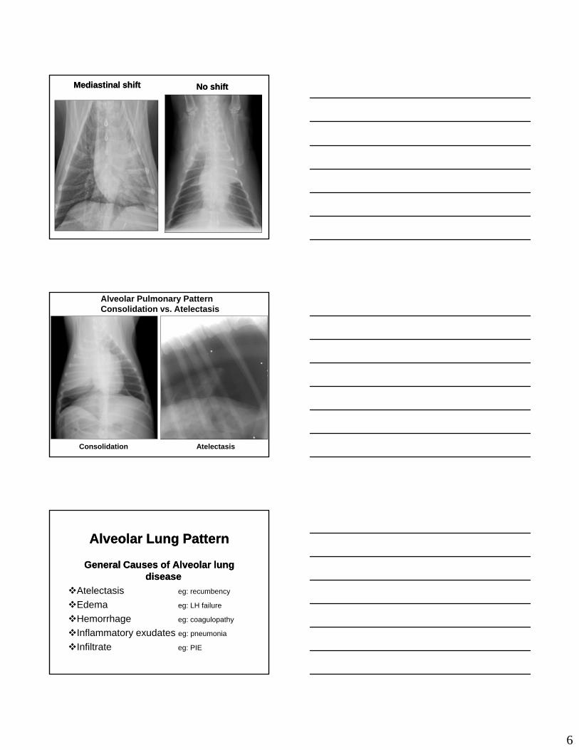
Mediastinal shiftMediastinal shift No shiftNo shift
Alveolar Pulmonary PatternConsolidation vs. Atelectasis
Consolidation Atelectasis
Alveolar Lung PatternAlveolar Lung Pattern
General Causes of Alveolar lung General Causes of Alveolar lung diseasedisease
Atelectasis eg: recumbencyAtelectasis eg: recumbency
Edema eg: LH failure
Hemorrhage eg: coagulopathy
Inflammatory exudates eg: pneumonia
Infiltrate eg: PIE

7
Alveolar Lung PatternAlveolar Lung Pattern
Most common cause of generalized Most common cause of generalized alveolar lung disease isalveolar lung disease is
Pulmonary EdemaPulmonary EdemaPulmonary EdemaPulmonary Edema
Increased Hydrostatic Pressure
Reduced Oncotic pressure
Increased Capillary Permeability
Alveolar Lung PatternAlveolar Lung Pattern
Focal alveolar lung diseaseFocal alveolar lung disease
Multiple causes
Differential diagnosis prioritizationDifferential diagnosis prioritization influenced by distribution and intensity of change
Accurate history important in ranking differentials
Alveolar Lung PatternAlveolar Lung Pattern
Important ‘distribution patterns’Important ‘distribution patterns’
Generalized perm/hydro/onc
Cranioventral pneumoniaCranioventral pneumonia
Perihilar hydrostatic
Caudodorsal perm/hydro/onc
Focal nonspecific

8
Alveolar PatternAlveolar Pattern
Pattern Distribution – Aspiration pneumonia
Cranioventral lung lobes
Right middle lung lobe most commonLook for summation sign over the cardiac silhouetteLook for summation sign over the cardiac silhouette
on left lateral view!
Usually intensity of opacification is
most severe with inhalation pneumonia
Alveolar PatternAlveolar PatternPattern Distribution – Pulmonary EdemaCardiogenic
– Perihilar – Can become generalized
Non-cardiogenic (or neurogenic)Non cardiogenic (or neurogenic)– Caudodorsal lung fields– Can become generalized
Caudodorsal lung fields generally most affected
Remember that distribution can change as disease progresses or resolves.
Alveolar PatternNon-cardiogenic Edema

9
Interstitial PatternsInterstitial Patterns
UnstructuredUnstructured StructuredStructured
Interstitial PatternsInterstitial PatternsUnstructuredUnstructured
Patient factors•Ageing changes•Inflammatory processes•Infiltrative processes•Vasogenic factors
Technical factors•Under exposure•Expiratory radiograph•Obesity
Usually generalized / diffuseUsually generalized / diffuse
Unstructured Interstitial Unstructured Interstitial PatternPattern
Technical factorsImportant that thoracic radiographs are made
on PEAK INSPIRATIONE i t fil tif t ll– Expiratory films may artifactually cause or enhance an unstructured interstitial pattern
Other factors that may cause an APPARENTunstructured interstitial pattern– Respiratory motion– Obesity– Underexposure

10
Expiration Inspiration
Expiration Inspiration Inspiration vs Expiration
Same dog, same views. Note difference in degree of soft tissue
lung opacity!
Unstructured Interstitial Unstructured Interstitial PatternPattern
Hazy/amorphous increase in
soft tissue opacityThe result of :– The result of : Fluid and/or cells in the interstitial space
Fibrosis in the interstitial space– Chronic inflammation
– Normal ageing change
Diffuse Unstructured Interstitial Pattern - LSA
Normal
11
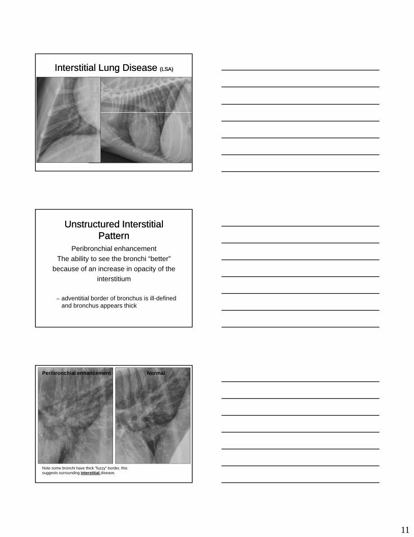
Interstitial Lung Disease Interstitial Lung Disease (LSA)(LSA)
Unstructured Interstitial Unstructured Interstitial PatternPattern
Peribronchial enhancement
The ability to see the bronchi “better”
because of an increase in opacity of thebecause of an increase in opacity of the
interstitium
– adventitial border of bronchus is ill-defined and bronchus appears thick
Peribronchial enhancement Normal
Note some bronchi have thick “fuzzy” border, this suggests surrounding interstitial disease.

12
Unstructured Interstitial Unstructured Interstitial PatternPattern
Overlap of Patterns
A severe unstructured interstitial pattern may mimic a mild alveolar patternmay mimic a mild alveolar pattern– Sometimes cannot distinguish between the
two patterns
– If in doubt identify the pattern as alveolar - being most severe
Interstitial PatternsInterstitial PatternsStructuredStructured
Cavitary•Abscess / cyst•Necrotic tumor•Parasitic
Non-cavitary•Extrathoracic•Fake out•Tumor 1 or 2•Granuloma
Focal or generalizedFocal or generalized
Interstitial PatternsInterstitial PatternsStructured Structured -- NonNon--cavitarycavitary
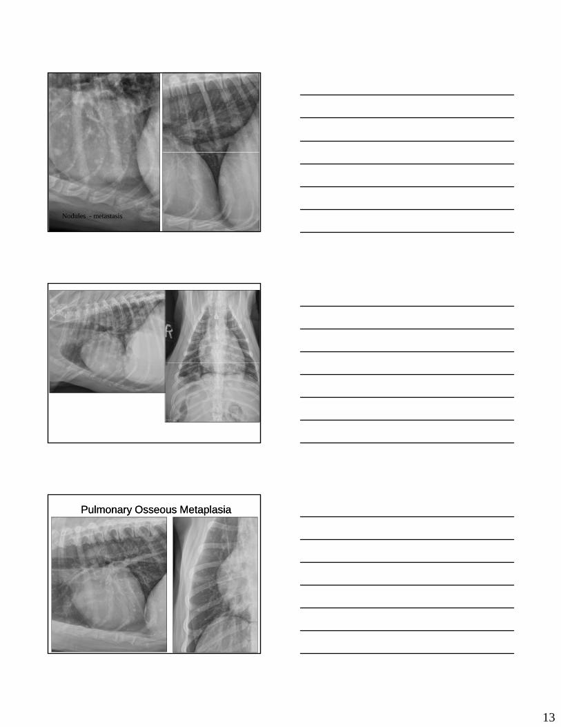
Focal or generalized – Nodular
Metastatic lung disease
Primary lung mass
Pulmonary osseous metaplasia
Granuloma fungal, eosinophilic, FB
Abscess
Fluid filled bulla
– Miliary Fungal – Blastomycosis
13
Nodules - metastasis
Pulmonary Osseous MetaplasiaPulmonary Osseous Metaplasia

14
Intense MiliaryIntense Miliary
–– with coalescencewith coalescence
Structured / Nodular InterstitialStructured / Nodular Interstitial
Radiographic appearanceCircumscribed lesions of various opacity, size
and number in the interstitial space of the lung
May be single or multipleMay be CAVITARY or NON-CAVITARY
– CAVITARY = CONTAINS GAS OPACITY– NON-CAVITARY = NO GAS OPACITY
Can have cavitary and non-cavitary lesions in the same patient
Structured or Nodular Structured or Nodular InterstitialInterstitial
Where are the nodules?The nodules are located in the interstitial
spaceTh i t titi l t i thThe interstitial space contains the:
– Vessels– Bronchi– Nerves– Lymphatics
The nodules are between or invade the structures of the interstitial space

15
Structured / Nodular InterstitialStructured / Nodular InterstitialEnd-on Vessel Located near other
vessels Same size or smaller
than associated l it di l l
Pulmonary Nodule Does not have to be
near vessels Can be any size Random in location
longitudinal vessels Tend to follow a patternMay be near a
bronchus Typically well-defined
smooth marginsMay have a “tail”More opaque than
expected for size
Does not have to be near a bronchus
Margins may be smooth or irregular
No “tail”
Structured / Nodular InterstitialStructured / Nodular Interstitial
Pulmonary Nodule or Something on Skin?
Ectoparasites (ticks), skin masses, nipples etc can mimic a pulmonary noduleetc. can mimic a pulmonary nodule
Structures on the surface often more opaque than expected due to air/soft tissue interface
Place radiopaque marker on “lesion” to determine location
Extrathoracic “Nodule”
Note large nipples on lateral view
“Perfect” alignment of “nodules” of the same size should be a clue that these structures may not be pulmonary.

16
Cavitary Structured Interstitial Pulmonary PatternHematocoele (Blood-filled bulla)
Horizontal beam radiograph - note the fluid lines in the pulmonary masses.
Structured Interstitial – cavitary
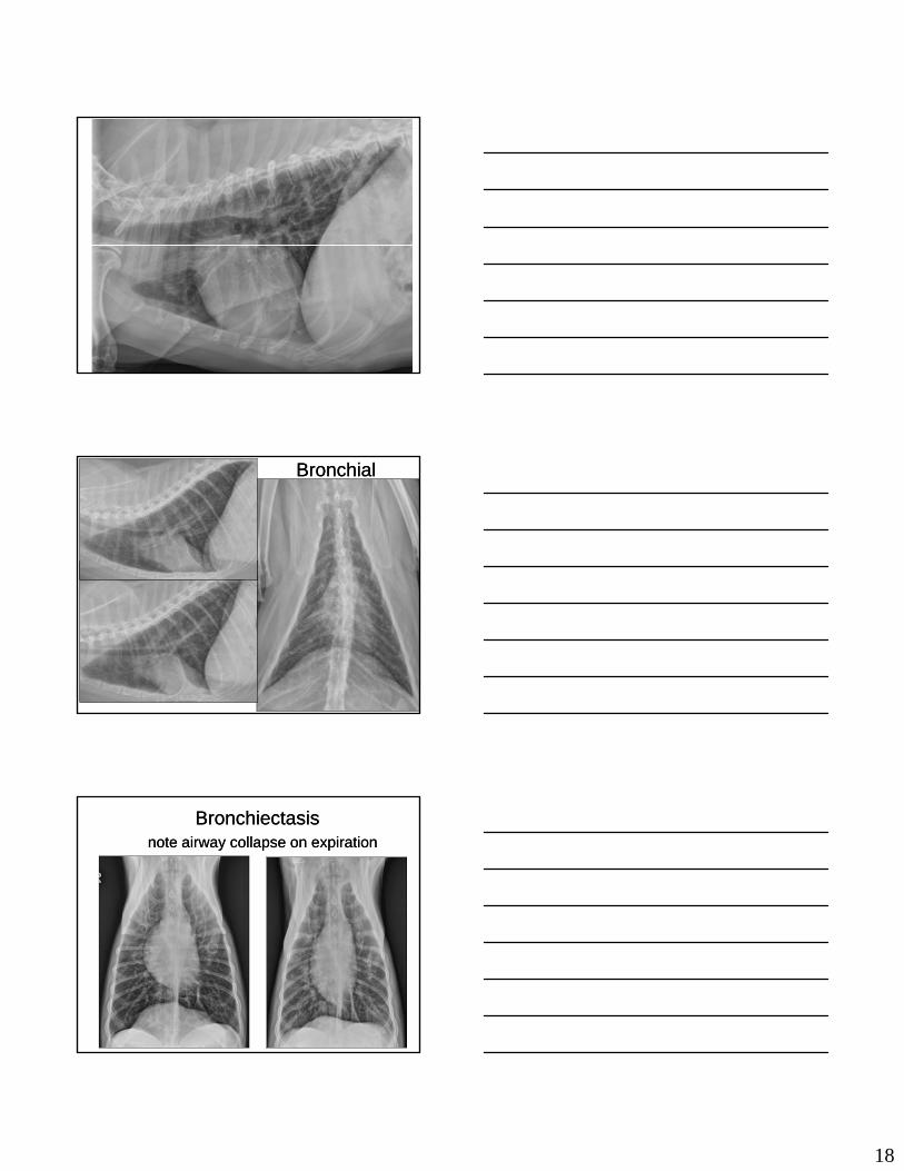
Bronchial PatternBronchial Pattern
Radiographic appearanceIncreased visualization of the bronchi
Typically most difficult pattern for students to recognize
Must look for normally visualized pulmonary structures– Bronchi– Vessels

17
Bronchial PatternBronchial PatternRadiographic FindingsIncrease in size of bronchiApparent increase in number of bronchi
– Due to increase in size of bronchi
Loss of taperBronchial walls become parallel– Bronchial walls become parallel
Bronchial wall thickening– Can be difficult to distinguish from peribronchial
enhancement
Special CircumstancesBronchial mineralization
– Can be a normal aging change If only finding, probably do NOT have a bronchial
pattern
Bronchial PatternBronchial Pattern
Radiographic FindingsIncrease in size is RELATIVELook in the periphery of the lung fields
– Look for end on bronchi– Should have a lucent center
Look VERY closely!Although identified as large, the bronchi are
still very small!– Especially in CATS!!!
Severe Bronchial Pattern
18
Bronchial Bronchial
BronchiectasisBronchiectasisnote airway collapse on expirationnote airway collapse on expiration
1
Lung Patterns Case Discussions
3/8/2011
Lung Model using Adobe PhotoshopLung Model using Adobe Photoshop
Case Discussions Case Discussions
Things we will cover Things we will cover
Case 1Case 1
1 year old German Shepherd 707221 year old German Shepherd 70722
Female spayed Female spayed
Febrile and dyspnea Febrile and dyspnea

2
L

3
L R
Case 1 Findings Case 1 Findings
There is soft tissue opacification with air bronchograms There is soft tissue opacification with air bronchograms in the right middle lung lobe, caudal part of the right in the right middle lung lobe, caudal part of the right cranialcranial lung lobe and to a lesser degree of the caudal lung lobe and to a lesser degree of the caudal portion of the left cranial lung lobe. portion of the left cranial lung lobe.
The heart and pulmonary vessels are small consistent The heart and pulmonary vessels are small consistent with hypovolemia possibly due to dehydration. with hypovolemia possibly due to dehydration.
What are the most likely differentials for this pattern?What are the most likely differentials for this pattern?
Case 2Case 2
12 year old Labrador Retriever 12 year old Labrador Retriever 6793367933
Male castrateMale castrate
Chronic progressive paraparesisChronic progressive paraparesis

4

5
Case 2 Findings Case 2 Findings
There is a moderate diffuse bronchial There is a moderate diffuse bronchial (bronchointerstitial)(bronchointerstitial) lung pattern with thickening and lung pattern with thickening and mineralization of bronchial walls.mineralization of bronchial walls.
Ple ral fiss re lines are presentPle ral fiss re lines are present probably aprobably a Pleural fissure lines are present Pleural fissure lines are present –– probably a probably a manifestation of pleural fibrosis. manifestation of pleural fibrosis.
Changes are consistent with chronic Changes are consistent with chronic inflammatory airway disease. inflammatory airway disease.
Bronchial Bronchial -- another example another example
Case 3Case 3
2 year old Chinese Crested 2 year old Chinese Crested 5116751167
Male Male
Rescued from a garage fire. Severe dyspneaRescued from a garage fire. Severe dyspnea

6

7
Case 3 Findings Case 3 Findings
Extensive air bronchograms are present Extensive air bronchograms are present throughout the lung parenchyma, indicating a throughout the lung parenchyma, indicating a diffuse, intense alveolar pattern. diffuse, intense alveolar pattern.
In light of the history p lmonary edemaIn light of the history p lmonary edema In light of the history, pulmonary edema In light of the history, pulmonary edema secondary to smoke inhalation is the most likely secondary to smoke inhalation is the most likely diagnosis.diagnosis.
Case 4Case 4
10 year old Golden Retriever 8690610 year old Golden Retriever 86906
Male Male
Osteosarcoma Osteosarcoma –– distal femur distal femur

8

9
Case 4 Findings Case 4 Findings
Multiple variablyMultiple variably--sized soft tissue pulmonary nodules sized soft tissue pulmonary nodules are present throughout the lungs. are present throughout the lungs.
Cardiovascular structures appear within normal limits. Cardiovascular structures appear within normal limits. Ca d ovascu a st uctu es appea w t o a ts.Ca d ovascu a st uctu es appea w t o a ts.
This pattern is typical of metastatic lung disease.This pattern is typical of metastatic lung disease.
What are other differentials for a nodular interstitial What are other differentials for a nodular interstitial pattern? pattern?
Case 5Case 5
5 year old Cocker Spaniel 5 year old Cocker Spaniel 6655866558
Constipation and tachypneaConstipation and tachypnea
Constipation thought due to pelvic canal mass Constipation thought due to pelvic canal mass
What are the radiographic findings? What are the radiographic findings?
What are the most likely differentials? What are the most likely differentials?

10
11
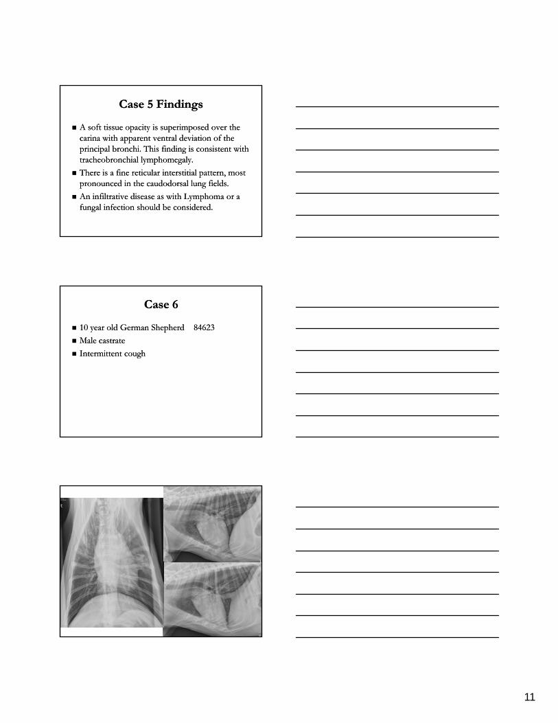
Case 5 Findings Case 5 Findings
A soft tissue opacity is superimposed over the A soft tissue opacity is superimposed over the carina with apparent ventral deviation of the carina with apparent ventral deviation of the principal bronchi. This finding is consistent with principal bronchi. This finding is consistent with tracheobronchial lymphomegalytracheobronchial lymphomegalytracheobronchial lymphomegaly. tracheobronchial lymphomegaly.
There is a fine reticular interstitial pattern, most There is a fine reticular interstitial pattern, most pronounced in the caudodorsal lung fields. pronounced in the caudodorsal lung fields.
An infiltrative disease as with Lymphoma or a An infiltrative disease as with Lymphoma or a fungal infection should be considered. fungal infection should be considered.
Case 6Case 6
10 year old German Shepherd 10 year old German Shepherd 8462384623
Male castrateMale castrate
Intermittent cough Intermittent cough

12
R

13
Case 6 Findings Case 6 Findings
An alveolar pattern is present in the caudal part of the An alveolar pattern is present in the caudal part of the left cranial lung lobe.left cranial lung lobe.
A patchy alveolar pattern is also present in the right A patchy alveolar pattern is also present in the right cranial lung lobe.cranial lung lobe.gg
The cardiovascular structures are within normal The cardiovascular structures are within normal limits.limits.
Dorsal deviation of the trachea is likely a manifestation Dorsal deviation of the trachea is likely a manifestation of head position.of head position.
What are the most likely differentials for this pattern?What are the most likely differentials for this pattern?