Embed Size (px)
Citation preview
Lung Imaging in the Mouse with SWIFT
Curt Corum, Djaudat Idiyatullin,Steen Moeller, Ryan Chamberlain,Deepali Sachdev, and Mike Garwood
Center for Magnetic Resonance Research and Masonic Cancer Center, School of MedicineUniversity of Minnesota, Minneapolis, Minnesota, USA
#204

5/4/2010 ISMRM 2010 #204 Lung Imaging in the Mouse with SWIFT
2
Declaration of Conflict of Interest or Relationship
Speaker Name: Curt Corum
I have the following conflict(s) of interest to disclose with regard to the subject matter of this presentation:
Dr. Corum is entitled to sales royalty from Steady State Imaging, which isdeveloping products related to the research described in this presentation.
5/4/2010 ISMRM 2010 #204 Lung Imaging in the Mouse with SWIFT
3
Motivation
Lung and especially lung parenchyma is difficult to image with MRI
Low proton density, susceptibility, T2, T
2*, cardiac and
respiratory motion all contribute At higher fields intrinsic (air to tissue and pathology)
and extrinsic (contrast injection) suseptibility effects become stronger
With T2
* sensitive sequences these cause signal dropout and phase effects due to the local field changes

5/4/2010 ISMRM 2010 #204 Lung Imaging in the Mouse with SWIFT
4
Lots of interest and great work... UTE approachHuman and AnimalBergin, Noll et al.1992Shattuck, Gewalt et al.1997
GRE approachHuman and AnimalMuch great work...
FID (BLAST, RUFIS)Animal ImagingKuethe et al. and others...
5/4/2010 ISMRM 2010 #204 Lung Imaging in the Mouse with SWIFT
5
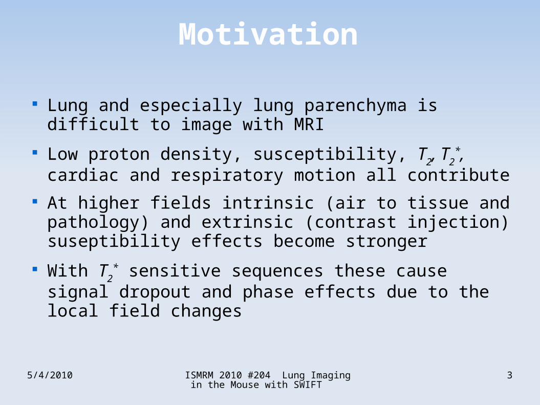
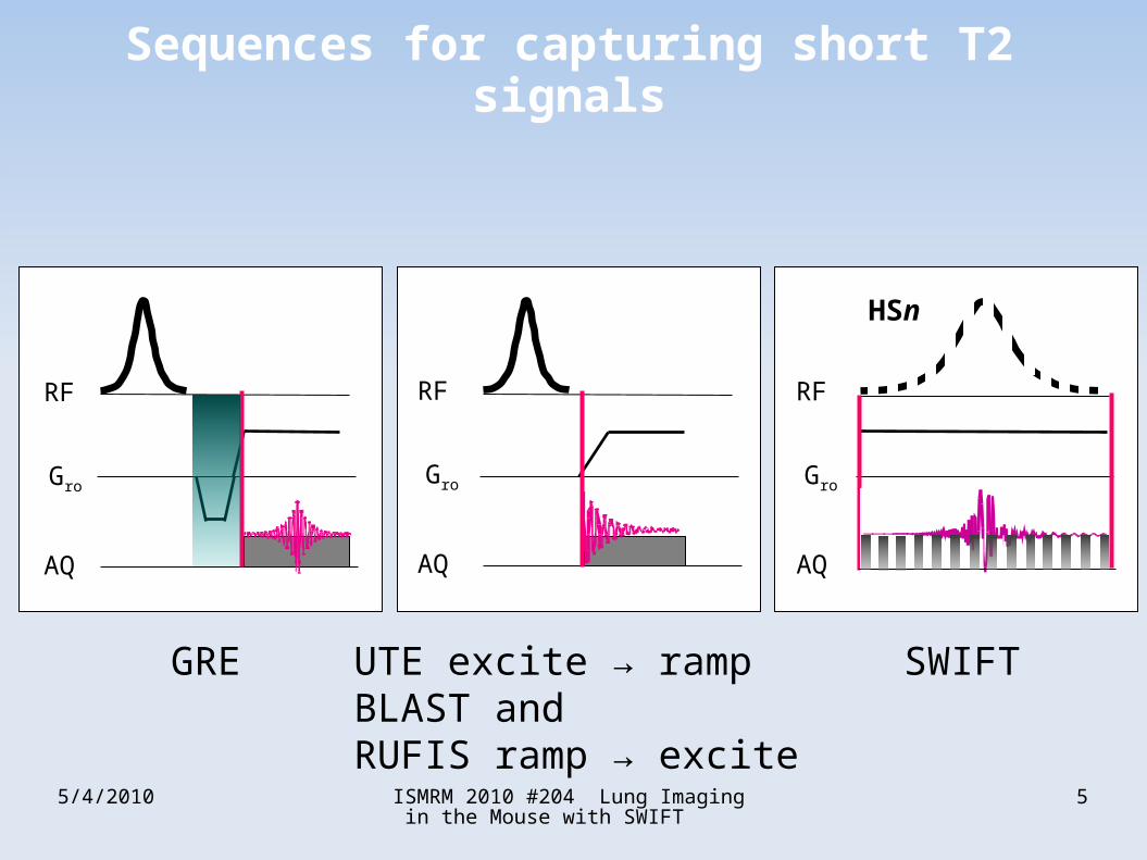
AQ
Gro
RF
UTE excite → rampBLAST andRUFIS ramp → excite
SWIFTGRE
AQ
Gro
RF
HSn
AQ
Gro
RF
Sequences for capturing short T2 signals

5/4/2010 ISMRM 2010 #204 Lung Imaging in the Mouse with SWIFT
6
T2
* of Parenchyma
Mouse LungGRE TE = 2msRespiratory gated
Clip level adjustment
5/4/2010 ISMRM 2010 #204 Lung Imaging in the Mouse with SWIFT
7
SWIFT Sequence
SWeep Imaging with Fourier Transform Acquisition occurs in the gaps of a frequency swept (usually
HSn) pulse Excitation and Acquisition nearly simultaneous
“dead time” ~2 µs No time for slice selection or phase encoding, is most naturally a
readout only, interleaved preparations are possible Aquired data are FIDs (after correlation with the RF pulse
shape) Operates in 2d projection mode or 3d image mode with radial
FID sampling scheme and gridding reconstruction Most similar to BLAST, RUFIS, etc. in concept

5/4/2010 ISMRM 2010 #204 Lung Imaging in the Mouse with SWIFT
8
SWIFT Sequence
Idiyatullin, D.; Corum, C.; Park, J. Y. & Garwood, M., Fast and quiet MRI using a swept radiofrequency., J Magn Reson, 2006, 181, 342-349

5/4/2010 ISMRM 2010 #204 Lung Imaging in the Mouse with SWIFT
9
Mouse Model of Lung Cancer
7-9 week old athymic mice
Lung seeking metastatic variant of MDA-MB-231 breast cancer cells injected into the tail vein.
D. Sachdev
Isoflurane O2 N
2O anesthesia
N=4 Healthy Animals and
N=2 Tumor animals so far...

5/4/2010 ISMRM 2010 #204 Lung Imaging in the Mouse with SWIFT
10
MRI parameters
• SWIFT TR 2.2 ms dead time 2 µs
• 125 kHz acquisition bandwidth
• 3072*16*2 = 96,304 unique FID views
• 256 complex points per view
• Prospective respiratory trigger and gating, 40-50% duty
• 100-200 views per trigger (typically 128)
• Reconstruction to 256^3 3D image
• ~0.12 mm nominal resolution
• 8-9 min (x4) for 32-36 min total imaging time per animal

5/4/2010 ISMRM 2010 #204 Lung Imaging in the Mouse with SWIFT
11
Healthy Mouse
Respiratory pillow gated, 125kHz SWIFT9.4 T Varian animal system Combination of 4x 8 min scans (32 min)Quadrature surface coil on backAnimal is prone, ~3x3x4 cm fov
Intensity flattening appliedReformatted to bronchial VNative intensity

5/4/2010 ISMRM 2010 #204 Lung Imaging in the Mouse with SWIFT
12
Tumor Mouse
Same parametersLeft lung has collapsed
Intensity flattening appliedReformatted to bronchial VNative intensity

5/4/2010 ISMRM 2010 #204 Lung Imaging in the Mouse with SWIFT
13
Tumor Montage

5/4/2010 ISMRM 2010 #204 Lung Imaging in the Mouse with SWIFT
14
Excised Lung
D. Sachdev

5/4/2010 ISMRM 2010 #204 Lung Imaging in the Mouse with SWIFT
15
Discussion
Preserves signal Lung Parenchyma and signal next to susceptibility boudaries (bronchii to parenchyma, vessels to parenchyma, tumor margin to parenchyma, etc...)
Avoidance of gradient ramp sampling required for UTE sequences
Avoidance of acuostic noise and eddy currents Low peak RF power compared to BLAST or RUFIS
(radial FID aquisition sequences), T1 weighting
obtainableScalable to Human Imaging

5/4/2010 ISMRM 2010 #204 Lung Imaging in the Mouse with SWIFT
16
Future Work
Histological and Pathological verificationnormal tissue, tumors, inflammation
Scalable to human imaging (no peak power or gradient performance limitations)
Contrast and DCE

5/4/2010 ISMRM 2010 #204 Lung Imaging in the Mouse with SWIFT
17
Support
NIH BTRR 5P41RR008079-17CMRR Center Grant, Core 3, PI Mike Garwood
Masonic Cancer Center (startup), PI Deepali Sachdev 1R21CA139688-01, PI Curt Corum
IMPROVED BREAST DCE MRI WITH SWIFT MN MED FDN/3932-9227-09, PI Curt Corum
MRI Utilizing SWIFT to Detect Breast Calcifications,Minnesota Medical Foundation

5/4/2010 ISMRM 2010 #204 Lung Imaging in the Mouse with SWIFT
18
Upcoming SWIFT presentations
• Theoretical Sensitivities of SWIFT and the Ideal Sequence (Delta Pulse-Acquire) for Ultra-Short T2Tuesday 4 May, 13:30 - 15:30 #2975
• Detection of Short T2 Component in Brain by SWIFTTuesday 4 May, 13:30 - 15:30 #2980
• In Vivo SWIFT Imaging of SPIO Labeled Stem Cells Grafted in the HeartTuesday 4 May, 14:30 - 15:00 #3747, Computer 40
• Measurement of T1 Relaxation Time in Articular Cartilage Using SWIFTWednesday 5 May, 13:30 - 15:30 #841
• Dipole Matched Filter with SWIFTWednesday 5 May, 14:30 - 15:00 #5113, Computer 126
• SWIFT Versus X-Ray In Dental ImagingThursday 6 May ,12:18 - 12:30 Room A5 #543

5/4/2010 ISMRM 2010 #204 Lung Imaging in the Mouse with SWIFT
19
Bonus: Detailed SWIFT Timing

5/4/2010 ISMRM 2010 #204 Lung Imaging in the Mouse with SWIFT
20
Bonus: Dipole Matched Filter
Bo
Unfiltered Magnitude Image Re ImageAfter dipole matched filter Fin k-space
Mag ImageAfter dipole matched filter Fin k-space
62kHz SWIFT at 4 T2 o'clock position has MRI compatible catheter tip
F=3cos2 θ k −1
2E-Poster #5113Thurs

5/4/2010 ISMRM 2010 #204 Lung Imaging in the Mouse with SWIFT
21
Bonus: Teeth
With Don Nixdorf, DDS, Hari Prasad, et al.See talk #543 Thurs 05/06, 12:18 PM, room A5