Embed Size (px)
Citation preview
Canadian Association of Radiologists 2005 Lake Louise, Alberta
© Ravi Bhargava 1
Prenatal Predictors of Postnatal Lung Hypoplasia: MRI Assessment
Ravi Bhargava MD FRCPC
Associate Professor, Radiology
University of Alberta
Chief, RadiologyStollery Children’s Hospital
Edmonton, Canada
Objectives
� Identify prenatal predictors of postnatal lung and vascular hypoplasia on fetal MRI.
� Review MRI literature
� Identify MR research areas in lung hypoplasia
Lung Hypoplasia
� Autopsy lung/body weight– < 0.012 in GA > 28 weeks
– < 0.015 in GA < 28 weeks
� Autopsy lung weight– Compared to Expected range
� Radial-alveolar count– Most complicated, not often used
� Clinical pulmonary insufficiency
How is pulmonary hypoplasiadiagnosed?
� Pathology– Lung weight/Body weight (most common)
– Pathology assessment of alveolar branching
� Clinically– Respiratory issues
– Death
Canadian Association of Radiologists 2005 Lake Louise, Alberta
© Ravi Bhargava 2
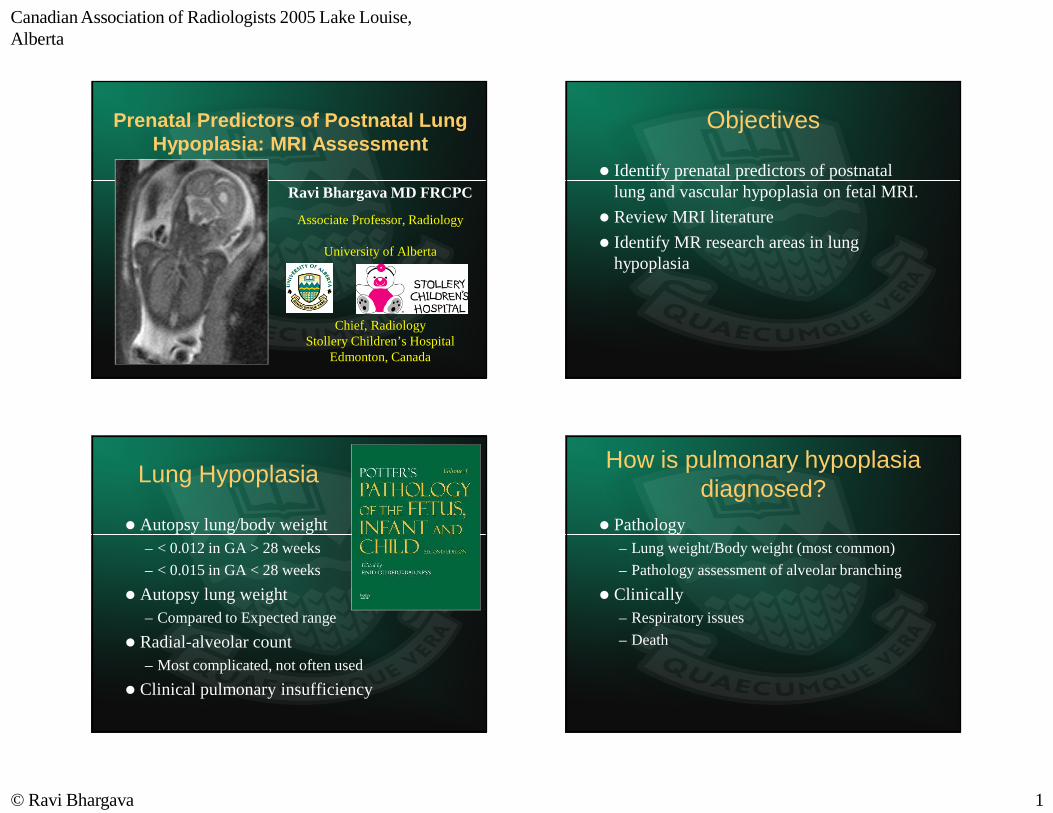
� Canalicular phase– Large influence of mechanical factors
�Transpulmonary pressue (Secretion of lung fluid)– Efflux of 50% of lung liquid contributes to about 30% of
amniotic fluid
– Led to treatment strategies such as FETO (Fetoscopic tracheal occlusion, Deprest J)
�Cyclic stretch, generated by fetal breathing movement
� Saccular phase– Start of surfactant production
� Alveolar phase– 85% of alveoli are formed AFTER birth
MRI Sequences
HASTE
Half-fourier single shot turbo spin echo
HASTE: T2 FLASH: T1Chest
� 4 chamber anatomy lost due to motion
� Aorta dark on HASTE due to flow voids
� Thymus of intermediate signal
Canadian Association of Radiologists 2005 Lake Louise, Alberta
© Ravi Bhargava 3
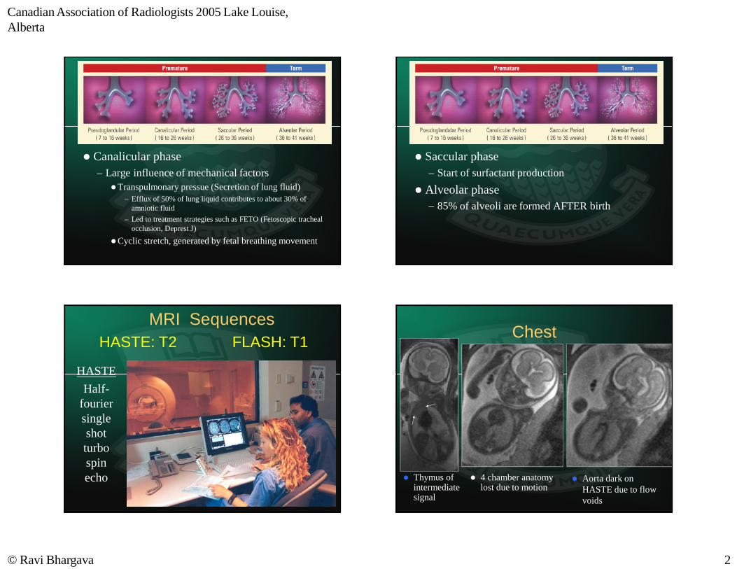
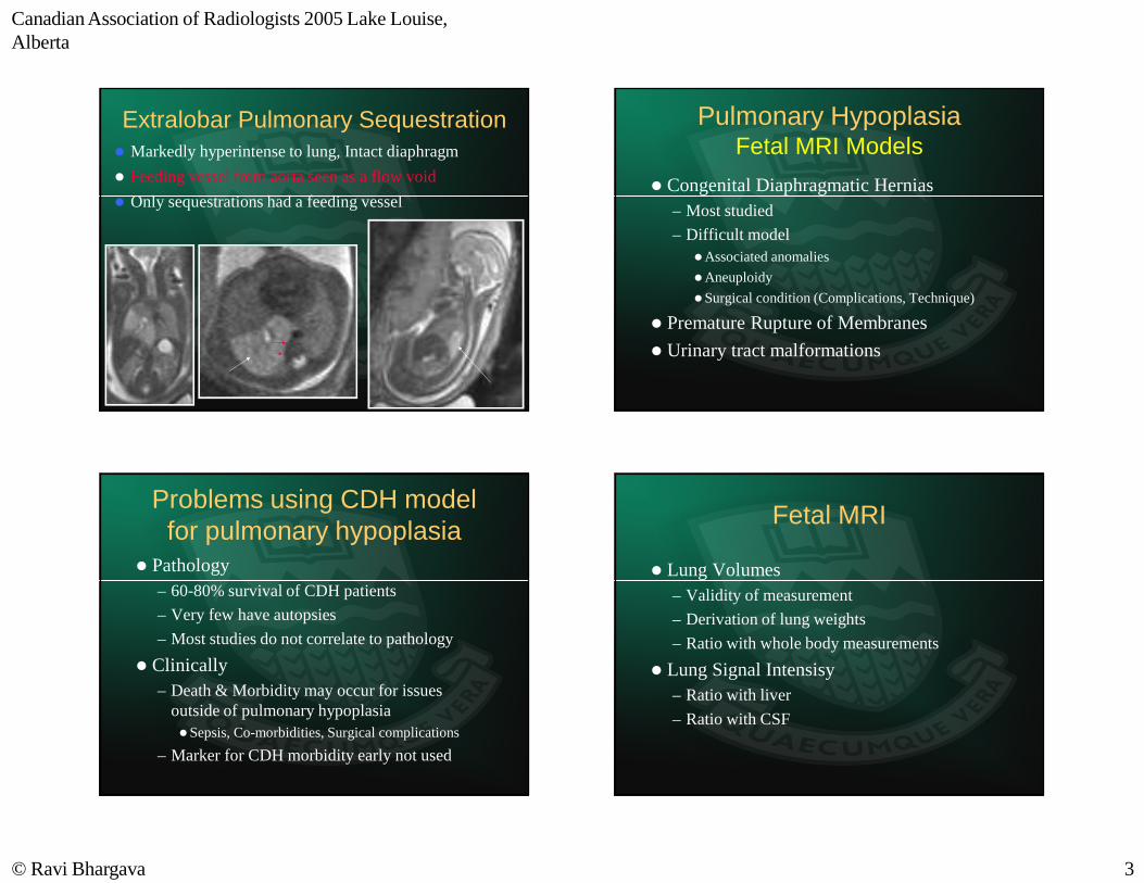
Extralobar Pulmonary Sequestration� Markedly hyperintense to lung, Intact diaphragm
� Feeding vessel from aorta seen as a flow void
� Only sequestrations had a feeding vessel
Pulmonary HypoplasiaFetal MRI Models
� Congenital Diaphragmatic Hernias– Most studied
– Difficult model�Associated anomalies
�Aneuploidy
�Surgical condition (Complications, Technique)
� Premature Rupture of Membranes
� Urinary tract malformations
Problems using CDH model for pulmonary hypoplasia
� Pathology– 60-80% survival of CDH patients
– Very few have autopsies
– Most studies do not correlate to pathology
� Clinically– Death & Morbidity may occur for issues
outside of pulmonary hypoplasia�Sepsis, Co-morbidities, Surgical complications
– Marker for CDH morbidity early not used
Fetal MRI
� Lung Volumes– Validity of measurement
– Derivation of lung weights
– Ratio with whole body measurements
� Lung Signal Intensisy– Ratio with liver
– Ratio with CSF
Canadian Association of Radiologists 2005 Lake Louise, Alberta
© Ravi Bhargava 4
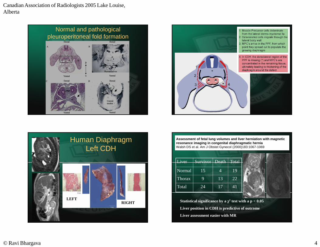
Normal and pathological pleuroperitoneal fold formation
LEFTRIGHT
Human Diaphragm Left CDH
Assessment of fetal lung volumes and liver herniation with magnetic resonance imaging in congenital diaphragmatic herniaWalsh DS et al. Am J Obstet Gynecol (2000)183:1067-1069
Liver Survivor Death Total
Normal 15 4 19
Thorax 9 13 22
Total 24 17 41
Statistical significance by a χ2 test with a p < 0.05
Liver position in CDH is predictive of outcome
Liver assessment easier with MR
Canadian Association of Radiologists 2005 Lake Louise, Alberta
© Ravi Bhargava 5
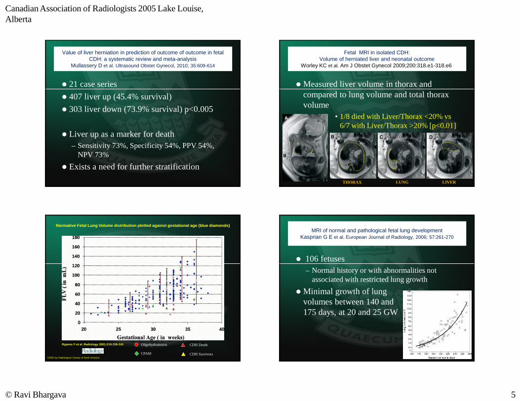
Value of liver herniation in prediction of outcome of outcome in fetal CDH: a systematic review and meta-analysis
Mullassery D et al. Ultrasound Obstet Gynecol, 2010; 35:609-614
� 21 case series
� 407 liver up (45.4% survival)
� 303 liver down (73.9% survival) p<0.005
� Liver up as a marker for death– Sensitivity 73%, Specificity 54%, PPV 54%,
NPV 73%
� Exists a need for further stratification
Fetal MRI in isolated CDH: Volume of herniated liver and neonatal outcome
Worley KC et al. Am J Obstet Gynecol 2009;200:318.e1-318.e6.
� Measured liver volume in thorax and compared to lung volume and total thorax volume
• 1/8 died with Liver/Thorax <20% vs6/7 with Liver/Thorax >20% [p<0.01]
THORAX LUNG LIVER
Normative Fetal Lung Volume distribution plotted against gestational age (blue diamonds)
Rypens F et al. Radiology 2001;219:236-241
©2001 by Radiological Society of North America
Oligohydramnios
CPAM
CDH Death
CDH Survivors
MRI of normal and pathological fetal lung developmentKasprian G E et al. European Journal of Radiology, 2006; 57:261-270
� 106 fetuses– Normal history or with abnormalities not
associated with restricted lung growth
� Minimal growth of lung volumes between 140 and 175 days, at 20 and 25 GW
Canadian Association of Radiologists 2005 Lake Louise, Alberta
© Ravi Bhargava 6
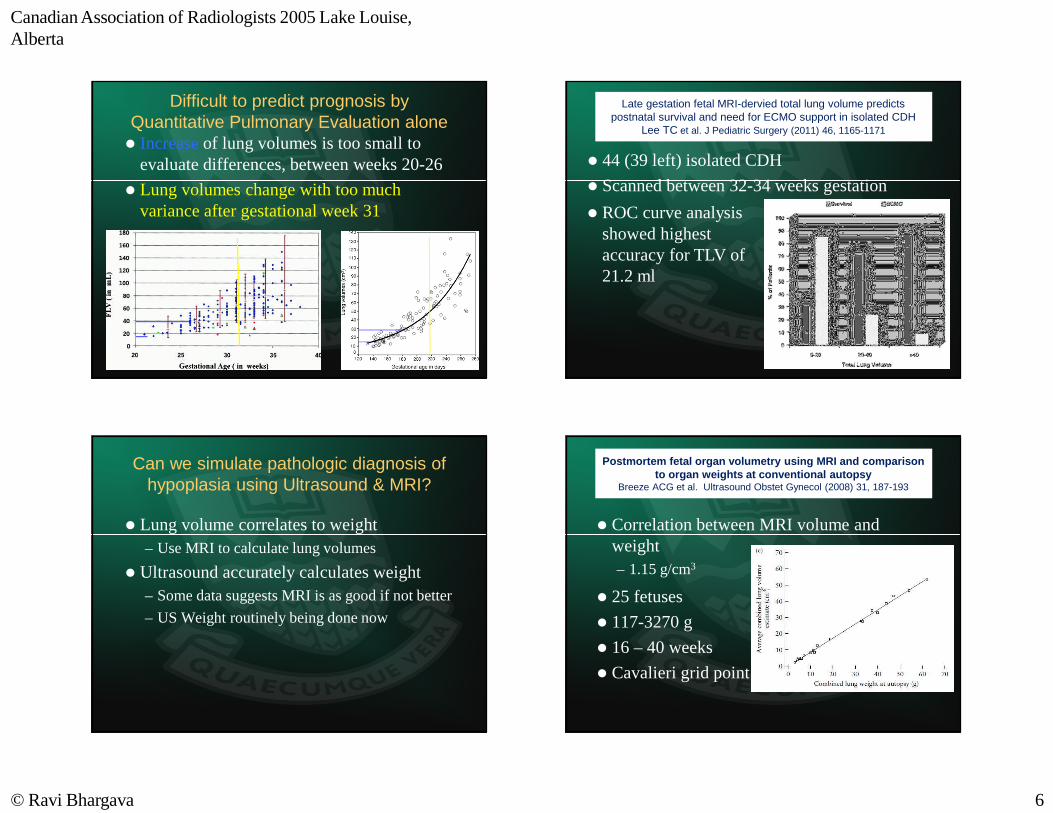
Difficult to predict prognosis by Quantitative Pulmonary Evaluation alone� Increaseof lung volumes is too small to
evaluate differences, between weeks 20-26
� Lung volumes change with too much variance after gestational week 31
Late gestation fetal MRI-dervied total lung volume predicts postnatal survival and need for ECMO support in isolated CDH
Lee TC et al. J Pediatric Surgery (2011) 46, 1165-1171
� 44 (39 left) isolated CDH
� Scanned between 32-34 weeks gestation
� ROC curve analysis showed highest accuracy for TLV of 21.2 ml
Can we simulate pathologic diagnosis of hypoplasia using Ultrasound & MRI?
� Lung volume correlates to weight– Use MRI to calculate lung volumes
� Ultrasound accurately calculates weight– Some data suggests MRI is as good if not better
– US Weight routinely being done now
Postmortem fetal organ volumetry using MRI and comparison to organ weights at conventional autopsy
Breeze ACG et al. Ultrasound Obstet Gynecol (2008) 31, 187-193
� Correlation between MRI volume and weight– 1.15 g/cm3
� 25 fetuses
� 117-3270 g
� 16 – 40 weeks
� Cavalieri grid point
Canadian Association of Radiologists 2005 Lake Louise, Alberta
© Ravi Bhargava 7
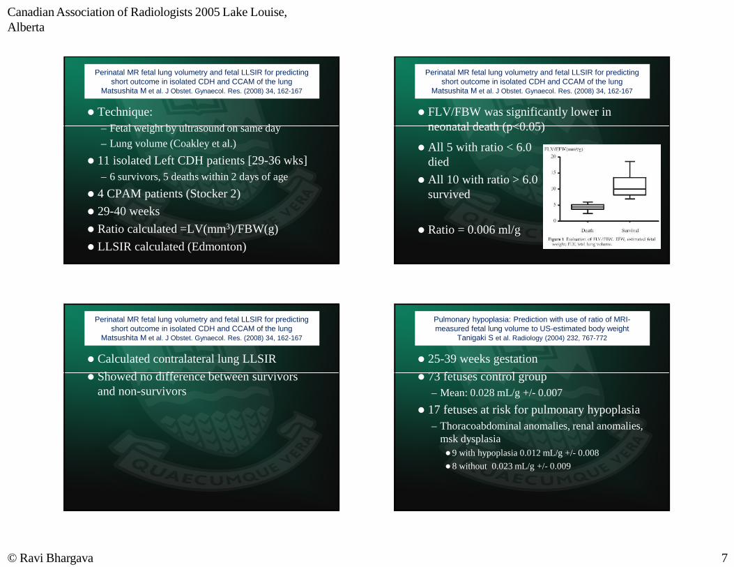
Perinatal MR fetal lung volumetry and fetal LLSIR for predicting short outcome in isolated CDH and CCAM of the lung
Matsushita M et al. J Obstet. Gynaecol. Res. (2008) 34, 162-167
� Technique: – Fetal weight by ultrasound on same day
– Lung volume (Coakley et al.)
� 11 isolated Left CDH patients [29-36 wks]– 6 survivors, 5 deaths within 2 days of age
� 4 CPAM patients (Stocker 2)
� 29-40 weeks
� Ratio calculated =LV(mm3)/FBW(g)
� LLSIR calculated (Edmonton)
Perinatal MR fetal lung volumetry and fetal LLSIR for predicting short outcome in isolated CDH and CCAM of the lung
Matsushita M et al. J Obstet. Gynaecol. Res. (2008) 34, 162-167
� FLV/FBW was significantly lower in neonatal death (p<0.05)
� All 5 with ratio < 6.0 died
� All 10 with ratio > 6.0 survived
� Ratio = 0.006 ml/g
Perinatal MR fetal lung volumetry and fetal LLSIR for predicting short outcome in isolated CDH and CCAM of the lung
Matsushita M et al. J Obstet. Gynaecol. Res. (2008) 34, 162-167
� Calculated contralateral lung LLSIR
� Showed no difference between survivors and non-survivors
Pulmonary hypoplasia: Prediction with use of ratio of MRI-measured fetal lung volume to US-estimated body weight
Tanigaki S et al. Radiology (2004) 232, 767-772
� 25-39 weeks gestation
� 73 fetuses control group– Mean: 0.028 mL/g +/- 0.007
� 17 fetuses at risk for pulmonary hypoplasia– Thoracoabdominal anomalies, renal anomalies,
msk dysplasia �9 with hypoplasia 0.012 mL/g +/- 0.008
�8 without 0.023 mL/g +/- 0.009
Canadian Association of Radiologists 2005 Lake Louise, Alberta
© Ravi Bhargava 8
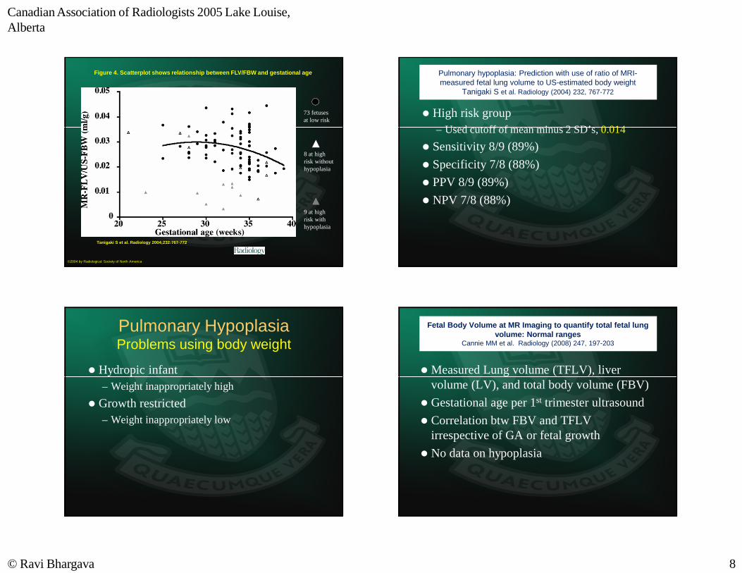
Figure 4. Scatterplot shows relationship between FLV/FBW and gestational age.
Tanigaki S et al. Radiology 2004;232:767-772
©2004 by Radiological Society of North America
73 fetuses at low risk
8 at high risk without hypoplasia
9 at high risk with hypoplasia
Pulmonary hypoplasia: Prediction with use of ratio of MRI-measured fetal lung volume to US-estimated body weight
Tanigaki S et al. Radiology (2004) 232, 767-772
� High risk group– Used cutoff of mean minus 2 SD’s, 0.014
� Sensitivity 8/9 (89%)
� Specificity 7/8 (88%)
� PPV 8/9 (89%)
� NPV 7/8 (88%)
Pulmonary HypoplasiaProblems using body weight
� Hydropic infant– Weight inappropriately high
� Growth restricted– Weight inappropriately low
Fetal Body Volume at MR Imaging to quantify total fetal lung volume: Normal ranges
Cannie MM et al. Radiology (2008) 247, 197-203
� Measured Lung volume (TFLV), liver volume (LV), and total body volume (FBV)
� Gestational age per 1st trimester ultrasound
� Correlation btw FBV and TFLV irrespective of GA or fetal growth
� No data on hypoplasia
Canadian Association of Radiologists 2005 Lake Louise, Alberta
© Ravi Bhargava 9
Observed Fetal Lung VolumeExpected Fetal Lung Volume
� 20-33 week GA, 77 fetuses with isolated CDH
� R < 25%, 19 % Survival vs 40.3% (p=0.008)
� 30-32 weeks, 22 fetuses, 10 survivors
� R < 30%, Sensitivity =0.83, Specificity = 1 for death
Prenatal prognosis of CDH using MRI measurement of FLV Gorincour G et al. Ultrasound Obstet Gynecol (2005) 26, 738-744
Fetal lung volume in CDHBonfils M et al. Arch Dis Child Fetal Neonatal Ed (2006) 91, F363-F364
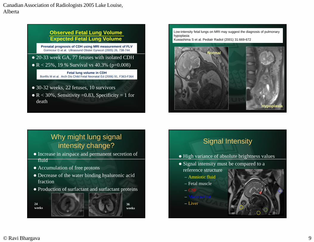
Low-intensity fetal lungs on MRI may suggest the diagnosis of pulmonary hypoplasiaKuwashima S et al. Pediatr Radiol (2001) 31:669-672
Normal
Hypoplasia
Why might lung signal intensity change?
� Increase in airspace and permanent secretion of fluid
� Accumulation of free protons
� Decrease of the water binding hyaluronic acid fraction
� Production of surfactant and surfactant proteins
24 weeks
36 weeks
Signal Intensity
� High variance of absolute brightness values
� Signal intensity must be compared to a reference structure– Amniotic fluid
– Fetal muscle
– CSF
– Maternal fat
– Liver
Canadian Association of Radiologists 2005 Lake Louise, Alberta
© Ravi Bhargava 10
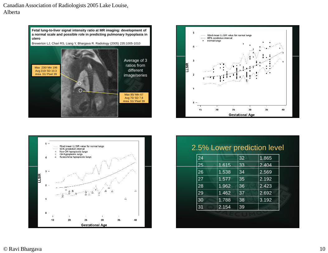
Fetal lung-to-liver signal intensity ratio at MR imaging: development of a normal scale and possible role in predicting pulmonary hypoplasia in uteroBrewerton LJ, Chari RS, Liang Y, Bhargava R. Radiology (2005) 235:1005-1010
Max 230/ Min 196Avg 210/ SD 10.3Area .51/ Pixel 39
Max 85/ Min 67Avg 75/ SD 7.8
Area .51/ Pixel 39
Average of 3 ratios from
different image/series
2.5% Lower prediction level24 32 1.865
25 1.615 33 2.40426 1.538 34 2.569
27 1.577 35 2.192
28 1.962 36 2.42329 1.462 37 2.692
30 1.788 38 3.192
31 2.154 39
Canadian Association of Radiologists 2005 Lake Louise, Alberta
© Ravi Bhargava 11
Does Liver-to Lung signal intensity ratio (LLSIR) measured by fetal magnetic resonance imaging (MRI) predict the severity of pulmonary hypoplasia in congenital diaphragmatic hernia?
Hsi B, Skarsgard ED, Chari RS, Pugash D, and Bhargava R CAPS 2005
� 18 fetuses with isolated CDH– 16 liveborn, 4 (25%) of which survived
� LLSIR values for CDH fetuses were significantly lower than gestationally adjusted controls (p<0.05)
� LLSIR < 2.5%ile predicted mortality with a positive predictive value (PPV) of 83%.
Concerns with LLSIR
� Liver signal intensity changes more dramatically then Lung signal intensity
� Liver not in same slice as lungs
� Liver not homogeneous
� Problems could be overcome using CSF
Demonstration of changes in fetal liver erythropoiesis using echo-planar MRI
Duncan KR et al. J Physiol Gastrintest Liver Physiol 1997; 273:965-967
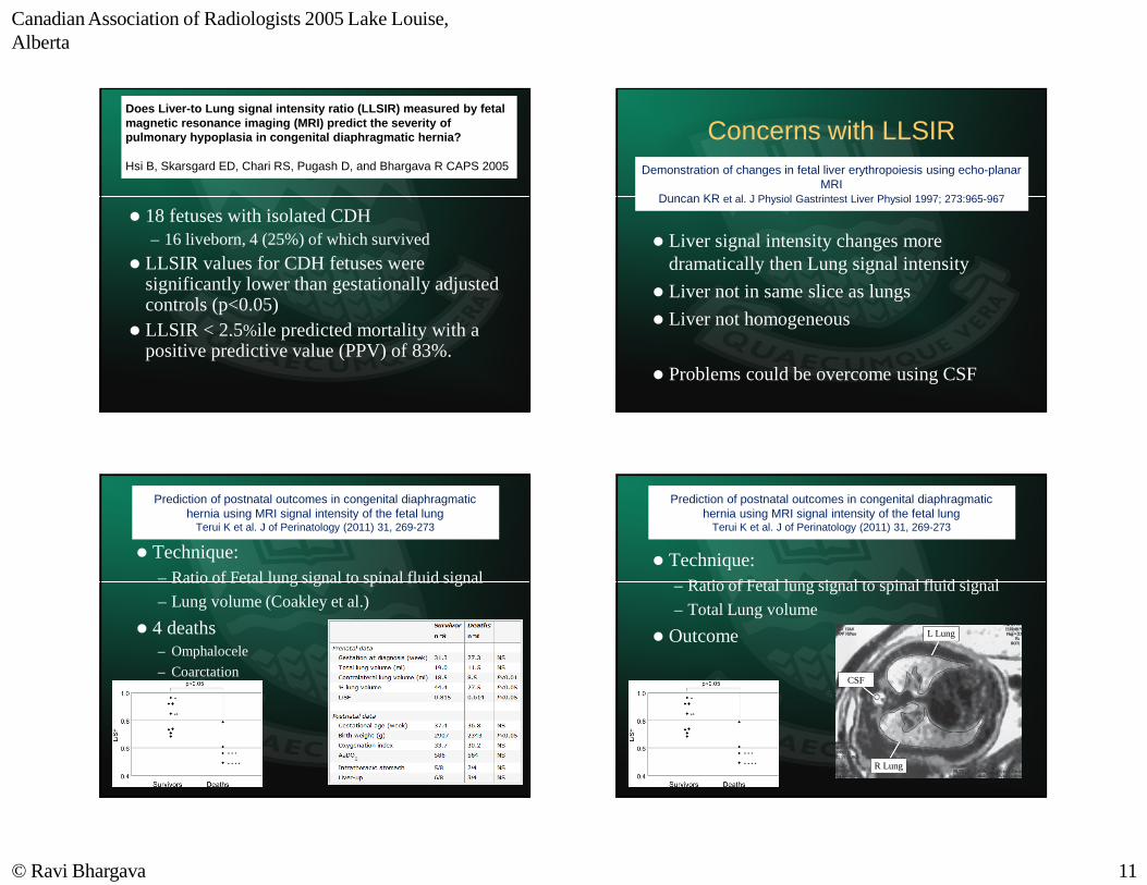
Prediction of postnatal outcomes in congenital diaphragmatic hernia using MRI signal intensity of the fetal lung
Terui K et al. J of Perinatology (2011) 31, 269-273
� Technique: – Ratio of Fetal lung signal to spinal fluid signal
– Lung volume (Coakley et al.)
� 4 deaths– Omphalocele
– Coarctation
Prediction of postnatal outcomes in congenital diaphragmatic hernia using MRI signal intensity of the fetal lung
Terui K et al. J of Perinatology (2011) 31, 269-273
� Technique: – Ratio of Fetal lung signal to spinal fluid signal
– Total Lung volume
� Outcome L Lung
CSF
R Lung
Canadian Association of Radiologists 2005 Lake Louise, Alberta
© Ravi Bhargava 12
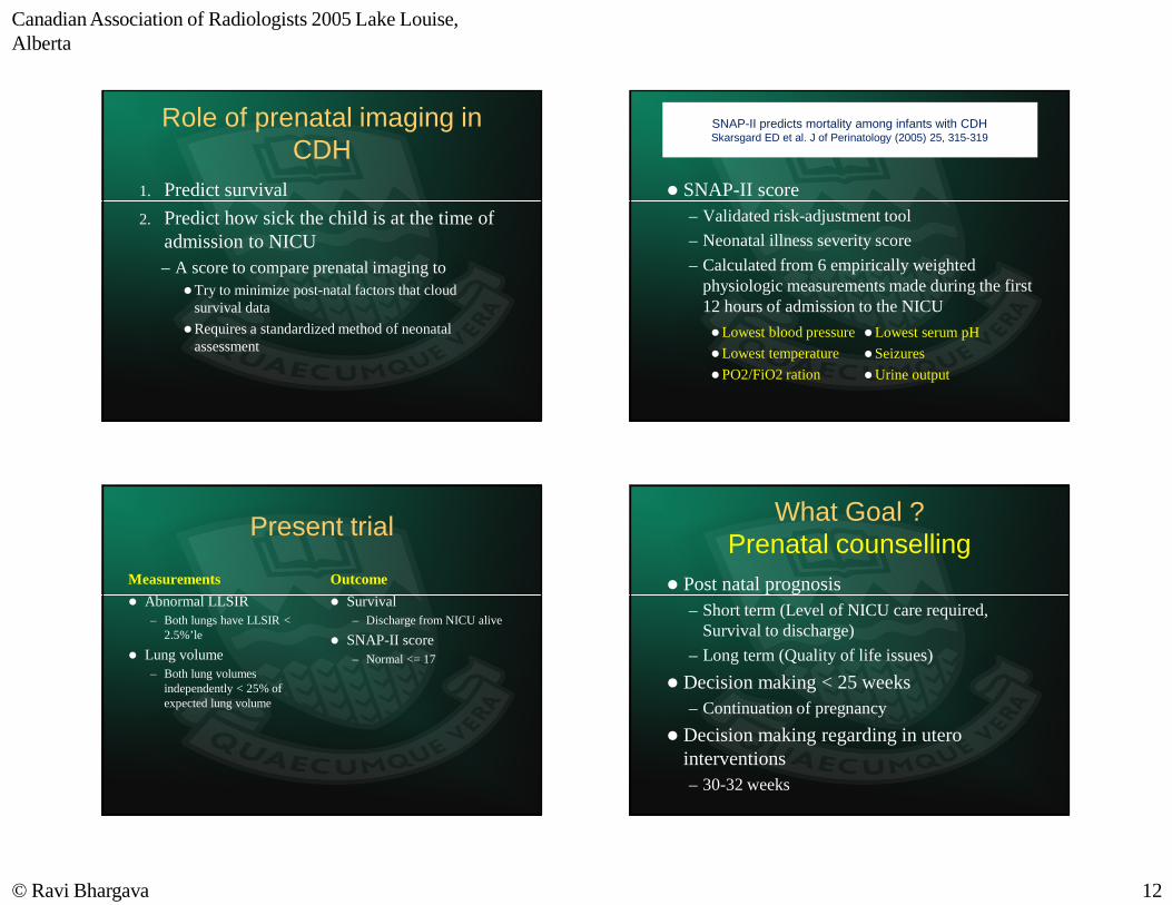
Role of prenatal imaging in CDH
1. Predict survival
2. Predict how sick the child is at the time of admission to NICU– A score to compare prenatal imaging to
�Try to minimize post-natal factors that cloud survival data
�Requires a standardized method of neonatal assessment
� SNAP-II score– Validated risk-adjustment tool
– Neonatal illness severity score
– Calculated from 6 empirically weighted physiologic measurements made during the first 12 hours of admission to the NICU
�Lowest serum pH
�Seizures
�Urine output
�Lowest blood pressure
�Lowest temperature
�PO2/FiO2 ration
SNAP-II predicts mortality among infants with CDHSkarsgard ED et al. J of Perinatology (2005) 25, 315-319
Present trial
Measurements
� Abnormal LLSIR– Both lungs have LLSIR <
2.5%’le
� Lung volume– Both lung volumes
independently < 25% of expected lung volume
Outcome
� Survival– Discharge from NICU alive
� SNAP-II score– Normal <= 17
What Goal ?Prenatal counselling
� Post natal prognosis– Short term (Level of NICU care required,
Survival to discharge)
– Long term (Quality of life issues)
� Decision making < 25 weeks– Continuation of pregnancy
� Decision making regarding in uterointerventions– 30-32 weeks
Canadian Association of Radiologists 2005 Lake Louise, Alberta
© Ravi Bhargava 13
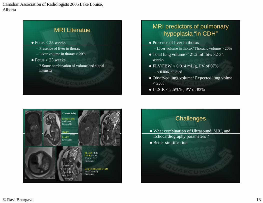
MRI Literatue
� Fetus < 25 weeks– Presence of liver in thorax
– Liver volume in thorax > 20%
� Fetus > 25 weeks– ? Some combination of volume and signal
intensity
MRI predictors of pulmonary hypoplasia “in CDH”
� Presence of liver in thorax– Liver volume in thorax/ Thoracic volume > 20%
� Total lung volume < 21.2 mL btw 32-34 weeks
� FLV/FBW < 0.014 mL/g, PV of 87%– < 0.006, all died
� Observed lung volume/ Expected lung volme< 25%
� LLSIR < 2.5%’le, PV of 83%
RLLSIR =1.96LLSIR = 1.882.5% = 1.577Favourable
Lung volume/Fetal weight = 0.0234 mL/g Favourable
27 week 6 day
Liver position: Abdomen Favourable
Obs LV--------- >30%Exp LV Favourable
Challenges
� What combination of Ultrasound, MRI, and Echocardiography parameters ?
� Better stratification