Embed Size (px)
Citation preview
Disease Markers 24 (2008) 257–266 257IOS Press
Lung cancer stem cells
Sharon R. Pinea, Blair Marshallb and Lyuba Varticovskia,∗aCenter for Cancer Research, NCI, NIH, Bethesda, MD, USAbGeorgetown University, Washington, DC, USA
Abstract. Lung cancer remains a major cause of cancer-related lethality because of high incidence and recurrence in spiteof significant advances in staging and therapies. Recent data indicates that stem cells situated throughout the airways mayinitiate cancer formation. These putative stem cells maintain protumorigenic characteristics including high proliferative capacity,multipotent differentiation, drug resistance and long lifespan relative to other cells. Stem cell signaling and differentiationpathways are maintained within distinct cancer types, and destabilization of this machinery may participate in maintenance ofcancer stem cells. Characterization of lung cancer stem cells is an area of active research and is critical for developing noveltherapies. This review summarizes the current knowledge on stem cell signaling pathways and cell markers used to identify thelung cancer stem cells.
Keywords: ABC transporters, drug resistance, hedgehog, notch, non-small cell lung cancer, side population, small cell lungcancer, stem cells, Wnt
1. Introduction
Lung cancer is one of the most frequently occurringmalignancies accounting for 18% of cancers in menworld wide and 21% in Western countries [70]. Ap-proximately 213,380 new cases of lung cancer are pro-jected to be diagnosed in 2007 and at least 160,000deaths are expected to occur next year from this dis-ease in the United States. Annual deaths from lungcancer exceed deaths due to breast, colon, and prostatecancers combined [45]. Despite numerous therapeuticadvances during the last thirty years, the majority ofpatients present with advanced disease because earlystage lung cancer is asymptomatic and there are no stan-dard screening procedures in place for high risk popu-lations. Even with the most up-to-date imaging, stag-ing, and surgical modalities, the five-year survival ratefollowing adjuvant therapy for patients with stage IBto IIIA ranges only from 5–15% [48,101]. In addition,patients with small cell lung cancer (SCLC) frequentlypresent with metastases regardless of the primary tu-
∗Corresponding author: Lyuba Varticovski, MD, Center for Can-cer Research, LHC, Building 37, Room 3060, National Cancer Insti-tute, Bethesda, MD 20892, USA. E-mail: [email protected].
mor size [74]. These data support the notion that earlydisseminated disease in patients with lung cancer is dueto enrichment of cancer stem cells in these tumors.
Commonly thought of as a single disease, lung can-cer actually represents a group of neoplasias arisingfrom phenotypically diverse cells [9,98]. These encom-pass several distinct tumor types: squamous cell car-cinoma (SCC), adenocarcinoma/bronchoalveolar(non-small cell lung cancer, NSCLC), and neuroendocrinecarcinomas. Despite the histological and biologic dif-ferences, squamous cell and NSCLC are treated as ahomogeneous group and patients presenting with theselesions are treated similarly. The neuroendocrine car-cinomas include small cell (SCLC), large cell, atypicaland typical carcinoids. Although these tumors havecommon neuroedocrine origin, they are histologicallydiverse with widely varying survival rates from lessthan 5% for SCLC to greater than 90% for well differ-entiated, slow growing carcinoid tumors [59,74].
Ninety percent of lung cancer is caused, at leastin part, by cigarette smoking [64]. Despite increasedawareness of the carcinogenic potential of cigarettesmoke, the incidence of lung cancer continues to in-crease in parallel with an increase in predominantlyyoung smokers. There has been a shift in lung cancer
ISSN 0278-0240/08/$17.00 2008 – IOS Press and the authors. All rights reserved
258 S.R. Pine et al. / Lung cancer stem cells
pathology over the past few decades due to changesin cigarette design and subsequent changes in inhala-tion patterns [102]. In the 1950’s, less than 1% of allcigarettes contained filters and by the late 1990’s, morethan 95% contained filters. Prior to this change, squa-mous cell was the predominant cell type. Squamouscells populate the proximal and central airways. Withdeeper inhalation techniques associated with the use offiltered cigarettes, peripheral adenocarcinoma has tak-en over as the most common non-small lung cancersubtype.
Despite the fact that the majority of lung cancers canbe attributed to smoking, 10% arise in never-smokers,suggesting that several genetic and environmental fac-tors participate in lung carcinogenesis, including ge-netic polymorphisms, and exposure to radon, cookingfumes, asbestos, heavy metals, and passive smoking. Adisproportionate number of never-smoking lung cancerpatients are women, and the incidence of lung cancerin never-smoking Asian women in particular is increas-ing [30]. A high indoor concentration of benzo pyrenesdue to indoor coal burning, other environmental causes,and possibly human papilloma virus (HPV) have beenlinked to this trend [54].
The prime role of the airways (trachea, bronchi,bronchioles and terminal bronchioles) is to conduct airinto and out of the lung and to form a first line ofdefense against undesired constituents of inhaled air.The airways are continuously exposed to pathogens,irritants, pollutants and agents that produce oxidativestress; therefore, the composition of the respiratorytract surface is very important. The upper airways con-tain specialized cell types such as ciliated cells and mu-cous secreting goblet cells. The lower conducting air-ways (respiratory bronchioles, alveolar ducts and alve-olar sacs) participate in gas exchange by diffusion. Thealveolar epithelial surface comprises essentially twocell types, the alveolar epithelial type I cell and thecuboidal alveolar epithelial type II cell. Type I cellsflatten out and in this way constitute approximately95% of the total alveolar surface, whereas type II cellsare more numerous and produce surfactant [64].
In the last several years, there is growing evidencethat solid tumors are composed of cells with differ-ent biological properties, and the capability to sus-tain tumor growth resides in a small fraction of tu-mor cells, termed cancer stem cells or tumor-initiatingcells [18,69,78]. Tumor-initiating cells have been iden-tified in many types of cancers by sorting cell subpop-ulations based on surface marker expression patterns,and transplantation into animal models. These stud-
ies have shown that tumor-initiating cells are respon-sible for tumor formation and progression, and havestem/progenitor cell properties such as self-renewal,long-term proliferation and expression of genes associ-ated with stem cells of the organ [87,100]. The purposeof this review is to provide recent information aboutidentification and characterization of cancer stem cellswithin the lung.
2. Pathways in stem cell development and cancerstem cells
2.1. Common pathways
Much understanding of cancer stem cell biologycomes from earlier studies of normal and malignanthematopoiesis. Recent information on normal embry-onic and neuronal stem cells, as well as breast and braincancer stem cells describes common features that de-fine normal and malignant stem cells. This informa-tion may be applicable to cancer stem cells from manyother organs, including the lung. Cancer stem cellsderived either from normal stem cells or from differen-tiated cells that reverted to a stem cell-like phenotyperesult in deregulation of the fundamental characteristicof stem cell: the capacity for asymmetric division andself-renewal. The Notch, Hedgehog and Wnt pathwaysdefine normal stem cells and guide the behavior of nor-mal pulmonary precursors within several different lin-eages. Lung cancer of specific phenotypes or locationswithin the lung that arise from inappropriate expan-sion of pulmonary cancer stem cell lineages may becaused by abnormal signaling in these pathways. Here,we examine the Notch, Hedgehog and Wnt pathwaysin lung development and integrate them with currentknowledge about lung cancer stem cells. Details of thesignaling pathways can be found in [67].
2.2. Notch pathway
Notch signaling requires asymmetry between neigh-boring cells and is initiated by activation of Notch re-ceptors by ligands present on adjoining cells. Prote-olytic cleavage steps generate a Notch intracellular do-main fragment (Notch ICD) that associates with thelatent transcriptional activator CBF-1 (also referred toas RBP-Jk and SuH), rendering it active. The Notchpathway participates in development and maintenanceof normal tissues, by regulation of differentiation andcell cycle progression. Notch pathway is present in
S.R. Pine et al. / Lung cancer stem cells 259
the developing lung and lung cancer cells at the re-ceptor (Notch1-Notch4), ligand (Jagged1,2 and Delta-like, Dll1,3,4), and downstream effector (Hes1, Hey1and HeyL) levels. In the mouse, Notch1 is expressedin the distal lung endoderm as early as E11.5 [44,76].Notch2 and Notch3 are expressed in the developinglung mesenchyme that surrounds the primitive epitheli-um, Notch3 is expressed in epithelial cells [44,76] andNotch4 in endothelial cells [76]. Notch 1 and 3 arealso expressed in Clara cells of adult lung. The Notchligand, Jagged1, is expressed in lung mesenchyme andlung vessels, and Jagged2 is in the peripheral lung mes-enchyme [76] suggesting that Jagged2 interacts withNotch1-expressing airway epithelial cells in the devel-oping lung. Expression of Dll1 is restricted to pul-monary neuroendocrinecells [10,44,76] and may inter-act with Notch1 and 3 expressed on airway epithelialClara cell progenitors.
Among Notch effectors, Hes1 is expressed in fetalmouse lung and is co-expressed with Notch 1 and 3 inClara cells [44]. Pulmonary expression of other Notchpathway effectors in the lung is not known, althoughHey1 and Hey2 are expressed in the adult lung [90] andHeyL is expressed in lung vasculature [57]. The Notchpathway effectors act as transcriptional repressors toinhibit tissue-specific differentiation proteins, such asmASH1 in mice [36]. mASH1 expression is confinedto neuroepithelial bodies and pulmonary neuroepithe-lial cells, and mice lacking maSH1 in the lungs haveno neuroendocrine cells [13]. This suggests that neu-roendocrine cell differentiation is regulated by Notch1.Further information on the role of Notch in lung devel-opment can be found elsewhere [17].
There are several reasons to suggest that Notch par-ticipates in maintaining lung cancer stem cells. Notch1and 3 and the downstream Notch effector, Hes-1, areexpressed in NSCLC cell lines, although they are rarelydetected in SCLC cell lines [14,17,19]. SAGE ex-pression profiling data collected in a range of cancercell lines also implicates the Notch effectors Hes1,Hey1 and HeyL in NSCLC (Gene expression OmnibusDataset 217, Cancer Genome Anatomy Project SAGElibrary collection, (http://www.ncbi.nlm.nih.gov/geo).hASH1 expression is inhibited by Notch signaling andhASH1 is elevated in neuroendocrine SCLC, but israrely expressed in NSCLC [4]. Thus, hASH1 ex-pression in the lung neuroendocrine cells may be reg-ulated by Notch signaling. Deregulated Notch signal-ing may play a role in SCLC pathogenesis. SCLCcells are growth-inhibited by overexpression of activat-ed Notch1 and 2 [88,89]. Because Notch signaling is a
key pathway in Clara and neuroendocrine cell develop-ment and is dysregulated in NSCLC and SCLC, Notchpathway may be an important therapeutic target in lungcancer.
2.3. Hedgehog pathway
Hedgehog (Hh) is a morphogen that acts in a short-or long-range fashion on various tissue types [26]. Inmammals, there are three Hh proteins: Sonic Hh, In-dian Hh, and Desert Hh. During lung embryogenesisSHh, expressed in the budding airway epithelium, isrequired for lung development and is a key mediatorof epithelial-mesenchymal interactions [7]. SHh-nullmouse embryos fail to separate trachea from esopha-gus, resulting in formation of a rudimentary lung sacfrom a single tracheo-esophageal tube due to failure ofbranching and growth after formation of primary lungbuds [60]. Over-expression of SHh from the SP-C pro-moter in transgenic mice results in increased epithelialand mesenchymal proliferation [7]. Patched (PTCH) isa surface membrane receptor that is required for SHhsignaling that is highly expressed in mesenchyme sur-rounding the terminal lung buds, and knock-down ofPTCH in mice also results in decreased lung branchingmorphogenesis. Upon SHh binding, PTCH receptorsrelease the smoothened (SMO) protein resulting in ac-tivation of GLI, which functions as transcriptional reg-ulator. Knockdown of SMO, GLI2 and GLI3 in miceresults in absence of lung development [98].
Dysregulation of the Hh pathway has been well-documented in SCLC. The Hh pathway can be con-stitutively activated by mutations in SMO and ampli-fication of GLI in several cancer types [2]. GLI ex-pression in normal cells is restricted to precursor cells,and may, therefore, participate in cancer stem cell self-renewal. Persistent SHh pathway activation in SCLC,driven by a high expression of SHh, PTCH and GLI1,is observed in primary SCLC, but rarely in NSCLCcell lines [95]. The Hh pathway can be inhibited bycyclopamine, a naturally-occurring compound whichspecifically targets SMO [99]. Treatment of SCLCcell lines and cell line xenografts with cyclopamineleads to tumor growth arrest, and SCLC cell lines thatoverexpress GLI1 can be protected from inhibition bycyclopamine [99]. Thus, vulnerability of SCLC to ablockade of SHh signaling may provide a new thera-peutic target approach.
260 S.R. Pine et al. / Lung cancer stem cells
2.4. Wnt pathway
Wnt signaling is a critical component of early lungorganogenesis and disease [75,97]. Several of 19known Wnts are expressed in the adult lung: Wnt2,Wnt5a, and Wnt 11 are expressed in the mesenchyme,and Wnt5a and Wnt7b are expressed in the pulmonaryepithelium [53,58,86,96]. Studies of knockout mice re-veal the importance of Wnts in lung development [39].The canonical Wnt pathway involves binding to one of10 known Frizzled (Fzd) receptors leading to nucleartranslocation ofβ-catenin [51] and transcriptional ac-tivaton. Expression of the Wnt inhibitor Dickkopf-1(Dkk-1) has been observed in the distal epithelium, andDkk1 inhibits branching morphogenesis [20].
Disruption of the Wnt pathway has been implicatedin non-small cell lung cancer. Wnt2 is overexpressed inNSCLC, and inhibition of Wnt2-mediated signaling bysiRNA or a monoclonal antibody results in apoptosis ofNSCLC cell lines [105]. Wnt inhibitory factor (WIF1),a secreted Wnt antagonist, inhibits NSCLC cell linegrowthin vitro andin vivo, and is also downregulated inNSCLC [50]. WIF1 silencing is associated with hyper-methylation of its promoter. Methylation-silencing ofWIF1 is a common mechanism for aberrant activationof Wnt signaling in lung cancer [39]. Disheveled (dvl)proteins are positive mediators of Wnt signaling. Dvl3 is overexpressed in 75% of freshly microdissectedNSCLC tissue specimens [92]. Inhibition of dvl1, 2and 3 resulted in decreasedβ-catenin, TCF-mediatedtranscription and inhibition of cell growth in NSCLCcell lines [92]. One can speculate that blocking the Wntsignaling pathway may be also an attractive target fortherapeutic agents.
3. Stem cell gene expression
In addition to genes involved in the Notch, Wntand HH pathways, other genetic markers are associat-ed with cancer stem cells. One of the most interest-ing genes, Oct-4 (also known as OCT3 or POU5F1),is a mammalian POU family transcription factor re-quired for maintenance of embryonic stem (ES) cellpluripotentcy [73]. Oct-4 expression is downregulatedin all differentiated somatic cell typesin vitro as wellas in vivo by increased DNA methylation and struc-tural changes involving immediate upstream regulatoryregion [8,93].
Oct-4 may be regulated by the Wnt pathway andhas the ability to reprogram committed somatic cells,
inducing their dedifferentiation by reverting them to amore developmentally potent state [33]. Keratinocytesthat overexpress Oct-4 can differentiate into other celltypes, based on expression of genes characteristic ofneuronal cells: nestin, neuN, and Sox-1. This indicatesthat Oct-4 may be a master regulator of the pluripotentstate in mammalian cells.
Recently, miRNAs have been implicated in thecontrol of self-renewal in stem cells [38]. In par-ticular, studies inDrosophila and mice suggest thatmiRNAs are important regulators of stem cell dif-ferentiation, self-renewal and division [38]. Geor-gantas et al. recently found 33 miRNAs expressedin CD34+ hematopoietic stem-progenitor cells thatwere important for haematopoietic differentiation [31]and miRNA-221, -222 and -223 have been implicat-ed in the inhibition of granulopoiesis and erythro-poiesis while miRNA-155 plays a role in normal hu-man myelopoiesis, lung differentiation, and erythro-poieis [31]. Also, recent work from NCI has shownthat high hsa-mir-155 and low hsa-let-7a-2 expressionhave been associated with poor outcome in lung can-cer [104].
4. Stem cells and drug resistance
Another important characteristic of cancer and nor-mal stem cells is resistance to toxic compounds [78].This concept is specifically applicable to airborne pol-lutants in reference to lung cancer. Bronchiolarprogen-itor cells localized within neuroepithelial body (NEB)express reduced levels of cytochrome p450 xenobioticmetabolizing enzymes [79] which reduces intracellularlevels of lipophilic toxic metabolites allowing airwaysstem cells to survive frequent airborne injuries. Thesesame cells also exhibit efficient drug efflux or a ‘sidepopulation’ (SP) phenotype as detected by efflux ofHoechst and other dyes due to ATP Binding Cassette(ABC) transporters [91]. Many ABC transporters, suchas ABCB1 (P-glycoprotein, MDR1), ABCC1 (mul-tidrug resistance-associated protein 1, MRP1), ABCG2(breast cancer resistance protein, BCRP,) and others arehighly expressed in lung bronchial epithelium and alve-olar type II cells [93,106]. Over-expression of MDR-1is detected in lung tumors prior to therapy [82] and en-hanced efflux of doxorubicin, cisplatinum, and mitox-antrone is associated with resistance to chemotherapy.
It is not known whether SP cells isolated from lungtruly exhibit bona-fide lung stem cell function [63,91] but SP-associated drug efflux and resistance to
S.R. Pine et al. / Lung cancer stem cells 261
chemotherapies occur in lung and other tissue tumorcell linesin vitro. SP cells have stem cell characteris-tics including enhanced clonogenicity [80] and tumorformationin vivo [11,41,52,65] (Pine and Varticovsky,unpublished).
Recently, a side population (SP) has been identifiedin the lungs of PTEN deficient mice [103] and a va-riety of lung cancer cell lines [42]. The latter are tu-morigenicin vivo, display increased invasive capabili-ty and have increased expression of ABC transporters.In addition, Eramo et al. [25] showed that, similar tocolon and brain cancer, lung cancer CD133+ cells areresistant to conventional chemotherapy, including cis-platin, etoposide, gemcitabine, vinorelbine, docetaxel,doxorubicin, and daunorubicin.
Aldehyde Dehydrogenase (ALDH), a drug-resistancegene found in normal hematopoietic stem cells [46],was recently found in several types of cancer initiatingcells, including NSCLC [71,83]. Expression of ALDHis regulated by the Hh pathway [27] and blockade ofhedgehog signaling inhibits pancreatic cancer invasionand metastases [28]. ALDH1 participates in the oxida-tion of retinal to all-trans retinoic acid [81], and con-fers drug resistance to chemotherapeutic agents, suchas cyclophosphamide, by an uncertain mechanism [5].
5. Evidence of cancer stem cells in lung cancermouse models
In the mouse, stem cell niches have been identifiedin the lung that may be targets for cancer initiation andpromotion. These stem cell niches maintain epithelialdifferentiation in the airways [32,43]. Of particular in-terest is a sub-type of Clara cells residing at the bro-chioalveolar junction that is able to regenerate Claraand alveolar cells lining the terminal bronchioles andalveoli [32]. A recent study [49] provided exciting in-sights about how these bronchioalveolarstem cells maybe involved in normal lung-tissue homeostasis and de-velopment of lung cancer. The bronchioalveolar stemcells co-express Clara, alveolar cell markers, and stemcell markers, Sca-1 and CD34, and are refractory tothe lung-damaging agent, naphthalene. These cells arelikely candidates for NSCLC stem cells because of theircapacity for self-renewal and tissue regeneration. Ex-pression of oncogenic K-ras in mouse broncheoalve-olar stem cells resulted in heightened proliferation ascompared to differentiated alveolar cells. These fea-tures are similar to expansion of broncheoalveolarstemcells in adenocarcinoma [49]. Validation of these cellsas tumor-repopulatingcells and identification of humanbroncheoalveolar stem cells have not been reported.
6. Methods used for identification andcharacterization of cancer stem cells
6.1. Cell surface markers
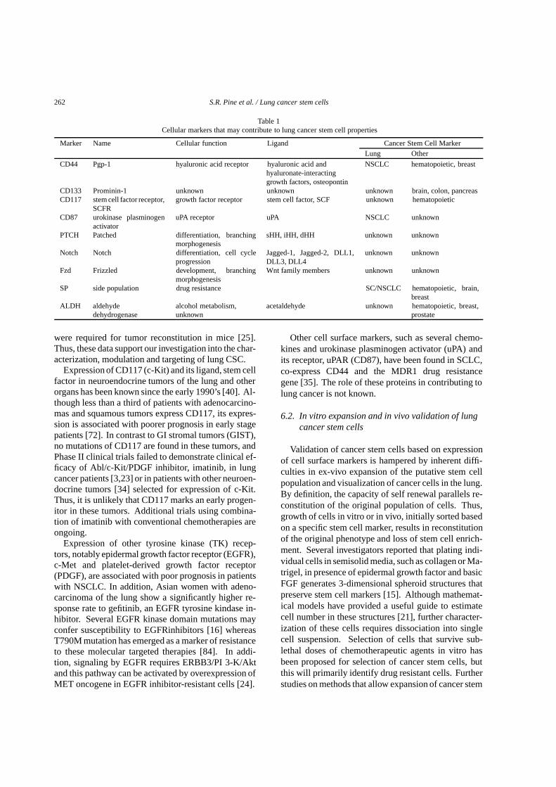
Although we tend to think of a tumor as a sin-gle type of tissue, even undifferentiated hematopoiet-ic tumors contain cells that differ by surface markersthat mark the capacity for self-renewal and differen-tiation. Theories proposing that cancers contain stemcells were published over 20 years ago [62], but on-ly ten years ago experimental evidence was provid-ed that tumor-initiating cells in human acute myeloidleukaemia (AML) share cell surface (CD34+CD38−)phenotype with normal hematopoietic stem cells [12].Identification of hematopoietic cells with long-term andshort-term proliferative capacity has provided informa-tion on stem cells and lineage-specific progenitor cells.Several cell surface markers indicate the potential stemcell characteristics of epithelial tumors. Table 1 listsknown or potential stem cell markers in lung cancer.
CD44 is a ubiquitously expressed multispanningtransmembrane cell-surface adhesion glycoporoteinthat mediates cell-matrix and cell-cell interactions. Ex-pression of CD44 correlates with drug resistance andpoor prognosis in many malignancies [61]. In addi-tion to hyaluronic acid, CD44 can be bound by fibrino-gen, fibronectin, collagen, laminin, FGF-2, and oth-er heparin-binding growth factors [61,66]. CD44 al-so binds to, and is upregulated by ostopeontin, an in-flammatory cytokine associated with metastatic pro-gression, and this interaction may constitute a feed-back loop for survival of disseminated cells closely re-sembling the origin of cancer stem cells [22]. CD44is found in hematopoietic, breast, and other malignantstem cells, including SCLC and NSCLC [55]. Alter-native splicing of this complex proteoglican was corre-lated with survival in patients with NSCLC [56].
CD133 (prominin-1) is a glycoprotein with fivemembrane-spanningdomains [85] that was initially de-tected in endothelial cells, but recently found aloneor in combination with other cell surface markers inbrain [94], colon and pancreatic cancer-initiating stemcells. CD133 expression correlates with drug resis-tance [68] and brain tumor-derivedand cell lines-sortedCD133 positive cells have a distinct gene expressionprofile [6]. A recent paper identified CD133 as a lungCSC marker. Using CD133+ cells isolated from hu-man lung tumors of diverse histological type, the au-thors reported that these cells grew indefinitely as tu-mor spheres, although a large number of cells, i.e., 104,
262 S.R. Pine et al. / Lung cancer stem cells
Table 1Cellular markers that may contribute to lung cancer stem cell properties
Marker Name Cellular function Ligand Cancer Stem Cell MarkerLung Other
CD44 Pgp-1 hyaluronic acid receptor hyaluronic acid andhyaluronate-interactinggrowth factors, osteopontin
NSCLC hematopoietic, breast
CD133 Prominin-1 unknown unknown unknown brain, colon, pancreasCD117 stem cell factor receptor,
SCFRgrowth factor receptor stem cell factor, SCF unknown hematopoietic
CD87 urokinase plasminogenactivator
uPA receptor uPA NSCLC unknown
PTCH Patched differentiation, branchingmorphogenesis
sHH, iHH, dHH unknown unknown
Notch Notch differentiation, cell cycleprogression
Jagged-1, Jagged-2, DLL1,DLL3, DLL4
unknown unknown
Fzd Frizzled development, branchingmorphogenesis
Wnt family members unknown unknown
SP side population drug resistance SC/NSCLC hematopoietic, brain,breast
ALDH aldehydedehydrogenase
alcohol metabolism,unknown
acetaldehyde unknown hematopoietic, breast,prostate
were required for tumor reconstitution in mice [25].Thus, these data support our investigation into the char-acterization, modulation and targeting of lung CSC.
Expression of CD117 (c-Kit) and its ligand, stem cellfactor in neuroendocrine tumors of the lung and otherorgans has been known since the early 1990’s [40]. Al-though less than a third of patients with adenocarcino-mas and squamous tumors express CD117, its expres-sion is associated with poorer prognosis in early stagepatients [72]. In contrast to GI stromal tumors (GIST),no mutations of CD117 are found in these tumors, andPhase II clinical trials failed to demonstrate clinical ef-ficacy of Abl/c-Kit/PDGF inhibitor, imatinib, in lungcancer patients [3,23] or in patients with other neuroen-docrine tumors [34] selected for expression of c-Kit.Thus, it is unlikely that CD117 marks an early progen-itor in these tumors. Additional trials using combina-tion of imatinib with conventional chemotherapies areongoing.
Expression of other tyrosine kinase (TK) recep-tors, notably epidermal growth factor receptor (EGFR),c-Met and platelet-derived growth factor receptor(PDGF), are associated with poor prognosis in patientswith NSCLC. In addition, Asian women with adeno-carcinoma of the lung show a significantly higher re-sponse rate to gefitinib, an EGFR tyrosine kindase in-hibitor. Several EGFR kinase domain mutations mayconfer susceptibility to EGFRinhibitors [16] whereasT790M mutation has emerged as a marker of resistanceto these molecular targeted therapies [84]. In addi-tion, signaling by EGFR requires ERBB3/PI 3-K/Aktand this pathway can be activated by overexpression ofMET oncogene in EGFR inhibitor-resistant cells [24].
Other cell surface markers, such as several chemo-kines and urokinase plasminogen activator (uPA) andits receptor, uPAR (CD87), have been found in SCLC,co-express CD44 and the MDR1 drug resistancegene [35]. The role of these proteins in contributing tolung cancer is not known.
6.2. In vitro expansion and in vivo validation of lungcancer stem cells
Validation of cancer stem cells based on expressionof cell surface markers is hampered by inherent diffi-culties in ex-vivo expansion of the putative stem cellpopulation and visualization of cancer cells in the lung.By definition, the capacity of self renewal parallels re-constitution of the original population of cells. Thus,growth of cells in vitro or in vivo, initially sorted basedon a specific stem cell marker, results in reconstitutionof the original phenotype and loss of stem cell enrich-ment. Several investigators reported that plating indi-vidual cells in semisolid media, such as collagen or Ma-trigel, in presence of epidermal growth factor and basicFGF generates 3-dimensional spheroid structures thatpreserve stem cell markers [15]. Although mathemat-ical models have provided a useful guide to estimatecell number in these structures [21], further character-ization of these cells requires dissociation into singlecell suspension. Selection of cells that survive sub-lethal doses of chemotherapeutic agents in vitro hasbeen proposed for selection of cancer stem cells, butthis will primarily identify drug resistant cells. Furtherstudies on methods that allow expansion of cancer stem
S.R. Pine et al. / Lung cancer stem cells 263
cells in vitro are critical for their characterization anddevelopment of stem cell-targeted therapies.
Validation that cancer stem cells form tumors in vi-vo requires implantation into immunosuppressed mice.Orthotopic implantation of lung cancer into the mousehas been shown to be superior to commonly used subcu-taneous (Orthopic) methods for tumor growth and drugresponse. Intrabronchial and percutaneous implanta-tion into lung parenchyma has been used with variablesuccess [47]. In addition, visualization of tumor growthis best when cells are labeled with fluorescent or biolu-minescent markers. In vivo generation of lung cancerfrom isolated cancer stem cells using a combination ofthese methods provides a valuable preclinical modelfor testing novel cancer stem-cell targeted therapies.
7. Conclusions
Although molecular analysis of gene expression [1,29,37] has contributed greatly over the past 10 years tounderstanding molecular mechanisms in developmentof lung cancer, we have not impacted disease survival,altered treatment strategies, or refined staging system.Recent microarray data, the lung metagene model, maypermit more accurate staging for these cancers and de-fine those which will benefit from which adjuvant ther-apy [77]. Combination of these techniques with devel-opment of specific agents that target cancer stem cellswill provide much needed information for improvingthe outcome of patients with lung cancer.
Acknowledgments
The authors thank Curtis C. Harris for review ofthe manuscript and support of this work, and KarenMacPherson for bibliographical assistance.
References
[1] S.A. Ahrendt, S.C. Yang, L. Wu, C.M. Roig, P. Russell,W.H. Westra, J. Jen, M.V. Brock, R.F. Heitmiller and D.Sidransky, Molecular assessment of lymph nodes in patientswith resected stage I non-small cell lung cancer: preliminaryresults of a prospective study,J Thorac Cardiovasc Surg 123(2002), 466–473.
[2] A. Altaba, P. Sanchez and N. Dahmane, Gli and hedgehog incancer: tumours, embryos and stem cells,Nat Rev Cancer 2(2002), 361–372.
[3] O. Altundag, K. Altundag, C. Boruban, Y.S. Silay and S.Turen, Imatinib mesylate lacks activity in small cell lungcarcinoma expressing c-kit protein: a Phase II clinical trial,Cancer 104 (2005), 2033–2034.
[4] D.W. Ball, C.G. Azzoli, S.B. Baylin, D. Chi, S. Dou, H.Donis-Keller, A. Cumaraswamy, M. Borges and B.D. Nelkin,Identification of a human achaete-scute homolog highly ex-pressed in neuroendocrine tumors,Proc Natl Acad Sci USA90 (1993), 5648–5652.
[5] C. Baum, L.J. Fairbairn, M. Hildinger, L.S. Lashford, S.Hegewisch-Becker and J.A. Rafferty, New perspectives forcancer chemotherapy by genetic protection of haematopoiet-ic cells,Expert Rev Mol Med 1999 (1999), 1–28.
[6] D. Beier, P. Hau, M. Proescholdt, A. Lohmeier, J. Wis-chhusen, P.J. Oefner, L. Aigner, A. Brawanski, U. Bogdahnand C.P. Beier, CD133(+) and CD133(−) glioblastoma-derived cancer stem cells show differential growth charac-teristics and molecular profiles,Cancer Res 67(9) (1 May2007), 4040–4015.
[7] S. Bellusci, J. Grindley, H. Emoto, N. Itoh and B.L. Hogan,Fibroblast growth factor 10 (FGF10) and branching mor-phogenesis in the embryonic mouse lung,Development 124(1997), 4867–4878.
[8] E. Ben Shushan, H. Sharir, E. Pikarsky and Y. Bergman, A dy-namic balance between ARP-1/COUP-TFII, EAR-3/COUP-TFI, and retinoic acid receptor:retinoid X receptor het-erodimers regulates Oct-3/4 expression in embryonal carci-noma cells,Mol Cell Biol 15 (1995), 1034–1048.
[9] A. Berns, Stem cells for lung cancer?Cell 121 (2005), 811–813.
[10] B. Bettenhausen, d.A. Hrabe, D. Simon, J.L. Guenet and A.Gossler, Transient and restricted expression during mouseembryogenesis of Dll1, a murine gene closely related toDrosophila Delta,Development 121 (1995), 2407–2418.
[11] R.I. Bhatt, M.D. Brown, C.A. Hart, P. Gilmore, V.A. Ramani,N.J. George and N.W. Clarke, Novel method for the isola-tion and characterisation of the putative prostatic stem cell,Cytometry A 54 (2003), 89–99.
[12] D. Bonnet and J.E. Dick, Human acute myeloid leukemiais organized as a hierarchy that originates from a primitivehematopoietic cell,Nat Med 3 (1997), 730–737.
[13] M. Borges, R.I. Linnoila, H.J. van de Velde, H. Chen, B.D.Nelkin, M. Mabry, S.B. Baylin and D.W. Ball, An achaete-scute homologue essential for neuroendocrine differentiationin the lung,Nature 386 (1997), 852–855.
[14] H. Chen, A. Thiagalingam, H. Chopra, M.W. Borges, J.N.Feder, B.D. Nelkin, S.B. Baylin and D.W. Ball, Conserva-tion of the Drosophila lateral inhibition pathway in humanlung cancer: a hairy-related protein (HES-1) directly repress-es achaete-scute homolog-1 expression,Proc Natl Acad SciUSA 94 (1997), 5355–5360.
[15] R.B. Clarke, Isolation and characterization of human mam-mary stem cells,Cell Prolif 38 (2005), 375–386.
[16] C.D. Coldren, B.A. Helfrich, S.E. Witta, M. Sugita, R. La-padat, C. Zeng, A. Baron, W.A. Franklin, F.R. Hirsch, M.W.Geraci and P.A. Bunn, Jr., Baseline gene expression predictssensitivity to gefitinib in non-small cell lung cancer cell lines,Mol Cancer Res 4 (2006), 521–528.
[17] B.J. Collins, W. Kleeberger and D.W. Ball, Notch in lungdevelopment and lung cancer,Semin Cancer Biol 14 (2004),357–364.
[18] J.D. Crapo, B.E. Barry, P. Gehr, M. Bachofen and E.R.Weibel, Cell number and cell characteristics of the normalhuman lung,Am Rev Respir Dis 126 (1982), 332–337.
[19] T.P. Dang, A.F. Gazdar, A.K. Virmani, T. Sepetavec, K.R.Hande, J.D. Minna, J.R. Roberts and D.P. Carbone, Chromo-some 19 translocation, overexpression of Notch3, and humanlung cancer,J Natl Cancer Inst 92 (2000), 1355–1357.
264 S.R. Pine et al. / Lung cancer stem cells
[20] S.P. De Langhe, F.G. Sala, P.M. Del Moral, T.J. Fairbanks,K.M. Yamada, D. Warburton, R.C. Burns and S. Bellusci,Dickkopf-1 (DKK1) reveals that fibronectin is a major targetof Wnt signaling in branching morphogenesis of the mouseembryonic lung,Dev Biol 277 (2005), 316–331.
[21] P.P. Delsanto, M. Griffa, C.A. Condat, S. Delsanto and L.Morra, Bridging the Gap between mesoscopic and macro-scopic models: the case of multicellular tumor spheroids,Phys Rev Lett 94 (2005), 148105.
[22] B. Desai, M.J. Rogers and M.A. Chellaiah, Mechanisms ofosteopontin and CD44 as metastatic principles in prostatecancer cells,Mol Cancer 6 (2007), 18.
[23] G.K. Dy, A.A. Miller, S.J. Mandrekar, M.C. Aubry, R.M.Langdon, Jr., R.F. Morton, S.E. Schild, J.R. Jett and A.A.Adjei, A phase II trial of imatinib (ST1571) in patients withc-kit expressing relapsed small-cell lung cancer: a CALGBand NCCTG study,Ann Oncol 16 (2005), 1811–1816.
[24] J.A. Engelman, K. Zejnullahu, T. Mitsudomi, Y. Song, C. Hy-land, J.O. Park, N. Lindeman, C.M. Gale, X. Zhao, J. Chris-tensen, T. Kosaka, A.J. Holmes, A.M. Rogers, F. Cappuzzo,T. Mok, C. Lee, B.E. Johnson, L.C. Cantley and P.A. Janne,MET Amplification Leads to Gefitinib Resistance in LungCancer by Activating ERBB3 Signaling,Science (2007).
[25] A. Eramo, F. Lotti, G. Sette, E. Pilozzi, M. Biffoni, A. DiVirgilio, C. Conticello, L. Ruco, C. Peschle and R. De Maria,Identification and expansion of the tumorigenic lung cancerstem cell population,Cell Death Differ 15 (2008), 504–514.
[26] M. Evangelista, H. Tian and F.J. De Sauvage, The hedgehogsignaling pathway in cancer,Clin Cancer Res 12 (2006),5924–5928.
[27] G. Feldmann, S. Dhara, V. Fendrich, D. Bedja, R. Beaty, M.Mullendore, C. Karikari, H. Alvarez, C. Iacobuzio-Donahue,A. Jimeno, K.L. Gabrielson, W. Matsui and A. Maitra, Block-ade of hedgehog signaling inhibits pancreatic cancer invasionand metastases: a new paradigm for combination therapy insolid cancers,Cancer Res 67 (2007), 2187–2196.
[28] C.M. Ferrell, S.T. Dorsam, H. Ohta, R.K. Humphries, M.K.Derynck, C. Haqq, C. Largman and H.J. Lawrence, Activa-tion of stem-cell specific genes by HOXA9 and HOXA10homeodomain proteins in CD34+ human cord blood cells,Stem Cells 23 (2005), 644–655.
[29] I. Fishel, A. Kaufman and E. Ruppin, Meta-Analysis of GeneExpression Data: A Predictor-Based Approach,Bioinfor-matics (2007).
[30] E. Gabrielson, Worldwide trends in lung cancer pathology,Respirology 11 (2006), 533–538.
[31] R.W. Georgantas, III, R. Hildreth, S. Morisot, J. Alder, C.G.Liu, S. Heimfeld, G.A. Calin, C.M. Croce and C.I. Civin,CD34+ hematopoietic stem- progenitor cell microRNA ex-pression and function: a circuit diagram of differentiationcontrol,Proc Natl Acad Sci USA 104 (2007), 2750–2755.
[32] A. Giangreco, S.D. Reynolds and B.R. Stripp, Terminal bron-chioles harbor a unique airway stem cell population that lo-calizes to the bronchoalveolar duct junction,Am J Pathol 161(2002), 173–182.
[33] K.L. Grinnell, B. Yang, R.L. Eckert and J.R. Bickenbach,De-differentiation of mouse interfollicular keratinocytes bythe embryonic transcription factor Oct-4,J Invest Dermatol127 (2007), 372–380.
[34] D.J. Gross, G. Munter, M. Bitan, T. Siegal, A. Gabizon,R. Weitzen, O. Merimsky, A. Ackerstein, A. Salmon, A.Sella and S. Slavin, The role of imatinib mesylate (Glivec)for treatment of patients with malignant endocrine tumors
positive for c-kit or PDGF-R,Endocr Relat Cancer 13 (2006),535–540.
[35] M. Gutova, J. Najbauer, A. Gevorgyan, M.Z. Metz, Y. Weng,C.C. Shih and K.S. Aboody, Identification of uPAR-positiveChemoresistant Cells in Small Cell Lung Cancer,PLoS ONE2 (2007), e243.
[36] E.M. Hansson, U. Lendahl and G. Chapman, Notch signalingin development and disease,Semin Cancer Biol 14 (2004),320–328.
[37] D.H. Harpole and S.L. Meyerson, Lung cancer staging: pro-teomics,Thorac Surg Clin 16 (2006), 339–343.
[38] S. Hatfield and H. Ruohola-Baker, microRNA and stem cellfunction,Cell Tissue Res 331 (2008), 57–66.
[39] B. He, R.N. Barg, L. You, Z. Xu, N. Reguart, I. Mikami, S.Batra, R. Rosell and D.M. Jablons, Wnt signaling in stemcells and non-small-cell lung cancer,Clin Lung Cancer 7(2005), 54–60.
[40] K. Hibi, T. Takahashi, Y. Sekido, R. Ueda, T. Hida, Y.Ariyoshi, H. Takagi and T. Takahashi, Coexpression of thestem cell factor and the c-kit genes in small-cell lung cancer,Oncogene 6(12) (Dec 1991), 2291–2296.
[41] C. Hirschmann-Jax, A.E. Foster, G.G. Wulf, J.G. Nuchtern,T.W. Jax, U. Gobel, M.A. Goodell and M.K. Brenner, A dis-tinct “side population” of cells with high drug efflux capacityin human tumor cells,Proc Natl Acad Sci USA 101 (2004),14228–14233.
[42] M.M. Ho, A.V. Ng, S. Lam and J.Y. Hung, Side populationin human lung cancer cell lines and tumors is enriched withstem-like cancer cells,Cancer Res 67 (2007), 4827–4833.
[43] K.U. Hong, S.D. Reynolds, S. Watkins, E. Fuchs and B.R.Stripp, Basal cells are a multipotent progenitor capable ofrenewing the bronchial epithelium,Am J Pathol 164 (2004),577–588.
[44] T. Ito, N. Udaka, T. Yazawa, K. Okudela, H. Hayashi, T.Sudo, F. Guillemot, R. Kageyama and H. Kitamura, Basichelix-loop-helix transcription factors regulate the neuroen-docrine differentiation of fetal mouse pulmonary epithelium,Development 127 (2000), 3913–3921.
[45] A. Jemal, R. Siegel, E. Ward, T. Murray, J. Xu and M.J. Thun,Cancer statistics, 2007,CA Cancer J Clin 57 (2007), 43–66.
[46] T.A. Juopperi, W. Schuler, X. Yuan, M.I. Collector, C.V.Dang and S.J. Sharkis, Isolation of bone marrow-derivedstem cells using density-gradient separation,Exp Hematol35 (2007), 335–341.
[47] Y. Kang, M. Omura, A. Suzuki, T. Oka, Y. Nakagami, C.Cheng, Y. Nagashima and T. Inoue, Development of an or-thotopic transplantation model in nude mice that simulatesthe clinical features of human lung cancer,Cancer Sci 97(2006), 996–1001.
[48] H. Kato, Y. Ichinose, M. Ohta, E. Hata, N. Tsubota, H.Tada, Y. Watanabe, H. Wada, M. Tsuboi, N. Hamajima andM. Ohta, A randomized trial of adjuvant chemotherapy withuracil-tegafur for adenocarcinoma of the lung,N Engl J Med350 (2004), 1713–1721.
[49] C.F. Kim, E.L. Jackson, A.E. Woolfenden, S. Lawrence, I.Babar, S. Vogel, D. Crowley, R.T. Bronson and T. Jacks,Identification of bronchioalveolar stem cells in normal lungand lung cancer,Cell 121 (2005), 823–835.
[50] J. Kim, L. You, Z. Xu, K. Kuchenbecker, D. Raz, B. He andD. Jablons, Wnt inhibitory factor inhibits lung cancer cellgrowth,J Thorac Cardiovasc Surg 133 (2007), 733–737.
[51] N. Kim and T.H. Vu, Parabronchial smooth muscle cells andalveolar myofibroblasts in lung development,Birth DefectsRes C Embryo Today 78 (2006), 80–89.
S.R. Pine et al. / Lung cancer stem cells 265
[52] T. Kondo, T. Setoguchi and T. Taga, Persistence of a smallsubpopulation of cancer stem-like cells in the C6 glioma cellline, Proc Natl Acad Sci USA 101 (2004), 781–786.
[53] M. Lako, T. Strachan, P. Bullen, D.I. Wilson, S.C. Robsonand S. Lindsay, Isolation, characterisation and embryonicexpression of WNT11, a gene which maps to 11q13.5 andhas possible roles in the development of skeleton, kidney andlung,Gene 219 (1998), 101–110.
[54] W.K. Lam, Lung cancer in Asian women-the environmentand genes,Respirology 10 (2005), 408–417.
[55] Q.T. Le, E. Chen, A. Salim, H. Cao, C.S. Kong, R. Whyte,J. Donington, W. Cannon, H. Wakelee, R. Tibshirani, J.D.Mitchell, D. Richardson, K.J. O’Byrne, A.C. Koong andA.J. Giaccia, An evaluation of tumor oxygenation and geneexpression in patients with early stage non-small cell lungcancers,Clin Cancer Res 12 (2006), 1507–1514.
[56] L.N. Lee, S.H. Kuo, Y.C. Lee, Y.L. Chang, H.C. Chang, I.S.Jan and P.C. Yang, CD44 splicing pattern is associated withdisease progression in pulmonary adenocarcinoma,J FormosMed Assoc 104 (2005), 541–548.
[57] C. Leimeister, A. Externbrink, B. Klamt and M. Gessler, Heygenes: a novel subfamily of hairy- and Enhancer of split re-lated genes specifically expressed during mouse embryoge-nesis,Mech Dev 85 (1999), 173–177.
[58] C. Li, J. Xiao, K. Hormi, Z. Borok and P. Minoo, Wnt5a par-ticipates in distal lung morphogenesis,Dev Biol 248 (2002),68–81.
[59] E. Lim, Y.K. Yap, B.L. De Stavola, A.G. Nicholson and P.Goldstraw, The impact of stage and cell type on the prognosisof pulmonary neuroendocrine tumors,J Thorac CardiovascSurg 130 (2005), 969–972.
[60] Y. Litingtung, L. Lei, H. Westphal and C. Chiang, Sonichedgehog is essential to foregut development,Nat Genet 20(1998), 58–61.
[61] J. Liu and G. Jiang, CD44 and hematologic malignancies,Cell Mol Immunol 3 (2006), 359–365.
[62] W.J. Mackillop, A. Ciampi, J.E. Till and R.N. Buick, A stemcell model of human tumor growth: implications for tumorcell clonogenic assays,J Natl Cancer Inst 70 (1983), 9–16.
[63] S.M. Majka, M.A. Beutz, M. Hagen, A.A. Izzo, N. Voelkeland K.M. Helm, Identification of novel resident pulmonarystem cells: form and function of the lung side population,Stem Cells 23 (2005), 1073–1081.
[64] J.D. Minna, J.A. Roth and A.F. Gazdar, Focus on lung cancer,Cancer Cell 1 (2002), 49–52.
[65] N. Mitsutake, A. Iwao, K. Nagai, H. Namba, A. Ohtsuru,V. Saenko and S. Yamashita, Characterization of side pop-ulation in thyroid cancer cell lines: cancer stem-like cellsare enriched partly but not exclusively,Endocrinology 148(2007), 1797–1803.
[66] D. Naor, S. Nedvetzki, I. Golan, L. Melnik and Y. Faitelson,CD44 in cancer,Crit Rev Clin Lab Sci 39 (2002), 527–579.
[67] T.G. Natarajan and K.T. FitzGerald, Markers in normal andcances stem cells,Cancer Biomarkers 3 (2007), 211–231.
[68] J. Neuzil, M. Stantic, R. Zobalova, J. Chladova, X. Wang,L. Prochazka, L. Dong, L. Andera and S.J. Ralph, Tumour-initiating cells vs. cancer ’stem’ cells and CD133: what’sin the name?Biochem Biophys Res Commun 355 (2007),855–859.
[69] R. Pardal, M.F. Clarke and S.J. Morrison, Applying the prin-ciples of stem-cell biology to cancer,Nat Rev Cancer 3(2003), 895–902.
[70] D.M. Parkin, P. Pisani and J. Ferlay, Estimates of the world-wide incidence of 25 major cancers in 1990,Int J Cancer 80(1999), 827–841.
[71] D.J. Pearce, D. Taussig, C. Simpson, K. Allen, A.Z. Ro-hatiner, T.A. Lister and D. Bonnet, Characterization of cellswith a high aldehyde dehydrogenase activity from cord bloodand acute myeloid leukemia samples,Stem Cells 23 (2005),752–760.
[72] G. Pelosi, M. Barisella, F. Pasini, M.E. Leon, G. Veronesi,L. Spaggiari, F. Fraggetta, A. Iannucci, M. Masullo, A. Son-zogni, F. Maffini and G. Viale, CD117 immunoreactivity instage I adenocarcinoma and squamous cell carcinoma of thelung: relevance to prognosis in a subset of adenocarcinomapatients,Mod Pathol 17 (2004), 711–721.
[73] M. Pesce and H.R. Scholer, Oct-4: gatekeeper in the be-ginnings of mammalian development,Stem Cells 19 (2001),271–278.
[74] E. Pisick, S. Jagadeesh and R. Salgia, Small cell lung cancer:from molecular biology to novel therapeutics,J Exp TherOncol 3 (2003), 305–318.
[75] J.E. Pongracz and R.A. Stockley, Wnt signalling in lungdevelopment and diseases,Respir Res 7 (2006), 15.
[76] L.C. Post, M. Ternet and B.L. Hogan, Notch/Delta expressionin the developing mouse lung,Mech Dev 98 (2000), 95–98.
[77] A. Potti, S. Mukherjee, R. Peterson, H.K. Dressman, A. Bild,J. Koontz, R. Kratzke, M.A. Watson, M. Kelley, G.S. Gins-burg, M. West, D.H. Harpole, Jr. and J.R. Nevins, A genomicstrategy to refine prognosis in early-stage non-small-cell lungcancer,The New England Journal of Medicine 355 (2006),570–580.
[78] T. Reya, S.J. Morrison, M.F. Clarke and I.L. Weissman, Stemcells, cancer, and cancer stem cells,Nature 414 (2001), 105–111.
[79] S.D. Reynolds, A. Giangreco, J.H. Power and B.R. Stripp,Neuroepithelial bodies of pulmonary airways serve as a reser-voir of progenitor cells capable of epithelial regeneration,AmJ Pathol 156 (2000), 269–278.
[80] S.D. Reynolds, H. Shen, P.R. Reynolds, T. Betsuyaku, J.M.Pilewski, F. Gambelli, M. Deguiseppe, L.A. Ortiz and B.R.Stripp, Molecular and functional properties of lung SP cells,Am J Physiol Lung Cell Mol Physiol 292 (2007), L972–L983.
[81] J. Russo, A. Barnes, K. Berger, J. Desgrosellier,J. Henderson, A. Kanters and L. Merkov, 4-(N,N-dipropylamino)benzaldehyde inhibits the oxidation of all-trans retinal to all-trans retinoic acid by ALDH1A1, but notthe differentiation of HL-60 promyelocytic leukemia cellsexposed to all-trans retinal,BMC Pharmacol 2 (2002), 4.
[82] G.V. Scagliotti, F. Michelotto, G. Kalikatzaros, E. Leonardo,S. Cappia, L. Gubetta, P. Borasio and E. Pozzi, Detection ofmultidrug resistance associated P-170 glycoprotein in previ-ously untreated non small cell lung cancer,Anticancer Res11 (1991), 2207–2210.
[83] J.B. Schnier, G. Kaur, A. Kaiser, S.F. Stinson, E.A. Sausville,J. Gardner, K. Nishi, E.M. Bradbury and A.M. Senderowicz,Identification of cytosolic aldehyde dehydrogenase 1 fromnon-small cell lung carcinomas as a flavopiridol-binding pro-tein,FEBS Lett 454 (1999), 100–104.
[84] L.V. Sequist, D.W. Bell, T.J. Lynch and D.A. Haber, Molecu-lar predictors of response to epidermal growth factor receptorantagonists in non-small-cell lung cancer,J Clin Oncol 25(2007), 587–595.
[85] S.V. Shmelkov, R. St Clair, D. Lyden and S. Rafii,AC133/CD133/Prominin-1,Int J Biochem Cell Biol 37(2005), 715–719.

266 S.R. Pine et al. / Lung cancer stem cells
[86] W. Shu, Y.Q. Jiang, M.M. Lu and E.E. Morrisey, Wnt7b reg-ulates mesenchymal proliferation and vascular developmentin the lung,Development 129 (2002), 4831–4842.
[87] G.H. Smith, Label-retaining epithelial cells in mouse mam-mary gland divide asymmetrically and retain their templateDNA strands,Development 132 (2005), 681–687.
[88] V. Sriuranpong, M.W. Borges, R.K. Ravi, D.R. Arnold, B.D.Nelkin, S.B. Baylin and D.W. Ball, Notch signaling inducescell cycle arrest in small cell lung cancer cells,Cancer Res61 (2001), 3200–3205.
[89] V. Sriuranpong, M.W. Borges, C.L. Strock, E.K. Nakakura,D.N. Watkins, C.M. Blaumueller, B.D. Nelkin and D.W. Ball,Notch signaling induces rapid degradation of achaete-scutehomolog 1,Mol Cell Biol 22 (2002), 3129–3139.
[90] C. Steidl, C. Leimeister, B. Klamt, M. Maier, I. Nanda, M.Dixon, R. Clarke, M. Schmid and M. Gessler, Characteri-zation of the human and mouse HEY1, HEY2, and HEYLgenes: cloning, mapping, and mutation screening of a newbHLH gene family,Genomics 66 (2000), 195–203.
[91] R. Summer, D.N. Kotton, X. Sun, B. Ma, K. Fitzsimmons andA. Fine, Side population cells and Bcrp1 expression in lung,Am J Physiol Lung Cell Mol Physiol 285 (2003), L97–104.
[92] K. Uematsu, B. He, L. You, Z. Xu, F. McCormick and D.M.Jablons, Activation of the Wnt pathway in non small cell lungcancer: evidence of dishevelled overexpression,Oncogene22 (2003), 7218–7221.
[93] D.M. van der, E.G. de Vries, W. Timens, R.J. Scheper,H. Timmer-Bosscha and D.S. Postma, ATP-binding cassette(ABC) transporters in normal and pathological lung,RespirRes 6 (2005), 59.
[94] A.L. Vescovi, R. Galli and B.A. Reynolds, Brain tumour stemcells,Nat Rev Cancer 6 (2006), 425–436.
[95] J. Vestergaard, M.W. Pedersen, N. Pedersen, C. Ensinger,Z. Tumer, N. Tommerup, H.S. Poulsen and L.A. Larsen,Hedgehog signaling in small-cell lung cancer: frequentinvivo but a rare eventin vitro, Lung Cancer 52 (2006), 281–290.
[96] Z. Wang, W. Shu, M.M. Lu and E.E. Morrisey, Wnt7b acti-vates canonical signaling in epithelial and vascular smoothmuscle cells through interactions with Fzd1, Fzd10, andLRP5,Mol Cell Biol 25 (2005), 5022–5030.
[97] D. Warburton, S. Bellusci, S. De Langhe, P.M. Del Moral, V.Fleury, A. Mailleux, D. Tefft, M. Unbekandt, K. Wang and W.
Shi, Molecular mechanisms of early lung specification andbranching morphogenesis,Pediatr Res 57 (2005), 26R–37R.
[98] D. Warburton, C. Wuenschell, G. Flores-Delgado and K. An-derson, Commitment and differentiation of lung cell lineages,Biochem Cell Biol 76 (1998), 971–995.
[99] D.N. Watkins, D.M. Berman, S.G. Burkholder, B. Wang, P.A.Beachy and S.B. Baylin, Hedgehog signalling within airwayepithelial progenitors and in small-cell lung cancer,Nature422 (2003), 313–317.
[100] I.L. Weissman, D.J. Anderson and F. Gage, Stem and pro-genitor cells: origins, phenotypes, lineage commitments,and transdifferentiations,Annu Rev Cell Dev Biol 17 (2001),387–403.
[101] T. Winton, R. Livingston, D. Johnson, J. Rigas, M. Johnston,C. Butts, Y. Cormier, G. Goss, R. Inculet, E. Vallieres, W. Fry,D. Bethune, J. Ayoub, K. Ding, L. Seymour, B. Graham, M.S.Tsao, D. Gandara, K. Kesler, T. Demmy and F. Shepherd,Vinorelbine plus cisplatin vs. observation in resected non-small-cell lung cancer,N Engl J Med 352 (2005), 2589–2597.
[102] E.L. Wynder and J.E. Muscat, The changing epidemiology ofsmoking and lung cancer histology,Environ Health Perspect103(Suppl 8) (1995), 143–148.
[103] S. Yanagi, H. Kishimoto, K. Kawahara, T. Sasaki, M. Sasa-ki, M. Nishio, N. Yajima, K. Hamada, Y. Horie, H. Kubo,J.A. Whitsett, T.W. Mak, T. Nakano, M. Nakazato and A.Suzuki, Pten controls lung morphogenesis, bronchioalveolarstem cells, and onset of lung adenocarcinomas in mice,JClin Invest 117 (2007), 2929–2940.
[104] N. Yanaihara, N. Caplen, E. Bowman, M. Seike, K. Kumamo-to, M. Yi, R.M. Stephens, A. Okamoto, J. Yokota, T. Tanaka,G.A. Calin, C.G. Liu, C.M. Croce and C.C. Harris, UniquemicroRNA molecular profiles in lung cancer diagnosis andprognosis,Cancer Cell 9 (2006), 189–198.
[105] L. You, B. He, Z. Xu, K. Uematsu, J. Mazieres, I. Mika-mi, N. Reguart, T.W. Moody, J. Kitajewski, F. McCormickand D.M. Jablons, Inhibition of Wnt-2-mediated signalinginduces programmed cell death in non-small-cell lung cancercells,Oncogene 23 (2004), 6170–6174.
[106] S. Zhou, J.J. Morris, Y. Barnes, L. Lan, J.D. Schuetz andB.P. Sorrentino, Bcrp1 gene expression is required for normalnumbers of side population stem cells in mice, and confersrelative protection to mitoxantrone in hematopoietic cellsinvivo, Proc Natl Acad Sci USA 99 (2002), 12339–12344.

Submit your manuscripts athttp://www.hindawi.com
Stem CellsInternational
Hindawi Publishing Corporationhttp://www.hindawi.com Volume 2014
Hindawi Publishing Corporationhttp://www.hindawi.com Volume 2014
MEDIATORSINFLAMMATION
of
Hindawi Publishing Corporationhttp://www.hindawi.com Volume 2014
Behavioural Neurology
EndocrinologyInternational Journal of
Hindawi Publishing Corporationhttp://www.hindawi.com Volume 2014
Hindawi Publishing Corporationhttp://www.hindawi.com Volume 2014
Disease Markers
Hindawi Publishing Corporationhttp://www.hindawi.com Volume 2014
BioMed Research International
OncologyJournal of
Hindawi Publishing Corporationhttp://www.hindawi.com Volume 2014
Hindawi Publishing Corporationhttp://www.hindawi.com Volume 2014
Oxidative Medicine and Cellular Longevity
Hindawi Publishing Corporationhttp://www.hindawi.com Volume 2014
PPAR Research
The Scientific World JournalHindawi Publishing Corporation http://www.hindawi.com Volume 2014
Immunology ResearchHindawi Publishing Corporationhttp://www.hindawi.com Volume 2014
Journal of
ObesityJournal of
Hindawi Publishing Corporationhttp://www.hindawi.com Volume 2014
Hindawi Publishing Corporationhttp://www.hindawi.com Volume 2014
Computational and Mathematical Methods in Medicine
OphthalmologyJournal of
Hindawi Publishing Corporationhttp://www.hindawi.com Volume 2014
Diabetes ResearchJournal of
Hindawi Publishing Corporationhttp://www.hindawi.com Volume 2014
Hindawi Publishing Corporationhttp://www.hindawi.com Volume 2014
Research and TreatmentAIDS
Hindawi Publishing Corporationhttp://www.hindawi.com Volume 2014
Gastroenterology Research and Practice
Hindawi Publishing Corporationhttp://www.hindawi.com Volume 2014
Parkinson’s Disease
Evidence-Based Complementary and Alternative Medicine
Volume 2014Hindawi Publishing Corporationhttp://www.hindawi.com