Embed Size (px)
Citation preview

.::VOLUME 13, LESSON 7::.
Low Level Exposure to Ionizing Radiation: Current Concepts and Concerns for Nuclear Pharmacists
Continuing Education for Nuclear Pharmacists and
Nuclear Medicine Professionals
By Stanley M. Shaw, Ph.D.
William R. Widmer, DVM, M.S.
Review and updated 2007 By
Jac Nickoloff, PhD
The University of New Mexico Health Sciences Center College of Pharmacy is accredited by the Accreditation Council for Pharmacy Education as a provider of continuing pharmaceutical education. Universal Activity Number (UAN) 0039-0000-10-137-H04-P 2.5 Contact Hours or .25 CEUs. Release date: 11/29/2010 Expiration date: 11/29/2013 This is an Application based program
- Page 1 of 29 -

-- Intentionally left blank --
- Page 2 of 29 -
Low Level Exposure to Ionizing Radiation: Current Concepts and Concerns for Nuclear Pharmacists
By Stanley M. Shaw, Ph.D.
William R. Widmer, DVM, M.S.
Review and updated 2007 By
Jac Nickoloff, PhD
Editor, CENP Jeffrey P. Norenberg, MS, PharmD, BCNP, FASHP, FAPhA
UNM College of Pharmacy
Editorial Board Samuel Augustine, R.P, PharmD, FAPhA
Stephen C. Dragotakes, RPh, BCNP, FAPhA Richard Kowalsky, PharmD, BCNP, FAPhA
Neil Petry, RPh, MS, BCNP, FAPhA James Ponto, MS, RPh, BCNP, FAPhA
Timothy Quinton, PharmD, BCNP, FAPhA S. Duann Vanderslice, RPh, BCNP, FAPhA
Guest Editor Jac Nickoloff, PhD
Professor, Department of Molecular Genetics and Microbiology University of New Mexico School of Medicine and Cancer Center
Advisory Board Dave Abbott, RPh, BCNP
Vivian Loveless, PharmD, BCNP, FAPhA Lisa Marmon, RPh, BCNP
Michael Mosley, RPh, BCNP Janet Robertson, BS, RPh, BCNP
Brantley Strickland, BCNP John Yuen, PharmD, BCNP
Director, CENP Administrator, CE & Web Publisher Kristina Wittstrom, RPh, BCNP, FAPhA Christina Muñoz, B.S.
UNM College of Pharmacy UNM College of Pharmacy
While the advice and information in this publication are believed to be true and accurate at the time of press, the author(s), editors, or the publisher cannot accept any legal responsibility for any errors or omissions that may be made. The publisher makes no warranty, expressed or
implied, with respect to the material contained herein.
Copyright 2007 University of New Mexico Health Sciences Center
Pharmacy Continuing Education Albuquerque, New Mexico
- Page 3 of 29 -
LOW-LEVEL EXPOSURE TO IONIZING RADIATION: CURRENT CONCEPTS AND CONCERNS FOR NUCLEAR
PHARMACISTS
STATEMENT OF OBJECTIVES The objective of this review is to provide an overview of some of the current concepts and concerns about low-level exposure to ionizing radiation. Topics considered are mechanisms of radiation injury, assessing risk, genetic (heritable) effects, radiation carcinogenesis, and effects upon the embryo/fetus. The review is not all-inclusive as the material available is voluminous. However, upon successful completion of the course the reader should be able:
1. Describe the initial change in DNA brought about by radiation. 2. Discuss the concepts of repair of DNA following irradiation. 3. Understand the concept of target theory and resultant terminology. 4. Describe cell survival vs. dose relationships for single-hit, multi-hit systems. 5. Compare the concept of the “Q” theory to target theory. 6. Describe the difference between a gene (point) mutation and a chromosome mutation. 7. Understand radiation-induced injury to chromosomes. 8. Discuss the controversy regarding the shape of the dose response curve for radiation-induced genetic
effects. 9. Discuss the importance of species, dose rate and dose as relevant to induction of mutations. 10. Describe two methods for assessment of the risk of genetic effects caused by ionizing radiation. 11. Understand the concept of the genetically significant dose. 12. Define the term radiogenic cancer. 13. List and explain three possible mechanisms regarding the induction of cancer by radiation. 14. Define the terms absolute risk and relative risk of radiogenic cancer. 15. Describe dose response models for cancer induction and relate the fundamental different between
models. 16. Explain the consequences of employing one dose response model for cancer induction vs. another. 17. Compare the risk of death from cancer from an acute dose to that from a fractionated dose. 18. List the potential effects of in utero exposure of the embryo/fetus to ionizing radiation. 19. Describe the risk of mental retardation from in utero exposure to radiation. 20. Discuss the concept of radiation induced life-span shortening vs. the concept of hormesis.
- Page 4 of 29 -

COURSE OUTLINE
INTRODUCTION........................................................................................................................................... 6
OVERVIEW: MECHANISM OF RADIATION INJURY ......................................................................... 7
GENETIC (HERITABLE) EFFECTS OF RADIATION ......................................................................... 10 INTRODUCTION ............................................................................................................................................ 10 MUTAGENESIS ............................................................................................................................................. 10
RADIATION CARCINOGENESIS ............................................................................................................ 15 INTRODUCTION ............................................................................................................................................ 15 MECHANISMS OF CANCER INDUCTION ......................................................................................................... 16 ASSESSING THE RISK.................................................................................................................................... 17 THE BEIR V REPORT ................................................................................................................................... 17
EFFECTS OF IONIZING RADIATION ON THE EMBRYO AND THE FETUS ............................... 20
OTHER LATE EFFECTS DUE TO LOW-LEVEL IONIZING RADIATION ..................................... 21
CONCLUSION ............................................................................................................................................. 22
REFERENCES .............................................................................................................................................. 24
QUESTIONS ................................................................................................................................................. 26
- Page 5 of 29 -
LOW-LEVEL EXPOSURE TO IONIZING RADIATION: CURRENT CONCEPTS AND CONCERNS FOR NUCLEAR PHARMACISTS
By
Stanley M. Shaw, Ph.D. William R. Widmer, DVM, M.S.
Review and updated 2007
By Jac Nickoloff, PhD
Professor, Department of Molecular Genetics and Microbiology University of New Mexico School of Medicine and Cancer Center
INTRODUCTION Understanding the current concepts and concerns about low-level exposure to ionizing radiation is of the
utmost importance to the nuclear pharmacist. It is the responsibility of the nuclear pharmacist to inform
ancillary personnel about the potentially harmful effects of radiation. As new knowledge is generated, the
nuclear pharmacist must be aware of changes in concepts and the implications for all personnel employed in
the pharmacy.
Because of the excellent radiation protection procedures utilized in a nuclear pharmacy, the biological
effects of low-level chronic exposure occurring over years of employment are the main concerns for
personnel in nuclear pharmacy practice. Concerns regarding low-level chronic exposure include
Carcinogenesis, life-span shortening, cataractogenesis, and genetic dysfunction. Of these effects, radiation
carcinogenesis is considered as the single most important consequence of low level exposure to ionizing
radiation (1).
Although there is an abundance of literature regarding the deleterious effects of low-level chronic exposure,
considerable controversy and uncertainty regarding the true hazard to personnel still exists. The
determination of appropriate dose-response relationships for low doses based upon evidence obtained from
higher dose risk assessment data continue to be a significant, controversial issue. The issue will not be
resolved until mechanisms of cancer induction and expression are fully understood.
The objective of this review is to provide an overview of several aspects relative to the potential problems
from low-level chronic exposure. Emphasis is places upon low linear energy transfer (LET) radiation since
- Page 6 of 29 -
current radiopharmaceuticals incorporate primarily gamma-emitting radionuclides. Upon completion of the
review, the pharmacists should be enhanced in the knowledge of the potential effects from low-level chronic
exposure and the controversy that exists regarding this important issue.
OVERVIEW: MECHANISM OF RADIATION INJURY
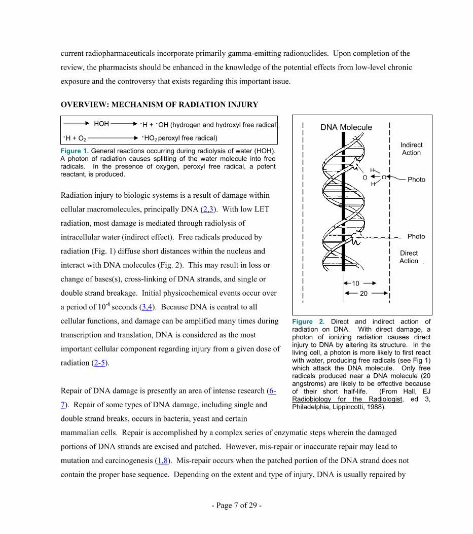
Radiation injury to biologic systems is a result of damage within
cellular macromolecules, principally DNA (2,3). With low LET
radiation, most damage is mediated through radiolysis of
intracellular water (indirect effect). Free radicals produced by
radiation (Fig. 1) diffuse short distances within the nucleus and
interact with DNA molecules (Fig. 2). This may result in loss or
change of bases(s), cross-linking of DNA strands, and single or
double strand breakage. Initial physicochemical events occur over
a period of 10-6 seconds (3,4). Because DNA is central to all
cellular functions, and damage can be amplified many times during
transcription and translation, DNA is considered as the most
important cellular component regarding injury from a given dose of
radiation (2-5).
Repair of DNA damage is presently an area of intense research (6-
7). Repair of some types of DNA damage, including single and
double strand breaks, occurs in bacteria, yeast and certain
mammalian cells. Repair is accomplished by a complex series of enzymatic steps wherein the damaged
portions of DNA strands are excised and patched. However, mis-repair or inaccurate repair may lead to
mutation and carcinogenesis (1,8). Mis-repair occurs when the patched portion of the DNA strand does not
contain the proper base sequence. Depending on the extent and type of injury, DNA is usually repaired by
·H + ·OH (hydrogen and hydroxyl free radical)
Figure 1. General reactions occurring during radiolysis of water (HOH). A photon of radiation causes splitting of the water molecule into free radicals. In the presence of oxygen, peroxyl free radical, a potent reactant, is produced.
HOH
·H + O2 ·HO2 peroxyl free radical) DNA Molecule
Indirect Action
Direct Action
Photo
Photo
Figure 2. Direct and indirect action of radiation on DNA. With direct damage, a photon of ionizing radiation causes direct injury to DNA by altering its structure. In the living cell, a photon is more likely to first react with water, producing free radicals (see Fig 1) which attack the DNA molecule. Only free radicals produced near a DNA molecule (20 angstroms) are likely to be effective because of their short half-life. (From Hall, EJ Radiobiology for the Radiologist, ed 3, Philadelphia, Lippincotti, 1988).
2010
HOO
H
- Page 7 of 29 -

cellular mechanisms within minutes to hours following exposure to ionizing radiation (Ref17). Post-
irradiation conditions, including oxygen concentration, temperature, Ph, and position in the cell cycle may
affect the outcome of DNA repair (8). For instance, repair of sublethal damage is favored by physiologic
pH and oxygen concentration, while repair of other forms of damage is most likely to occur when irradiated
cells are under the influence of low oxygen concentration and low temperature.
The importance of position within the cell cycle, frequency of mitosis, and cellular differentiation cannot be
neglected in considering the potential for repair of damage to DNA and other cellular components. For
instance, cells in late G2 and mitosis are less capable of repairing damage than are cells in G1 or S. The so-
called “Law of Bergonie and Tribondeau” still has considerable merit as cells that are radiosensitive may
not have the repair capability of less sensitive cells (5).
Many approaches have been used to determine the important components(s) in a cell that is sensitive to
radiation and important to function and replication. In early studies, microbeam irradiation and
microsurgical techniques were used to identify the putative “target” of radiation injury. A mathematical
expression of survival relationships for increased doses of radiation applied to cells in vitro was also used to
ascertain target size. As a result of the latter approach, the concept of the target theory evolved (2,8) as a
means of identifying the components(s) within a cell of importance. The target theory is a model in which it
is stated that the production of ionization in or very near a structure (target) is responsible for the observed
effect. The ionization in or near the structure is termed a hit. In the case of a single-hit model, it is stated
that a single event in or near the target will result in the effect observed. Mathematical expression of cell
survival vs. dose models let to the concepts of Do and D37- Do is identified as the dose that will give an
average of one hit per target in the cell population. The mathematical expression predicts that if a Do dose
is given, 37% of the original number of cells will survive (D37).
For the single-hit theory it is stated that there is a target that must be hit once to inactivate the cell.
According to the multi-hit theory, there is more than one target in the cell and each target must be hit once
to inactivate the cell. A semi-log expression of survival fraction data vs. dose for a system exhibiting the
characteristics of a two-hit system results in a survival curve that has a shoulder followed by a linear
decrease in survival from increasing dose (Fig. 3b). The shoulder region may be considered as an area of
- Page 8 of 29 -

sublethal damage where repair may occur and cells may survive. A single-hit system results in a linear
decrease in survival with no indication of repairable damage (Fig. 3a).
Another approach to interpretation of cell
survival vs. dose curves is that of a single
target or multi-target system. The single target
theory states that the system (cell) has a single
target that must be hit once to elicit the
response. The multi-target theory states that
the cell has two or more targets. In order to
inactivate the cell, each of the targets must be
hit once. The survival fraction vs. dose curve
appears the same for either the single-hit,
multi-hit or single-target, multi-target models.
It should be noted that although DNA is
considered the “target” of radiation, the exact
type and amount of damage (base deletion,
strand break, etc) that is sustained remains
obscure.
In addition to the target theory, the “Q” theory
has been offered as an explanation for
observations relating to cell killing. With the
Q theory, cells are considered to contain a single radiosensitive site (DNA). However, cells also are thought
to contain a protective “Q” substance that engenders repair of radiation damage before it can be expressed
(6). Therefore, Q substance must be inactivated by radiation before cell killing (reproductive death) occurs.
The Q substance has not been conclusively identified, but may be associated with sulfhydryl-rich
intracellular molecules (8). The Q theory assumes that repair of sublethal damage occurs and can be
demonstrated on the dose-response curves used for the target theory. When Q has been inactivated, cell
killing is represented by a straight line.
Figure 3. Dose-response model for cell killing (reproductive death) comparing single hit (a) and two hit radiation damage (b). A semi-log plot is used. The dose-response to cells with a single target for cell killing is linear (a). Note the lack of a shoulder in the first decade of cell killing. With a cell system requiring two hits for cell killing, there is a shoulder representing sublethal damage (b). The slop (D, or D37) for both dose responses is the same at higher doses.
- Page 9 of 29 -

GENETIC (HERITABLE) EFFECTS OF RADIATION Introduction Ionizing radiation is a potent mutagen. Numerous radiation-induced gene and chromosome mutation have
been documented in man and animals. Gamma and x-ray are known to increase the rate of spontaneous
mutation in man and animals. Mutations caused by ionizing radiation are no different than those that occur
naturally and are also the same as those caused by viruses and chemicals. Hence, their detection in human
population is a difficult task requiring sophisticated statistical methods.
Although the term “genetic effects” (mutation) usually implies injury to germ cells (reproductive tissue)
causing hereditary diseases, the genome of somatic cells can also undergo mutation and the damage can be
expressed in daughter cells of an organ or tissue. Therefore, exposure to ionizing radiation can result in
damage to future generations, that is, hereditary, or to the parent organism. For example, squamous cell
carcinoma of the hands is thought to result from genomic damage to somatic cells of an individual, while
dysproteinemia that has been observed in the offspring of atomic bomb survivors is caused by geneomic
damage to reproductive tissue of the parent(s). The important issue of cancer induction will be dealt with
under the heading “Radiation Carcinogenesis.”
While heritable effects of radiation have been extensively studied in animals, there is a paucity of data for
human beings. The major source of human data stems from the continuing evaluation of data collected from
offspring of survivors of the Hiroshima and Nagasaki atomic bomb detonations. Although dose rates were
high and LET varied, considerable effort has been made to normalize the data so that low dose and low dose
rate extrapolations can be made for low LET radiation exposure. Although the results have been questioned
repeatedly by the scientific community, atomic bomb survivors remain the best source of information
regarding human genetic effects resulting from exposure to ionizing radiation. Because future generations
of the survivors will be studied, these data will continue to yield meaningful conclusions in the years to
come.
Mutagenesis A gene (point) mutation is a change in the primary structure of DNA resulting from loss, gain or substitution
of a base. Gene mutation can be dominant, recessive, or sex-linked and range from being nearly
- Page 10 of 29 -
imperceptible to lethal. Chromosome mutations (aberrations) are more complex than gene mutations and
result from radiation-induced chromosome breaks. After a chromosome has been broken, the break may be
restored with no ill effect (restoration) or part of the chromosome may be lost during the next mitosis
(deletion). When there are multiple breaks in a chromosome, rearrangement of sections of chromosomes
may occur, changing the base sequence and the genetic code. Rejoining (restitution) of broken ends of the
same chromosome results in formation of rings, and rejoining of two different chromosomes may result in
formation of a dicentric; these events usually result in mitotic death.
Specific data on point mutation in human beings is lacking, but cytogenetic damage induced by gamma and
x-irradiation has been studied in man and animals. A high number of chromosomal mutations (dicentric and
rings) has been observed in the circulation lymphocytes of medical radiation workers and atomic bomb
survivors. Although these mutations may persist for years, cytogenetic damage to lymphocytes has not been
linked to an increase in cancer rate (2, 9). Because they are easily sampled and are uniquely sensitive,
lymophocytes have been used as a dosimeter for human exposure to gamma radiation. A linear-quadratic,
no threshold dose response has been generally described (2); however, there is other evidence that a
threshold clearly exists (9).
Ionizing radiation may also injure or cause dysfunction of the mitotic apparatus that either slows or impairs
mitosis (8). A serious imbalance of genetic material in daughter cells may result. Damage to the
centromere of a chromosome may slow migration of a chromosome (lagging) along the mitotic spindle so
that it is not included in a daughter cell. The leftover chromosome becomes a micronucleus and is lost to
future generations. Rearrangement following multiple breaks of chromosomal arms may result in location
of a centromere at the end of a chromosome. For unknown reasons, chromosomes do not function properly
during mitosis, when a terminal centromere is present. During mitosis, a chromosome with a terminal
centromere is also likely to be lost. Nondisjunction occurs when a centromere damaged by ionizing
radiation fails to separate at mitosis, causing an uneven chromosome number. Aneuploidy (change in the
number of one or more specific chromosomes) and polyploidy (multiple sets of chromosomes) are examples
of the result of nondisjunction.
The shape of the dose-response curve for radiation-induced genetic effects has been debated for years (10).
The results of various in vitro studies are confounding. Because limited human data is available, the basis
- Page 11 of 29 -

for estimation of genetic risk and dose-response has been extrapolated from high dose animal experiments
[doses greater than 10 rem (10)]. Moreover, many of these extrapolations to account for the “dose rate
effect” [for a given dose of radiation, high dose rate is generally considered 3-4 times as effective as low
dose rate radiation as occurs in chronic low-level exposure (11)].
Early work with Drosophila (fruit fly) gene
mutations indicated gamma radiation caused
a linear, no threshold response (Fig. 4)
(3,10). This model, e.g., the linear
hypothesis, implied that the frequency of
gene mutations was directly proportional to
dose and was independent of dose rate
(fractionation). Therefore, genetic effects
were considered to be cumulative, adding up
over a period of time and suggesting that
chronic, low-level exposure to radiation
resulted in a serious hazard to radiation
workers. From these experiments, it was
concluded that the dose required to double
the spontaneous mutation rate (doubling
dose) in Drosphila was in the range of 5-150 rem. The maximum permissible dose (MPD) concept evolved
from these data. The MPD may be considered conservative because it assumes no repair and is considered
to err on the “safe” side.
Figure 4. Linear dose-response model derived from experimental data on radiation-induced gene mutations in Drosophila. No threshold was identified; however, the low dose (dotted line) region has been extrapolated from the high-dose data.
Subsequent work, including the “megamouse∗” project (10) of the 1960’s and 1970’s, revealed a variety of
dose responses depending on the mutation and the germ cell line studied ( in the megamouse project, the
radiosensitivity of different mutations varied by a factor of 20). Mutation frequency was also affected by
the quality factor of the radiation (LET), the dose given, and the dose rate. In the megamouse project, a
dose-rate sparing effect was found with low LET radiation. This important conclusion suggested that,
∗ ”Megamouse” refers to the fact that over 7 million mice were used, not giant mice from mutation.
- Page 12 of 29 -
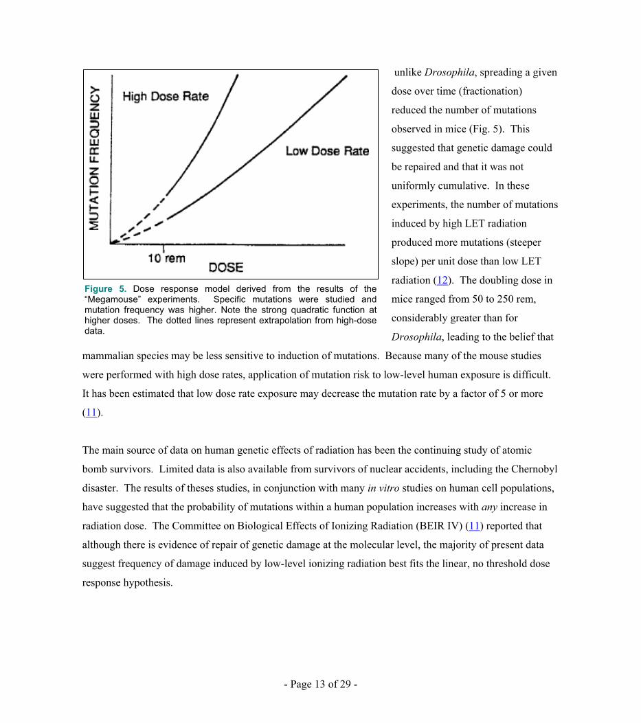
unlike Drosophila, spreading a given
dose over time (fractionation)
reduced the number of mutations
observed in mice (Fig. 5). This
suggested that genetic damage could
be repaired and that it was not
uniformly cumulative. In these
experiments, the number of mutations
induced by high LET radiation
produced more mutations (steeper
slope) per unit dose than low LET
radiation (12). The doubling dose in
mice ranged from 50 to 250 rem,
considerably greater than for
Drosophila, leading to the belief that
mammalian species may be less sensitive to induction of mutations. Because many of the mouse studies
were performed with high dose rates, application of mutation risk to low-level human exposure is difficult.
It has been estimated that low dose rate exposure may decrease the mutation rate by a factor of 5 or more
(11).
Figure 5. Dose response model derived from the results of the “Megamouse” experiments. Specific mutations were studied and mutation frequency was higher. Note the strong quadratic function at higher doses. The dotted lines represent extrapolation from high-dose data.
The main source of data on human genetic effects of radiation has been the continuing study of atomic
bomb survivors. Limited data is also available from survivors of nuclear accidents, including the Chernobyl
disaster. The results of theses studies, in conjunction with many in vitro studies on human cell populations,
have suggested that the probability of mutations within a human population increases with any increase in
radiation dose. The Committee on Biological Effects of Ionizing Radiation (BEIR IV) (11) reported that
although there is evidence of repair of genetic damage at the molecular level, the majority of present data
suggest frequency of damage induced by low-level ionizing radiation best fits the linear, no threshold dose
response hypothesis.
- Page 13 of 29 -

Assessing the Risk Two methods are generally used to assess the risk for genetic effects caused by ionizing radiation: absolute
risk and doubling dose. A third method, the genetically significant dose, is used to chart the impact of
ionizing radiation on the human population, particularly regarding medical exposure.
The absolute risk of genetic effects is expressed as the number of mutations per rem or rad of ionizing
radiation. With absolute risk, the incidence of mutation is quoted as a function of dose. Estimates in man,
based on animal experimentation, suggest that the absolute risk of mutation is approximately 10-7 per rem
per gene (8). Because the spontaneous mutation rate in man is 10E-5 per gene per generation, detection of
absolute changes in radiation-induced mutations is problematic and requires large populations and long-term
study. Therefore, it is often easier to use the doubling dose method to assess the relative genetic risk.
The doubling dose is the dose of radiation required to cause twice the number of spontaneous mutations
(2,8). Because this method takes into account the natural, or spontaneous mutation rate, it is known as the
“relative” mutation risk. Although extremely controversial, early evidence from atomic bomb data suggest
the median doubling dose in humans is 100 rem. This is in agreement with the results of animal studies
using various genetic endpoints, where the median doubling dose for low-level, low LET radiation was
found to be approximately 100 rem (11). These estimates fall within the 50-250 range originally suggested
by the megamouse project.
The genetically significant dose (GSD) is an index used to assess the presumed impact of gonadal exposure
to ionizing radiation on whole populations. The GSD is expressed in rems and relates to a population and
not the individual or the number of mutations produced. Only radiation doses to the gonads of the members
of the population who will reproduce are considered genetically significant. In other words, the GSD
merely determines the gonadal dose that is received by those that will likely bear children and averages it
over the entire population (gene pool). Therefore, people that receive gonadal radiation, but are not
considered likely to have children, do not contribute a genetically significant dose. Similarly, a gonadal
dose absorbed by young, healthy individuals contributes to the GSD. Finally, the GSD attempts to average
the impact of presumed genetic effects for the entire population. Thus, the GSD is the dose that if given to
each member of the population would result in the same genetic impact (effect on total gene pool) that
actually results form doses received by those producing offspring.
- Page 14 of 29 -
In summary, current evidence implies that any increase in the amount of radiation exposure is expected to
cause a proportional increase in the frequency of mutations and there is some “risk” for occupations workers
(9, 11). It has been suggested that a dose of 1 rem per generation might raise the spontaneous mutation rate
by 1% (10). Thus, for chronic low-level, low LET exposure, the increase in mutation frequency may be
undetectable in the human population (8). Although human data is sparse, it appears that human beings are
no more sensitive to heritable effects of radiation than animals; possibly, human beings are less sensitive
(10). Ionizing radiation is known to cause mutation, but a statistically significant increase in heritable
effects has not been documented when occupational exposure has remained within dose limits
recommended by the NCRP.
RADIATION CARCINOGENESIS Introduction
The potential for cancer induction is considered the most important concern resulting from chronic, low-
level exposure to ionizing radiation (8, 9). Although studied extensively, the mechanism, the dose-response
curve and the absolute risk of radiation carcinogenesis remain poorly understood. Because carcinogenesis is
a late effect that occurs years after exposure to radiation and because radiation-induced cancers are no
different than cancer caused by other agents, the epidemiological investigation of radiogenic cancer has
progressed slowly. Perhaps the most practical approach to the study of radiogenic cancer involves the
assessment of absolute risk of cancer from medical and background radiation sources. Quantization of the
risk of chronic low-level radiation exposure is a major endeavor of organization such as the International
Commission on Radiation Protection (IRCP) and the National Commission on Radiation Protection
(NCRP).
Unlike heritable effects, there is ample data on radiation-induced cancer in human beings. Many of the
early workers in the field of radiation, including Mme. Curie and her daughter, suffered from radiogenic
cancer. Historically, high doses of ionizing radiation have been associated with skin, lung, thyroid, breast,
bone and lymphatic tumors (1, 8). In addition, radiation is known to induce leukemia and other
hematopoietic neoplasms. Various cancers have been documented in people exposed to large doses of
gamma radiation at Chernobyl and atom bomb explosions. Scientists generally concede that exposure to
any dose of ionizing radiation carries some risk (probability) of cancer induction, although it may be
- Page 15 of 29 -

imperceptible at low doses (8, 10, 11). Unfortunately, few data are available for assessing the risk of human
cancer in the low dose range (<10 rem). The majority of risk assessments for low-dose, low-level exposure
have been extrapolated from high dose data. Risks for cancer types including leukemia, breast and thyroid
cancers associated with low-level exposure have been estimated (11). Regarding radiogenic cancer, the
bone marrow, thyroid gland, breast, and to a lesser extent, bone are considered the more sensitive organs of
the human body.
Mechanisms of Cancer Induction
While the exact mechanism of radiogenic cancer is unknown, there is an unequivocal association of
radiation exposure and cancer. Several theories have been proposed; three will be described here. The first is
the two-step initiation-promotion theory. It is believed that most cancers are a two step process (8). First, the
genome of the cell is injured in some way by a carcinogen (initiation). This injury, followed by a latent
period, is usually unapparent and cannot be measured by routine medical tests. However, cell injury can be
enhanced by an additional insult (promotion) which results in tumorigenesis, Ionizing radiation is thought to
be a carcinogen that causes initiation. Chemicals, viruses, mutation and free radicals are other examples of
initiators. Promoters include hormones and some carcinogens as well as the event of cell division.
A second theory regarding the mechanism of radiogenic cancer is the mutational theory. The mutational
theory is supported by evidence that radiation alone may cause somatic mutations which result in cancer
induction. While the exact genetic change associated with mutation is unknown, the altered genome results
in malignant transformation that establishes a clone of cancer cells. Portions of the cells genome that down-
regulate cell division are possibly altered by mutagenesis. Chromosomal aberrations are frequently observed
in cancer cells (8).
The oncogene theory proposes that all cells contain oncogenes that are normally suppressed by a regulator
gene. If chromosomes are broken by radiation and rejoined, rearrangement (translocation) may result so that
the oncogene is no longer located near a regulator gene. Thus, the oncogene can be turned-on, expressing a
malignancy. However, there is no direct evidence that oncogenes are activated by ionizing radiation (2).
- Page 16 of 29 -
Assessing the Risk
As with genetic effects, the risk of radiogenic cancer can be expressed in absolute and relative terms (8, 10).
Absolute risk is the number of excess cancers per unit of time in a specific population per unit of dose.
Absolute risk is usually derived by irradiated versus nonirradiated populations. Relative risk is the ratio
between the risk of cancer induction for a population receiving a given mean dose and an unexposed
population (similar to the doubling dose concept). The relative risk may be expressed as a multiple or
fraction of the spontaneous cancer rate for a population. Because the spontaneous rate of cancer is low,
expressing risk in this way may be quite misleading. For instance, if the spontaneous cancer rate is 2%,
doubling the risk would give a cancer rate of 4%, only representing a few cases. Therefore, risk is best
expressed in absolute terms.
The dose-response relationship for induction of radiogenic cancer is very complex and controversial (1, 8,
11, 12, 14, 15). In most animal and ceil culture models there is a dose-dependent increase in tumorigenesis
(steep) portion followed by a dose-dependent decrease when there is “saturation” at higher doses (> 300
rem). Data from human cancer resulting from high doses (atomic bomb detonations and nuclear accidents)
of radiation also fit a similar dose-response curve. Establishing the exact shape of the curve below 100 rem
is problematic because extrapolation from high dose data is needed (limited human or animal data is
available for low-dose exposure.) It is even more difficult to set the 0-10 rem region of the curve, the range
of low-level exposures. Compounding the issue, most high-dose data was associated with a high dose rate.
Therefore, extrapolation of high dose animal data and the available human data is one of the most
controversial issues in the field of radiobiology. Because of the complexity of the issue at hand, only the
dose response for low-level, low-LET radiation will be presented.
The BEIR V Report
The subject of dose-response and assessment of risk for radiogenic cancer was addressed in the BEIR V
report in 1990. The Committee used three general methods of extrapolation from the high dose region of
the dose-response curve (Fig. 6). The linear model, used for the MPD concept, assumes no threshold and
that the effectiveness of radiation per unit of dose is the same at high and low doses. This, again, is
consistent with the concept that any radiation exposure is harmful. Animal data suggests otherwise; that the
low part of the dose-effect curve bends downward, hence the “below linear model” (Fig 6). With this model,
- Page 17 of 29 -

the risk per dose unit is less at low,
doses and some degree of repair or
threshold effect is implied. An “above
linear model” was proposed, which has
a rapidly rising dose response rate
(slope) at low-levels. The above linear
assumption was used to fit data from
occupationally exposed workers at
nuclear power plants and the Portsmouth
Naval shipyard and from soldiers
exposed to nuclear weapons tests (8,
15). The model has been debated, and
was refuted by the 1980 BEIR III report.
Many scientists question the above
linear model because the population
sample was too small and may have
been at a higher risk for cancer
induction in the first place. The
possibility that this model is correct is alarming; the increased sensitivity to low-level radiation would imply
that 50-70% of all human cancer would be due to natural background radiation∗∗, rather than the present
estimate of 1% (8).
Figure 6. Generalized dose response model for induction of radiogenic cancer. At doses greater than 10 rem the incidence of cancer is usually a quadratic function of dose. Below 10 rem, the shape of the curve is unknown. There is data to suggest that the low dose region may be (a) linear, (b) above linear or (c) below linear. The BIER V report considers the below 10 rem portion of the curve to be linear. Dose response cures for specific radiogenic cancer are available (11).
For whole body doses below 10 rem, the BEIR V Committee concluded that the frequency of cancer
induction, like heritable effects, is best represented by the linear-quadratic model with no threshold. The
BEIR V Committee adopted this position after careful review of the life-span study of Japanese atomic
bomb survivors, people that received therapeutic x-rays for ankylosing spondylosis, tuberculosis,
dermatophytosis (tinea capitis) and cancer of the cervix. The Committee determined that the risk of
radiogenic cancer was 3 times higher for solid tumors and 4-5 times given in the BEIR III Report.
∗∗ The estimated dose form natural background radiation is approximately 80-250 mrem/year depending on geographic location. This is well below the 10 rad range that is considered to be low-level.
- Page 18 of 29 -

The estimated absolute lifetime risk of death from cancer following a 10 rem acute whole body dose was
stated to be 0.8%. For chronic (fractionated) exposure the risk decreased by a factor of 2, implying repair
may occur. Compared to previous reports, the committee stated that the risk for leukemia was also increased
for those subjected to in utero irradiation.
The principal conclusion of the BEIR V Committee is that if 100,000 people of all ages receive a whole-
body dose of 0.1 Gray (Gy) low LET radiation in a single brief exposure, 800 extra cancer deaths are
expected over their remaining lifetimes in addition to the nearly 20,000 cancer deaths in the absence of the
radiation. The BEIR V Report also lists the expected types of cancer and differences in risks due to sex and
age at time of exposure. Furthermore, it provides estimates of risk for chronic continuous low dose rate
exposures to all radiation qualities. For example, estimates of lifetime excess cancer mortality per 100,000
exposed persons are listed below:
a. continuous lifetime exposure to 1mSv/y (100 mrem/y)
Total % of normal* Leukemia Non-leukemia Male 520 2.5 70 450 female 600 3.4 60 540
b. continuous exposure to 0.01 Sv/y from age 18y to 65y
Total % of normal* Leukemia Non-leukemia Male 2880 14 400 2480 female 3070 17 310 2760
(*) “normal” refers to those individual normally expected to die from cancer, from all causes.
A linear dose-response model was preferred by the BEIR V Committee. The Committee stated that
departure from linearity could not be excluded at low doses beyond the range of observation. They also
reasoned that the departures could be in either direction, i.e., increased or decreased risk. Moreover, the
Committee noted that epidemiologic data cannot rigorously exclude the existence of a threshold in the
millisievert dose range.
While these findings indicate that the risk of radiogenic cancer is greater than previously thought, the BEIR
V Report has been questioned by leading radiobiologists and physicians (16). Much of the controversy
stems from the use of data gleaned from the atomic bomb survivors, who received acute (microsecond)
- Page 19 of 29 -

doses. No adjustment was made by the BEIR V Committee for the difference in dose rate effect when
extrapolating the data to represent the response to low-level irradiation. Because repair is known to occur at
lower dose rates, the frequency of dose related effects is usually considered to be reduced by a factor of 2-3
(16).
What practical conclusions can be drawn from these studies? First, risk assessments for low-level exposure
are really estimates, extrapolated from high dose data. High-dose data are available to health scientists for
studying dose-response. Data on low-level exposures, suitable for epidemiologic analysis, are not available.
This includes the 0.1-0.5 rem range used in nuclear medicine patients. This is because the difference
between the incidence of spontaneous and radiogenic cancers is so small that a sample population of
millions would be needed to conduct a meaningful study (16). Second the entire issue of risk due to low-
level exposure must be placed into perspective when people residing in areas of high natural background
radiation do not have a higher rate of solid tumors or leukemia when compared to other populations. Third,
if the recommended MPD were to be reduced by one-half, nuclear medicine workers would likely be
unaffected because most exposures are <2.5 rem per year (16).
EFFECTS OF IONIZING RADIATION ON THE EMBRYO AND THE FETUS Excessive exposure of the embryo or the fetus to ionizing radiation may cause a classic triad of embryonic
death, congenital malformation or growth retardation in mammals (8, 17). Exposure of the developing
embryo to ionizing radiation may cause major congenital defects, embryonic death or perinatal death.
During the period of a preimplantation (0-9 days in man), the conceptus is particularly sensitive to radiation.
Based on extrapolation from experimental animals, in utero doses of 10-25 rad may be lethal during
preimplantation. However, surviving embryos are at a low risk for congenital abnormalities and perinatal
death. Ionizing radiation is teratogenic and may cause major congenital malformations, including
microcephaly, microphthalmia, cerebral hypoplasia, and skeletal and dental defects. These malformations
are most common when exposure occurs during the period of organogenesis (10-50 days). Irradiation at this
time has been associated with a high incidence of growth retardation and perinatal death (8,11).
With in utero exposure to ionizing radiation during the period of the fetus (51 days-term), the most likely
deleterious effects are fetal growth and mental retardation, and maldevelopment of the central nervous
system. Although differentiation of most human tissues has ceased by 51 days post-conception, the central
- Page 20 of 29 -
nervous system continues to develop, even postnatally. The developing embryo/fetus is sensitive to
radiation-induced mental retardation, and there is little if any threshold for this effect between 48 and 112
days of gestation. This unique sensitivity can be explained by intense neuronal stem cell proliferation
during organogenesis. Analysis of data from atomic bomb survivors implies that the risk of severe mental
retardation at 48-112 days is 40-45% per seivert (100 rem) (9,11). Exposure after 112 days of gestation may
also cause mental retardation, but the risk is reduced by a factor of 4 because neuronal proliferation has
peaked. The occurrence of radiation-induced mental retardation often correlates with microcephaly (17).
The incidence of childhood leukemia might be increased by in utero exposure to ionizing radiation at
anytime during gestation (8, 9, 17). There is data both for and against this concept. Studies in the USA and
England claim an association between childhood malignancy and in utero irradiation, while results from
animal experiments and data from atomic bomb survivors do not (8).
OTHER LATE EFFECTS DUE TO LOW-LEVEL IONIZING RADIATION
Life span shortening, and cataractogenesis are other effects that may be linked to chronic exposure to low
doses of ionizing radiation. While there is abundant literature regarding these effects, their importance is
considered less than those effects previously described in this paper.
Although studies have suggested that low-level exposure to radiation causes life-span shortening, this issue
remains in doubt. In many of these studies, the decrease in life span was actually a secondary effect
resulting from cancer induction (1, 18). Life span shortening attributed solely to chronic radiation exposure
in people has not been conclusively demonstrated.
The lens of the eye is sensitive to ionizing radiation. In atomic bomb survivors, a single dose in the range of
60-150 rad of gamma radiation was considered the threshold for cataractogenesis. With highly fractionated
exposure, the threshold was 5000 rad (11). These doses are clearly beyond the occupational limit.
The mammalian gonads are also sensitive to the effects of ionizing radiation (11). In the human testis, an
acute dose of 15 rad is sufficient to cause temporary infertility. The human ovary is less radiosensitive than
the testis, where the threshold for acute exposure is approximately 65 rad. Fractionation increases the
- Page 21 of 29 -

tolerance to infertility (11). It should be stressed that the major risk to the gonads for low-level exposure to
ionizing radiation is mutation, as described under “Genetic Effects of Radiation.”
Paradoxically, there is scientific evidence that in some instances, chronic low-level exposure to ionizing
radiation causes a beneficial effect. These beneficial effects include enhanced resistance to disease,
improved viability, and increased life-span. Studies have shown that laboratory animals exposed to chronic
low doses of ionizing radiation may outlive control animals. At the cellular level, it has been suggested that
beneficial radiation-induced mutations may result in hormesis. Mechanisms are unclear, but may include
enhanced repair of DNA damage, improved scavenging of free radicals, stimulation of the immune
response, and improved maintenance of cell populations (19, 20). Although the concept of radiation
hormesis is fascinating, it has not gained widespread acceptance in the scientific community. However the
issue cannot be avoided and merits further investigative efforts.
CONCLUSION Knowledge of the biological effects of ionizing radiation is of contemporary importance to nuclear
pharmacists for three main reasons. First, it is necessary for nuclear pharmacy practitioners to retain
adequate radiation safety practices in the work place. Second, nuclear pharmacists are often the main source
of expertise regarding biological effects of radiation in laboratories where radiopharmaceuticals are used.
Lastly, those occupationally exposed to chronic low doses of ionizing radiation should be kept abreast of
new theories and concepts generated by research in radiobiology.
As expected, the jury is still out in regard to the true risk from low-level chronic exposure to ionizing
radiation. The data included herein are not intended to bring forth fear for personnel employed in a nuclear
pharmacy, but to reinforce the attitude of a healthy respect for the potential harm from overexposure to
radiation. However, the nuclear pharmacist must maintain a proper perspective. For example, the author of
an article in a health physics newsletter (21) presented comparisons of risk to nuclear power plant workers
to other occupations and situations. He cited an overall radiation risk inefficient of 3x10⁻⁴ per rem for
nuclear power plant workers as compared to an annual risk for crew members being involved in a aircraft
accident with a fatality as 4.5x10⁻⁴ for scheduled commercial carriers and 7.7x10⁻³ for scheduled commuter
carriers. Also, we accept a per capita annual risk greater than 2x10⁻⁴ for the privilege, convenience and
advantages of automobiles and trucks.
- Page 22 of 29 -
In summary, there is a risk from radiation of which occupational workers must be aware. However, the
harmful effects attributed to ionizing radiation at doses below the maximum permissible dose level (5
rem/yr) may never be truly resolved even by statistical methodology with large populations. Utilization of a
good practice technique and adherence to safety guidelines remain the best approach to assuring a safe
environment attaining conformity to the ALARA concept.
- Page 23 of 29 -

REFERENCES 1. Hall EJ. Late Effects of Radiation: carcinogenesis and life span shortening. In: Radiobiology for the
Radiologist. 3rd ed. Hagerstown, MD: Harper & Row; 1988:386-489. 2. Hall EJ. Cell survival curves. In: Radiobiology for the Radiologist. 2nd ed. Hagerstown, MD: Harper &
Row; 1978:18-37. 3. Sutherland RM, Mulcahy RT. Basic principles of radiation biology. IN: Clinical Oncology for Medical
Students and Physicians: A Multidisciplinary Approach. Rochester, NY: American Cancer Society; 1983:40-57.
4. Coggle J. The effect of radiation at the molecular and subcellular levels. In: Biological Effects of
Radiation. 2nd ed. New York, NY: International Publications Service; 1983:29-31. 5. Alper T, Cramp WA. The role of repair in radiobiology. Experentia 1989:45:21-33. 6. Frankenburg-Shwager M. Review of repair kinetics for DNA damage induced in eukaryotic cells in
vitro by ionizing radiation. Radiother and Oncol 1989;14:307-320. 7. Nias AHW. Clinical Radiobiology. 2nd ed. New York, NY: Churchill Livingstone; 1988:7-55. 8. Pizzarello WJ, Witcofski RL. Medical Radiation Biology. 2nd ed. Philadelphia, PA: Lea and Febiger;
1982 9. Coggle JE> Medical effects of low doses or ionizing radiation. Radiat Prot Dosim 1989;30:5-12 10. Hall EJ. Late effects of radiation: genetic changes. In: Radiobiology for Radiologist. 2nd ed. Hagerstown,
MD: Harper & Row; 1978:412-426. 11. Health effects of exposure to low levels of ionizing radiation. (BEIR V report). Genetic Effects of
Radiation. Washington DC; National Research Council; National Academic Press; 1990:65-125. 12. Upton AC. The biological effects of low-level ionizing radiation. Sci Am 1982;246:41-49. 13. Schull WJ, Otake M, Neel JV. Genetic effects of the atomic bombs: a reappraisal. Science
1981;213:1220-1227. 14. Upton AC. Environmental standards for ionizing radiation: theoretical bases for dose response curves.
Environ health Perspect 1983;53:31-39. 15. Bross IDJ, Ball M, Falen S. A dosage response curve for the one rad range: adult risks from diagnostic
radiation. Am J Public Health 1979;69:130-136. 16. Tilyou S. BEIR V Report: Experts urge cautious interpretation of higher risk estimates. J Nucl Med
April 1990;31:13A-19A. Newsline
- Page 24 of 29 -

17. Hall EJ. Effects of radiation on the embryo and the fetus. In: Radiobiology for the Radiologist. 3rd ed.
Hagerstwon, MD: Harper & Row; 1988:412-426. 18. Nias AHW. Clinical Radiobiology. 2nd ed. New York, NY: Churchill Livingstone; 1988:258-259. 19. Wolff S. Are radiation induced effects hormetic? Science 1989;245:574. 20. Segan LA. On radiation, paradigms and hormesis. Science 1989;245:574. 21. Alexander RE. Acceptable radiation risks for nuclear powerplant workers. The Health Physics Society’s
Newsletter 1986;14:1-44.
- Page 25 of 29 -

QUESTIONS
1. In regard to low-level chronic exposure, the single most important consequence is: A. Radiation carcinogenesis B. Life-span shortening C. Cataractogenesis D. Organ fibrosis
2. In determining the true risk of low-level exposure the greatest controversy continues over:
A. The length of time necessary to develop cataracts B. The risk of high doses C. The dose-response relationship for low doses D. The lack of high dose data to aid in drawing conclusions
3. With low LET radiation, initial damage to the DNA is mediated by: A. The direct hit on a single strand B. Ionization on purine bases C. Enzyme mediated reactions D. Radiolysis of intracellular water (indirect effect)
4. For the single hit dose response model the Do for a cell population is the dose that: A. Will give an average of one hit per target in the population B. Will result in the death of each cell C. Can be tolerated and repaired D. Will result in the death of 37% of the cells in the population
5. For a two-target dose response model for a cell population, the shoulder on the dose response curve indicates:
A. Cells cannot be repaired B. That both targets have been hit once C. A region of sub-lethal damage D. Nothing regarding effects upon targets with in the cell
6. Prior to the Chernobyl disaster the best source of human data for determining genetic (heritable) effects of radiation has been:
A. Patients exposed to diagnostic x-rays B. Survivors of exposure following atomic bombs C. Radiologists from the 1930s D. Radium dial painters
7. Gene mutations from early work with Drosophila (fruit fly) indicated that gamma radiation: A. Caused genetic effects that were not cumulative from doses received over time B. Caused gene mutations that were dependent upon dose rate C. Caused gene mutation directly proportional to dose D. Caused a non-linear, threshold response
- Page 26 of 29 -

8. The results of the megamouse project regarding radiation induced mutation indicated that: A. The degree of low LET radiation damage was not influence by dose rate B. Genetic damage could be repaired C. There was little difference in mutation produced by high LET compared to low LET radiation D. The doubling dose for mice was the same as for fruit flies.
9. Absolute risk is a method used to assess the risk for genetic effects caused by radiation. In absolute risk:
A. The incidence of spontaneous mutation is considered B. Th risk of mutation is approximately 10-1 per gene C. The determination of radiation-induced mutations can be accomplished in a few months D. The genetic effects are expressed as the number of mutations per rem
10. The doubling dose of radiation is defined as the dose that causes twice the number of spontaneous mutations. The acute doubling dose of radiation for humans, based upon atomic bomb survivors is ___________ rems.
A. 10 B. 100 C. 50 D. 500
11. The “below linear model” dose response curve, implies A. Risk per dose is less at low doses B. Risk per dose is more at low doses C. Risk per dose is the same for low and high doses D. All risk is linear
12. In the calculation of genetically significant dose, radiation doses __________ are considered. A. To the entire population B. To children C. To members of the population that will reproduce D. To nuclear medicine/nuclear pharmacy personnel only
13. Radiation-induced cancers have which of the following characteristics? A. Are common in low-level exposure situations B. Occur rather quickly after acute exposure C. Differ from cancers produced by other agents D. Cannot be clearly identified as radiation-induced
14. Regarding radiation-induced cancer, the ___ are considered as more radiosensitive. A. Skin and liver B. Eyes and skin C. Colon and eyes D. Breast and thyroid
- Page 27 of 29 -

15. In the two-step initiation-promotion theory for cancer induction: A. Radiation is considered as an initiator B. Radiation is considered to be a promoter C. The genome of the cell is not injured initially by the radiation D. The initial radiation injury to the cell can be measured by routine medial tests.
16. In the oncogene theory for cancer induction it is proposed that: A. Cells have regulator genes normally suppressed by an oncogene B. Radiation acts by activating the regulator genes C. Chromosomes are broken by radiation D. This is the main method of cancer induction by radiation
17. Relative risk for expressing the risk of radiogenic cancer: A. Is the number of excess cancers per unit of time in a specific population per unit of dose B. May provide rather misleading risk numbers when compared to the spontaneous cancer rate C. Is the ratio between the risk to medical exposure and reactor personnel D. Is the best way to express radiation-induced cancer risk
18. The dose-response relationship for induction of radiogenic cancer in humans is: A. Well established and accepted B. The same for low dose and high dose data C. Based upon animal data D. Very complex and controversial
19. The MPD concept is based upon a ___ dose-response model. A. Linear at all doses B. Below linear at low doses C. Above linear at low doses D. A linear-quadratic
20. For whole body doses below 10 rem, the BEIR V Committee concluded that the frequency of cancer induction is best represented by:
A. A linear model B. A below linear model at low doses C. An above linear model at low doses D. A linear quadratic model
21. The BEIR V Committee determined that the risk of radiogenic cancer was ______ times higher for solid tumors than given in the BEIR III report.
A. Two B. Three C. Four D. Five
- Page 28 of 29 -
- Page 29 of 29 -
22. In the BEIR V Committee report the absolute lifetime risk of death from cancer following a 10 rem acute whole-body dose was stated as _______percent.
A. 0.1 B. 0.5 C. 0.8 D. 1.0
23. Exposure of the embryo or fetus to ionizing radiation A. Invariably causes embryonic death B. Increases congenital malformation C. Is lethal at 10 – 25 rads at 0-9 days D. Is of little concern to regulators
24. Radiation exposure to the fetus may result in: A. Skeletal defects B. Retinitis pigmentosa C. Mental retardation D. Dental defects
25. Radiation hormesis: A. Is proposed as a possibility for higher doses given over a long period B. Causes blindness in higher mammalian species C. Is a concept that indicates the exceptional danger from radiation D. Proposes a potential beneficial effect from radiation exposure


![SOURCES AND EFFECTS OF IONIZING RADIATION AND EFFECTS OF IONIZING RADIATION ... aged the exchange of information on the effects of radiation exposure on non-human biota [I19, N6]](https://img.pdfslide.us/doc/110x75/5aba28597f8b9a684c8eaf66/sources-and-effects-of-ionizing-and-effects-of-ionizing-radiation-aged-the-exchange.jpg)