Embed Size (px)
Citation preview

LECITHINASE PRODUCTION BY GRAM-NEGATIVE BACTERIA
MARIE T. ESSELMANN AND PINGHUI V. LIU
Veterans Administration Hospital and Department of Microbiology, School of Medicine, University ofLouisville, Louisville, Kentucky
Received for publication December 5, 1960
Lecithinase production by microorganisms hasbeen investigated in an attempt to use it in theclassification of bacteria, and to associate forma-tion with toxicity and virulence. The first recordof this reaction was made when Nagler (1939)and Seifert (1939) observed, independently, theformation of an opalescence in human serum bythe a toxin of Clostridium perfringens. It wassoon found that human serum could be replacedwith egg yolk (Macfarlane, Oakley, and Ander-son, 1941), and the nature of the reaction wasidentified as the lecithinase activity which re-sulted in the liberation of phosphorus and choline(Macfarlane and Knight, 1941), with precipita-tion of fat which was the source of the opalescence.Other members of the genus Clostridium havesince been shown to produce this enzyme. C. oede-matiens (Crook, 1942; Hayward, 1943), C. bifer-mentans, C. sordellii (Hayward, 1943), andC. haemolyticum (Jasmin, 1947) are the wellknown lecithinase producers in the genus Clos-tridium. Some members of the genus Bacillus,such as B. cereus, B. mycoides, and B. anthracis(McGaughey and Chu, 1948; Colmer, 1948),and several different types of acid-fast bacilli(Toda and Urabe, 1936) were also reported toproduce lecithinase.Most of the organisms which produced leci-
thinase were gram-positive, and only a fewgram-negative bacteria were reported to have thisactivity. Lecithinase production by Vibrio commaand El Tor vibrio has been reported by Ruataand Caneva (1901), Kraaij and Wolff (1923),and Felsenfeld (1944). The lecithinase activitiesof the members of the genus Serratia (Monsourand Colmer, 1952) and of the pseudomonads(Klinge, 1957) have been reported recently.In view of the paucity of the literature on thelecithinase activity of gram-negative bacteria,a study was undertaken to see the extent oflecithinase activity by this group of organisms.The present communication describes thesefindings.
MATERIALS AND METHODS
Strains of gram-negative bacteria used. Twentystrains of Pseudomonas aeruginosa isolated fromhuman sources were used. Strain P-A-1, therepresentative of this species, was isolated froma case of otitis and was a good pyocyaninproducer. W. C. Haynes furnished Pseudomonaspseudomallei (B-1110), Pseudomonas reptilivora(B-963), Pseudomonas caviae (B-966), Pseudo-monas aureofaciens (B-1543P), and Pseudomonaschlororaphis (B-560). Several strains of Pseudo-monas fragi and Pseudomonas stutzeri were ob-tained from P. W. Wetmore and one strain ofPseudomonas viscosa was obtained from G.Knaysi. M. E. Rhodes supplied 11 strains ofPseudomonas fluorescens and several other strainsof this species were obtained from K. Klinge.S. F. Snieszko kindly supplied 6 strains ofAeromonas liquefaciens (including some strainsof A. hydrophila and A. punctata which wereconsidered by him to be identical with A.liquefaciens) and 2 strains of Aeromonas sal-monicida.W. H. Burkholder supplied Xanthomonas begonia
(XB-10), Xanthomonas campestris (XC-10), Xan-thomonas vesicatoria (XV-25), and Xanthomonas-phaseoli (XP-34). Several strains of Vibriocomma, El Tor vibrio, and a water vibrio wereobtained from W. Burrows and W. B. Cherry.P. R. Edwards furnished Salmonella typhi-murium, Salmonella typhosa, Salmonella hirsch-feldii, Salmonella paratyphi, Salmonella Schott-muelleri, Salmonella anatum, Salmonella newport,Shigella dysenteriae, Shigella boydii, Shigellaflexneri, and Shigella sonnei. L. S. McClung sup-plied Serratia marcescens, Serratia indica, Serratiaplymuthica, Serratia kiliensis, and Serratia marino-rubra. W. B. Cherry kindly supplied Pasteurellamultocida (4866), Pasteurella haemolytica (4960),Pasteurella pestis, and Pasteurella pseudotuber-culosis.The following stock cultures from our de-
partment were used: Escherichia coli, Aerobacter
939
on October 12, 2018 by guest
http://jb.asm.org/
Dow
nloaded from
ESSELMANN AND LIU
aerogenes, Serratia marcescens, Proteus vulgaris,Bordetella bronchiseptica, Bordetella pertussis,Brucella melitensis, Brucella abortus, Brucellasuis, Haemophilus influenzae, Haemophilus para-influenzae, Neisseria gonorrhoeae, Neisseria menin-gitidis, Neisseria catarrhalis, and Neisseria flava.
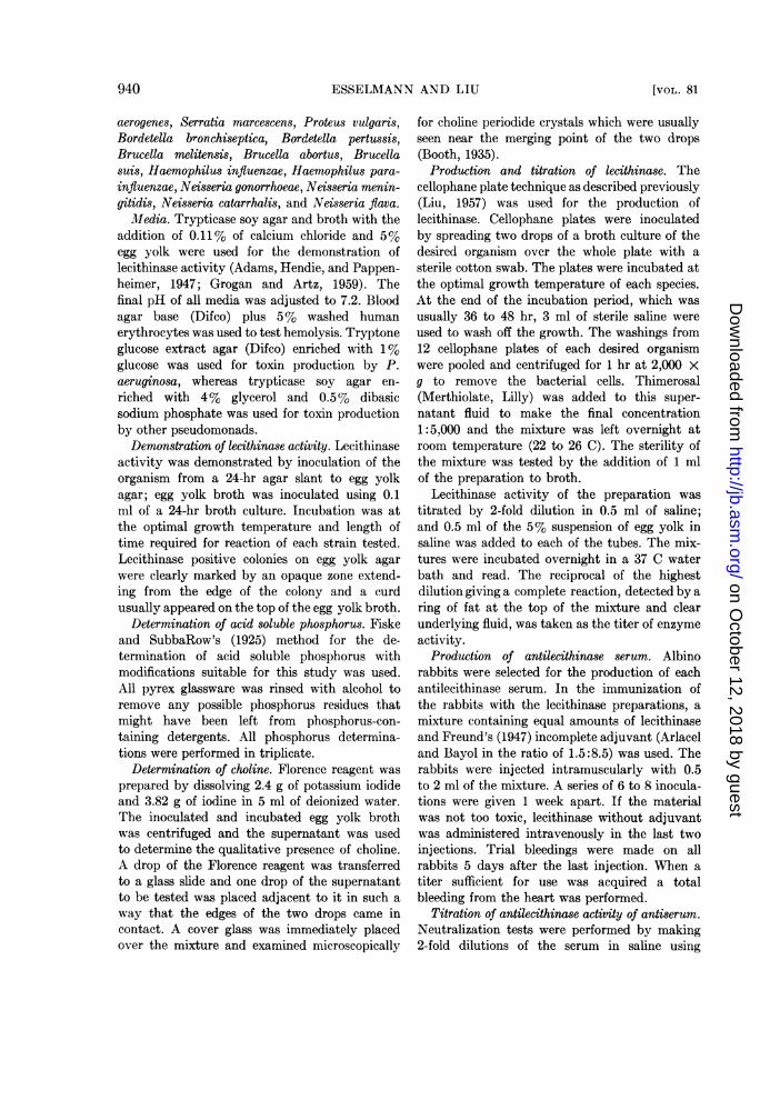
Jlledia. Trypticase soy agar and broth with theaddition of 0.11% of calcium chloride and 5%egg yolk were used for the demonstration oflecithinase activity (Adams, Hendie, and Pappen-heimer, 1947; Grogan and Artz, 1959). Thefinal pH of all media was adjusted to 7.2. Bloodagar base (Difco) plus 5% washed humanerythrocytes was used to test hemolysis. Tryptoneglucose extract agar (Difco) enriched with 1%glucose was used for toxin production by P.aeruginosa, whereas trypticase soy agar en-riched with 4% glycerol and 0.5% dibasicsodium phosphate was used for toxin productionby other pseudomonads.
Demonstration of lecithinase activity. Lecithinaseactivity was demonstrated by inoculation of theorganism from a 24-hr agar slant to egg yolkagar; egg yolk broth was inoculated using 0.1ml of a 24-hr broth culture. Incubation was atthe optimal growth temperature and length oftime required for reaction of each strain tested.Lecithinase positive colonies on egg yolk agarwere clearly marked by an opaque zone extend-ing from the edge of the colony and a curdusually appeared on the top of the egg yolk broth.
Determination of acid soluble phosphorus. Fiskeand SubbaRow's (1925) method for the de-termination of acid soluble phosphorus withmodifications suitable for this study was used.All pyrex glassware was rinsed with alcohol toremove any possible phosphorus residues thatmight have been left from phosphorus-con-taining detergents. All phosphorus determina-tions were performed in triplicate.
Determination of choline. Florence reagent wasprepared by dissolving 2.4 g of potassium iodideand 3.82 g of iodine in 5 ml of deionized water.The inoculated and incubated egg yolk brothwas centrifuged and the supernatant was usedto determine the qualitative presence of choline.A drop of the Florence reagent was transferredto a glass slide and one drop of the supernatantto be tested was placed adjacent to it in such away that the edges of the two drops came incontact. A cover glass was immediately placedover the mixture and examined microscopically
for choline periodide crystals which were usuallyseen near the merging point of the two drops(Booth, 1935).Production and titration of lecithinase. The
cellophane plate technique as described previously(Liu, 1957) was used for the production oflecithinase. Cellophane plates were inoculatedby spreading two drops of a broth culture of thedesired organism over the whole plate with asterile cotton swab. The plates were incubated atthe optimal growth temperature of each species.At the end of the incubation period, which wasusually 36 to 48 hr, 3 ml of sterile saline wereused to wash off the growth. The washings from12 cellophane plates of each desired organismwere pooled and centrifuged for 1 hr at 2,000 Xg to remove the bacterial cells. Thimerosal(Merthiolate, Lilly) was added to this super-natant fluid to make the final concentration1:5,000 and the mixture was left overnight atroom temperature (22 to 26 C). The sterility ofthe mixture was tested by the addition of 1 mlof the preparation to broth.
Lecithinase activity of the preparation wastitrated by 2-fold dilution in 0.5 ml of saline;and 0.5 ml of the 5% suspension of egg yolk insaline was added to each of the tubes. The mix-tures were incubated overnight in a 37 C waterbath and read. The reciprocal of the highestdilution giving a complete reaction, detected by aring of fat at the top of the mixture and clearunderlying fluid, was taken as the titer of enzymeactivity.
Production of antilecithinase serum. Albinorabbits were selected for the production of eachantilecithinase serum. In the immunization ofthe rabbits with the lecithinase preparations, amixture containing equal amounts of lecithinaseand Freund's (1947) incomplete adjuvant (Arlaceland Bayol in the ratio of 1.5:8.5) was used. Therabbits were injected intramuscularly with 0.5to 2 ml of the mixture. A series of 6 to 8 inocula-tions were given 1 week apart. If the materialwas not too toxic, lecithinase without adjuvantwas administered intravenously in the last twoinjections. Trial bleedings were made on allrabbits 5 days after the last injection. When atiter sufficient for use was acquired a totalbleeding from the heart was performed.
Titration of antilecithinase activity of antiserum.Neutralization tests were performed by making2-fold dilutions of the serum in saline using
940 [VOL. 81
on October 12, 2018 by guest
http://jb.asm.org/
Dow
nloaded from

LECITHINASE AND GRAM-NEGATIVE BACTERIA
TABLE 1Lecithinase activity among gram-negative bacteria
No. Posi-Family Genus and Species tive/No, of
StrainsTested
Pseudomo- Pseudomonas aeruginosa 20/20nadaceae Pseudomonas pseudo- 3/4
malleiPseudomonas reptilivora 1/1Pseudomonas caviae 1/1Pseudomonas aureofaciens 1/1Pseudomonas chlororaphis 1/1Pseudomonas fluorescens 13/16Pseudomonas fragi 0/1Pseudomonas stutzeri 0/1Pseudomonas viscosa 1/1Xanthomonas begoniae 1/1Xanthomonas campestris 0/1Xanthomonas vesicatoria 1/1Xanthomonas phaseoli 1/1Aeromonas liquefaciens 4/4Aeromonas punctata 1/2Aeromonas salmonicida 2/2
Spirillaceae Vibrio comma 6/6Vibrio El Tor 2/2Vibrio sp. ? water vibrio 1/1
Enterobac- Escherichia coli 0/2teriaceae Aerobacter aerogenes 0/4
Serratia sp. 14/16Proteus vulgaris 0/7Salmonella sp. 0/7Shigella sp. 0/7
Brucella- Pasteurella multocida 0/1ceae Pasteurella haemolytica 0/1
Pasteurella pestis 0/1Pasteurella pseudotuber- 0/1
culosisBordetella bronchiseptica 0/1Bordetella pertussis 0/1Brucella melitensis 0/1Brucella abortus 0/1Brucella suis 0/1Haemophilus influenzae 0/1Haemophilus parainflu- 0/1
enzae
Neisseria- Neisseria gonorrhoeae 0/1ceae Neisseria meningitidis 0/1
Neisseria catarrhalis 0/2Neisseria flava 0/1
* Egg yolk lecithin was used to test for lecithi-nase production by gram-negative bacteria. De-
0.25-ml amounts. The same amount of lecithinasepreparation, diluted to contain 4 units per ml,was added to each tube. These mixtures wereincubated at 37 C for 1 hr, and 0.5 ml of 5%egg yolk in saline was added. The tubes wereincubated again at 37 C overnight and read forinhibition of lecithinase activity. The reciprocalof the highest dilution showing complete inhibi-tion of the lecithinase activity was taken as thetiter of the antiserum.
RESULTS
Lecithinase activity of various organisms. Table 1indicates combined results of testing with allthree techniques, i.e., egg yolk agar, egg yolkbroth, and testing the cell-free extract from thecellophane plate. Most lecithinase positive or-ganisms will show activity with egg yolk agarand egg yolk broth with the exception of P.aeruginosa which shows no reaction on egg yolkagar and an atypical reaction on egg yolk broth.However, extracts from cellophane plates ofP. aeruginosa were strongly positive. The familyPseudomonadaceae has the largest number oflecithinase producers. P. pseudomallei was veryactive in this respect. P. reptilivora and P. caviaeproduced weak lecithinase activity on egg yolkagar. Of the parasitic organisms only P. pseudo-mallei showed a rather typical precipitatingzone around its colony in addition to displayinga definite clearing of the egg yolk media aroundthe colony (Fig. 1, top). P. reptilivora and P.caviae showed a more flaky-like flocculationaround their colonies in addition to a precedingzone of clearing of the egg yolk agar. P. fragiand P. stutzeri gave negative results.
P. aureofaciens and P. chlororaphis, bothphenazine pigment producers and found byLiu (1957) to be good producers of heat labilehemolysin, were active producers of lecithinase.Fig. 2 shows P. chlororaphis on egg yolk agarand blood agar. The zone of hemolysis is not aswide as the zone of precipitation on egg yolkagar and it is not certain whether the lecithinaseis identical with the heat labile hemolysin.P. aureofaciens and P. chlororaphis gave a typicallecithinase reaction on egg yolk agar whichfeatures a dense, fine, cream-colored precipitate
terminations of acid soluble phosphorus and ofcholine were made to verify the hydrolysis of eggyolk lecithin by all reactive lecithinase producinggram-negative organisms.
1961] 941
on October 12, 2018 by guest
http://jb.asm.org/
Dow
nloaded from

ESSELMANN AND LIU
with an even border, regardless of the size ormorphology of the colony (Fig. 2, right). As theculture aged, a clearing of the egg yolk mediumdirectly adjacent to the colony itself appeared.P. chlororaphis formed chlororaphine crystals after5 to 6 days of incubation. These appeared asfine emerald green needles that sometimes
Fig. 1. Egg yolk agar plate. Reading clockwisefrom top: three colonies of Pseudomonas pseudo-mallei strain B-1110, showing a zone of opacitydue to lecithinase surrounded by a zone of clearingof the media; three colonies of El Tor vibriostrain 34-D-23, showing a zone of opacity with aslight zone of clearing of the medium; threecolonies of Escherichia coli showing a negativereaction.
radiated from the center. These crystals appearednear the surface of the egg yolk agar and eggyolk broth.
Table 1 shows many strains of P. fluorescensto be good lecithinase producers, with the excep-tion of three strains which were received fromDr. Rhodes. All the positive P. fluorescensstrains gave typical lecithinase reactions onegg yolk agar with no preceding zone of clearing.The only difference between P. aureofaciens
and the lecithinase-producing P. fluorescens isthe fact that P. aureofaciens produced a phenazinepigment (phenazine a-carboxylic acid). Therefore,if a strain of P. aureofaciens loses its ability toform this pigment it becomes indistinguishablefrom P. fluorescemns. Many of the strains ofP. fluorescens used in this study produced smallamounts of orange pigment which appearedsimilar to the pigment of P. aureofaciens.The genera Xanthomonas and Aeromonas were
active producers of lecithinase but were muchslower to show visible activity than the genusPseudomonas. One species of Xanthomonas failedto show any lecithinase activity or hemolysis ofhuman erythrocytes, whereas three members ofthis genus did. All members of the genus Xantho-monas tested developed a yellow nondiffusablecarotenoid pigment. The lecithinase-active mem-bers of this genus showed a flaky fluocculationtype of reaction around the colony which wasuneven and preceded by a clearing zone of theegg yolk agar.
Fig. 2. Pseudomonas chlororaphis strain B-560 on human blood agar plate (left) and egg yolk plate(right). The zone of hemolysis is not as wide as the zone of precipitation on egg yolk agar. There isa clearing of the agar around the colonies of P. chlororaphis on egg yolk agar.
942 [VOL. 81
on October 12, 2018 by guest
http://jb.asm.org/
Dow
nloaded from

LECITHINASE AND GRAM-NEGATIVE BACTERIA
All the strains of A. liquefaciens producedlecithinase after 1 to 2 weeks. One strain ofA. punctata (U-21) did not show hemolyticactivity toward human erythrocytes nor did thisorganism exhibit a lecithinase reaction on eggyolk agar or egg yolk broth. However, anotherstrain of A. punctata (U-23) hemolyzed humanred blood cells and was an active lecithinaseproducer. A. salmonicida formed a soluble,brown melanin-like pigment. This organism wasslow to produce lecithinase on egg yolk agar andegg yolk broth.
Findings of Felsenfeld (1944) were confirmedsince two strains of V. comma, El Tor vibrio,and the water vibrio showed the production oflecithinase on egg yolk agar and egg yolk brothin 4 to 7 days. Fig. 1 shows the reaction of ElTor vibrio on an egg yolk agar plate. The zoneof precipitation is not as even as the typicalzone formed around colonies of the reactivePseudomonas species as seen in Fig. 2 (right).The zones around all the vibrios tested werepreceded by a zone of clearing of the mediawhich might have been due to its proteolyticactivity.Only the genus Serratia of the Enterobacteriaceae
family showed a precipitate on egg yolk agarin 1 to 4 days. Five strains of S. marcescensproduced typical zones of precipitation on eggyolk agar, whereas two strains did not produce azone. Two strains of S. indica were slow toproduce a slight zone of precipitation on eggyolk agar and choline could not be demonstrated.Three strains of S. plymuthica and two strains ofS. marinorubra produced marked zones of pre-cipitation on the agar substrate, whereas twostrains of S. kiliensis produced partial zones.All the reactive Serratia species tested showedzones of precipitation preceded by zones ofclearing of the media. The members of the familyBrucellaceae and of the genus Neisseria failed todemonstrate any lecithinase activity.
Data on the demonstration of phosphate andcholine. Each milliliter of filtrate used in thephosphorus determination was equivalent to0.01 ml of egg yolk. P. aeruginosa strain 20showed the liberation of 16.1 mg of phosphateper 100 ml in 10 days and choline was demon-strated in 4 days. P. aeruginosa strain P-A-7did not produce choline in a 5% egg yolk brothculture. However, the demonstration of cholinewas possible by mixing equal amounts of 5%
egg yolk saline and its lecithinase produced bythe cellophane plate technique. Therefore, thefailure to demonstrate choline may be due to theutilization of choline by the organism. P. pseudo-mallei (B-1110) and P. caviae (B-966) freedphosphate equal to 17.3 and 8.3 mg per 100 ml,respectively, in 10 days, whereas P. reptilivora(B-963) liberated 11.2 mg per 100 ml in 16 days.Choline was demonstrated in the two formerorganisms in 4 to 5 days, whereas the latter didnot produce choline.
P. aureofaciens and P. chlororaphis liberated16.4 and 18.0 mg acid soluble phosphorus per100 ml, respectively, in 8 days. Choline crystalswere present on the 6th day of incubation in thecase of P. aureofaciens, whereas P. chlororaphisdid not show choline until the 14th day. P.fluorescens strain 28/4 liberated 16.1 mg of acidsoluble phosphorus per 100 ml in 10 days, whereasthe demonstration of choline was possible in 3to 5 days of incubation. P. viscosa freed 20.9mg of acid soluble phosphorus per 100 ml in12 days, and choline was demonstrated in 8days.The Florence test was positive in all the
lecithinase producers of the genera Xanthomonasand Aeromonas, thus proving that choline wasliberated. Acid soluble phosphorus values after22 days of incubation were 12.4 mg for X.phaseoli (XP-34) and 11.6 mg for A. punctata(U-23), per 100 ml.
V. cholerae (Hikojima 18) and El Tor vibrio(34-D-23) were strong producers of acid solublephosphorus, liberating 24.0 and 16.8 mg per 100ml, respectively, and choline crystals weredemonstrated in 8 days. S. marcescens (16a)and S. plymuthica (146) liberated 16.8 mg ofacid soluble phosphorus per 100 ml in 4 days.The former organism showed choline periodidecrystals in 1 day, whereas the latter showed thepresence of choline in 2 days. S. marinorubra(60) liberated 18.4 mg of acid soluble phosphorusper 100 ml in 4 days and choline was demon-strated in 1 day. S. indica (11) and S. kiliensis(113) did not produce choline. The formerorganism liberated only 10.0 mg of phosphate,whereas the latter liberated 9.5 mg of phosphate,per 100 ml.
Cross neutralization of antilecithinase of closelyrelated organisms. Normal rabbit serum did notneutralize the lecithinase of P. aeruginosa,P. pseudomallei, and P. aureofaciens but did
190-1 ] 943
on October 12, 2018 by guest
http://jb.asm.org/
Dow
nloaded from
ESSELMANN AND LIU
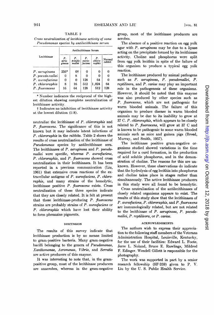
TABLE 2Cross neutralization of lecithinase activity of somePseudomonas species by antilecithinase serum
Antilecithinase Serum
Lecithinase P P. P Nmaeru- pseudo- aureo- chloro- Nrbbalginosa mallei faciens raphis rabbt
P. aeruyginosa 256* 0t 0 0 0P. pseudo,nallei 0 8 0 0 0P. aureofaciens 0 0 128 64 0P. chlororaphis 8 16 512 1,024 64P. fluorescens 16 64 128 512 128
* Number indicates the reciprocal of the high-est dilution showing complete neutralization oflecithinase activity.
t Indicates no inhibition of lecithinase activityat the lowest dilution (1:8).
neutralize the lecithinase of P. chlororaphis andP. fluorescens. The significance of this is notknown but it may indicate latent infections ofP. chlororaphis in the rabbits. Table 2 shows theresults of cross neutralization of the lecithinase ofPseudomonas species by antilecithinase sera.The lecithinases of P. aeruginosa and P. pseudo-mallei were specific, whereas P. aureofaciens,P. chlororaphis, and P. fluorescens showed crossneutralization in their lecithinases. It has beenreported in a previous communication (Liu,1961) that extensive cross reactions of the ex-tracellular antigens of P. aureofaciens, P. chloro-raphis, and many strains of the hemolytic,lecithinase positive P. fluorescens exists. Crossneutralization of these three species indicatethat they are closely related. It is felt at presentthat these lecithinase-producing P. fluorescensstrains are probably strains of P. aureofaciens orP. chlororaphis which have lost their abilityto form phenazine pigments.
DISCUSSION
The results of this survey indicate thatlecithinase production is by no means limitedto gram-positive bacteria. Many gram-negativebacilli belonging to the genera of Pseudomonas,Xanthomonas, Aeromonas, Vibrio, and Serratiaare active producers of this enzyme.
It was interesting to note that, in the gram-positive group, most of the lecithinase producersare anaerobes, whereas in the gram-negative
group, most of the lecithinase producers areaerobes.The absence of a positive reaction on egg yolk
agar with P. aeruginosa may be due to a lipaseacting on the precipitate formed by its lecithinaseactivity. Choline and phosphorus were splitfrom egg yolk lecithin in spite of the failure ofthis organism to produce a typical egg yolkreaction.The lecithinase produced by animal pathogens
such as P. aeruginosa, P. pseudomallei, P.reptilivora, and P. caviae may play an importantrole in the pathogenesis of these organisms.However, it should be noted that this enzymewas also produced by other species such asP. fluorescens, which are not pathogenic forwarm blooded animals. The failure of thisorganism to produce disease in warm bloodedanimals may be due to its inability to grow at37 C. P. chlororaphis, which appears to be closelyrelated to P. fluorescens, will grow at 37 C andis known to be pathogenic to some warm bloodedanimals such as mice and guinea pigs (Breed,Murray, and Smith, 1957).The lecithinase positive gram-negative or-
ganisms studied showed variations in the timerequired for a curd formation, in the productionof acid soluble phosphorus, and in the demon-stration of choline. The reasons for this are un-known. However, these observations do indicatethat the hydrolysis of egg lecithin into phosphorusand choline takes place in stages rather thansimultaneously. The active lecithinase producersin this study were all found to be hemolytic.
Cross neutralization of the antilecithinases ofclosely related organisms appears to exist. Theresults of this study show that the lecithinases ofP. aureofaciens, P. chlororaphis, and P. fluorescensare immunologically related, but are not relatedto the lecithinase of P. aeruginosa, P. pseudo-mallei, P. reptilivora, or P. caviae.
ACKNOWLEDGMENTS
The authors wish to express their apprecia-tion to the following staff members of the VeteransAdministration Hospital, Louisville, Kentucky,for the use of their facilities: Edward L. Foote,Jerre L. Noland, Bruce E. Rawlings, MildredF. Edinger. Wendell Gillett is responsible for thephotography.The work was supported in part by a senior
research fellowship (SF-259) given to P. V.Liu by the U. S. Public Health Service.
944 [VOL. 81
on October 12, 2018 by guest
http://jb.asm.org/
Dow
nloaded from

LECITHINASE AND GRAM-NEGATIVE BACTERIA
SUMMARY
A survey of lecithinase production among
gram-negative bacteria was made using egg
yolk agar and egg yolk broth.The families Pseudomonadaceae and Spirillaceae
contained the most active lecithinase producers.Pseudomonas and Vibrio species were the mostactive, whereas the Xanthomonas and Aeromonasspecies showed less activity.Pseudomonas aeruginosa, Pseudomonas pseudo-
mallei, Pseudomonas reptilivora, Pseudomonascaviae, Pseudomonas aureofaciens, Pseudomonaschlororaphis, Pseudomonas fluorescens, and Pseu-domonas viscosa were active lecithinase producers,whereas Pseudomonas fragi and Pseudomonasstutzeri gave negative results.
Lecithinases of P. aeruginosa, P. pseudomallei,P. reptilivora, and P. caviae were immunologicallydistinct and species specific. Antilecithinasesera of P. aureofaciens and P. chlororaphisnot only neutralized their own lecithinase butcross neutralized. Their antilecithinase sera alsoneutralized the lecithinase of many strains ofP. fluorescens.
In the family Enterobacteriaceae, Serratia was
the only genus producing lecithinase. The mem-
bers of the family Brucellaceae and of the genus
Neisseria failed to produce lecithinase.
REFERENCES
ADAMS, M. H., E. D. HENDIE, AND A. M. PAP-PENHEIMER, JR. 1947 Factors involved inproduction of Clostridium welchii a toxin.J. Exptl. Med., 85, 701-703.
BOOTH, F. J. 1935 A microchemical test forcholine and its esters in tissue extracts.Biochem. J., 29, 2064-2066.
BREED, R. S., E. D. G. MURRAY, AND N. R.SMITH 1957 Bergey's manual of determinativebacteriology, 7th ed. The Williams & WilkinsCo., Baltimore.
COLMER, A. R. 1948 The action of Bacilluscereus and related species on the lecithincomplex of egg yolk. J. Bacteriol., 55,
777-785.CROOK, E. M. 1942 Nagler reaction; break-
down of lipoprotein complexes by bacterialtoxins. Brit. J. Exptl. Pathol., 23, 37-55.
FELSENFELD, 0. 1944 The lecithinase activityof Vibrio comma and the El Tor vibrio. J.Bacteriol., 48, 155-157.
FISKE, C. H., AND Y. SUBBAROW 1925 Thecolorimetric determination of phosphorus.J. Biol. Chem., 66, 375-400.
FREUND, J. 1947 Some aspects of active im-
munization. Ann. Rev. Microbiol., 1, 291-308.
GROGAN, J. B., AND C. P. ARTZ 1959 The efficacyof a simple egg yolk test on determiningphagetypability of staphylococci. Clin. Re-search, 7, 159.
HAYWARD, N. J. 1943 The rapid identificationof Clostridium welchii by Nagler tests inplate culture. J. Pathol. Bacteriol., 55,285-293.
JASMIN, A. M. 1947 Enzyme activity in Clostrid-ium haemolyticum toxin. Am. J. Vet. Re-search, 8, 289-293.
KLINGE, V. K. 1957 Differenzierung der inWasser vorkommenden Bakterien des GenusPseudomonas durch die Eigelbreaktion. Arch.Hyg. U. Bakteriol., 141, 348-360.
KRAAJI, G. M., AND L. K. WOLFF 1923 Overde splitzing van lipoiden door bacterien.Koninkl. Akad. Wetenschap. Amsterdam,32, 624-625.
Liu, P. V. 1957 Survey of hemolysin productionamong species of Pseudomonads. J. Bac-teriol., 74, 718-727.
Liu, P. V. 1961 Identification of pathogenicpseudomonads by extracellular antigens. J.Bacteriol., 81, 28-35.
MACFARLANE, M. G., AND B. C. J. G. KNIGHT1941 The biochemistry of bacterial toxins.I. The lecithinase activity of Clostridiumwelchii toxins. Biochem. J., 35, 884-902.
MACFARLANE, R. G., C. L. OAKLEY, AND C. G.ANDERSON 1941 Haemolysis and the pro-duction of opalescence in serum and lecitho-vitellin by the a-toxin of Clostridium welchii.J. Pathol. Bacteriol., 52, 99-102.
McGAUGHEY, C. A., AND H. P. CHU 1948 Theegg yolk reaction of aerobic sporing Bacilluts.J. Gen. Microbiol., 2, 334-340.
MONSOUR, V., AND A. R. COLMER 1952 Theaction of some members of the genus Serratiaon egg yolk complex. J. Bacteriol., 63,597-603.
NAGLER, F. P. 0. 1939 Observation on a reac-tion between the lethal toxin of Clostridiumwelchii (type A) and human serum. Brit.J. Exptl. Pathol., 20, 473-485.
RUATA, G. Q., AND G. CANEVA 1901 Dellascomposizione delle lecitine. Ann. Hyg.,5, 79-86.
SEIFERT, G. 1939 Eine Reaktion menschlicherSera mit Perfringens-Toxin. Z. Immuni-tatsforsch., 96, 515-520.
TODA, T., AND K. URABE 1936 Comparativeinvestigation of the biological properties oftubercle bacilli, lepra bacilli of man and ratand other nonpathogenic bacilli. Chem.Abstr., 30, 8286.
1961] 945
on October 12, 2018 by guest
http://jb.asm.org/
Dow
nloaded from