Embed Size (px)
Citation preview
Loss of Function of the Cik1/Kar3 Motor Complex Resultsin Chromosomes with Syntelic Attachment That AreSensed by the Tension CheckpointFengzhi Jin1., Hong Liu2.¤, Ping Li2, Hong-Guo Yu2, Yanchang Wang1*
1 Department of Biomedical Sciences, College of Medicine, Florida State University, Tallahassee, Florida, United States of America, 2 Department of Biological Science,
Florida State University, Tallahassee, Florida, United States of America
Abstract
The attachment of sister kinetochores by microtubules emanating from opposite spindle poles establishes chromosomebipolar attachment, which generates tension on chromosomes and is essential for sister-chromatid segregation. Syntelicattachment occurs when both sister kinetochores are attached by microtubules from the same spindle pole and thisattachment is unable to generate tension on chromosomes, but a reliable method to induce syntelic attachments is notavailable in budding yeast. The spindle checkpoint can sense the lack of tension on chromosomes as well as detachedkinetochores to prevent anaphase onset. In budding yeast Saccharomyces cerevisiae, tension checkpoint proteins Aurora/Ipl1 kinase and centromere-localized Sgo1 are required to sense the absence of tension but are dispensable for thecheckpoint response to detached kinetochores. We have found that the loss of function of a motor protein complex Cik1/Kar3 in budding yeast leads to syntelic attachments. Inactivation of either the spindle or tension checkpoint enablespremature anaphase entry in cells with dysfunctional Cik1/Kar3, resulting in co-segregation of sister chromatids. Moreover,the abolished Kar3-kinetochore interaction in cik1 mutants suggests that the Cik1/Kar3 complex mediates chromosomemovement along microtubules, which could facilitate bipolar attachment. Therefore, we can induce syntelic attachments inbudding yeast by inactivating the Cik1/Kar3 complex, and this approach will be very useful to study the checkpointresponse to syntelic attachments.
Citation: Jin F, Liu H, Li P, Yu H-G, Wang Y (2012) Loss of Function of the Cik1/Kar3 Motor Complex Results in Chromosomes with Syntelic Attachment That AreSensed by the Tension Checkpoint. PLoS Genet 8(2): e1002492. doi:10.1371/journal.pgen.1002492
Editor: Gregory P. Copenhaver, The University of North Carolina at Chapel Hill, United States of America
Received August 9, 2011; Accepted December 6, 2011; Published February 2, 2012
Copyright: � 2012 Jin et al. This is an open-access article distributed under the terms of the Creative Commons Attribution License, which permits unrestricteduse, distribution, and reproduction in any medium, provided the original author and source are credited.
Funding: This work was supported by American Cancer Society Research Scholar Grant (RSG-08-104-010CCG), NIH grant (R15GM097326-01), and NSF grant(MCB-1121771). The funders had no role in study design, data collection and analysis, decision to publish, or preparation of the manuscript.
Competing Interests: The authors have declared that no competing interests exist.
* E-mail: [email protected]
¤ Current address: Department of Pharmacology, University of Texas Southwestern Medical Center, Dallas, Texas, United States of America
. These authors contributed equally to this work.
Introduction
One of the most important events during the cell cycle is
chromosome segregation and errors in this process will lead to
chromosome missegregation. To separate sister chromatids into
daughter cells, sister kinetochores must be attached to the
microtubules emanating from opposite spindle poles in order to
establish bipolar attachment. Even though this process is highly
regulated, incorrect attachment takes place occasionally. Syntelic
attachment occurs when both sister kinetochores are connected to
microtubules from the same spindle pole. In monotelic attach-
ment, only one of the sister kinetochores connects to the
microtubules from a spindle pole [1]. It is also possible for both
sister kinetochores to be detached. These incorrect attachments
have to be corrected before anaphase entry, or chromosome
missegregation will occur.
The kinetochore is a multi-protein complex that connects
chromosomes to microtubules. More than 60 kinetochore proteins
have been identified in budding yeast. The CBF3 (centromere
binding factor) complex associates directly with centromeric DNA,
while the DASH/Dam1 complex residues at the kinetochore-
microtubule interface. As a ten-protein complex including Dam1
and Ask1, the DASH can form a ring structure around a single
microtubule and mediate the kinetochore-microtubule interaction
[2,3,4,5]. Ndc80 (Ndc80, Nuf2, Spc24, Spc25), COMA (Ctf19-
Okp1-Mcm21-Ame1), and MIND (Mtw1p including Nnf1-Nsl1-
Dsn1) complexes bridge the gap between centromere-bound
CBF3 and microtubule-associated DASH [6,7].
Chromosome attachment is monitored by the spindle check-
point which includes Bub1, Bub3, Mad1, Mad2, Mad3, and Mps1
[8,9,10,11]. Detached kinetochores activate the checkpoint by
allowing the formation of a Mad2-Mad3/BubR1-Bub3-Cdc20
complex. Because Cdc20 is an essential activator of the anaphase-
promoting complex (APC), the binding of Cdc20 by the spindle
checkpoint components blocks APCCdc20 activity [12,13].
APCCdc20 mediates the ubiquitination and the subsequent
degradation of the anaphase inhibitor securin, known as Pds1 in
budding yeast [14]. Pds1 protein inhibits anaphase by binding to
separase Esp1 and preventing Esp1-dependent cleavage of
cohesin, a protein complex that holds sister chromatids together
[15,16]. Therefore, the activation of the spindle checkpoint
prevents anaphase entry by blocking Pds1 degradation, and
PLoS Genetics | www.plosgenetics.org 1 February 2012 | Volume 8 | Issue 2 | e1002492
brought to you by COREView metadata, citation and similar papers at core.ac.uk
provided by PubMed Central
stabilized Pds1 protein indicates the activation of the spindle
checkpoint.
Chromosome bipolar attachment generates tension on sister
kinetochores. The observation that the application of tension on
an improperly attached chromosome in grasshopper cells abolishes
the anaphase entry delay directly demonstrates the role of tension
in cell cycle regulation [17]. To analyze the response to the
absence of tension in yeast cells, tension defects can be induced by
the block of DNA synthesis or by the abrogation of sister
chromatid cohesion [18,19]. In both situations, the lack of tension
prevents anaphase entry as indicated by the stabilized Pds1 protein
levels. Ipl1 and Sgo1 were found to be required to sense tension
defects and prevent anaphase entry, but they are dispensable for
cell cycle arrest induced by the disruption of the spindle structure
[19,20]. In addition to its checkpoint function, Ipl1 kinase also
promotes the turnover of kinetochore-microtubule interaction
when tension is absent [21,22]. Therefore, it is speculated that Ipl1
may activate the checkpoint by generating detached chromosomes
when tension is absent. In contrast, Sgo1 does not play a role in
destabilizing kinetochore attachment and its checkpoint function
remains unclear at the molecular level [22].
As one of the six kinesin-related proteins in budding yeast, Kar3
was identified as being essential for yeast nuclear fusion during
mating [23]. Unlike other kinesins, Kar3 protein contains a motor
domain at its carboxy terminus that possesses minus-end-directed
motility [24]. Recent evidence indicates that Kar3 localizes at the
spindle midzone and may also function as an interpolar-
microtubule cross-linker to prevent spindle collapse [25]. More-
over, Kar3 protein promotes the poleward transport of chromo-
somes along astral microtubules [26,27]. Two proteins, Cik1 and
Vik1, associate with Kar3 through coiled-coil domains to form
Cik1/Kar3 or Vik1/Kar3 heterodimers. Both kar3D and cik1Dmutants show defects in mating, spindle morphogenesis, and
chromosome segregation [28], but their direct role in mitosis
remains unclear.
We previously showed that cik1D and kar3D mutants are
sensitive to hydroxyurea (HU), a DNA synthesis inhibitor, and
these mutants exhibit chromosome bipolar attachment defects
after HU treatment [29]. We recently found that cik1D and kar3Dmutants are synthetically lethal with tension checkpoint mutants
ipl1-321 and sgo1D, indicating a role for Cik1/Kar3 in
chromosome segregation. To further study the function of Cik1/
Kar3, we constructed a plasmid PGALCIK1-CC that contains the
coiled-coil domain of Cik1. Our results indicate that overexpres-
sion of CIK1-CC can competitively disrupt the Cik1-Kar3
interaction, which allows us to conditionally abolish Cik1/Kar3
function. With this method, we show that dysfunctional Cik1/
Kar3 results in significant co-segregation of sister chromatids in
the absence of the spindle checkpoint. Strikingly, dysfunctional
Cik1/Kar3 also causes co-segregation of sister chromatids in ipl1-
321 and sgo1D cells. Given the role of Ipl1 and Sgo1 in sensing
chromosomes that lack tension, these data suggest that the loss of
function of Cik1/Kar3 results in an increased frequency of syntelic
attachment. Results with live-cell imaging and cohesin mutants
further support this conclusion. Therefore, syntelic attachments
can be induced in budding yeast by inactivating Cik1/Kar3
complex and this method will be a very useful tool for studying the
response to tension defects.
Results
Overexpression of CIK1-CC mimics the phenotype ofcik1D and kar3D
Our previous study indicates that the Cik1/Kar3 complex
facilitates chromosome bipolar attachment after treatment with
HU, an inhibitor of DNA synthesis [29]. We also noticed that
cik1D and kar3D mutants exhibited an anaphase entry delay in the
absence of HU, suggesting the presence of improper chromosome
attachments. Previous work shows that both cik1 and kar3 mutants
are synthetically lethal with spindle checkpoint mutants, bub1,
mad1, mad2, and mad3 [30,31]. Interestingly, we found that cik1Dand kar3D are also synthetically lethal with tension checkpoint
mutants sgo1D and ipl1-321. This genetic interaction with tension
checkpoint mutants suggests that the Cik1/Kar3 complex may
facilitate the establishment of chromosome bipolar attachment
that generates tension on chromosomes.
To further study the role of Cik1/Kar3 in chromosome bipolar
attachment, we need to examine chromosome segregation in cik1Dand kar3D mutants in the absence of the spindle checkpoint, which
allows anaphase entry in spite of incorrect chromosome attach-
ments. Because of the synthetic lethality, we have to develop a way
to conditionally inactivate the Cik1/Kar3 complex. Kar3 and
Cik1 associate with each other through their respective coiled-coil
domains [32], thus overexpression of this domain may compet-
itively disrupt the Cik1-Kar3 interaction. We constructed a
plasmid PGALCIK1-CC that contains the coiled-coil domain of
CIK1 under control of a galactose inducible promoter and the
Cik1-Kar3 interaction in cells overexpressing CIK1-CC was
examined.
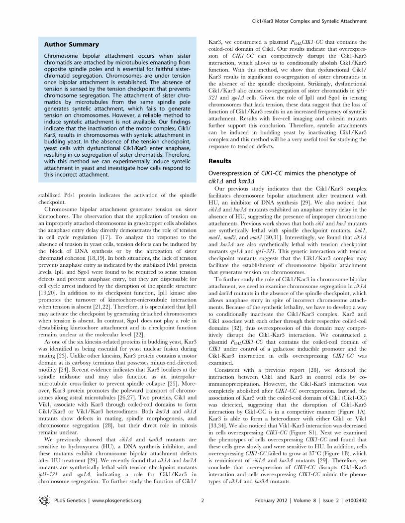
Consistent with a previous report [28], we detected the
interaction between Cik1 and Kar3 in control cells by co-
immunoprecipitation. However, the Cik1-Kar3 interaction was
completely abolished after CIK1-CC overexpression. Instead, the
association of Kar3 with the coiled-coil domain of Cik1 (Cik1-CC)
was detected, suggesting that the disruption of Cik1-Kar3
interaction by Cik1-CC is in a competitive manner (Figure 1A).
Kar3 is able to form a heterodimer with either Cik1 or Vik1
[33,34]. We also noticed that Vik1-Kar3 interaction was decreased
in cells overexpressing CIK1-CC (Figure S1). Next we examined
the phenotypes of cells overexpressing CIK1-CC and found that
these cells grew slowly and were sensitive to HU. In addition, cells
overexpressing CIK1-CC failed to grow at 37uC (Figure 1B), which
is reminiscent of cik1D and kar3D mutants [29]. Therefore, we
conclude that overexpression of CIK1-CC disrupts Cik1-Kar3
interaction and cells overexpressing CIK1-CC mimic the pheno-
types of cik1D and kar3D mutants.
Author Summary
Chromosome bipolar attachment occurs when sisterchromatids are attached by microtubules emanating fromopposite spindle poles and is essential for faithful sister-chromatid segregation. Chromosomes are under tensiononce bipolar attachment is established. The absence oftension is sensed by the tension checkpoint that preventschromosome segregation. The attachment of sister chro-matids by microtubules from the same spindle polegenerates syntelic attachment, which fails to generatetension on chromosomes. However, a reliable method toinduce syntelic attachment is not available. Our findingsindicate that the inactivation of the motor complex, Cik1/Kar3, results in chromosomes with syntelic attachment inbudding yeast. In the absence of the tension checkpoint,yeast cells with dysfunctional Cik1/Kar3 enter anaphase,resulting in co-segregation of sister chromatids. Therefore,with this method we can experimentally induce syntelicattachment in yeast and investigate how cells respond tothis incorrect attachment.
Cik1/Kar3 Motor Complex and Syntelic Attachment
PLoS Genetics | www.plosgenetics.org 2 February 2012 | Volume 8 | Issue 2 | e1002492
Tension checkpoint mutants abolish the anaphase entrydelay induced by CIK1-CC overexpression
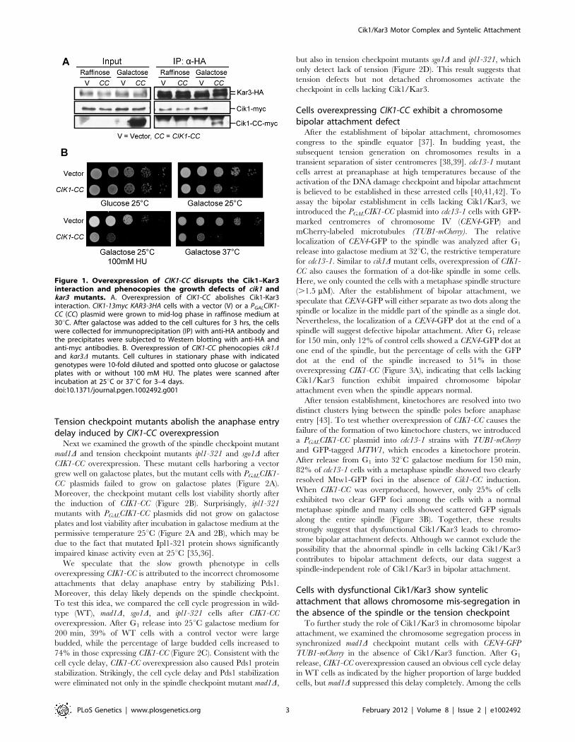
Next we examined the growth of the spindle checkpoint mutant
mad1D and tension checkpoint mutants ipl1-321 and sgo1D after
CIK1-CC overexpression. These mutant cells harboring a vector
grew well on galactose plates, but the mutant cells with PGALCIK1-
CC plasmids failed to grow on galactose plates (Figure 2A).
Moreover, the checkpoint mutant cells lost viability shortly after
the induction of CIK1-CC (Figure 2B). Surprisingly, ipl1-321
mutants with PGALCIK1-CC plasmids did not grow on galactose
plates and lost viability after incubation in galactose medium at the
permissive temperature 25uC (Figure 2A and 2B), which may be
due to the fact that mutated Ipl1-321 protein shows significantly
impaired kinase activity even at 25uC [35,36].
We speculate that the slow growth phenotype in cells
overexpressing CIK1-CC is attributed to the incorrect chromosome
attachments that delay anaphase entry by stabilizing Pds1.
Moreover, this delay likely depends on the spindle checkpoint.
To test this idea, we compared the cell cycle progression in wild-
type (WT), mad1D, sgo1D, and ipl1-321 cells after CIK1-CC
overexpression. After G1 release into 25uC galactose medium for
200 min, 39% of WT cells with a control vector were large
budded, while the percentage of large budded cells increased to
74% in those expressing CIK1-CC (Figure 2C). Consistent with the
cell cycle delay, CIK1-CC overexpression also caused Pds1 protein
stabilization. Strikingly, the cell cycle delay and Pds1 stabilization
were eliminated not only in the spindle checkpoint mutant mad1D,
but also in tension checkpoint mutants sgo1D and ipl1-321, which
only detect lack of tension (Figure 2D). This result suggests that
tension defects but not detached chromosomes activate the
checkpoint in cells lacking Cik1/Kar3.
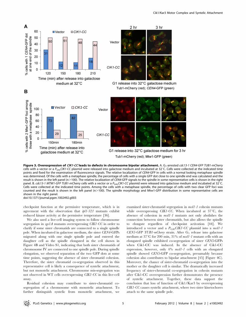
Cells overexpressing CIK1-CC exhibit a chromosomebipolar attachment defect
After the establishment of bipolar attachment, chromosomes
congress to the spindle equator [37]. In budding yeast, the
subsequent tension generation on chromosomes results in a
transient separation of sister centromeres [38,39]. cdc13-1 mutant
cells arrest at preanaphase at high temperatures because of the
activation of the DNA damage checkpoint and bipolar attachment
is believed to be established in these arrested cells [40,41,42]. To
assay the bipolar establishment in cells lacking Cik1/Kar3, we
introduced the PGALCIK1-CC plasmid into cdc13-1 cells with GFP-
marked centromeres of chromosome IV (CEN4-GFP) and
mCherry-labeled microtubules (TUB1-mCherry). The relative
localization of CEN4-GFP to the spindle was analyzed after G1
release into galactose medium at 32uC, the restrictive temperature
for cdc13-1. Similar to cik1D mutant cells, overexpression of CIK1-
CC also causes the formation of a dot-like spindle in some cells.
Here, we only counted the cells with a metaphase spindle structure
(.1.5 mM). After the establishment of bipolar attachment, we
speculate that CEN4-GFP will either separate as two dots along the
spindle or localize in the middle part of the spindle as a single dot.
Nevertheless, the localization of a CEN4-GFP dot at the end of a
spindle will suggest defective bipolar attachment. After G1 release
for 150 min, only 12% of control cells showed a CEN4-GFP dot at
one end of the spindle, but the percentage of cells with the GFP
dot at the end of the spindle increased to 51% in those
overexpressing CIK1-CC (Figure 3A), indicating that cells lacking
Cik1/Kar3 function exhibit impaired chromosome bipolar
attachment even when the spindle appears normal.
After tension establishment, kinetochores are resolved into two
distinct clusters lying between the spindle poles before anaphase
entry [43]. To test whether overexpression of CIK1-CC causes the
failure of the formation of two kinetochore clusters, we introduced
a PGALCIK1-CC plasmid into cdc13-1 strains with TUB1-mCherry
and GFP-tagged MTW1, which encodes a kinetochore protein.
After release from G1 into 32uC galactose medium for 150 min,
82% of cdc13-1 cells with a metaphase spindle showed two clearly
resolved Mtw1-GFP foci in the absence of Cik1-CC induction.
When CIK1-CC was overproduced, however, only 25% of cells
exhibited two clear GFP foci among the cells with a normal
metaphase spindle and many cells showed scattered GFP signals
along the entire spindle (Figure 3B). Together, these results
strongly suggest that dysfunctional Cik1/Kar3 leads to chromo-
some bipolar attachment defects. Although we cannot exclude the
possibility that the abnormal spindle in cells lacking Cik1/Kar3
contributes to bipolar attachment defects, our data suggest a
spindle-independent role of Cik1/Kar3 in bipolar attachment.
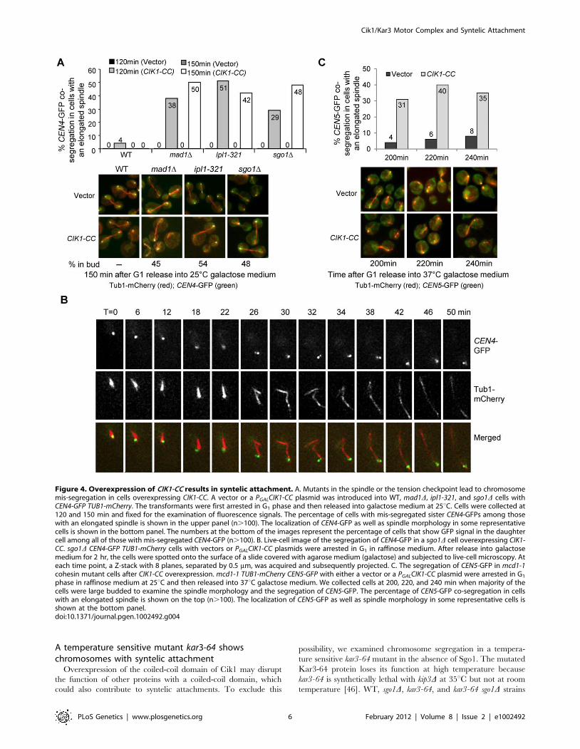
Cells with dysfunctional Cik1/Kar3 show syntelicattachment that allows chromosome mis-segregation inthe absence of the spindle or the tension checkpoint
To further study the role of Cik1/Kar3 in chromosome bipolar
attachment, we examined the chromosome segregation process in
synchronized mad1D checkpoint mutant cells with CEN4-GFP
TUB1-mCherry in the absence of Cik1/Kar3 function. After G1
release, CIK1-CC overexpression caused an obvious cell cycle delay
in WT cells as indicated by the higher proportion of large budded
cells, but mad1D suppressed this delay completely. Among the cells
Figure 1. Overexpression of CIK1-CC disrupts the Cik1–Kar3interaction and phenocopies the growth defects of cik1 andkar3 mutants. A. Overexpression of CIK1-CC abolishes Cik1-Kar3interaction. CIK1-13myc KAR3-3HA cells with a vector (V) or a PGALCIK1-CC (CC) plasmid were grown to mid-log phase in raffinose medium at30uC. After galactose was added to the cell cultures for 3 hrs, the cellswere collected for immunoprecipitation (IP) with anti-HA antibody andthe precipitates were subjected to Western blotting with anti-HA andanti-myc antibodies. B. Overexpression of CIK1-CC phenocopies cik1Dand kar3D mutants. Cell cultures in stationary phase with indicatedgenotypes were 10-fold diluted and spotted onto glucose or galactoseplates with or without 100 mM HU. The plates were scanned afterincubation at 25uC or 37uC for 3–4 days.doi:10.1371/journal.pgen.1002492.g001
Cik1/Kar3 Motor Complex and Syntelic Attachment
PLoS Genetics | www.plosgenetics.org 3 February 2012 | Volume 8 | Issue 2 | e1002492
with an elongated spindle after G1 release for 150 min, more than
40% of mad1D cells showed CEN4-GFP co-segregation when Cik1-
CC was overproduced, where one or two GFP dots were close to
only one of the spindle poles. However, in mad1D cells with a
vector control, no co-segregation of sister chromatids was observed
(Figure 4A), indicating that Cik1-CC overproduction causes a
kinetochore attachment defects.
The co-segregation of chromosome IV in mad1D mutant cells
overexpressing CIK1-CC could be a consequence of syntelic
attachment, monotelic attachment, or chromosome detachment.
For a detached chromosome, both sister chromatids will stay in the
mother cell after spindle elongation. For a chromosome with
monotelic attachment, after anaphase onset the detached
chromatid will stay in the mother cell but the attached one will
move along with the connected spindle poles to either the mother
or the daughter cell. Hence, it is impossible for both sister
chromatids to move to the daughter cell together when a
chromosome is either detached or with monotelic attachment.
For a chromosome with syntelic attachment, however, the sister
chromatids will co-segregate to either the mother or the daughter
cell. Therefore, co-segregation of sister chromatids to the daughter
cell will be an indication of syntelic attachment. We examined the
frequency of sister-CEN4-GFP co-segregation into daughter cells
in mad1D mutants overexpressing CIK1-CC. Mother cells are
usually bigger in size and show a shmoo-like morphology because
a-factor was used for G1 synchronization. Among the mad1D cells
that show CEN4-GFP co-segregation after Cik1-CC induction,
45% of them have the GFP signal in the daughter cell (Figure 4A),
indicating the presence of syntelic attachment. As this number is
close to 50%, the chance of syntelic attachment to either spindle
pole is similar.
If syntelic attachment in cells lacking Cik1/Kar3 function leads
to a cell cycle delay, we expect that this delay depends on the
tension checkpoint, because chromosomes with syntelic attach-
ment are not under tension. Thus, we examined the chromosome
segregation in ipl1-321 and sgo1D cells at 25uC after CIK1-CC
overexpression. Strikingly, more than 40% ipl1-321 and sgo1D cells
with an elongated spindle exhibited co-segregation of sister CEN4-
GFPs after G1 release for 150 min, which is similar to mad1Dcheckpoint mutants. Among them, 54% of ipl1-321 and 48% of
sgo1D cells showed exclusive daughter cell localization of CEN4-
GFP signal. In contrast, no co-segregation was observed in the
mutant cells with a vector control (Figure 4A). Given the fact that
the loss of function of Ipl1 or Sgo1 fails to abolish the cell cycle
arrest in response to detached chromosomes [19,20], this result
further indicates that dysfunctional Cik1/Kar3 induces syntelic
attachment. Since we performed the experiments at 25uC, the
data demonstrate that ipl1-321 mutant cells lose tension
Figure 2. Overexpression of CIK1-CC results in checkpoint-dependent anaphase entry delay. A. Overexpression of CIK1-CC is lethal tomad1D, ipl1-321, and sgo1D mutants. Serial 10-fold dilutions of WT and mutant cells with a vector or a PGALCIK1-CC plasmid were spotted ontoglucose and galactose plates and incubated for 3 days at 25uC. B. mad1D, ipl1-321, and sgo1D mutant cells lose viability after CIK1-CC overexpression.G1-arrested cells with the indicated genotypes were released into galactose medium and incubated at 25uC. Cells were collected at the indicated timepoints and spread onto YPD plates. After overnight growth, the plating efficiency was determined and the percentage of viable cells is shown. C andD. Checkpoint mutants alleviate the Pds1 degradation delay induced by CIK1-CC overexpression. G1-arrested WT, mad1D, ipl1-321, and sgo1D cellscontaining Pds1-18myc as well as a vector or a PGALCIK1-CC plasmid were released into galactose medium and incubated at 25uC. a-factor wasrestored after budding to block the second round of cell cycle. Cells were collected at the indicated time points and protein samples were preparedfor Western blotting. The budding index is shown in C and Pds1 levels are shown in D. Pgk1 protein levels were used as a loading control.doi:10.1371/journal.pgen.1002492.g002
Cik1/Kar3 Motor Complex and Syntelic Attachment
PLoS Genetics | www.plosgenetics.org 4 February 2012 | Volume 8 | Issue 2 | e1002492
checkpoint function at the permissive temperature, which is in
agreement with the observation that ipl1-321 mutants exhibit
reduced kinase activity at the permissive temperature [36].
We also used a live-cell imaging system to follow chromosome
segregation in sgo1D mutants overexpressing CIK1-CC in order to
clarify if some sister chromatids are connected to a single spindle
pole. When incubated in galactose medium, the sister CEN4-GFPs
migrated along with one single spindle pole and entered the
daughter cell as the spindle elongated in the cell shown in
Figure 4B and Video S1, indicating that both sister chromatids of
chromosome IV are connected to one spindle pole. During spindle
elongation, we observed separation of the two GFP dots at some
time points, suggesting the absence of sister chromatid cohesion.
Therefore, the sister chromatid co-segregation observed in this
representative cell is likely a consequence of syntelic attachment,
but not monotelic attachment. Chromosome mis-segregation was
not observed in WT cells overexpressing CIK1-CC in this live-cell
assay.
Residual cohesion may contribute to sister-chromatid co-
segregation of a chromosome with monotelic attachment. To
further distinguish syntelic from monotelic attachment, we
examined sister-chromatid segregation in mcd1-1 cohesin mutants
while overexpressing CIK1-CC. When incubated at 37uC, the
absence of cohesion in mcd1-1 mutants not only abolishes the
connection between sister chromatids, but also allows the spindle
to elongate regardless of checkpoint activation [44]. We
introduced a vector and a PGALCIK1-CC plasmid into a mcd1-1
CEN5-GFP TUB1-mCherry strain. After G1 release into galactose
medium at 37uC for 200 min, 31% of mcd1-1 mutant cells with an
elongated spindle exhibited co-segregation of sister CEN5-GFPs
when Cik1-CC was induced. In the absence of Cik1-CC
expression, however, only 4% mcd1-1 cells with an elongated
spindle showed CEN5-GFP co-segregation, presumably because
cohesion also contributes to bipolar attachment [45] (Figure 4C).
Moreover, the chance of sister-chromatid co-segregation into the
mother or the daughter cell is similar. The dramatically increased
frequency of sister-chromatid co-segregation in cohesin mutants
after Cik1-CC overexpression further demonstrates the presence
of syntelic attachment. Together, these data support the
conclusion that loss of function of Cik1/Kar3 by overexpressing
CIK1-CC causes syntelic attachment, where two sister kinetochores
attach to the same spindle pole.
Figure 3. Overexpression of CIK1-CC leads to defects in chromosome bipolar attachment. A. G1-arrested cdc13-1 CEN4-GFP TUB1-mCherrycells with a vector or a PGALCIK1-CC plasmid were released into galactose medium and incubated at 32uC. Cells were collected at the indicated timepoints and fixed for the examination of fluorescence signals. The relative localization of CEN4-GFP in cells with a normal looking metaphase spindlewas determined. Of the cells with a metaphase spindle, the percentage of cells with a single GFP dot close to one spindle end was calculated and theresult is shown in the left panel (n.100). The relative localization of CEN4-GFP signals to the spindle in some representative cells is shown in the rightpanel. B. cdc13-1 MTW1-GFP TUB1-mCherry cells with a vector or a PGALCIK1-CC plasmid were released into galactose medium and incubated at 32uC.Cells were collected at the indicated time points. Among the cells with a metaphase spindle, the percentage of cells with two clear GFP foci wascounted and the result is shown in the left panel (n.100). The spindle morphology and Mtw1-GFP distribution in some representative cells areshown in the right panel.doi:10.1371/journal.pgen.1002492.g003
Cik1/Kar3 Motor Complex and Syntelic Attachment
PLoS Genetics | www.plosgenetics.org 5 February 2012 | Volume 8 | Issue 2 | e1002492
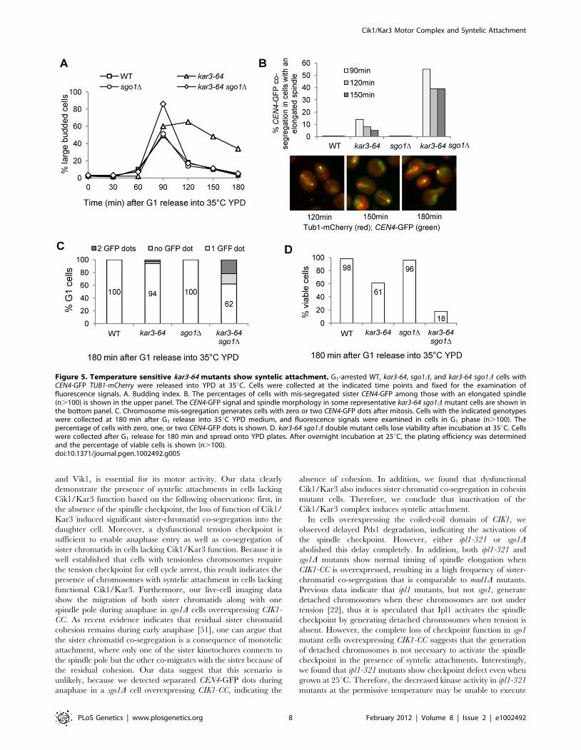
A temperature sensitive mutant kar3-64 showschromosomes with syntelic attachment
Overexpression of the coiled-coil domain of Cik1 may disrupt
the function of other proteins with a coiled-coil domain, which
could also contribute to syntelic attachments. To exclude this
possibility, we examined chromosome segregation in a tempera-
ture sensitive kar3-64 mutant in the absence of Sgo1. The mutated
Kar3-64 protein loses its function at high temperature because
kar3-64 is synthetically lethal with kip3D at 35uC but not at room
temperature [46]. WT, sgo1D, kar3-64, and kar3-64 sgo1D strains
Figure 4. Overexpression of CIK1-CC results in syntelic attachment. A. Mutants in the spindle or the tension checkpoint lead to chromosomemis-segregation in cells overexpressing CIK1-CC. A vector or a PGALCIK1-CC plasmid was introduced into WT, mad1D, ipl1-321, and sgo1D cells withCEN4-GFP TUB1-mCherry. The transformants were first arrested in G1 phase and then released into galactose medium at 25uC. Cells were collected at120 and 150 min and fixed for the examination of fluorescence signals. The percentage of cells with mis-segregated sister CEN4-GFPs among thosewith an elongated spindle is shown in the upper panel (n.100). The localization of CEN4-GFP as well as spindle morphology in some representativecells is shown in the bottom panel. The numbers at the bottom of the images represent the percentage of cells that show GFP signal in the daughtercell among all of those with mis-segregated CEN4-GFP (n.100). B. Live-cell image of the segregation of CEN4-GFP in a sgo1D cell overexpressing CIK1-CC. sgo1D CEN4-GFP TUB1-mCherry cells with vectors or PGALCIK1-CC plasmids were arrested in G1 in raffinose medium. After release into galactosemedium for 2 hr, the cells were spotted onto the surface of a slide covered with agarose medium (galactose) and subjected to live-cell microscopy. Ateach time point, a Z-stack with 8 planes, separated by 0.5 mm, was acquired and subsequently projected. C. The segregation of CEN5-GFP in mcd1-1cohesin mutant cells after CIK1-CC overexpression. mcd1-1 TUB1-mCherry CEN5-GFP with either a vector or a PGALCIK1-CC plasmid were arrested in G1
phase in raffinose medium at 25uC and then released into 37uC galactose medium. We collected cells at 200, 220, and 240 min when majority of thecells were large budded to examine the spindle morphology and the segregation of CEN5-GFP. The percentage of CEN5-GFP co-segregation in cellswith an elongated spindle is shown on the top (n.100). The localization of CEN5-GFP as well as spindle morphology in some representative cells isshown at the bottom panel.doi:10.1371/journal.pgen.1002492.g004
Cik1/Kar3 Motor Complex and Syntelic Attachment
PLoS Genetics | www.plosgenetics.org 6 February 2012 | Volume 8 | Issue 2 | e1002492
with CEN4-GFP TUB1-mCherry were first arrested in G1 phase and
then released into 35uC medium to inactivate Kar3. The
accumulation of large budded cells in kar3-64 mutants indicates
the loss of Kar3 function (Figure 5A). Similar to the cells
overexpressing CIK1-CC, the cell cycle delay in kar3-64 mutant was
abolished by sgo1D. We also found that about 40% of kar3-64
sgo1D double mutant cells with an elongated spindle showed
CEN4-GFP co-segregation after release for 120 and 150 min at
35uC (Figure 5B), which is comparable to sgo1D cells overexpress-
ing CIK1-CC. The majority of kar3-64 sgo1D mutant cells exited
mitosis after release for 180 min. At this time point, 38% of the G1
cells were either absent for CEN4-GFP signal or showed two
CEN4-GFP dots, suggesting the gain or loss of chromosome IV
after mitosis (Figure 5C). It is also possible that some G1 cells have
two CEN4-GFP dots but they are too close to be distinguished by
microscopy. Consistently, only 18% of kar3-64 sgo1D mutants were
viable after G1 release for 180 min, but 95% of WT and sgo1Dcells as well as 61% of kar3-64 cells were viable (Figure 5D). kar3-
64 cells exhibited partial viability loss presumably due to the
inability to recover from mitotic arrest. These results validate the
conclusion that the loss of function of Kar3 causes syntelic
attachment.
The bipolar attachment defects in kar3 mutants or in cells
overexpressing CIK1-CC could be a result of dysfunctional Cik1/
Kar3 or Vik1/Kar3, because overexpression of CIK1-CC also
partially disrupts Vik1-Kar3 interactions (Figure S1). Moreover,
previous observation that vik1D mutant is synthetically lethal with
ipl1-321 indicates a possible role of Vik1 in chromosome
segregation [45]. To test whether dysfunctional Vik1 also
contributes to syntelic attachment, we examined the establishment
of bipolar attachment in cdc13-1 vik1D cells. Like cdc13-1 single
mutant, more than 80% of cdc13-1 vik1D cells showed either
separated CEN4-GFP dots or one GFP dot at the center region of
the spindle after G1 release for 90 min (Figure S2). We further
examined the segregation of sister chromatids in vik1D mad1Ddouble mutant cells and no mis-segregation was observed. We also
crossed vik1D with ipl1-321 and sgo1D. Surprisingly, we obtained
vik1D ipl1-321 and vik1D sgo1D double mutants and these mutants
did not show co-segregation of sister chromatids (Figure S3).
Therefore, vik1D mutants exhibit distinct phenotypes from cik1D.
It is likely that only the Cik1/Kar3 complex is required for the
establishment of chromosome bipolar attachment.
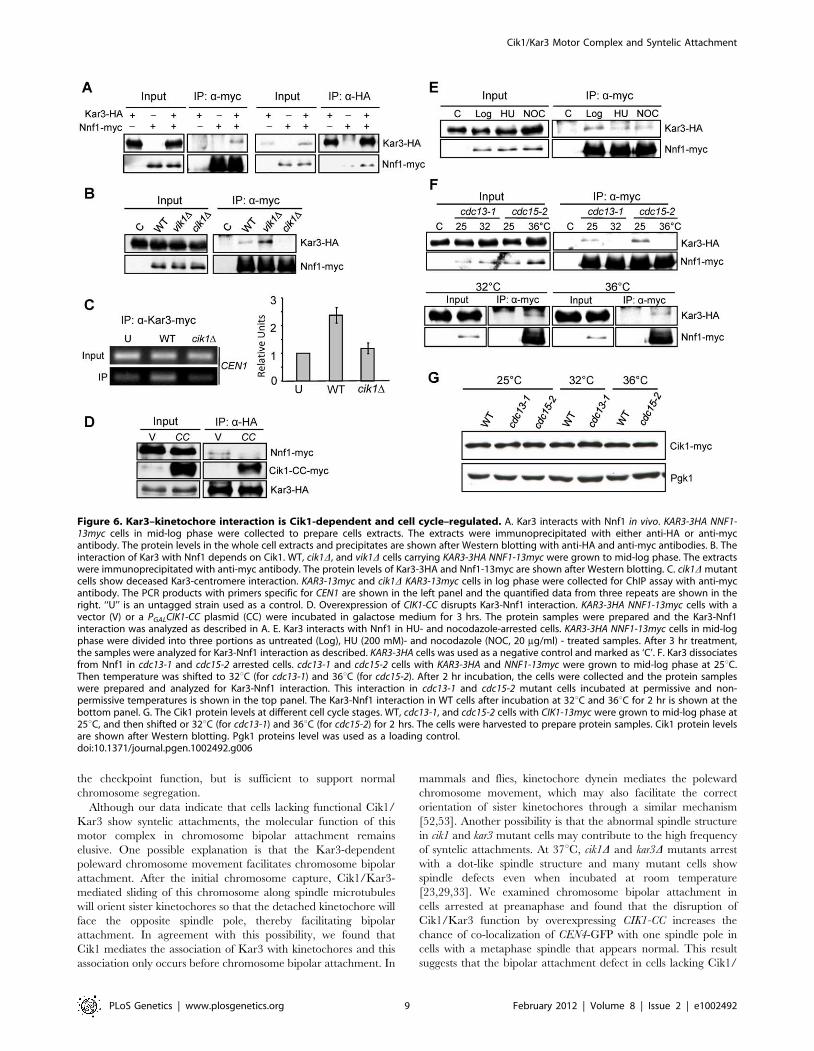
Cik1 mediates Kar3–kinetochore interactionCik1 and Vik1 are the two Kar3 partners in budding yeast and
Cik1/Kar3 localizes along the length of the spindle, and probably
at interpolar microtubule plus ends [25,33]. Kar3 was also found
to bind to the kinetochore to promote its transport along astral
microtubules towards spindle poles [26]. The similar mitotic
defects in cik1 and kar3 mutants suggest that Cik1 likely mediates
the association of Kar3 with kinetochores. Based on the genome-
wide yeast two-hybrid assay, Kar3 was shown to interact with
Nnf1, a kinetochore protein in the MIND complex, [7,47,48].
Using a co-immunoprecipitation (co-IP) approach we found that
Nnf1-myc was able to pull down Kar3-HA, and vice versa,
confirming the Kar3-kinetochore interaction in vivo, although it
remains inconclusive whether this Kar3-Nnf1 interaction is direct
(Figure 6A). Then, we examined Kar3-Nnf1 interaction in the
absence of either Cik1 or Vik1. As shown in Figure 6B, deletion of
CIK1 but not VIK1 abolished this interaction completely,
suggesting that the Kar3-kinetochore interaction is dependent on
Cik1. Interestingly, this interaction was obviously increased in
vik1D mutant cells, presumably because more Kar3 protein is
available for the binding to kinetochores. Consistently, chromatin
immunoprecipitation (ChIP) data shows diminished Kar3-centro-
mere association in cik1D cells (Figure 6C). If Cik1 mediates Kar3-
Nnf1 interaction, the overexpression of the coiled-coil domain of
Cik1 should disrupt this interaction, because CIK1-CC overex-
pression disrupts Cik1-Kar3 interaction (Figure 1A). Indeed,
Kar3-HA failed to pull down Nnf1-myc in cells overexpressing
CIK1-CC (Figure 6D).
Data from the Sorger lab suggest that Kar3 may associate with
detached kinetochores [43]. Moreover, Kar3 is essential for the
lateral sliding of chromosomes towards spindle poles during S-
phase [27]. Therefore, the association of Kar3 with kinetochores
might be cell cycle regulated. To test this possibility, we compared
Kar3-Nnf1 interaction in different cell cycle stages. Kar3
interacted with Nnf1 in both HU- and nocodazole-arrested cells
(Figure 6E), when bipolar attachment has not established yet.
cdc13-1 mutant cells arrest at preanaphase with established bipolar
attachment [40]. Interestingly, the Kar3-Nnf1 interaction was not
detected in cdc13-1 mutant cells after 2 hr incubation at 32uC(Figure 6F). Similarly, Kar3 did not associate with Nnf1 in cdc15-2-
arrested telophase cells. The decreased Kar3-Nnf1 interaction in
cdc13-1 or cdc15-2 mutant cells could be due to the degradation of
Cik1 [49], thus the Cik1 protein levels were examined in WT,
cdc13-1, and cdc15-2 mutant cells incubated at permissive or no-
permissive temperatures. It is clear that the mutant cells exhibit
Cik1 protein levels comparable to WT cells when incubated at
high temperatures (Figure 6G). The results suggest that the Cik1/
Kar3 complex associates with kinetochores before the establish-
ment of bipolar attachment. This association might be essential for
chromosome transport as well as the achievement of bipolar
attachment, but lack of this mechanism will contribute to syntelic
attachment.
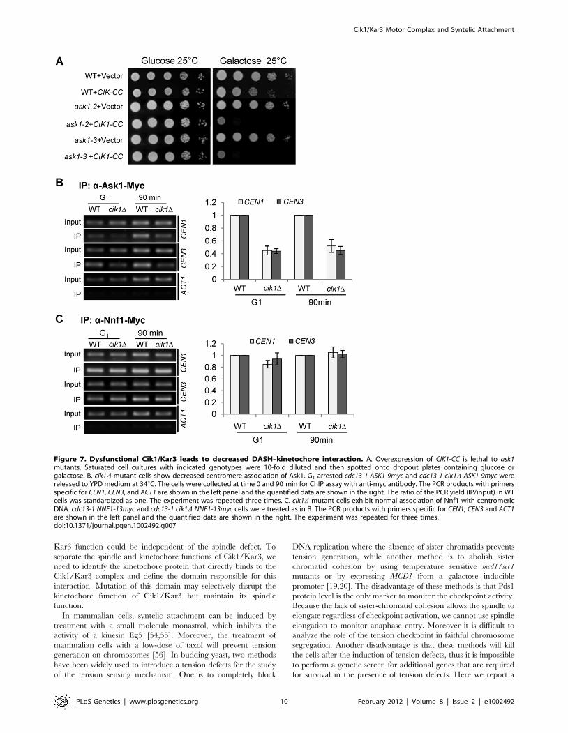
The Cik1/Kar3 complex is required for efficient DASH–centromere binding
The DASH kinetochore complex contains 10 protein subunits
including Dam1 and Ask1. Unlike other kinetochore proteins, the
association of the DASH complex with kinetochores occurs only
after kinetochore-microtubule interaction [2]. Interestingly, kar3Dhas been shown to be synthetically lethal with dam1-1 mutant [50].
We found that overexpression of CIK1-CC caused lethality to ask1-2
and ask1-3 mutants (Figure 7A). One possibility is that the Cik1/
Kar3 complex promotes bipolar attachment by inducing the
DASH-kinetochore interaction. Therefore, we performed ChIP
assays to examine DASH-centromere interaction in synchronous
cell cultures. cdc13-1 mutants were used to arrest cells at
preanaphase, when DASH complexes have already been loaded
onto centromeres [2]. Interestingly, cdc13-1 cik1D cells exhibited
reduced Ask1-centromere interaction in G1 phase as well as after G1
release for 90 min (Figure 7B). In order to determine whether the
Ask1 binding defect is a result of impaired kinetochore integrity, we
also compared the centromere binding of another kinetochore
protein Nnf1. In contrast to Ask1, the association of Nnf1 with
centromeric DNA was similar in synchronous cdc13-1 and cdc13-1
cik1D cells either before or after G1 release (Figure 7C), indicating
that the core kinetochore structure is intact. These results suggest
that the Cik1/Kar3 complex facilitates the association of the DASH
complex with the core kinetochore proteins. It is our future interest
to determine if decreased DASH-kinetochore interaction is the
cause or a consequence of syntelic attachments.
Discussion
In budding yeast, Kar3 is the only kinesin with minus-end-
directed motor activity and the interaction with its partners, Cik1
Cik1/Kar3 Motor Complex and Syntelic Attachment
PLoS Genetics | www.plosgenetics.org 7 February 2012 | Volume 8 | Issue 2 | e1002492
and Vik1, is essential for its motor activity. Our data clearly
demonstrate the presence of syntelic attachments in cells lacking
Cik1/Kar3 function based on the following observations: first, in
the absence of the spindle checkpoint, the loss of function of Cik1/
Kar3 induced significant sister-chromatid co-segregation into the
daughter cell. Moreover, a dysfunctional tension checkpoint is
sufficient to enable anaphase entry as well as co-segregation of
sister chromatids in cells lacking Cik1/Kar3 function. Because it is
well established that cells with tensionless chromosomes require
the tension checkpoint for cell cycle arrest, this result indicates the
presence of chromosomes with syntelic attachment in cells lacking
functional Cik1/Kar3. Furthermore, our live-cell imaging data
show the migration of both sister chromatids along with one
spindle pole during anaphase in sgo1D cells overexpressing CIK1-
CC. As recent evidence indicates that residual sister chromatid
cohesion remains during early anaphase [51], one can argue that
the sister chromatid co-segregation is a consequence of monotelic
attachment, where only one of the sister kinetochores connects to
the spindle pole but the other co-migrates with the sister because of
the residual cohesion. Our data suggest that this scenario is
unlikely, because we detected separated CEN4-GFP dots during
anaphase in a sgo1D cell overexpressing CIK1-CC, indicating the
absence of cohesion. In addition, we found that dysfunctional
Cik1/Kar3 also induces sister chromatid co-segregation in cohesin
mutant cells. Therefore, we conclude that inactivation of the
Cik1/Kar3 complex induces syntelic attachment.
In cells overexpressing the coiled-coil domain of CIK1, we
observed delayed Pds1 degradation, indicating the activation of
the spindle checkpoint. However, either ipl1-321 or sgo1Dabolished this delay completely. In addition, both ipl1-321 and
sgo1D mutants show normal timing of spindle elongation when
CIK1-CC is overexpressed, resulting in a high frequency of sister-
chromatid co-segregation that is comparable to mad1D mutants.
Previous data indicate that ipl1 mutants, but not sgo1, generate
detached chromosomes when these chromosomes are not under
tension [22], thus it is speculated that Ipl1 activates the spindle
checkpoint by generating detached chromosomes when tension is
absent. However, the complete loss of checkpoint function in sgo1
mutant cells overexpressing CIK1-CC suggests that the generation
of detached chromosomes is not necessary to activate the spindle
checkpoint in the presence of syntelic attachments. Interestingly,
we found that ipl1-321 mutants show checkpoint defect even when
grown at 25uC. Therefore, the decreased kinase activity in ipl1-321
mutants at the permissive temperature may be unable to execute
Figure 5. Temperature sensitive kar3-64 mutants show syntelic attachment. G1-arrested WT, kar3-64, sgo1D, and kar3-64 sgo1D cells withCEN4-GFP TUB1-mCherry were released into YPD at 35uC. Cells were collected at the indicated time points and fixed for the examination offluorescence signals. A. Budding index. B. The percentages of cells with mis-segregated sister CEN4-GFP among those with an elongated spindle(n.100) is shown in the upper panel. The CEN4-GFP signal and spindle morphology in some representative kar3-64 sgo1D mutant cells are shown inthe bottom panel. C. Chromosome mis-segregation generates cells with zero or two CEN4-GFP dots after mitosis. Cells with the indicated genotypeswere collected at 180 min after G1 release into 35uC YPD medium, and fluorescence signals were examined in cells in G1 phase (n.100). Thepercentage of cells with zero, one, or two CEN4-GFP dots is shown. D. kar3-64 sgo1D double mutant cells lose viability after incubation at 35uC. Cellswere collected after G1 release for 180 min and spread onto YPD plates. After overnight incubation at 25uC, the plating efficiency was determinedand the percentage of viable cells is shown (n.100).doi:10.1371/journal.pgen.1002492.g005
Cik1/Kar3 Motor Complex and Syntelic Attachment
PLoS Genetics | www.plosgenetics.org 8 February 2012 | Volume 8 | Issue 2 | e1002492
the checkpoint function, but is sufficient to support normal
chromosome segregation.
Although our data indicate that cells lacking functional Cik1/
Kar3 show syntelic attachments, the molecular function of this
motor complex in chromosome bipolar attachment remains
elusive. One possible explanation is that the Kar3-dependent
poleward chromosome movement facilitates chromosome bipolar
attachment. After the initial chromosome capture, Cik1/Kar3-
mediated sliding of this chromosome along spindle microtubules
will orient sister kinetochores so that the detached kinetochore will
face the opposite spindle pole, thereby facilitating bipolar
attachment. In agreement with this possibility, we found that
Cik1 mediates the association of Kar3 with kinetochores and this
association only occurs before chromosome bipolar attachment. In
mammals and flies, kinetochore dynein mediates the poleward
chromosome movement, which may also facilitate the correct
orientation of sister kinetochores through a similar mechanism
[52,53]. Another possibility is that the abnormal spindle structure
in cik1 and kar3 mutant cells may contribute to the high frequency
of syntelic attachments. At 37uC, cik1D and kar3D mutants arrest
with a dot-like spindle structure and many mutant cells show
spindle defects even when incubated at room temperature
[23,29,33]. We examined chromosome bipolar attachment in
cells arrested at preanaphase and found that the disruption of
Cik1/Kar3 function by overexpressing CIK1-CC increases the
chance of co-localization of CEN4-GFP with one spindle pole in
cells with a metaphase spindle that appears normal. This result
suggests that the bipolar attachment defect in cells lacking Cik1/
Figure 6. Kar3–kinetochore interaction is Cik1-dependent and cell cycle–regulated. A. Kar3 interacts with Nnf1 in vivo. KAR3-3HA NNF1-13myc cells in mid-log phase were collected to prepare cells extracts. The extracts were immunoprecipitated with either anti-HA or anti-mycantibody. The protein levels in the whole cell extracts and precipitates are shown after Western blotting with anti-HA and anti-myc antibodies. B. Theinteraction of Kar3 with Nnf1 depends on Cik1. WT, cik1D, and vik1D cells carrying KAR3-3HA NNF1-13myc were grown to mid-log phase. The extractswere immunoprecipitated with anti-myc antibody. The protein levels of Kar3-3HA and Nnf1-13myc are shown after Western blotting. C. cik1D mutantcells show deceased Kar3-centromere interaction. KAR3-13myc and cik1D KAR3-13myc cells in log phase were collected for ChIP assay with anti-mycantibody. The PCR products with primers specific for CEN1 are shown in the left panel and the quantified data from three repeats are shown in theright. ‘‘U’’ is an untagged strain used as a control. D. Overexpression of CIK1-CC disrupts Kar3-Nnf1 interaction. KAR3-3HA NNF1-13myc cells with avector (V) or a PGALCIK1-CC plasmid (CC) were incubated in galactose medium for 3 hrs. The protein samples were prepared and the Kar3-Nnf1interaction was analyzed as described in A. E. Kar3 interacts with Nnf1 in HU- and nocodazole-arrested cells. KAR3-3HA NNF1-13myc cells in mid-logphase were divided into three portions as untreated (Log), HU (200 mM)- and nocodazole (NOC, 20 mg/ml) - treated samples. After 3 hr treatment,the samples were analyzed for Kar3-Nnf1 interaction as described. KAR3-3HA cells was used as a negative control and marked as ‘C’. F. Kar3 dissociatesfrom Nnf1 in cdc13-1 and cdc15-2 arrested cells. cdc13-1 and cdc15-2 cells with KAR3-3HA and NNF1-13myc were grown to mid-log phase at 25uC.Then temperature was shifted to 32uC (for cdc13-1) and 36uC (for cdc15-2). After 2 hr incubation, the cells were collected and the protein sampleswere prepared and analyzed for Kar3-Nnf1 interaction. This interaction in cdc13-1 and cdc15-2 mutant cells incubated at permissive and non-permissive temperatures is shown in the top panel. The Kar3-Nnf1 interaction in WT cells after incubation at 32uC and 36uC for 2 hr is shown at thebottom panel. G. The Cik1 protein levels at different cell cycle stages. WT, cdc13-1, and cdc15-2 cells with CIK1-13myc were grown to mid-log phase at25uC, and then shifted or 32uC (for cdc13-1) and 36uC (for cdc15-2) for 2 hrs. The cells were harvested to prepare protein samples. Cik1 protein levelsare shown after Western blotting. Pgk1 proteins level was used as a loading control.doi:10.1371/journal.pgen.1002492.g006
Cik1/Kar3 Motor Complex and Syntelic Attachment
PLoS Genetics | www.plosgenetics.org 9 February 2012 | Volume 8 | Issue 2 | e1002492
Kar3 function could be independent of the spindle defect. To
separate the spindle and kinetochore functions of Cik1/Kar3, we
need to identify the kinetochore protein that directly binds to the
Cik1/Kar3 complex and define the domain responsible for this
interaction. Mutation of this domain may selectively disrupt the
kinetochore function of Cik1/Kar3 but maintain its spindle
function.
In mammalian cells, syntelic attachment can be induced by
treatment with a small molecule monastrol, which inhibits the
activity of a kinesin Eg5 [54,55]. Moreover, the treatment of
mammalian cells with a low-dose of taxol will prevent tension
generation on chromosomes [56]. In budding yeast, two methods
have been widely used to introduce a tension defects for the study
of the tension sensing mechanism. One is to completely block
DNA replication where the absence of sister chromatids prevents
tension generation, while another method is to abolish sister
chromatid cohesion by using temperature sensitive mcd1/scc1
mutants or by expressing MCD1 from a galactose inducible
promoter [19,20]. The disadvantage of these methods is that Pds1
protein level is the only marker to monitor the checkpoint activity.
Because the lack of sister-chromatid cohesion allows the spindle to
elongate regardless of checkpoint activation, we cannot use spindle
elongation to monitor anaphase entry. Moreover it is difficult to
analyze the role of the tension checkpoint in faithful chromosome
segregation. Another disadvantage is that these methods will kill
the cells after the induction of tension defects, thus it is impossible
to perform a genetic screen for additional genes that are required
for survival in the presence of tension defects. Here we report a
Figure 7. Dysfunctional Cik1/Kar3 leads to decreased DASH–kinetochore interaction. A. Overexpression of CIK1-CC is lethal to ask1mutants. Saturated cell cultures with indicated genotypes were 10-fold diluted and then spotted onto dropout plates containing glucose orgalactose. B. cik1D mutant cells show decreased centromere association of Ask1. G1-arrested cdc13-1 ASK1-9myc and cdc13-1 cik1D ASK1-9myc werereleased to YPD medium at 34uC. The cells were collected at time 0 and 90 min for ChIP assay with anti-myc antibody. The PCR products with primersspecific for CEN1, CEN3, and ACT1 are shown in the left panel and the quantified data are shown in the right. The ratio of the PCR yield (IP/input) in WTcells was standardized as one. The experiment was repeated three times. C. cik1D mutant cells exhibit normal association of Nnf1 with centromericDNA. cdc13-1 NNF1-13myc and cdc13-1 cik1D NNF1-13myc cells were treated as in B. The PCR products with primers specific for CEN1, CEN3 and ACT1are shown in the left panel and the quantified data are shown in the right. The experiment was repeated for three times.doi:10.1371/journal.pgen.1002492.g007
Cik1/Kar3 Motor Complex and Syntelic Attachment
PLoS Genetics | www.plosgenetics.org 10 February 2012 | Volume 8 | Issue 2 | e1002492
new approach to induce syntelic attachments by inactivating the
Cik1/Kar3 motor complex, which prevents tension generation on
chromosomes but maintains intact kinetochore attachment. We
have demonstrated that overexpression of the coiled-coil domain
of Cik1 from a GAL promoter disrupts Cik1-Kar3 interaction,
which allows us to conditionally induce syntelic attachment by
growing cells with PGALCIK1-CC plasmid in galactose medium.
This approach will be a critical tool to study the response to
tension defects in budding yeast.
Materials and Methods
Strains, plasmids, and growth conditionsThe strains used in this study are derivatives of W303 and listed
in Table S1. Gene deletions and epitope tagging were performed
by using a PCR-based protocol [57]. The PGALCIK1-CC plasmid
was constructed by inserting the CIK1 coiled-coil fragment into a
CEN-TRP-GAL-myc vector.
To arrest yeast cells in G1 phase, 5 mg/ml a-factor was added
into mid-log phase cells grown in YPD or in TRP dropout
medium containing 2% raffinose at 25uC for 2.5 hr. G1-arrested
cells were centrifuged and washed twice with water to release into
YPD at 32uC for cdc13-1 arrest or TRP dropout medium
containing 2% galactose at 25uC for CIK1-CC overexpression.
To block the next cell cycle, 15 mg/ml a-factor was added when
majority of the cells were budded. Hydroxyurea was purchased
from ACROS Organics and the final concentration was 100 mM
for HU plates.
Protein techniquesThe yeast protein samples were separated and detected as
described previously [58]. Protein samples were prepared using an
alkaline method and were resolved by 10% SDS- PAGE. Primary
antibodies (anti-myc and anti-HA) were purchased from Covance
(Madison, WI), and anti-Pgk1 antibody was from Molecular
Probes (Eugene, OR). The HRP-conjugated secondary antibody
was purchased from Jackson ImmunoResearch (West Grove, PA).
Fluorescence microcopyCells were collected and fixed with 3.7% formaldehyde for
15 min at room temperature. The cells were washed once with
16PBS (pH7.2) and then resuspended in 16PBS buffer to
examine fluorescence signals with a microscope (Zeiss Axioplan 2).
Co-immunoprecipitation (co-IP) and chromatinimmunoprecipitation (ChIP) assay
Cell cultures were collected and washed once with water. After
being resuspended in RIPA buffer (25 mM Tris PH7.5, 10 mM
EDTA, 150 mM NaCl and 0.05% Tween-20) supplied with
protease inhibitors, cells were homogenized with a bead-beater.
The resulting cell extracts were incubated with primary antibody
overnight at 4uC. The cell extracts were then incubated with
protein-A conjugated agarose beads (Santa Cruz Biotechnology),
which was pre-incubated with BSA at 4uC. After incubation for
1 hr, the beads were collected by centrifugation and washed with
RIPA buffer for three times. Equal volume RIPA and protein
loading buffer were added and the protein samples were boiled for
5 min for Western blot analysis. The ChIP assay was performed as
described previously [59].
Live-cell fluorescence microscopyFor live-cell microscopy, we used a concave glass slide as a
culture chamber, which was filled with 2% agarose dissolved in
galactose medium. The agarose pad was solidified for 5 min at
room temperature before use. Cells were first arrested in G1 phase
in raffinose medium. After release into galactose medium for 2 hr,
1.5 ml concentrated cells were laid on the top of the agarose pad,
which was then sealed with a piece of cover glass. Live-cell
microscopy was carried out on a DeltaVision imaging system
equipped with an environmental chamber (Applied Precision,
Inc.). All live-cell images were acquired at 25uC with a 1006(NA = 1.41) objective lens on an Olympus ix71 microscope. A total
of 8 z-stacks were collected at each time point and each optical
section was 0.5 mm thick. Exposure time for each optical section
was set between 60 and 100 ms and the time-lapse interval was set
at 2 min. Projected images were used for display.
Supporting Information
Figure S1 Overexpression of CIK1-CC decreases Kar3-Vik1
interaction. KAR3-3HA VIK1-13myc cells with a vector or a
PGALCIK1-CC plasmid were grown to mid-log phase in raffinose
medium at 30uC. After 2% galactose was added to the cell cultures
to for 3 hr, the cells were collected for immunoprecipitation assay
with anti-HA antibody and the precipitates were subjected to
Western blotting following SDS-PAGE.
(TIF)
Figure S2 The chromosome bipolar attachment is normal in
vik1D mutant cells. G1-arrested cdc13-1 CEN4-GFP TUB1-mCherry
and vik1D cdc13-1 CEN4-GFP TUB1-mCherry cells were released
into YPD medium at 32uC. Cells were collected at the indicated
time points and fixed for the examination of fluorescence signals.
The relative localization of CEN4-GFP to the metaphase spindle
was determined. The percentage of cells with separated CEN4-
GFP dots or with a CEN4-GFP dot localized at the middle part of
the spindle is shown in A. The spindle morphology and CEN4-
GFP distribution in some representative cells are shown in B.
(TIF)
Figure S3 vik1D cells exhibit normal sister chromatid segrega-
tion in the absence of the spindle or the tension checkpoint. vik1Dsingle and vik1D mad1D, vik1D sgo1D, vik1D ipl1-321 double
mutants with TUB1-mCherry CEN4-GFP were arrested in G1 phase
and then released into YPD medium at 25uC. Cells were collected
for the budding index and the examination of CEN4-GFP
segregation. The budding index is shown in the top panel; the
localization of CEN4-GFP and spindle morphology are shown in
the bottom panel.
(TIF)
Table S1 Strains used in this study.
(DOCX)
Video S1 The co-segregation of sister CEN4-GFP in a sgo1D cell
overexpressing CIK1-CC. sgo1D CEN4-GFP TUB1-mCherry cells
with PGALCIK1-CC plasmids were arrested in G1 phase in raffinose
medium. After release into galactose medium for 2 hr, the cells
were laid onto the surface of an agarose pad (galactose medium)
and subjected to live-cell microscopy. Every 2 min, a Z-stack with
8 planes, separated by 0.5 mm, was acquired and subsequently
projected.
(AVI)
Acknowledgments
We thank Drs. Elledge, Hoyt, and Biggins for yeast strains and plasmid.
We are grateful to Dr. Kerry Maddox, Daniel Richmond, and Kelly
McKnight who read through the manuscript.
Cik1/Kar3 Motor Complex and Syntelic Attachment
PLoS Genetics | www.plosgenetics.org 11 February 2012 | Volume 8 | Issue 2 | e1002492
Author Contributions
Conceived and designed the experiments: FJ HL PL H-GY YW.
Performed the experiments: FJ HL PL. Analyzed the data: FJ HL PL H-
GY YW. Contributed reagents/materials/analysis tools: FJ HL PL H-GY
YW. Wrote the paper: FJ YW.
References
1. Pinsky BA, Biggins S (2005) The spindle checkpoint: tension versus attachment.
Trends Cell Biol 15: 486–493.
2. Li Y, Bachant J, Alcasabas AA, Wang Y, Qin J, et al. (2002) The mitotic spindle
is required for loading of the DASH complex onto the kinetochore. Genes Dev
16: 183–197.
3. Westermann S, Avila-Sakar A, Wang HW, Niederstrasser H, Wong J, et al.
(2005) Formation of a dynamic kinetochore- microtubule interface through
assembly of the Dam1 ring complex. Mol Cell 17: 277–290.
4. Westermann S, Wang HW, Avila-Sakar A, Drubin DG, Nogales E, et al. (2006)
The Dam1 kinetochore ring complex moves processively on depolymerizing
microtubule ends. Nature 440: 565–569.
5. Janke C, Ortiz J, Tanaka TU, Lechner J, Schiebel E (2002) Four new subunits of
the Dam1-Duo1 complex reveal novel functions in sister kinetochore biorienta-
tion. EMBO J 21: 181–193.
6. Westermann S, Drubin DG, Barnes G (2007) Structures and Functions of Yeast
Kinetochore Complexes. Annu Rev Biochem.
7. De Wulf P, McAinsh AD, Sorger PK (2003) Hierarchical assembly of the
budding yeast kinetochore from multiple subcomplexes. Genes Dev 17:
2902–2921.
8. Hoyt MA, Totis L, Roberts BT (1991) S. cerevisiae genes required for cell cycle
arrest in response to loss of microtubule function. Cell 66: 507–517.
9. Li R, Murray AW (1991) Feedback control of mitosis in budding yeast. Cell 66:
519–531.
10. Hardwick KG, Weiss E, Luca FC, Winey M, Murray AW (1996) Activation of
the budding yeast spindle assembly checkpoint without mitotic spindle
disruption. Science 273: 953–956.
11. Wang Y, Burke DJ (1995) Checkpoint genes required to delay cell division in
response to nocodazole respond to impaired kinetochore function in the yeast
Saccharomyces cerevisiae. Mol Cell Biol 15: 6838–6844.
12. Hardwick KG, Johnston RC, Smith DL, Murray AW (2000) MAD3 encodes a
novel component of the spindle checkpoint which interacts with Bub3p, Cdc20p,
and Mad2p. J Cell Biol 148: 871–882.
13. Chen RH (2002) BubR1 is essential for kinetochore localization of other spindle
checkpoint proteins and its phosphorylation requires Mad1. J Cell Biol 158:
487–496.
14. Cohen-Fix O, Peters JM, Kirschner MW, Koshland D (1996) Anaphase
initiation in Saccharomyces cerevisiae is controlled by the APC-dependent
degradation of the anaphase inhibitor Pds1p. Genes Dev 10: 3081–3093.
15. Ciosk R, Zachariae W, Michaelis C, Shevchenko A, Mann M, et al. (1998) An
ESP1/PDS1 complex regulates loss of sister chromatid cohesion at the
metaphase to anaphase transition in yeast. Cell 93: 1067–1076.
16. Uhlmann F, Lottspeich F, Nasmyth K (1999) Sister-chromatid separation at
anaphase onset is promoted by cleavage of the cohesin subunit Scc1. Nature
400: 37–42.
17. Li X, Nicklas RB (1995) Mitotic forces control a cell-cycle checkpoint. Nature
373: 630–632.
18. Keating P, Rachidi N, Tanaka TU, Stark MJ (2009) Ipl1-dependent
phosphorylation of Dam1 is reduced by tension applied on kinetochores. J Cell
Sci 122: 4375–4382.
19. Biggins S, Murray AW (2001) The budding yeast protein kinase Ipl1/Aurora
allows the absence of tension to activate the spindle checkpoint. Genes Dev 15:
3118–3129.
20. Indjeian VB, Stern BM, Murray AW (2005) The centromeric protein Sgo1 is
required to sense lack of tension on mitotic chromosomes. Science 307: 130–133.
21. Tanaka TU, Rachidi N, Janke C, Pereira G, Galova M, et al. (2002) Evidence
that the Ipl1-Sli15 (Aurora kinase-INCENP) complex promotes chromosome bi-
orientation by altering kinetochore-spindle pole connections. Cell 108: 317–329.
22. Pinsky BA, Kung C, Shokat KM, Biggins S (2006) The Ipl1-Aurora protein
kinase activates the spindle checkpoint by creating unattached kinetochores. Nat
Cell Biol 8: 78–83.
23. Meluh PB, Rose MD (1990) KAR3, a kinesin-related gene required for yeast
nuclear fusion. Cell 60: 1029–1041.
24. Endow SA, Kang SJ, Satterwhite LL, Rose MD, Skeen VP, et al. (1994) Yeast
Kar3 is a minus-end microtubule motor protein that destabilizes microtubules
preferentially at the minus ends. EMBO J 13: 2708–2713.
25. Gardner MK, Haase J, Mythreye K, Molk JN, Anderson M, et al. (2008) The
microtubule-based motor Kar3 and plus end-binding protein Bim1 provide
structural support for the anaphase spindle. J Cell Biol 180: 91–100.
26. Tanaka K, Mukae N, Dewar H, van Breugel M, James EK, et al. (2005)
Molecular mechanisms of kinetochore capture by spindle microtubules. Nature
434: 987–994.
27. Tanaka K, Kitamura E, Kitamura Y, Tanaka TU (2007) Molecular mechanisms
of microtubule-dependent kinetochore transport toward spindle poles. J Cell Biol
178: 269–281.
28. Page BD, Snyder M (1992) CIK1: a developmentally regulated spindle pole
body-associated protein important for microtubule functions in Saccharomyces
cerevisiae. Genes Dev 6: 1414–1429.
29. Liu H, Jin F, Liang F, Tian X, Wang Y (2011) The Cik1/Kar3 motor complex is
required for the proper kinetochore-microtubule interaction after stressful DNA
replication. Genetics 187: 397–407.
30. Daniel JA, Keyes BE, Ng YP, Freeman CO, Burke DJ (2006) Diverse functions of
spindle assembly checkpoint genes in Saccharomyces cerevisiae. Genetics 172: 53–65.
31. Tong AH, Lesage G, Bader GD, Ding H, Xu H, et al. (2004) Global mapping of
the yeast genetic interaction network. Science 303: 808–813.
32. Barrett JG, Manning BD, Snyder M (2000) The Kar3p kinesin-related protein
forms a novel heterodimeric structure with its associated protein Cik1p. Mol Biol
Cell 11: 2373–2385.
33. Manning BD, Barrett JG, Wallace JA, Granok H, Snyder M (1999) Differential
regulation of the Kar3p kinesin-related protein by two associated proteins, Cik1p
and Vik1p. J Cell Biol 144: 1219–1233.
34. Sproul LR, Anderson DJ, Mackey AT, Saunders WS, Gilbert SP (2005) Cik1
targets the minus-end kinesin depolymerase kar3 to microtubule plus ends. Curr
Biol 15: 1420–1427.
35. Kotwaliwale CV, Frei SB, Stern BM, Biggins S (2007) A pathway containing the
Ipl1/aurora protein kinase and the spindle midzone protein Ase1 regulates yeast
spindle assembly. Dev Cell 13: 433–445.
36. Makrantoni V, Stark MJ (2009) Efficient chromosome biorientation and the
tension checkpoint in Saccharomyces cerevisiae both require Bir1. Mol Cell Biol 29:
4552–4562.
37. Gardner MK, Bouck DC, Paliulis LV, Meehl JB, O’Toole ET, et al. (2008)
Chromosome congression by Kinesin-5 motor-mediated disassembly of longer
kinetochore microtubules. Cell 135: 894–906.
38. He X, Asthana S, Sorger PK (2000) Transient sister chromatid separation and
elastic deformation of chromosomes during mitosis in budding yeast. Cell 101:
763–775.
39. Goshima G, Yanagida M (2000) Establishing biorientation occurs with
precocious separation of the sister kinetochores, but not the arms, in the early
spindle of budding yeast. Cell 100: 619–633.
40. Bachant J, Alcasabas A, Blat Y, Kleckner N, Elledge SJ (2002) The SUMO-1
isopeptidase Smt4 is linked to centromeric cohesion through SUMO-1
modification of DNA topoisomerase II. Mol Cell 9: 1169–1182.
41. Tang X, Wang Y (2006) Pds1/Esp1-dependent and -independent sister
chromatid separation in mutants defective for protein phosphatase 2A. Proc
Natl Acad Sci U S A 103: 16290–16295.
42. Liang F, Wang Y (2007) DNA damage checkpoints inhibit mitotic exit by two
different mechanisms. Mol Cell Biol 27: 5067–5078.
43. Tytell JD, Sorger PK (2006) Analysis of kinesin motor function at budding yeast
kinetochores. J Cell Biol 172: 861–874.
44. Guacci V, Koshland D, Strunnikov A (1997) A direct link between sister
chromatid cohesion and chromosome condensation revealed through the
analysis of MCD1 in S. cerevisiae. Cell 91: 47–57.
45. Ng TM, Waples WG, Lavoie BD, Biggins S (2009) Pericentromeric sister
chromatid cohesion promotes kinetochore biorientation. Mol Biol Cell 20:
3818–3827.
46. Cottingham FR, Gheber L, Miller DL, Hoyt MA (1999) Novel roles for
Saccharomyces cerevisiae mitotic spindle motors. J Cell Biol 147: 335–350.
47. Newman JR, Wolf E, Kim PS (2000) A computationally directed screen
identifying interacting coiled coils from Saccharomyces cerevisiae. Proc Natl
Acad Sci U S A 97: 13203–13208.
48. Wong J, Nakajima Y, Westermann S, Shang C, Kang JS, et al. (2007) A protein
interaction map of the mitotic spindle. Mol Biol Cell 18: 3800–3809.
49. Benanti JA, Matyskiela ME, Morgan DO, Toczyski DP (2009) Functionally
distinct isoforms of Cik1 are differentially regulated by APC/C-mediated
proteolysis. Mol Cell 33: 581–590.
50. Jones MH, Bachant JB, Castillo AR, Giddings TH, Jr., Winey M (1999) Yeast
Dam1p is required to maintain spindle integrity during mitosis and interacts with
the Mps1p kinase. Mol Biol Cell 10: 2377–2391.
51. Renshaw MJ, Ward JJ, Kanemaki M, Natsume K, Nedelec FJ, et al. (2010)
Condensins promote chromosome recoiling during early anaphase to complete
sister chromatid separation. Dev Cell 19: 232–244.
52. Sharp DJ, Rogers GC, Scholey JM (2000) Cytoplasmic dynein is required for
poleward chromosome movement during mitosis in Drosophila embryos. Nat
Cell Biol 2: 922–930.
53. Bader JR, Vaughan KT (2010) Dynein at the kinetochore: Timing, Interactions
and Functions. Semin Cell Dev Biol 21: 269–275.
54. Mayer TU, Kapoor TM, Haggarty SJ, King RW, Schreiber SL, et al. (1999)
Small molecule inhibitor of mitotic spindle bipolarity identified in a phenotype-
based screen. Science 286: 971–974.
Cik1/Kar3 Motor Complex and Syntelic Attachment
PLoS Genetics | www.plosgenetics.org 12 February 2012 | Volume 8 | Issue 2 | e1002492
55. Kapoor TM, Mayer TU, Coughlin ML, Mitchison TJ (2000) Probing spindle
assembly mechanisms with monastrol, a small molecule inhibitor of the mitotickinesin, Eg5. J Cell Biol 150: 975–988.
56. Famulski JK, Chan GK (2007) Aurora B kinase-dependent recruitment of
hZW10 and hROD to tensionless kinetochores. Curr Biol 17: 2143–2149.57. Longtine MS, McKenzie A, 3rd, Demarini DJ, Shah NG, Wach A, et al. (1998)
Additional modules for versatile and economical PCR-based gene deletion andmodification in Saccharomyces cerevisiae. Yeast 14: 953–961.
58. Liu H, Wang Y (2006) The function and regulation of budding yeast Swe1 in
response to interrupted DNA synthesis. Mol Biol Cell 17: 2746–2756.
59. Liu H, Liang F, Jin F, Wang Y (2008) The coordination of centromere
replication, spindle formation, and kinetochore-microtubule interaction in
budding yeast. PLoS Genet 4: e1000262. doi:10.1371/journal.pgen.1000262.
Cik1/Kar3 Motor Complex and Syntelic Attachment
PLoS Genetics | www.plosgenetics.org 13 February 2012 | Volume 8 | Issue 2 | e1002492


![A General and Adaptive Robust Loss Function · 2019-05-03 · loss ( = 1), Cauchy loss ( = 0), Geman-McClure loss ( = 2), and Welsch loss ( = 1 ). 1 arXiv:1701.03077v10 [cs.CV] 4](https://img.pdfslide.us/doc/110x75/5f8285a03eed9b085a0fd28c/a-general-and-adaptive-robust-loss-function-2019-05-03-loss-1-cauchy-loss.jpg)