Embed Size (px)
Citation preview

Loss of cks1 homeostasis deregulates cell division cycle
Anand Krishnan, S. Asha Nair *, M. Radhakrishna Pillai
Translational Cancer Research Laboratory, Department of Molecular Medicine, Rajiv Gandhi Centre for Biotechnology, Kerala, India
Received: October 17, 2008; Accepted: January 21, 2009
Abstract
Genetic and biochemical studies have provided considerable insight into the multiple functions of cyclin-dependent kinase subunit(cks)1 in cell division cycle. In addition to enhanced substrate targeting by specific ubiquitin ligases SCFskp2 and APC/C, its direct inter-action with proteasome components normalizes multiple cell cycle regulators. Importantly, it also acts as a transcriptional regulator.cks1 overexpression reflects poor prognosis in malignancy thus indicating its possible role in tumour diagnosis and management. Thepresent review compiles the multiple functional roles of cks1 in cell division with specific emphasis on its molecular mechanisms. Itsdocking functions and the possible downstream proteolytic and transcriptional targets are described. The spatial configuration ofcks1–cdk2 complex and the structural organization of cks1–p27–skp2 assembly required for p27 ubiquitination are discussed in detail.In addition to enhanced p27 degradation, the possible other mechanisms which underlie its pathological functions in human cancer pro-gression are also discussed. Though there are apparent gaps in information, the turnover mechanism of cks1 is well addressed andpresents opportunity to exploit the target for disease management.
Keywords: cks1 • ubiquitin ligase • cell cycle • p27 • cancer
J. Cell. Mol. Med. Vol 14, No 1-2, 2010 pp. 154-164
© 2009 The AuthorsJournal compilation © 2010 Foundation for Cellular and Molecular Medicine/Blackwell Publishing Ltd
doi:10.1111/j.1582-4934.2009.00698.x
• Introduction• Functional analyses of cks1• cks1 linking the unlinked• cks1 acts as a transcriptional regulator• cks1 turnover• Human cks1
- Structural basis for docking functions- Targeting function of skp2-p27 interaction- Other targets of human cks1- Mitotic functions of human cks1- Human cks1 and cancer
• Future directions
*Correspondence to: Dr. S. Asha NAIR, Scientist E1, Translational Cancer Research Laboratory, Department of Molecular Medicine, Rajiv Gandhi Centre for Biotechnology,
Trivandrum, Kerala 695 014, India.Tel.: �91 471 2529501Fax: �91 471 2349303E-mail: [email protected]
Cellular Medicine
Introduction
Cyclin-dependent kinase subunit (cks)1* was initially identified in1986 in fission yeast by virtue of its ability to rescue certain tem-perature sensitive mutants of fission yeast cyclin-dependentkinase (cdc2) [1] and further biochemical studies revealed its vitalrole in the regulation of cell division cycle [2–3]. Its human homo-logue was identified in 1987 in complex with cdc2 [4] followed byidentification of its budding yeast homologue in 1989. SubsequentPCR analysis, using cDNA libraries from HeLa cells, probed withdegenerated oligonucleotides designed based on amino acidsequences of fission yeast and budding yeast cks1 homologuesrevealed two identical clones in human beings (cks1 and cks2)which show 81% identity among their amino acid sequences [5].
Both fission yeast and budding yeast homologues show 67%amino acid sequence identity with a single difference being thepresence of additional 16 consecutive glutamine codons in bud-ding yeast cks1 gene [6]. cks1 and cks2 show 53% and 57% iden-tity, respectively, with both budding yeast and fission yeast homo-logues [5] whereas the xenopus homologue of cks1 shows 91%identity with cks2 [7]. Expression analysis shows cks1 abundancethroughout cell division cycle. It forms a complex with both partial(in absence of regulatory cyclins) and complete forms of cdc2active kinase which is maintained even when the cells enter intostationary phase and during abnormal cell division states (S phasearrest) [6–8].

J. Cell. Mol. Med. Vol 14, No 1-2, 2010
155© 2009 The AuthorsJournal compilation © 2010 Foundation for Cellular and Molecular Medicine/Blackwell Publishing Ltd
*To bring more clarity and uniformity, the general names‘cks1’ and ‘cdc2’ is used throughout the manuscript to representall the cks1 and cdc2 homologues, respectively, in differentspecies.
Functional analyses of cks1
The essential functions of cks1 for normal cell division and growthhave been demonstrated through various genetic and biochemicalexperiments in different species. cks1 deletion confers mitoticarrest phenotype that eventually lose viability whereas its overex-pression produced another abnormal phenotype due to delayedmitotic entry [2, 9, 10]. These observations and the demonstrationthat cks1 deleted fission yeast could enter mitosis [9] initially sug-gested the physiological requirement of cks1 exclusive for mitoticexit. However, conditional mutation and functional inactivationexperiments have indicated the requirement of proper functions ofcks1 for both G1/S and G2/M transitions and maintenance of cellviability [6, 11]. Interestingly, consistent with the production ofabnormal mitotic phenotype, cks1 overexpression has inducedabnormal effects in meiosis also (production of two spored asciwhich failed to complete second meiotic nuclear division) under-scoring the pathological functions of cks1 when it is expressed atphysiologically high levels [2, 10]. Thus, it is evident that cks1 func-tions at multiple levels, depending upon the cell division status, fornormal check point transitions, mitotic progression and mitotic exit.Similarly, it is interesting to observe that both deletion and overex-pression of cks1 could confer a similar phenotype (delayed mitoticentry) [6, 7, 11]. The docking functions of cks1 could explain this.Collectively, in addition to the observation that cks1 is associatedwith mitotic cdk, functional analyses of cks1 indicate that, irrespec-tive of species differences, the following is apparent (i ) cks1 isessential to maintain cell viability (ii ) remarkable changes in itsexpression could break the rhythm of cell division cycle. However,the underlying mechanism that breaks the rhythm of cell cycle atconditions of abnormal cks1 kinetics has remained elusive andformed the basis for further research on cks1.
Cell cycle kinetics depends upon changes in the kinase activityof cyclin-dependent kinases [12, 13]. The influence of cks1 on celldivision cycle and its complex formation with mitotic cdks hasthus put forth substantial hint for cks1 having critical roles in reg-ulating the kinase activity of cdks. Substantiating this hypothesis,fission yeast studies demonstrated that only 5% of total cdc2,which is found to be in complex with cks1, is active as a kinase[8]. Similarly, cks1 association was observed in both partial andcompletely active cdc2 kinase complex in budding yeast [6].Depletion of cks1 from xenopus egg interphase extract had a neg-ative effect on cdc2 kinase activity to perform G2/M transition [7].However, as in the case of cks1 depleted/overexpressed functionalphenotypes, quite distinct observations were obvious. In particu-lar, cks1 deleted cells arrest at M phase with high cdc2 kinaseactivity [7, 9, 11]. Furthermore, addition of exogenous cks1 to
interphase extract had remarkably reduced cdc2 kinase activityand prevents mitotic entry [7]. A probable role of cks1 to inacti-vate cdc2 kinase activity has also been observed in fission yeast[9]. Overall, these findings suggest that, though in complex withmitotic cdks, cks1 may not be strictly a positive or negative regu-lator of cdks and could be considered as a cdk modulator at itsphysiological levels. However questions still remain regarding thespecific molecular mechanisms leading to G2 phase arrest whenthere is excess cks1 or how excess of cks1 at interphase reducescdc2 kinase activity. Similarly, how its deletion alters cdc2 kinaseactivity and arrests cells at check point transitions (except in fis-sion yeast: the cells enter mitosis even when cks1 is suppressed),mechanisms that delay the cells from mitotic exit when cks1 isdepleted from mitotic extract or how cks1 deletion from mitoticextract shoot up cdc2 kinase activity. The finding that cks1 doesnot alter cdc2 kinase activity directly [9, 14] had refocused theresearch on molecules that interacts with cks1, apart from cdc2,and the quest is still on to respond to the queries.
cks1: linking the unlinked
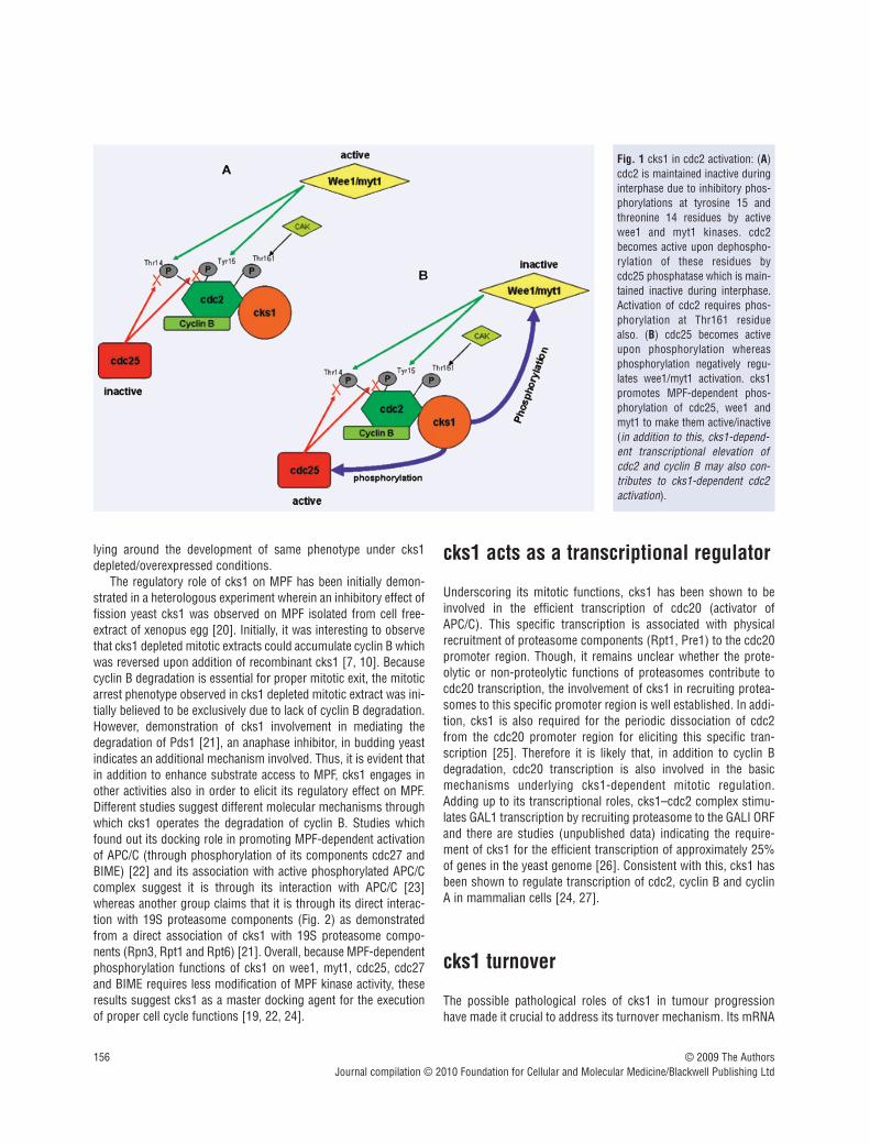
cdc2 is the sole kinase protein which mediates all stages of celldivision in yeast [15] whereas in higher eukaryotes it is primarilyengaged in mediating G2/M phase transition and mitotic progres-sion [13, 16]. Under normal conditions, cdc2 is maintained inac-tive during interphase due to inhibitory phosphorylation at tyro-sine 15 and threonine 14 residues by wee1 and myt1 kinases andcdc2 activation occurs after dephosphorylation of these residuesby cdc25 phosphatase [17]. Once cells have entered mitosis, pro-gression depends upon MPF (M phase-promoting factor[cdc2/cyclin B complex]) dependent activation of a specific ubiq-uitin ligase (E3) APC/C. In turn, exit from mitosis requires APC/Cmediated ubiquitination and subsequent proteasomal degradationof cyclin B [17, 18]. cdc25 becomes active upon phosphorylationwhereas phosphorylation negatively regulates wee1/myt1 kinaseactivity [17].
Molecular mechanistic studies show lack of Tyr 15 dephospho-rylation in cks1-depleted interphase extract and this moleculardefect correlates with the functional phenotype (lack of mitoticentry) observed in cks1 depleted cells [7]. This indeed indicatesthe requirement of cks1 activity for Tyr-15 dephosphorylation andcorrelates well with the role of cks1 in promoting MPF-dependentphosphorylation of cdc25, wee1 and myt1 [19]. Interestingly,addition of excess recombinant cks1 to interphase extract has alsoshowed similar phenotype associated with lack of cdc25 phospho-rylation and Tyr-15 dephosphorylation [7]. This reveals the opti-mum docking theory for cks1 wherein cks1 at normal levels mayserve for the interaction/or acts as a docking factor between MPFand cdc25 or Wee1/Myt1 for eliciting normal cell cycle functions(Fig. 1) and higher concentrations of cks1 may disrupt such aninteraction [7]. Even though further validation of this optimumdocking theory is missing, it could partially resolve the confusions
156 © 2009 The AuthorsJournal compilation © 2010 Foundation for Cellular and Molecular Medicine/Blackwell Publishing Ltd
lying around the development of same phenotype under cks1depleted/overexpressed conditions.
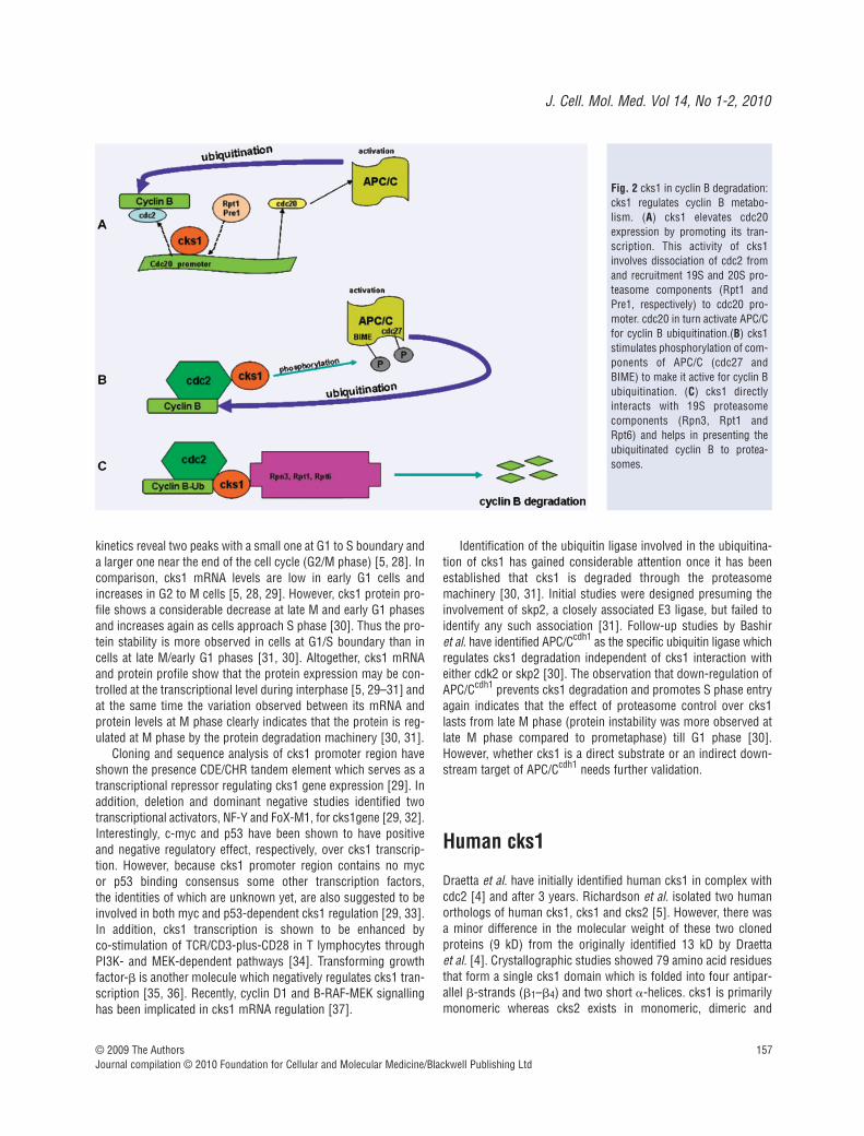
The regulatory role of cks1 on MPF has been initially demon-strated in a heterologous experiment wherein an inhibitory effect offission yeast cks1 was observed on MPF isolated from cell free-extract of xenopus egg [20]. Initially, it was interesting to observethat cks1 depleted mitotic extracts could accumulate cyclin B whichwas reversed upon addition of recombinant cks1 [7, 10]. Becausecyclin B degradation is essential for proper mitotic exit, the mitoticarrest phenotype observed in cks1 depleted mitotic extract was ini-tially believed to be exclusively due to lack of cyclin B degradation.However, demonstration of cks1 involvement in mediating thedegradation of Pds1 [21], an anaphase inhibitor, in budding yeastindicates an additional mechanism involved. Thus, it is evident thatin addition to enhance substrate access to MPF, cks1 engages inother activities also in order to elicit its regulatory effect on MPF.Different studies suggest different molecular mechanisms throughwhich cks1 operates the degradation of cyclin B. Studies whichfound out its docking role in promoting MPF-dependent activationof APC/C (through phosphorylation of its components cdc27 andBIME) [22] and its association with active phosphorylated APC/Ccomplex suggest it is through its interaction with APC/C [23]whereas another group claims that it is through its direct interac-tion with 19S proteasome components (Fig. 2) as demonstratedfrom a direct association of cks1 with 19S proteasome compo-nents (Rpn3, Rpt1 and Rpt6) [21]. Overall, because MPF-dependentphosphorylation functions of cks1 on wee1, myt1, cdc25, cdc27and BIME requires less modification of MPF kinase activity, theseresults suggest cks1 as a master docking agent for the executionof proper cell cycle functions [19, 22, 24].
cks1 acts as a transcriptional regulator
Underscoring its mitotic functions, cks1 has been shown to beinvolved in the efficient transcription of cdc20 (activator ofAPC/C). This specific transcription is associated with physicalrecruitment of proteasome components (Rpt1, Pre1) to the cdc20promoter region. Though, it remains unclear whether the prote-olytic or non-proteolytic functions of proteasomes contribute tocdc20 transcription, the involvement of cks1 in recruiting protea-somes to this specific promoter region is well established. In addi-tion, cks1 is also required for the periodic dissociation of cdc2from the cdc20 promoter region for eliciting this specific tran-scription [25]. Therefore it is likely that, in addition to cyclin Bdegradation, cdc20 transcription is also involved in the basicmechanisms underlying cks1-dependent mitotic regulation.Adding up to its transcriptional roles, cks1–cdc2 complex stimu-lates GAL1 transcription by recruiting proteasome to the GALI ORFand there are studies (unpublished data) indicating the require-ment of cks1 for the efficient transcription of approximately 25%of genes in the yeast genome [26]. Consistent with this, cks1 hasbeen shown to regulate transcription of cdc2, cyclin B and cyclinA in mammalian cells [24, 27].
cks1 turnover
The possible pathological roles of cks1 in tumour progressionhave made it crucial to address its turnover mechanism. Its mRNA
Fig. 1 cks1 in cdc2 activation: (A)cdc2 is maintained inactive duringinterphase due to inhibitory phos-phorylations at tyrosine 15 andthreonine 14 residues by activewee1 and myt1 kinases. cdc2becomes active upon dephospho-rylation of these residues bycdc25 phosphatase which is main-tained inactive during interphase.Activation of cdc2 requires phos-phorylation at Thr161 residuealso. (B) cdc25 becomes activeupon phosphorylation whereasphosphorylation negatively regu-lates wee1/myt1 activation. cks1promotes MPF-dependent phos-phorylation of cdc25, wee1 andmyt1 to make them active/inactive(in addition to this, cks1-depend-ent transcriptional elevation ofcdc2 and cyclin B may also con-tributes to cks1-dependent cdc2activation).
J. Cell. Mol. Med. Vol 14, No 1-2, 2010
157© 2009 The AuthorsJournal compilation © 2010 Foundation for Cellular and Molecular Medicine/Blackwell Publishing Ltd
kinetics reveal two peaks with a small one at G1 to S boundary anda larger one near the end of the cell cycle (G2/M phase) [5, 28]. Incomparison, cks1 mRNA levels are low in early G1 cells andincreases in G2 to M cells [5, 28, 29]. However, cks1 protein pro-file shows a considerable decrease at late M and early G1 phasesand increases again as cells approach S phase [30]. Thus the pro-tein stability is more observed in cells at G1/S boundary than incells at late M/early G1 phases [31, 30]. Altogether, cks1 mRNAand protein profile show that the protein expression may be con-trolled at the transcriptional level during interphase [5, 29–31] andat the same time the variation observed between its mRNA andprotein levels at M phase clearly indicates that the protein is reg-ulated at M phase by the protein degradation machinery [30, 31].
Cloning and sequence analysis of cks1 promoter region haveshown the presence CDE/CHR tandem element which serves as atranscriptional repressor regulating cks1 gene expression [29]. Inaddition, deletion and dominant negative studies identified twotranscriptional activators, NF-Y and FoX-M1, for cks1gene [29, 32].Interestingly, c-myc and p53 have been shown to have positiveand negative regulatory effect, respectively, over cks1 transcrip-tion. However, because cks1 promoter region contains no myc or p53 binding consensus some other transcription factors, the identities of which are unknown yet, are also suggested to beinvolved in both myc and p53-dependent cks1 regulation [29, 33].In addition, cks1 transcription is shown to be enhanced by co-stimulation of TCR/CD3-plus-CD28 in T lymphocytes throughPI3K- and MEK-dependent pathways [34]. Transforming growthfactor-� is another molecule which negatively regulates cks1 tran-scription [35, 36]. Recently, cyclin D1 and B-RAF-MEK signallinghas been implicated in cks1 mRNA regulation [37].
Identification of the ubiquitin ligase involved in the ubiquitina-tion of cks1 has gained considerable attention once it has beenestablished that cks1 is degraded through the proteasomemachinery [30, 31]. Initial studies were designed presuming theinvolvement of skp2, a closely associated E3 ligase, but failed toidentify any such association [31]. Follow-up studies by Bashir et al. have identified APC/Ccdh1 as the specific ubiquitin ligase whichregulates cks1 degradation independent of cks1 interaction witheither cdk2 or skp2 [30]. The observation that down-regulation ofAPC/Ccdh1 prevents cks1 degradation and promotes S phase entryagain indicates that the effect of proteasome control over cks1lasts from late M phase (protein instability was more observed atlate M phase compared to prometaphase) till G1 phase [30].However, whether cks1 is a direct substrate or an indirect down-stream target of APC/Ccdh1 needs further validation.
Human cks1
Draetta et al. have initially identified human cks1 in complex withcdc2 [4] and after 3 years. Richardson et al. isolated two humanorthologs of human cks1, cks1 and cks2 [5]. However, there wasa minor difference in the molecular weight of these two clonedproteins (9 kD) from the originally identified 13 kD by Draetta et al. [4]. Crystallographic studies showed 79 amino acid residuesthat form a single cks1 domain which is folded into four antipar-allel �-strands (�1–�4) and two short �-helices. cks1 is primarilymonomeric whereas cks2 exists in monomeric, dimeric and
Fig. 2 cks1 in cyclin B degradation:cks1 regulates cyclin B metabo-lism. (A) cks1 elevates cdc20expression by promoting its tran-scription. This activity of cks1involves dissociation of cdc2 fromand recruitment 19S and 20S pro-teasome components (Rpt1 andPre1, respectively) to cdc20 pro-moter. cdc20 in turn activate APC/Cfor cyclin B ubiquitination.(B) cks1stimulates phosphorylation of com-ponents of APC/C (cdc27 andBIME) to make it active for cyclin Bubiquitination. (C) cks1 directlyinteracts with 19S proteasomecomponents (Rpn3, Rpt1 andRpt6) and helps in presenting theubiquitinated cyclin B to protea-somes.
158 © 2009 The AuthorsJournal compilation © 2010 Foundation for Cellular and Molecular Medicine/Blackwell Publishing Ltd
hexameric forms. The �-hinge region (residues Glu61 to His65)which forms a �-bend between �3 and �4 in cks1 is sequenceconserved, but is conformationally different in cks2 where it formsan extended conformation promoting a �-strand exchange thatinterlocks two subunits into a dimer. The cks1 single domain foldexposes conserved aromatic side chains from �1, �2 and �3 forpossible kinase interactions whereas the hexameric cks2 structuresequesters these side chains within an internal channel [38].
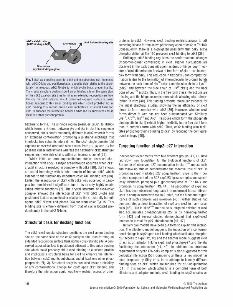
While initial co-immunoprecipitation studies revealed cks1interaction with cdc2, a major breakthrough occurred when cks1crystal structure resolved in complex with cdk2 [39]. cks1 showsstructural homology with N-lobe domain of human cdk2 whichextends to the functionally important cdk2 ATP binding site [38].Earlier, the association of cks1 with cdk2 was observed in xeno-pus but considered insignificant due to its already highly estab-lished mitotic functions [7]. The crystal structure of cks1/cdk2complex showed the interaction of cks1 with cdk2 C-lobe andpositioned it at an opposite side relative to the structurally homol-ogous cdk2 N-lobe and placed 26A far from cdk2 Tyr-15. Thisbinding site is entirely different from that of cyclin located pre-dominantly in the cdk2 N-lobe.
Structural basis for docking functions
The cdk2–cks1 crystal structure positions the cks1 anion bindingsite on the same side of the cdk2 catalytic site, thus forming anextended recognition surface flanking the cdk2 catalytic site. A con-served exposed surface is positioned adjacent to this anion bindingsite which could probably aid in cks1 binding to a second proteinand implicates a structural basis for cks1 to enhance the interac-tion between cdk2 and its substrates and at least one other phos-phoprotein (Fig. 3). Structural analysis predicted lesser probabilityof any conformational change for cdk2 upon cks1 binding andtherefore the interaction could less likely restrict access of other
proteins to cdk2. However, cks1 binding restricts access to cdkactivating kinase for the active phosphorylation of cdk2 at Thr160.Consequently, there is a highlighted possibility that cdk2 activephosphorylation at Thr 160 precedes cks1 binding to cdk2 [39].
Strikingly, cdk2 binding regulates the conformational changes(monomer–dimer conversion) in cks1. Higher fluctuations areobserved in the back bone nitrogen residues of hinge loop (medi-ator of cks1 dimerization in vitro) in free form of cks1 than in com-plex form with cdk2. This reduction in flexibility upon complex for-mation is due to the formation of intermolecular hydrogen bondsbetween the back bone of His60 (cks1) and the side chain of Lys237
(cdk2) and between the side chain of His60(cks1) and the backbone of Leu174 (cdk2). Thus, in the free form these interactions aremissing and the hinge becomes more stable allowing cks1 dimer-ization in vitro [40]. This finding presents molecular evidence forthe initial structural studies showing the in efficiency of cks1dimer to form complex with cdk2 [39]. However, whether cks1forms dimer in vivo has not been substantiated yet. Similarly,Lys11, Arg20, Trp54 and Arg71 (residues which form the phosphatebinding site in cks1) exhibit higher flexibility in the free cks1 formthan in complex form with cdk2. Thus, cdk2 binding also facili-tates phosphoproteins binding to cks1 by reducing the configura-tional entropy [40].
Targeting function of skp2–p27 interaction
Independent experiments from two different groups [41, 42] havelaid down new foundation for the biological functions of cks1.
Spruck et al. observed p27 accumulation in cks1�/– mouse cellsand follow-up studies demonstrated the essential role of cks1 inpromoting skp2 mediated p27 ubiquitination. Skp2 is the F boxprotein component of the SCF skp2 E3 ligase complex and specif-ically identifies phospho-p27 (phosphorylated at Thr-187) andpromotes its ubiquitination [43, 44]. The association of skp2 andcks1 has been observed long back in transformed human fibrob-lasts in complex form with cyclin A-cdk2, but the functional signif-icance of such complex was unknown [45]. Further studies haddemonstrated a direct interaction of skp2 and cks1 in mammaliancells [46]. Like in skp2�/– murine cells, targeted deletion of cks1also accumulates phosphorylated p27 in its non-ubiquitinatedform [42] and several studies demonstrated that skp2–cks1 interaction is vital for p27 ubiquitination [41, 47].
Initially two models have been put forth to explain this interac-tion. The allosteric model suggests the induction of a conforma-tional change in skp2 upon cks1 binding which facilitates phospho-p27 access to skp2 [42, 48] and the adaptor model suggests cks1to act as an adaptor linking skp2 and phospho-p27 and therebyfacilitating the interaction [41, 49]. In addition the structuralrequirement of cyclin E/A-cdk2 complex is also suggested for thisbiological interaction [50]. Combining all these, a new model hasbeen proposed by Sitry et al. in an attempt to identify differentbinding sites on cks1 which are required for p27-ubiquitination[51]. In this model, which actually is a compiled form of bothallosteric and adaptor models, cks1 binding to skp2 creates an
Fig. 3 cks1 as a docking agent for cdk2 and its substrates: cks1 interactswith cdk2 C-lobe and positioned at an opposite side relative to the struc-turally homologous cdk2 N-lobe to which cyclin binds predominantly.The crystal structure positions cks1 anion binding site on the same sideof the cdk2 catalytic site thus forming an extended recognition surfaceflanking the cdk2 catalytic site. A conserved exposed surface is posi-tioned adjacent to this anion binding site which could probably aid incks1 binding to a second protein and implicates a structural basis forcks1 to enhance the interaction between cdk2 and its substrates and atleast one other phosphoprotein.
J. Cell. Mol. Med. Vol 14, No 1-2, 2010
159© 2009 The AuthorsJournal compilation © 2010 Foundation for Cellular and Molecular Medicine/Blackwell Publishing Ltd
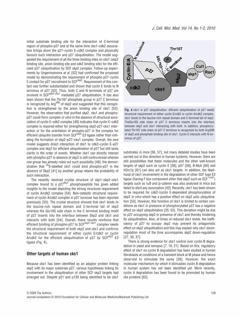
initial substrate binding site for the interaction of C-terminalregion of phospho-p27 and at the same time cks1–cdk2 associa-tion brings down the p27–cyclin E–cdk2 complex and physicallyfavours such interaction and p27 ubiquitination. The model sug-gested the requirement of all the three binding sites on cks1 (skp2binding site, anion binding site and cdk2 binding site) for the effi-cient p27 ubiquitination by SCF skp2 complex. Follow-up experi-ments by Ungermannova et al. [52] had confirmed the proposedmodel by demonstrating the requirement of phospho-p27–cyclinE contact for p27 recruitment to SCFskp2. Requirement of this con-tact was further substantiated and shown that cyclin E binds to Nterminus of p27 [53]. Thus, both C and N terminals of p27 areinvolved in SCFskp2–cks1 mediated p27 ubiquitination. It has alsobeen shown that the Thr187 phosphate group in p27 C terminusis recognized by Arg306 of skp2 and suggested that this recogni-tion is strengthened by the anion binding site of cks1 [52].However, the observation that purified skp2, cks1 and phospho-p27 could form complex in vitro in the absence of structural asso-ciation of cyclin E–cdk2 complex [48] indicates that cyclin E–cdk2complex is required either for strengthening skp2-p27-cks1 inter-action or for the orientation of phospho-p27 in the complex forefficient ubiquitin transfer from SCFskp2 E3 ligase rather than initi-ating the formation of skp2–p27–cks1 complex. Overall, the newmodel suggests direct interaction of cks1 to cdk2–cyclin E–p27complex and skp2 for efficient ubiquitination of p27 but still lacksclarity in the order of events. Whether cks1 can directly interactwith phospho-p27 in absence of skp2 is still controversial whereinone group has already ruled out such possibility [48], the demon-stration that 35S-labelled cks1 could bind phospho-p27 in theabsence of Skp2 [41] by another group retains the probability ofsuch interaction.
The recently resolved crystal structure of skp1–skp2–cks1complex bound to a p27Kip1 phosphopeptide has given addedinsights to the model depicting the strong structural requirementof cyclin A/cdk2 complex [54]. Non-catalytic structural require-ment of cyclin A/cdk2 complex in p27 turnover has been reportedpreviously [55]. The crystal structure showed that cks1 binds tothe leucine-rich repeat domain and C-terminal tail of skp2whereas the Glu185 side chain in the C terminal binding motifof p27 inserts into the interface between Skp2 and cks1 andinteracts with both [54]. Overall, these results reinforce thatefficient binding of phospho-p27 to SCFskp2–cks1 complex needsthe structural requirement of both skp2 and cks1 and confirmsthe structural requirement of either cyclin E/cdk2 or cyclinA/cdk2 for the efficient ubiquitination of p27 by SCFskp2 E3 ligase (Fig. 4).
Other targets of human cks1
Because cks1 has been identified as an adaptor protein linkingskp2 with its major substrate p27, various hypotheses linking itsinvolvement in the ubiquitination of other SCF skp2 targets hademerged out. Despite p21 and p130 being identified to be cks1
substrates in mice [56, 57], not many detailed studies have beencarried out in this direction in human systems. However, there arestill possibilities that these molecules and the other well-knowntargets of skp2 such as cyclin E [58], p57 [59], B-Myb [60] andhOrc1p [61] can also act as cks1 targets. In addition, the likeli-hood of cks1 involvement in the degradation of other SCF type E3ligase (having F box component other that skp2 such as SCF Fwd1)targets such as I�B and �-catenin was also analysed in mice butfailed to elicit any association [42]. Recently, cks1 has been shownto be required for cdk2-/cyclin E-dependent phosphorylation ofskp2 in vitro which has a positive effect on skp2 auto ubiquitina-tion [53]. However, this function of cks1 is limited to certain con-ditions as cks1 in presence of phosphorylated p27 has a negativeeffect on skp2 ubiquitination [35, 53]. This deviation might be dueto p27 occupying skp2 in presence of cks1 and thereby hinderingits ubiquitination. Also, at times of reduced cks1 levels, the ineffi-ciency of p27 to occupy skp2 may prevent its antagonistic effect on skp2 ubiquitination and this may explain why cks1 down-regulation most of the time accompanies skp2 down-regulation[27, 30, 37].
There is strong evidence for cks1 control over cyclin B degra-dation in yeast and xenopus [7, 10, 21]. Based on this, regulatoryeffect of cks1 on cyclin B degradation has been studied in humanfibroblasts at conditions of a transient block at M phase and henceobserved to stimulate the same [28]. However, the exact molecular mechanism by which it stimulates cyclin B degradationin human system has not been identified yet. More recently, cyclin A degradation has been found to be promoted by humancks proteins [62].
Fig. 4 cks1 in p27 ubiquitination: efficient ubiquitination of p27 needsstructural requirement of either cyclin E/cdk2 or cyclin A/cdk2 complex.cks1 binds to the leucine-rich repeat domain and C-terminal tail of skp2.TheGlu185 side chain of p27 C terminus inserts into the interfacebetween skp2 and cks1 interacting with both. In addition, phosphory-lated Thr187 side chain of p27 C terminus is recognized by both Arg306of skp2 and phosphate binding site of cks1. Cyclin E interacts with N ter-minus of p27.

160 © 2009 The AuthorsJournal compilation © 2010 Foundation for Cellular and Molecular Medicine/Blackwell Publishing Ltd
Mitotic functions of human cks1
Although p27 ubiquitination has been identified as the key func-tion of mammalian cks1 its possible mitotic functions has neverbeen ruled out in light of the well-established mitotic roles of itshomologues in lower eukaryotes. Crystal structure analysis showsthat cks1 binds to continuous sequence of residues that forms �helix 5 and loop L14 at the carboxy terminal lobe of the cdk2which are evolutionarily conserved in a subset of human cdks:cdc2, cdk2 and cdk3 [63]. This indicates its possible associationwith human cdc2. A reminder at this point is mandatory that cks1was initially identified in complex with mitotic cdc2 in HeLa cells[4]. In addition, cks1 was found to be present in cdc2-cyclin Bimmunoprecipitates from human fibroblasts [45].
Determining its importance in mitosis, an interesting study byHixon et al. has demonstrated the development of a polyploidy pheno-type in human fibroblasts containing mutated form of p53. p53 muta-tion in such cell type has up-regulated cks1 expression which in turnenhanced cyclin B degradation. This ultimately has led the cells escapea transient mitotic delay induced by colcemide and resulted in poly-ploidy. The results were also confirmed in cks1 overexpressed mousemyoblast cells. This indicates the pathological effect of cks1 overex-pression during spindle cell cycle check point signals [28]. However,the results are quite contradictory to the finding wherein cks1 knock-out cells have been observed to induce polyploidy [24, 42].
The regulatory role of human cks1 on MPF has also beendemonstrated indirectly [28]. cks1 kinetics in human fibroblastsshows peak levels at the onset of cyclin B degradation confirmingits regulatory role on cyclin B degradation and, in addition, a cellcycle dependent association of cks1 and cdc2 was observed at 40-to 56-hr period following cell cycle entry in fibroblasts. Inductionof a transient spindle cell cycle arrest by colcemide resulted in65% decrease in the amount of such association indicating therequirement of this complex for mitotic progression. Down- regulation of cdc2 kinase activity and induction of G2/M arrestobserved in cks1 depleted lung and breast cancer cells furthersubstantiate the possibility of mitotic roles of human cks1 [27,64]. More recently, cks1 and cks2 double knock out MEF cellshave been shown to be arrested at G2 phase accompanied by lowtranscript levels of cyclin A, B and cdc2 leading to polyploidy [24].This finding may explain the molecular basis for previouslyobserved polyploidy phenotype in cks1 knock out cells [24, 42].
Human cks1 and cancer
The importance of cks1 in cancer pathology is reflected from itsoverexpression in tumours that have poor prognosis. cks1 over-expression has been observed in prostate cancer [65] and com-bined overexpression of cks1 and skp2 has been observed inbreast, oral, urothelial, colon, gastric and lung cancers [66–71].cks1 expression has been observed to be inversely correlated withp27 levels in some cancer biopsies [66, 67, 69, 70] whereas biopsies from other few cancer types showed no such correlation
[68, 71]. This indicates that different mechanisms may be involvedin cks1 mediated tumour initiation and progression.
Following the identification of cks1 as a growth stimulatoryagent, in particular its regulatory role at G1/S transition, researchershave been keen in looking into its involvement in various small mol-ecules’/drugs’ pharmacology. Retinoic acid [72], oncostatin M [73]and fluoxetine [74] are some candidates identified that mediate itsaction via cks1 pathway either directly or indirectly, thus exhibitingtheir potential as anticancer agents. LY294002 and UO126 are theother potential candidates which inhibit cks1 at the transcriptionallevel through inhibition of PI3K and MEK signalling, respectively[34]. Further attempts are ongoing to look for new small moleculeswith potential to disrupt cks1–skp2 interaction.
Future directions
Basic research on cks1 for the last two decades has revealed itscritical importance in cell cycle regulation. Findings in yeast andxenopus systems have established multiple functional roles forcks1 with most of them pointing to its specific roles in mitoticentry, progression and exit. In contrast, except for a few studies,most of the studies in human cell lines and tissues establish itsspecific role in p27 degradation, an essential event associated withG1/S transition. However, its mRNA and protein kinetics as well asknockdown and overexpression studies have clearly suggested itspossible mitotic roles in human cells also. Although most of thecks1 overexpressed human tumours negatively correlate with p27levels there are few cancer types which show no such correlationbut still have poor prognosis [68, 71]. These p27-independentpathological roles of cks1 in human tumours might possibly befrom its uncontrolled mitotic functions. Development of polyploidyupon cks1 over activity at mitotic stop signals [28] and the poly-ploidy phenotype observed in cks1 and cks2 double knock out cellsstrongly supports this hypothesis [24]. However, in spite of identi-fication of its regulatory roles on cyclin A, B and cdc2 turnover [24, 27, 28, 62] and cdc2 kinase activity [64], its implications incancer progression has not been evaluated in clinical samples.
Another interesting question to be addressed is whether cks1acts as a tumour initiator or enhances the tumour progression byacting as secondary tumour triggering agent for some primary dis-ease cause. Crystal structure studies proposed the inefficiency ofcks1 dimer conformation to form complex with cdk2 [39]. Becausep27 ubiquitination requires cdk2–cks1 assembly and also protea-some-dependent cks1 turnover mainly occurs at M phase [30, 31],its possible dimer formation at G1/S transition phase under in vivoconditions may serve for its self-regulation in controlling p27 degra-dation. This indeed indicates that prolonged monomer state of themolecule may stimulate abnormal cell division due to uncontrolledp27 degradation. Therefore, mutational analysis and conformationalstudies on cks1 in tumour biopsies may provide clarity about itspossible primary role in tumour initiation. However, there are multi-ple reasons to believe its role as a secondary tumour triggeringagent. Its link with p53 [28] indicates that it may act downstream of
J. Cell. Mol. Med. Vol 14, No 1-2, 2010
161© 2009 The AuthorsJournal compilation © 2010 Foundation for Cellular and Molecular Medicine/Blackwell Publishing Ltd
p53 mutation, but no studies have so far been done to check out thispossibility in human tumour samples. In addition, cks1 involvementin fibroblast growth factor receptor kinase [75] and myc signallingwarrants an assessment of its implications in cancer progression.Whether cks1 overexpression in tumours is due to abnormalities inthe specific ubiquitin ligase for cks1 or defects in the proteasomecomponents need to be assessed. Even if APC/Ccdh1 is proposed tobe the specific ubiquitin ligase responsible for cks1 ubiquitination[30], its inefficiency in promoting cks1 ubiquitination in vitro sug-gests the involvement of additional molecules in this pathway.Therefore, confirmatory evidence of the specific ubiquitin ligase andthe accessory molecules involved need to be known in order to
analyse whether cks1 overexpression in tumours is secondary toabnormalities in the proteasome machinery.
The molecular biology of cks1 at G1/S and G2/M phases differand this indicates that cks1 has distinct and specific roles at dif-ferent time-points of cell division cycle. The efficiency of cks1 tophosphorylate cdc25, wee1 and myt1 and at the same time itsinability to phosphorylate the same molecules at times/conditionswhen it promotes cdc27 and BIME phosphorylation [22] strength-ened this hypothesis. Induction of hyper degradation of p27, p21,p130, cyclin B and Pds1 by cks1 when overexpressed is critical interms of its role in tumour progression as all these functionaldefects trigger hyper/abnormal cell division from different points
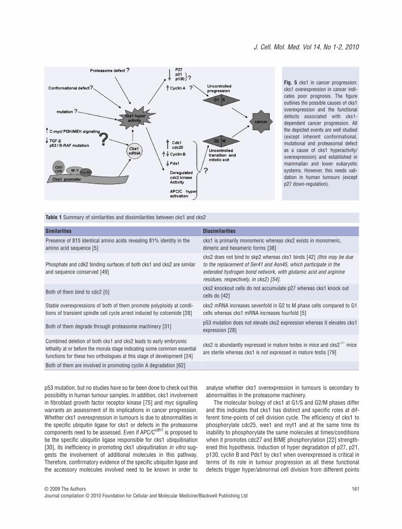
Fig. 5 cks1 in cancer progression:cks1 overexpression in cancer indi-cates poor prognosis. The figureoutlines the possible causes of cks1overexpression and the functionaldefects associated with cks1-dependent cancer progression. Allthe depicted events are well studied(except inherent conformational,mutational and proteasomal defectas a cause of cks1 hyperactivity/overexpression) and established inmammalian and lower eukaryoticsystems. However, this needs vali-dation in human tumours (exceptp27 down-regulation).
Similarities Dissimilarities
Presence of 815 identical amino acids revealing 81% identity in theamino acid sequence [5]
cks1 is primarily monomeric whereas cks2 exists in monomeric,dimeric and hexameric forms [38]
Phosphate and cdk2 binding surfaces of both cks1 and cks2 are similarand sequence conserved [49]
cks2 does not bind to skp2 whereas cks1 binds [42] (this may be dueto the replacement of Ser41 and Asn45, which participate in theextended hydrogen bond network, with glutamic acid and arginineresidues, respectively, in cks2) [54].
Both of them bind to cdc2 [5]cks2 knockout cells do not accumulate p27 whereas cks1 knock outcells do [42]
Stable overexpressions of both of them promote polyploidy at condi-tions of transient spindle cell cycle arrest induced by colcemide [28]
cks2 mRNA increases sevenfold in G2 to M phase cells compared to G1cells whereas cks1 mRNA increases fourfold [5]
Both of them degrade through proteasome machinery [31]p53 mutation does not elevate cks2 expression whereas it elevates cks1expression [28]
Combined deletion of both cks1 and cks2 leads to early embryoniclethality at or before the morula stage indicating some common essentialfunctions for these two orthologues at this stage of development [24]
cks2 is abundantly expressed in mature testes in mice and cks2–/– miceare sterile whereas cks1 is not expressed in mature testis [79]
Both of them are involved in promoting cyclin A degradation [62]
Table 1 Summary of similarities and dissimilarities between cks1 and cks2

162 © 2009 The AuthorsJournal compilation © 2010 Foundation for Cellular and Molecular Medicine/Blackwell Publishing Ltd
References
1. Hayles J, Beach DH, Durkacz B, et al.The fission yeast cell cycle control genecdc2�; isolation of a sequence sucl� thatsuppresses cdc2 mutant function. MolGen Genet. 1986; 202: 291–3.
2. Hayles J, Aves S, Nurse P. sucl� is anessential gene involved in both the cellcycle and growth in fission yeast. EMBO J.1986; 5: 3373–9.
3. Hindley J, Phear G, Stein M, et al. Sucl�encodes a predicted 13-kilodalton proteinthat is essential for cell viability anddirectly involved in the division cycle ofSchizosaccharomyces pombe. Mol CellBiol. 1987; 7: 504–11.
4. Draetta G, Brizuela L, Potashkin J, et al.Identification of p34 and p13, humanhomologs of the cell cycle regulators offission yeast encoded by cdc2� and suc1� . Cell. 1987; 50: 319–25.
5. Richardson HE, Stueland CS, Thomas J,et al. Human cDNAs encoding homologsof the small p34cdc28/cdc2-associated protein of Saccharomyces cerevisiae andSchizosac charomyces pombe. Genes Dev.1990; 4: 1332–44.
6. Hadwiger JA, Wittenberg C, MendenhallMD, et al. The Saccharomyces cere-visiae CKS1 gene, a homolog of theSchizosaccharomyces pombe sucl� gene,encodes a subunit of the cdc28 protein
kinase complex. Mol Cell Biol. 1989; 9:2034–41.
7. Patra D, Dunphy WG. Xe-p9, a XenopusSuc1/Cks homolog, has multiple essentialroles in cell cycle control. Genes Dev.1996; 10: 1503–15.
8. Brizuela L, Draetta G, Beach D. pl3suclacts in the fission yeast cell division cycleas a component of the p34cdc2 proteinkinase. EMBO J. 1987; 6: 3507–14.
9. Moreno S, Hayles J, Nurse P. Regulationof p34cdc2 protein kinase during mitosis.Cell. 1989; 58: 361–72.
10. Basi G, Draetta G. p13suc1 ofSchizosaccharomyces pombe regulatestwo distinct forms of the mitotic cdc2kinase. Mol Cell Biol. 1995; 15: 2028–36.
11. Tang Y, Reed SI. The Cdk-associated pro-tein Cks1 functions both in G1 and G2 inSaccharomyces cerevisiae. Genes Dev.1993; 7: 822–32.
12. Sherr CJ, Roberts JM. CDK inhibitors:positive and negative regulators of G1-phase progression. Genes Dev. 1999; 13:1501–12.
13. Senderowics AM, Sausville EA.Preclinical and clinical development ofcyclin dependent kinase modulators. J Natl Cancer Inst. 2000; 92: 376–87.
14. Arion D, Meijer L, Brizuela L, et al.cdc2 is a component of the M-phase-
specific histone H1 kinase: evidence foridentity with MPF. Cell. 1988; 55: 371–8.
15. Mendenhall MD, Hodge AE. Regulation ofCdc28 cyclin-dependent protein kinaseactivity during the cell cycle of the yeastSaccharomyces cerevisiae. Microbio MolBio Re. 1998; 62: 1191–243.
16. Murray AW. Cyclin-dependent kinases:regulators of the cell cycle and more.Chem Biol. 1994; 4: 191–5.
17. Smits VAJ, Medema RH. Checking out theG2/M transition. Biochim Biophys Acta.2001; 1519: 1–12.
18. Peters JM. The anaphase-promoting com-plex: proteolysis in mitosis and beyond.Mol Cell. 2002; 9: 931–43.
19. Patra D, Wang SX, Kumagai A, et al.The xenopus suc1/cks protein promotesthe phosphorylation of G2/M regulators.J Biol Chem. 1999; 274: 36839–42.
20. Dunphy WG, Brizuela L, Beach D, et al.The Xenopus cdc2 protein is a componentof MPF, a cytoplasmic regulator of mitosis.Cell. 1988; 54: 423–31.
21. Kaiser P, Moncollin V, Clarke DJ, et al.Cyclin-dependent kinase and Cks/Suc1interact with the proteasome in yeast tocontrol proteolysis of M-phase targets.Genes Dev. 1999; 13: 1190–202.
22. Patra D, Dunphy WG. Xe-p9, a XenopusSuc1/Cks protein, is essential for the
of the cell cycle. In addition, considering the transcriptional roles ofcks1 on cdc2, cyclin B, cyclin A and cdc20 it is likely that cks1overexpressed tumours progress through unregulated expressionof these proteins. Therefore, it becomes critical in analysing thelevels of all these proteins in cks1 overexpressed tumours toreveal the exact pathological behaviour of cks1 beyond p27 degra-dation (Fig. 5). The molecular basis of cks1-dependent cyclin Bdegradation in human system is another area of research interest.
Even though few structural and functional dissimilarities pre-vail, human orthologues cks1 and cks2 are 81% identical [5]. Theobserved important similarities and dissimilarities between thesetwo orthologues are presented in a tabular format (Table 1). Thesefindings indicate some critical biological functions for cks2 also inhuman system independent of cks1 functions. A mutual function-ing scenario for a specific biological effect also cannot be ruledout, in particular, at mitotic phase of the cell cycle. Substantiatingits mitotic roles, cks2 has been shown to directly interact with thegenes and promoters of cyclin B and cdc2 [24] to enhance theirtranscription. Therefore, future studies with a closer look on to thebasic biology of cks2 should also be done in parallel to cks1 insimilar systems in order to clearly distinguish the biology of thesetwo orthologues to have better understanding of their regulatoryroles in mammalian cell division and cancer pathology.
Does cks1 inhibition alone kill cancer cells? cks1 knockout micehave been shown to be viable with small sized phenotypes [42, 76].In addition, cks1 null cells retain viability with a slow proliferationrate [42, 77]. Though cks1 disruption up-regulates p27 and dis-rupts G2/M phase transition, this may not be ultimate for the induc-tion of apoptosis. Interestingly, combined knockdown of both cks1and cks2 conferred lethality in mouse embryo and induced apopto-sis in HeLa cells [24]. More recently, an independent role of cks2to protect cells from apoptosis has been demonstrated [65]. Thisindeed indicates that independent targeting of either cks1 or cks2may be utilized in repairing the disrupted molecular mechanismassociated with cell proliferation or apoptosis whereas combinedtargeting can be adopted to drive cytotoxicity in tumour cells.Interestingly, cks1 has now been presented as a unique model tostudy the molecular mechanism of poly (Q) aggregation associatedwith polyglutamine deposition diseases thus extending its biologi-cal importance beyond the horizon of cancer [78].
Acknowledgment
We thank the Council of Scientific and Industrial Research (CSIR) for supporting with a Senior Research Fellowship.
J. Cell. Mol. Med. Vol 14, No 1-2, 2010
163© 2009 The AuthorsJournal compilation © 2010 Foundation for Cellular and Molecular Medicine/Blackwell Publishing Ltd
Cdc2-dependent phosphorylation of theanaphase promoting complex at mitosis.Genes Dev. 1998; 12: 2549–59.
23. Sudakin V, Shteinberg M, Ganoth D, et al. Binding of activated cyclosome top13(suc1). Use for affinity purification. J Biol Chem. 1997; 272: 18051–9.
24. Martinsson-Ahlzen HS, Liberal V,Grunenfelder B, et al. Cyclin-dependentkinase-associated proteins Cks1 and Cks2are essential during early embryogenesisand for cell cycle progression in somaticcells. Mol Cell Biol. 2008; 28: 5698–709.
25. Morris MC, Kaiser P, Rudyak S, et al.Cks1-dependent proteasome recruitmentand activation of CDC20 transcription inbudding yeast. Nature. 2003; 423:1009–13.
26. Yu VPCC, Baskerville C, Grunenfelder B,et al. A kinase-independent function ofcks1 and cdk1 in regulation of transcrip-tion. Mol Cell. 2005; 17: 145–51.
27. Westbrook L, Manuvakhova M, Kern FG,et al. Cks1 regulates cdk1 expression: anovel role during mitotic entry in breastcancer cells. Cancer Res. 2007; 67:11393–401.
28. Hixon ML, Flores AI, Wagner MW, et al.Ectopic expression of cdc2/cdc28 kinasesubunit Homo sapiens 1 uncouples cyclinB metabolism from the mitotic spindle cellcycle checkpoint. Mol Cell Biol. 1998; 18:6224–37.
29. Rother K, Li YY, Tschop K, et al.Expression of cyclin-dependent kinasesubunit 1 (Cks1) is regulated during thecell cycle by a CDE/CHR tandem elementand is down regulated by p53 but not byp63 or p73. Cell Cycle. 2007; 6: 853–62.
30. Bashir T, Dorrello NV, Amador V, et al.Control of the SCF(Skp2-Cks1) ubiquitinligase by the APC/C(Cdh1) ubiquitin ligase.Nature. 2004; 428: 190–3.
31. Hattori T, Kitagawa K, Uchida C, et al.Cks1 is degraded via the ubiquitin-protea-some pathway in a cell cycle-dependentmanner. Genes Cells. 2003; 8: 889–96.
32. Wang IC, Chen YJ, Hughes D, et al.Forkhead Box M1 regulates the transcrip-tional network of genes essential formitotic progression and genes encodingthe SCF (Skp2-Cks1) ubiquitin ligase. MolCell Biol. 2005; 25: 10875–94.
33. Keller UB, Old JB, Dorsey FC, et al. Myctargets cks1 to provoke the suppression ofp27Kip1, proliferation and lymphomagen-esis. EMBO J. 2007; 26: 2562–74.
34. Appleman LJ, Chernova I, Li L, et al.CD28 costimulation mediates transcriptionof skp2 and cks1, the substrate recogni-
tion components of SCFSkp2 ubiquitin lig-ase that leads p27kip1 to degradation. CellCycle. 2006; 5: 2123–9.
35. Wang W, Ungermannova D, Jin J, et al.Negative regulation of SCFSkp2 ubiquitinligase by TGF beta signaling. Oncogene.2004; 23: 1064–75.
36. Simon KE, Cha HH, Firestone GL.Transforming growth factor � down-regu-lation of ckshs1 transcripts in growth-inhibited epithelial cells. Cell Growth Differ.1995; 6: 1261–69.
37. Bhatt KV, Hu R, Spofford LS, et al. MutantB-RAF signaling and cyclin D1 regulateCks1/S-phase kinase-associated protein 2-mediated degradation of p27Kip1 inhuman melanoma cells. Oncogene. 2007;26: 1056–66.
38. Arvai AS, Bourne Y, Hickey MJ, et al.Crystal structure of the human cell cycleprotein CksHs1: single domain fold withsimilarity to kinase N-lobe domain. J Mol Biol. 1995; 249: 835–42.
39. Bourne Y, Watson MH, Hickey MJ, et al.Crystal structure and mutational analysisof the human cdk2 kinase complex withcell cycle–regulatory protein ckshs1. Cell.1996; 84: 863–74.
40. Seeliger MA, Spichty M, Kelly SE, et al.Role of conformational heterogeneity indomain swapping and adapter function ofthe cks proteins. J Biol Chem. 2005; 280:30448–59.
41. Ganoth D, Bornstein G, Ko TK, et al.The cell-cycle regulatory protein Cks1 isrequired for SCF(Skp2)-mediated ubiqui-tinylation of p27. Nat Cell Biol. 2001; 3:321–4.
42. Spruck C, Strohmaier H, Watson M, et al.A cdk-independent function of mammaliancks1: targeting of SCFskp2 to the cdkinhibitor p27Kip1. Mol Cell. 2001; 7:639–50.
43. Tsvetkov LM, Yeh KH, Lee SJ, et al.p27(kip1) ubiquitination and degradationis regulated by the SCF(skp2) complexthrough phosphorylated Thr187 in p27.Curr Biol. 1999; 9: 661–4.
44. Carrano AC, Eytan E, Hershko A, et al.Skp2 is required for ubiquitin-mediateddegradation of the cdk inhibitor p27. NatCell Biol. 1999; 4: 193–9.
45. Zhang H, Kobayashi R, Galaktionov K, et al. p19 skpl and p45 skp2 are essentialelements of the cyclin A-cdk2 S phasekinase. Cell. 1995; 82: 915–25.
46. Mongay L, Plaza S, Vigorito E, et al.Association of the cell cycle regulatoryproteins p45skp2 and ckshs1. Functionaleffect on cdk2 complex formation and
kinase activity. J Biol Chem. 2001; 276:25030–6.
47. Wang W, Ungermannova D, Chen L, et al. A negatively charged amino acid inskp2 is required for skp2-cks1 interactionand ubiquitination of p27kip1. J BiolChem. 2003; 278: 32390–6.
48. Xu K, Belunis C, Chu W, et al.Protein–protein interactions involved inthe recognition of p27 by E3 ubiquitin ligase. Biochem J. 2003; 371: 957–64.
49. Harper JW. Protein destruction: adaptingroles for cks1 proteins. Curr Biol. 2001;11: 431–5.
50. Montagnoli A, Fiore F, Eytan E, et al.Ubiquitination of p27 is regulated by cdk-dependent phosphorylation and trimericcomplex formation. Genes Dev. 1999; 13:1181–9.
51. Sitry D, Seeliger MA, Ko TK, et al. Threedifferent binding sites of cks1 are requiredfor p27-ubiquitin ligation. J Biol Chem.2002; 277: 42233–40.
52. Ungermannova D, Gao Y, Liu X.Ubiquitination of p27kip1 requires physicalinteraction with cyclin E and probablephosphate recognition by skp2. J BiolChem. 2005; 280: 30301–9.
53. Xu S, Abbasian M, Patel P, et al.Substrate recognition and ubiquitination of SCFskp2/cks1 ubiquitin-protein isopep-tide ligase. J Biol Chem. 2007; 282:15462–70.
54. Hao B, Zheng N, Schulman BA, et al.Structural basis of the cks1-dependentrecognition of p27Kip1 by the SCFSkp2 ubiq-uitin ligase. Mol Cell. 2005; 20: 9–19.
55. Zhu XH, Nguyen H, Halicka HD, et al.Noncatalytic requirement for cyclin A-cdk2in p27 turnover. Mol Cell Biol. 2004; 24:6058–66.
56. Bornstein G, Bloom J, Sitry-Shevah D, et al. Role of the SCFSkp2 ubiquitin ligasein the degradation of p21cip1 in S phase. J Biol Chem. 2003; 278: 25752–7.
57. Tedesco D, Lukas J, Reed SI. The pRb-related protein p130 is regulated by phos-phorylation-dependent proteolysis via theprotein–ubiquitin ligase SCFskp2. GenesDev. 2002; 16: 2946–57.
58. Nakayama K, Nagahama H,Minamishima YA, et al. Targeted disrup-tion of skp2 results in accumulation ofcyclin E and p27(kip1), polyploidy andcentrosome overduplication. EMBO J.2000; 19: 2069–81.
59. Kamura T, Hara T, Kotoshiba S, et al.Degradation of p57kip2 mediated bySCFskp2-dependent ubiquitylation. PNAS.2003; 100: 10231–6.

164 © 2009 The AuthorsJournal compilation © 2010 Foundation for Cellular and Molecular Medicine/Blackwell Publishing Ltd
60. Charrasse S, Carena I, Brondani V, et al.Degradation of B-Myb by ubiquitin-medi-ated proteolysis: involvement of thecdc34-SCFp45skp2 pathway. Oncogene.2000; 19: 2986–95.
61. Mendez J, Zou-Yang XH, Kim SY, et al.Human Origin Recognition Complex largesubunit is degraded by ubiquitin-mediatedproteolysis after initiation of DNA replica-tion. Mol Cell. 2002; 9: 481–91.
62. Wolthuis R, Farrace LC, Zon WV, et al.cdc20 and cks direct the spindle check-point-independent destruction of cyclin A.Mol Cell. 2008; 30: 290–302.
63. Pines J. Cell cycle: reaching for a role forthe Cks proteins. Curr Biol. 1996; 6:1399–402.
64. Tsai YS, Chang HC, Chuang LY, et al.RNA silencing of cks1 induced G2/M arrestand apoptosis in human lung cancer cells.IUBMB Life. 2005; 57: 583–9.
65. Lan Y, Zhang Y, Wang J, et al.Aberrant expression of cks1 and cks2 contributes to prostate tumorigenesis bypromoting proliferation and inhibiting programmed cell death. Int J Cancer.2008; 123: 543–51.
66. Slotky M, Shapira M, Ben-Izhak O, et al.The expression of the ubiquitin ligase sub-unit cks1 in human breast cancer. BreastCancer Res. 2005; 7: 737–44.
67. Kitajima S, Kudo Y, Ogawa I, et al. Roleof cks1 overexpression in oral squamouscell carcinomas. Cooperation with skp2 inpromoting p27 degradation. Am J Pathol.2004; 165: 2147–55.
68. Kawakami K, Enokida H, Tachiwada T, et al. Increased skp2 and cks1 geneexpression contributes to the progressionof human urothelial carcinoma. J Urol.2007; 178: 301–7.
69. Shapira M, Ben-Izhak O, Linn S, et al.The prognostic impact of the ubiquitin lig-ase subunits skp2 and cks1 in colorectalcarcinoma. Cancer. 2005; 103: 1336–46.
70. Masuda TA, Inoue H, Nishida K, et al.Cyclin-dependent kinase 1 gene expres-sion is associated with poor prognosis ingastric carcinoma. Clin Cancer Res. 2003;9: 5693–8.
71. Inui N, Kitagawa K, Miwa S, et al. Highexpression of cks1 in human non-smallcell lung carcinomas. Biochem BiophysRes Commun. 2003; 303: 978–84.
72. Zancai P, Dal Col J, Piccinin S, et al.Retinoic acid stabilizes p27kip1 in EBV-immortalized lymphoblastoid B cell linesthrough enhanced proteasome-dependentdegradation of the p45skp2 and cks1 pro-teins. Oncogene. 2005; 24: 2483–94.
73. Halfter H, Friedrich M, Resch A, et al.Oncostatin M induces growth arrest by
inhibition of skp2, cks1, and cyclin Aexpression and induced p21 expression.Cancer Res. 2006; 66: 6530–9.
74. Krishnan A, Hariharan R, Nair SA, et al.Fluoxetine mediates G0/G1 arrest byinducing functional inhibition of cyclindependent kinase subunit (CKS) 1.Biochem Pharmacol. 2008; 75: 1924–34.
75. Zhang Y, Lin Y, Bowles C, et al. Direct cellcycle regulation by the fibroblast growthfactor receptor (FGFR) kinase throughphosphorylation-dependent release of cks1from FGFR substrate 2. J Biol Chem. 2004;279: 55348–54.
76. Donovan PJ, Reed SI. Germline exclusion of cks1 in the mouse reveals ametaphase I role for cks proteins in maleand female meiosis. Cell cycle. 2003; 2:275–6.
77. Yu VPCC, Reed SI. Cks1 is dispensable forsurvival in Saccharomyces cerevisiae. Cellcycle. 2004; 3: 1402–4.
78. Bader R, Seeliger MA, Kelly SE, et al.Folding and fibril formation of the cell cycleprotein cks1. J Biol Chem. 2006; 281:18816–24.
79. Spruck CH, de Miguel MP, Smith APL, et al. Requirement of cks2 for the firstmetaphase/anaphase transition of mam-malian meiosis. Science. 2003; 300:647–50.