1
analyzed by imaging rhodamine-labeled microtubules that were sampled by hot-spots to provide sub-diffraction limited images at 76nm resolution in the direction of movement and 135nm orthogonally. Degradation of signal-to- noise ratio due to side lobe modes was measured to be 6-dB. Intensity distribu- tion generated by the NLS was measured to be broader than that of conven- tional imaging, which is consistent with the enhancement of imaging resolution. We have also studied correlation analysis between neighboring nanoantennas. This proves the possibil- ity of measuring mi- crotubular transport dynamics. NLS can be useful for mov- ing objects that have a high labeling density or for per- forming fluctuation spectroscopy in small volumes and may allow ‘super-res- olution on demand’ by customizing nanostructures. 3558-Pos Board B419 Loop 8 Plays a Role in Controlling S. Cerevisiae Kinesin-5 Cin8 Motility and Function Adina Gerson Gurwitz 1 , Christina Thiede 2 , Natalia Movshovich 3 , Vladimir Fridman 4 , Maria Podolskaya 4 , Stefan Laka ¨mper 2 , Dieter R. Klopfenstein 2 , Christoph F. Schmidt 2 , Larisa Gheber 3,4 . 1 LICR, UCSD, san diego, CA, USA, 2 Drittes Physikalisches Institut, Georg-August-Universita ¨t, Go ¨ttingen, Germany, 3 Department of Chemistry, Ben-Gurion University of the Negev, Beer-Sheva, Israel, 4 Department of Clinical Biochemistry, Ben-Gurion University of the Negev, Beer-Sheva, Israel. Kinesin-5 proteins are microtubule associated motors, which are highly con- served from yeast to human cells. They share high homology in their catalytic motor domain sequence, fulfill similar essential mitotic roles in spindle assem- bly and anaphase B spindle elongation and, until recently (Roostalu et al., Sci- ence, 2011), were all thought to move towards plus ends of microtubules. Mechanisms that regulate Kinesin-5 function, specifically during anaphase B, are not well understood. S. cerevisiae cells express two Kinesin-5 homologues, Cin8 and Kip1, which overlap in function. Here we have examined in vitro and in vivo functions and regulation of Cin8 during anaphase B. We followed Cin8 localization and carried out single molecule fluorescence motility assays to study Cin8 mo- tile properties. We found that in vitro, Cin8 molecules are able to switch direc- tionality along a single microtubule as a function of ionic strength conditions and that during anaphase B, Cin8 moves not only towards the plus, but also to- wards the minus ends of spindle microtubules. Compared to kinesin-5 homologues of higher eukaryotes, S. cerevisiae Cin8 carries a uniquely large insert in loop 8 in its motor domain. To probe the role of the large loop 8 in the directionality switch of Cin8, we studied a con- struct in which this segment was replaced with the seven amino acids of loop 8 in the related S. cerevisiae kinesin-5 Kip1 (Cin8D99) (Hoyt et al.,J Cell Biol, 1992). We examined the anaphase B localization and in vitro motile properties of the Cin8D99 variant. Using combined in vitro and in vivo approaches, we were able to characterize the role of loop 8 in controlling Cin8 motility and function during S. cerevisiae anaphase. 3559-Pos Board B420 Microtubules Search for Lost Kinetochores by Pivoting Around the Spindle Pole Damien Ramunno-Johnson 1 , Iana Kalinina 1 , Amitabha Nandi 2 , Alexander Krull 1 , Benjamin Lindner 2 , Nenad Pavin 1,3 , Iva M. Toli c-Nørrelykke 1 . 1 Max Planck Institute of Molecular Cell Biology and Genetics, Dresden, Germany, 2 Max Planck Institute for the Physics of Complex Systems, Dresden, Germany, 3 University of Zagreb, Zagreb, Croatia. During cell division, proper segregation of genetic material between the two daughter cells requires that the spindle microtubules attach to the chromosomes via kinetochores, protein complexes on the chromosome. In fission yeast, kinet- ochore capture by microtubules can be observed when kinetochores are lost in the nucleoplasm, which can be induced by spindle disassembly during meta- phase. It is, however, unknown how microtubules find lost kinetochores. We observed that lost kinetochores are captured by microtubules pivoting around the spindle pole body, instead of extending towards the kinetochores. By intro- ducing a theoretical model, we show that the observed random movement of microtubules is sufficient to explain the process of kinetochore capture. We thus reveal a mechanism where microtubules explore space by pivoting, as they search for intracellular targets. 3560-Pos Board B421 Novel Roles of Kinesin-8 in Organizing Mitotic Spindles Xiaolei Su, David Pellman. Harvard Medical School, Boston, MA, USA. The kinesin-8 family of microtubule motors plays a critical role in microtubule length control in cells. The budding yeast kinesin-8 Kip3 is a microtubule plus end-specific depolymerase, which apparently destabilizes microtubules and mi- totic spindles. We identified a secondary tubulin-binding domain on the C-ter- minal tail of Kip3. With the tail-binding to tubulin, Kip3 can transport tubulin dimers along microtubules. Kip3 can also slide apart anti-parallel microtubules whereas parallel microtubules display a tug-of-war behavior in the presence of Kip3. To investigate the physiological role of this newly found microtubule- crosslinking activity of Kip3, we made a tail-less mutant form of Kip3, which does not contain the crosslinking activity. We expressed this mutant at the de- polymerase activity similar to the wild-type Kip3. We found spindles are frag- ile and prematurely broke during anaphase. On the other hand, cells expressing a Kip3 mutant that loses the depolymerase activity but maintains motility and the crosslinking-activity, have more stabilized spindles, compared to kip3-null cells. These data suggest that Kip3 has a tail-mediated stabilizing effect on mitotic spindles. Combining the previously found depolymerase activity, we propose a ‘‘slide, crosslink and chew’’ model to describe the roles of Kip3/ kinesin-8 in organizing mitotic spindles. 3561-Pos Board B422 Gamma Tubulin Phospho Regulation: Insights into Spindle Assembly Elena Nazarova 1 , Eileen O’Toole 2 , Daici Chen 1 , Susi Kaitna 1 , Michelle Jones 2 , Jamie Keck 2 , Conrad Hall 1 , Eric Yen 1 , Paul Francois 1 , Mark Winey 2 , Jackie Vogel 1 . 1 McGill University, Montreal, QC, Canada, 2 University of Colorado, Boulder, CO, USA. The mitotic spindle is composed of spindle poles, chromatin, and pole-to-pole and pole-kinetochore microtubules. Pole-kinetochore microtubules are an- chored at the spindle poles, with the opposite end attached to centromeric chro- matin. Pole-to-pole microtubules are represented by cross-linked microtubules from the opposite poles, and form the microtubule core bundle (CMB), that plays an important role in stability of pre-anaphase mitotic spindle. The mitotic spindle is a highly coupled mechanical system, whose dynamic behavior changes in response of Cdk1-dependent signaling. In cells, the majority of spindle microtubules are assembled from nucleation sites located at the spindle poles, centrosomes in animal cells and spindle pole bodies in yeast. g-tubulin is an essential part of g-TuRC (g-Tubulin Ring Complex), a template for nucleating the majority of microtubules in the cell. Here, we show the coupling of Cdk1-phosphorylation state of g-tubulin with microtubule organization during assembly of the mitotic spindle. We use confocal microscopy and high-resolution analysis of fluorescently labeled proteins to characterize in vivo the dynamic behavior of the spindle during its assembly in wild-type cells, phospho-mimicking and phosphor-inhibiting mu- tants. We reconstructed short (1-2.5 mm) spindles using EM tomography and could establish the relative stoichiometry of g -TuRC : microtubules in phosphor-mutants of g-tubulin. Our results suggest that g tubulin is a key reg- ulator of number and organization of microtubules. Here, we propose a model, where the phosphorylation state of g -tubulin contributes to determination of microtubule fate during spindle assembly and stabilizing the spindle prior and during anaphase. 3562-Pos Board B423 Live Cell Imaging of the Human Cells Depleted with Kinesin Family Mem- ber C1 (KIFC1) and Stathmin/Op18 shows that these Cells Process Mitosis with Lagging Chromosome and Micronuclei, Suggesting a Critical Role of KIFC1 and Stathmin/Op18 in Genomic Stability during Mitosis Kiwon Song. Yonsei University, Seoul, Korea, Republic of. The kinesin superfamily proteins (KIFs) take part in chromosomal and spindle movements during mitosis and meiosis. Most KIFs have plus-end directed mo- tility. However, the kinesin family member C1 (KIFC1) has a catalytic core at the C-terminus and has minus-end directed motility (C-type motors). In this study, we examined the function of KIFC1 during mitosis using live cell imag- ing. The number of cells with multiple microtubule organizing centers were in- creased when KIFC1 protein level was decreased by small interfering RNA in the primary IMR90 cell. Live cell images also confirmed that cell cycle was de- layed in early mitosis and multiple poles were induced when KIFC1 was knock- downed by lentiviral shRNA in IMR90 cells. MDA-MB-231 breast cancer cells with increased multiple poles by siRNA of KIFC1 managed to recluster the multiple poles to pseudo-bipoles to finish mitosis when observed using timelaps microscopy. KIFC1 depletion also increases micronuclei and lagging 702a Wednesday, February 29, 2012

Loop 8 Plays a Role in Controlling S. Cerevisiae Kinesin-5 Cin8 Motility and Function

  • Upload
    larisa

  • View
    212

  • Download
    0

Embed Size (px)

Citation preview

702a Wednesday, February 29, 2012

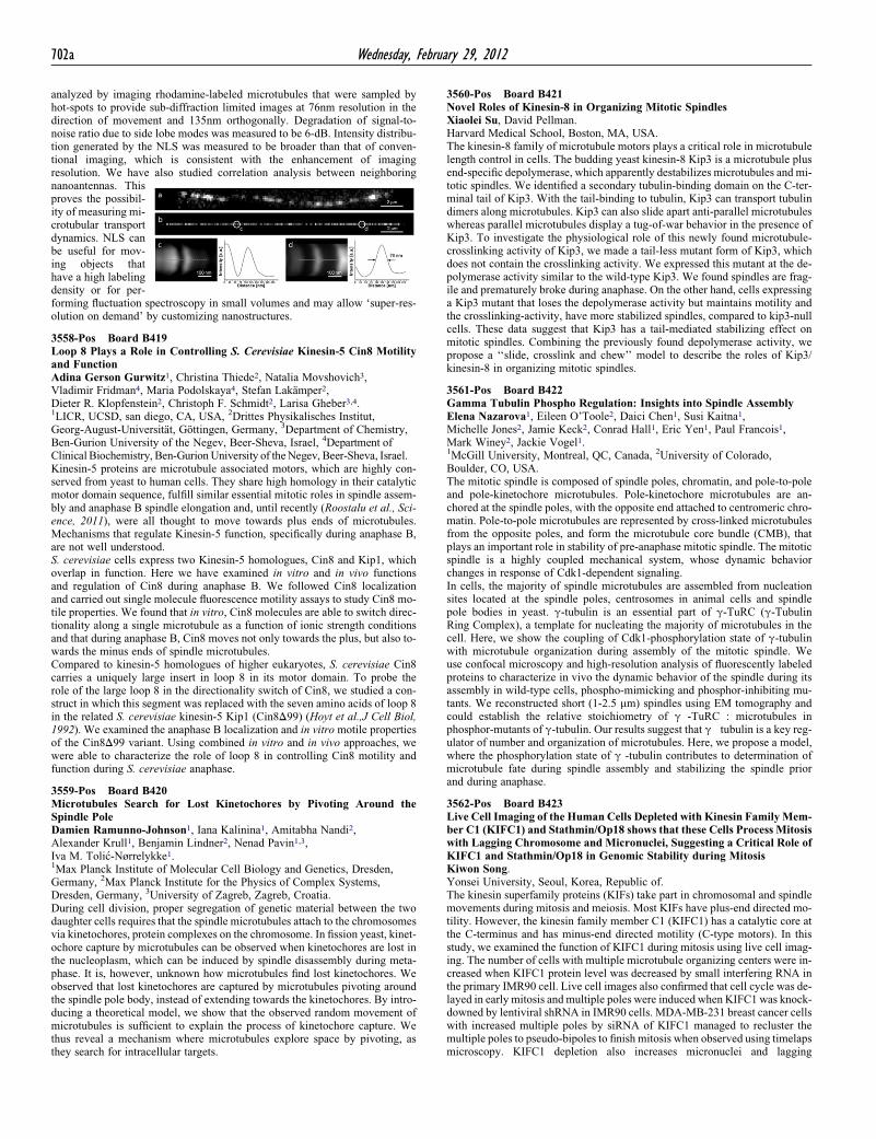
analyzed by imaging rhodamine-labeled microtubules that were sampled byhot-spots to provide sub-diffraction limited images at 76nm resolution in thedirection of movement and 135nm orthogonally. Degradation of signal-to-noise ratio due to side lobe modes was measured to be 6-dB. Intensity distribu-tion generated by the NLS was measured to be broader than that of conven-tional imaging, which is consistent with the enhancement of imagingresolution. We have also studied correlation analysis between neighboring

nanoantennas. Thisproves the possibil-ity of measuring mi-crotubular transportdynamics. NLS canbe useful for mov-ing objects thathave a high labelingdensity or for per- forming fluctuation spectroscopy in small volumes and may allow ‘super-res-olution on demand’ by customizing nanostructures.

3558-Pos Board B419Loop 8 Plays a Role in Controlling S. Cerevisiae Kinesin-5 Cin8 Motilityand FunctionAdina Gerson Gurwitz1, Christina Thiede2, Natalia Movshovich3,Vladimir Fridman4, Maria Podolskaya4, Stefan Lakamper2,Dieter R. Klopfenstein2, Christoph F. Schmidt2, Larisa Gheber3,4.1LICR, UCSD, san diego, CA, USA, 2Drittes Physikalisches Institut,Georg-August-Universitat, Gottingen, Germany, 3Department of Chemistry,Ben-Gurion University of the Negev, Beer-Sheva, Israel, 4Department ofClinical Biochemistry, Ben-GurionUniversity of theNegev, Beer-Sheva, Israel.Kinesin-5 proteins are microtubule associated motors, which are highly con-served from yeast to human cells. They share high homology in their catalyticmotor domain sequence, fulfill similar essential mitotic roles in spindle assem-bly and anaphase B spindle elongation and, until recently (Roostalu et al., Sci-ence, 2011), were all thought to move towards plus ends of microtubules.Mechanisms that regulate Kinesin-5 function, specifically during anaphase B,are not well understood.S. cerevisiae cells express two Kinesin-5 homologues, Cin8 and Kip1, whichoverlap in function. Here we have examined in vitro and in vivo functionsand regulation of Cin8 during anaphase B. We followed Cin8 localizationand carried out single molecule fluorescence motility assays to study Cin8 mo-tile properties. We found that in vitro, Cin8 molecules are able to switch direc-tionality along a single microtubule as a function of ionic strength conditionsand that during anaphase B, Cin8 moves not only towards the plus, but also to-wards the minus ends of spindle microtubules.Compared to kinesin-5 homologues of higher eukaryotes, S. cerevisiae Cin8carries a uniquely large insert in loop 8 in its motor domain. To probe therole of the large loop 8 in the directionality switch of Cin8, we studied a con-struct in which this segment was replaced with the seven amino acids of loop 8in the related S. cerevisiae kinesin-5 Kip1 (Cin8D99) (Hoyt et al.,J Cell Biol,1992). We examined the anaphase B localization and in vitro motile propertiesof the Cin8D99 variant. Using combined in vitro and in vivo approaches, wewere able to characterize the role of loop 8 in controlling Cin8 motility andfunction during S. cerevisiae anaphase.

3559-Pos Board B420Microtubules Search for Lost Kinetochores by Pivoting Around theSpindle PoleDamien Ramunno-Johnson1, Iana Kalinina1, Amitabha Nandi2,Alexander Krull1, Benjamin Lindner2, Nenad Pavin1,3,Iva M. Toli�c-Nørrelykke1.1Max Planck Institute of Molecular Cell Biology and Genetics, Dresden,Germany, 2Max Planck Institute for the Physics of Complex Systems,Dresden, Germany, 3University of Zagreb, Zagreb, Croatia.During cell division, proper segregation of genetic material between the twodaughter cells requires that the spindle microtubules attach to the chromosomesvia kinetochores, protein complexes on the chromosome. In fission yeast, kinet-ochore capture by microtubules can be observed when kinetochores are lost inthe nucleoplasm, which can be induced by spindle disassembly during meta-phase. It is, however, unknown how microtubules find lost kinetochores. Weobserved that lost kinetochores are captured by microtubules pivoting aroundthe spindle pole body, instead of extending towards the kinetochores. By intro-ducing a theoretical model, we show that the observed random movement ofmicrotubules is sufficient to explain the process of kinetochore capture. Wethus reveal a mechanism where microtubules explore space by pivoting, asthey search for intracellular targets.

3560-Pos Board B421Novel Roles of Kinesin-8 in Organizing Mitotic SpindlesXiaolei Su, David Pellman.Harvard Medical School, Boston, MA, USA.The kinesin-8 family of microtubule motors plays a critical role in microtubulelength control in cells. The budding yeast kinesin-8 Kip3 is a microtubule plusend-specific depolymerase, which apparently destabilizes microtubules and mi-totic spindles. We identified a secondary tubulin-binding domain on the C-ter-minal tail of Kip3. With the tail-binding to tubulin, Kip3 can transport tubulindimers along microtubules. Kip3 can also slide apart anti-parallel microtubuleswhereas parallel microtubules display a tug-of-war behavior in the presence ofKip3. To investigate the physiological role of this newly found microtubule-crosslinking activity of Kip3, we made a tail-less mutant form of Kip3, whichdoes not contain the crosslinking activity. We expressed this mutant at the de-polymerase activity similar to the wild-type Kip3. We found spindles are frag-ile and prematurely broke during anaphase. On the other hand, cells expressinga Kip3 mutant that loses the depolymerase activity but maintains motility andthe crosslinking-activity, have more stabilized spindles, compared to kip3-nullcells. These data suggest that Kip3 has a tail-mediated stabilizing effect onmitotic spindles. Combining the previously found depolymerase activity, wepropose a ‘‘slide, crosslink and chew’’ model to describe the roles of Kip3/kinesin-8 in organizing mitotic spindles.

3561-Pos Board B422Gamma Tubulin Phospho Regulation: Insights into Spindle AssemblyElena Nazarova1, Eileen O’Toole2, Daici Chen1, Susi Kaitna1,Michelle Jones2, Jamie Keck2, Conrad Hall1, Eric Yen1, Paul Francois1,Mark Winey2, Jackie Vogel1.1McGill University, Montreal, QC, Canada, 2University of Colorado,Boulder, CO, USA.The mitotic spindle is composed of spindle poles, chromatin, and pole-to-poleand pole-kinetochore microtubules. Pole-kinetochore microtubules are an-chored at the spindle poles, with the opposite end attached to centromeric chro-matin. Pole-to-pole microtubules are represented by cross-linked microtubulesfrom the opposite poles, and form the microtubule core bundle (CMB), thatplays an important role in stability of pre-anaphase mitotic spindle. The mitoticspindle is a highly coupled mechanical system, whose dynamic behaviorchanges in response of Cdk1-dependent signaling.In cells, the majority of spindle microtubules are assembled from nucleationsites located at the spindle poles, centrosomes in animal cells and spindlepole bodies in yeast. g-tubulin is an essential part of g-TuRC (g-TubulinRing Complex), a template for nucleating the majority of microtubules in thecell. Here, we show the coupling of Cdk1-phosphorylation state of g-tubulinwith microtubule organization during assembly of the mitotic spindle. Weuse confocal microscopy and high-resolution analysis of fluorescently labeledproteins to characterize in vivo the dynamic behavior of the spindle during itsassembly in wild-type cells, phospho-mimicking and phosphor-inhibiting mu-tants. We reconstructed short (1-2.5 mm) spindles using EM tomography andcould establish the relative stoichiometry of g -TuRC : microtubules inphosphor-mutants of g-tubulin. Our results suggest that g�tubulin is a key reg-ulator of number and organization of microtubules. Here, we propose a model,where the phosphorylation state of g -tubulin contributes to determination ofmicrotubule fate during spindle assembly and stabilizing the spindle priorand during anaphase.

3562-Pos Board B423Live Cell Imaging of the Human Cells Depleted with Kinesin FamilyMem-ber C1 (KIFC1) and Stathmin/Op18 shows that these Cells Process Mitosiswith Lagging Chromosome and Micronuclei, Suggesting a Critical Role ofKIFC1 and Stathmin/Op18 in Genomic Stability during MitosisKiwon Song.Yonsei University, Seoul, Korea, Republic of.The kinesin superfamily proteins (KIFs) take part in chromosomal and spindlemovements during mitosis and meiosis. Most KIFs have plus-end directed mo-tility. However, the kinesin family member C1 (KIFC1) has a catalytic core atthe C-terminus and has minus-end directed motility (C-type motors). In thisstudy, we examined the function of KIFC1 during mitosis using live cell imag-ing. The number of cells with multiple microtubule organizing centers were in-creased when KIFC1 protein level was decreased by small interfering RNA inthe primary IMR90 cell. Live cell images also confirmed that cell cycle was de-layed in early mitosis and multiple poles were induced when KIFC1 was knock-downed by lentiviral shRNA in IMR90 cells. MDA-MB-231 breast cancer cellswith increased multiple poles by siRNA of KIFC1 managed to recluster themultiple poles to pseudo-bipoles to finish mitosis when observed using timelapsmicroscopy. KIFC1 depletion also increases micronuclei and lagging