Embed Size (px)
Citation preview

LOOKING FOR LIPID PEROXIDATION IN VITRO AND IN VIVO: IS SEEING
BELIEVING?
Vanderbilt University School of Medicine
Jason D. Morrow MD

Question?
Which of the following assays of lipid peroxidation may be useful and accurate for the quantification of oxidant stress in vivo? More than one may be correct.
Plasma isoprostanes
Urinary malonaldehyde
Exhaled pentane
Total plasma antioxidant capacity
Plasma glutathione levels

Question?
In which of the following diseases has lipid peroxidation definitively been shown to be involved in disease pathogenesis?
Atherosclerosis
Scleroderma
Pulmonary hypertension
Stroke
All of the above
None of the above
Question?
A potential disadvantage of measuring isoprostanes as an index of oxidant stress include all of the following except: They can be formed ex vivo.
They are unstable and rapidly degrade ex vivo.
Accurate assay methods are complex and expensive.
They represent only one of a myriad of end products of lipid peroxidation.
Their quantification in a particular body fluid may not be index of systemic (total body) oxidant stress.
Immunoassay kits to measure isoprostanes are of questionable accuracy.

Goals of Lecture
Critical appraisal of methods to quantify lipid peroxidation in vitro and in vivo.
Theoretical considerations regarding quantification of lipid peroxidation.
Assays currently available to measure lipid peroxidation.
» Advantages
» Disadvantages

Background
Oxygen free radicals are implicated in many diseases. Ischemia-reperfusion injury
Cancer
Inflammation
Aging
Underlying mechanisms responsible for sequelae of oxidant stress are poorly understood.
Reliable methods of assessment have not been available.

Targets of Free Radical Injury
Lipids
Proteins
DNA

Cellular Sources of Free Radicals and Lipid Peroxidation Products

Lipid Peroxidation

Methods to Quantify Lipid Peroxidation
Measurement of substrate loss.
Quantification of lipid peroxidation products.
Primary end products
Secondary end products

The Ideal Assay of Lipid Peroxidation
Assay is accurate, specific, and sensitive index of lipid peroxidation.
Compounds to be quantified are stable.
Assay applicable to in vitro and in vivo studies.
Assay easy to perform with high throughput.
Assay economical.
Assays of Lipid Peroxidation
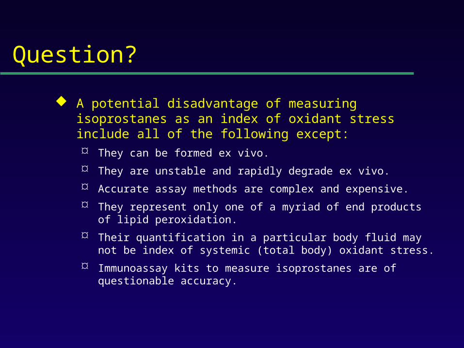
No assay is ideal.
Most assays are more accurate when quantifying lipid peroxidation in vitro than in vivo.
Little data exist comparing various methods in vivo.
Few assays accurately provide an integrated assessment of lipid peroxidation in an animal or human as a whole.

Assays of Potential Use to Quantify Lipid Peroxidation in Vitro and in Vivo
Fatty acid analysis
Conjugated dienes
Lipid hydroperoxides
Thiobarbituric acid-reactive substances (TBARS) or malondialdehyde (MDA)
Alkanes
F2-Isoprostanes
Fatty Acid Analysis
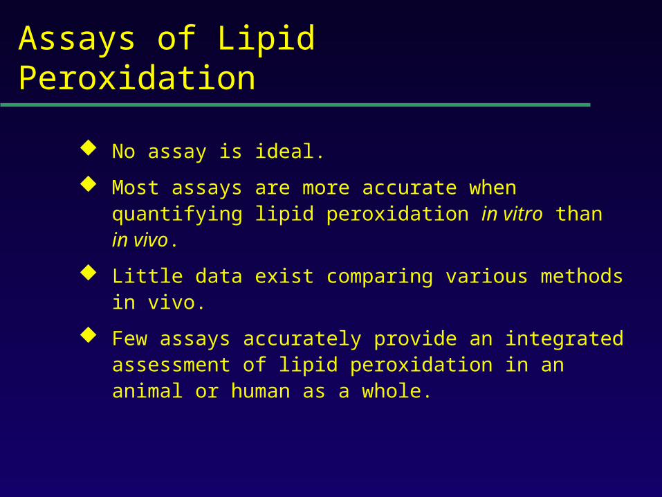
Commonly used to assess fatty acid content in biological fluids or tissues.
Method
Lipid extraction of biological fluid or tissue
Transmethylation of fatty acids
Separation by GC (or HPLC)
Quantification of fatty acid with flame ionization

Analysis of Fatty Acids in Peroxidizing Microsomes
Disappearance of arachidonic acid is associated with accumulation of peroxidation end products.

Fatty Acid Analysis
Advantages Easy to perform.
Equipment readily available.
Data complement generation of peroxidation products in vitro and in vivo.
Disadvantages Impractical for a number of in vivo situations.
Provides information on disappearance of substrate only.
May see very little change associated with oxidation in vivo.

Conjugated Dienes
Peroxidation of unsaturated fatty acids results in the formation of conjugated diene structures that absorb light in the wavelength of 230-235 nm which can be quantified spectrometrically.

Conjugated Dienes
Useful for studying oxidation of lipids in vitro. Enhanced assay sensitivity using second derivative
spectroscopy, HPLC, or GC/MS.
Studies have shown increases in animals and humans associated with oxidant stress.
Inaccurate index of lipid peroxidation when applied to complex biological fluids. A primary diene conjugate from human body fluids is an non-
oxygen containing isomer of linoleic acid, octadeca-9-cis-11-trans-dienoic acid.
Other compounds-purines, pyrimidines, heme proteins absorb at 234 nm.

Conjugated Dienes
Advantages
Relatively easy to perform
Provides useful information primarily regarding oxidation of pure lipids in vitro.
Disadvantages
Virtually useless for analysis of lipid peroxidation products in complex biological fluids.

Lipid Hydroperoxides
Primary products of lipid peroxidation.
Several quantitative methods exist (iodometric, electrochemical, mass spectrometric) Most accurate-chemiluminescence-based HPLC detection.
» Sensitive (pmol or less) and specific
» Information regarding which lipid class is oxidized can be obtained.

Plasma Lipid Hydroperoxides in Patients with Subarachnoid Hemorrhage
Polidori et al. Free Rad. Biol. Med. 23: 762-7, 1997

Lipid Hydroperoxides
Advantages
Assays more specific and sensitive than for other peroxidation products.
Disadvantages
Products are unstable.
Ex vivo oxidation a major concern.
Lack of detectable levels in some fluids and tissues.
Equipment expensive.

Thiobarbituric Acid-Reactive Substances (TBARS)/MDA
Most commonly used method to assess lipid peroxidation. Measures malondialdehyde (MDA) which is a breakdown
product of lipid peroxidation.
Method: Sample to be tested is heated with thiobarbituric acid at
low pH and a pink chromogen (believed to be a TBA-MDA adduct) is formed.
Quantification-absorbance at 532 nm or fluorescence at 553 nm.
TBARS/MDA
Quantification of TBARS is an accurate measure of peroxidation in oxidizing systems in vitro.
TBARS quantification in body fluids is inaccurate.
Substances other than MDA form chromogens at 532 nm.
MDA is formed during the assay procedure.
Antioxidants can interfere with the assay.
MDA can be derived from the diet.

TBARS/MDA
Assays exist to measure TBARs by HPLC.
MDA, HNE, and other aldehydes can be quantified by HPLC or GC/MS.
These assays are generally more specific than TBARs although not necessarily more accurate as an index of lipid peroxidation.

TBARS/MDA
Levels of TBARS vary widely. Plasma levels
» Regular assay 4-35 uM.
» HPLC-coupled 0-0.18 uM.
TBARS increased in various disorders. Hypercholesterolemia (Chirico et al., Free Rad. Res.
Comm. 19:51, 1993).
» Controls 0.10 + 0.08 uM
» Hypercholesterolemics 0.61 + 0.25 uM

TBARS/MDA
Summary The TBARS assays are important because they
are easy to perform and widely available.
They are a reasonably accurate index of lipid peroxidation in a number of in vitro oxidizing systems.
They are less reliable as an index of lipid peroxidation in complex biological fluids or in vivo.

Alkanes
Volatile hydrocarbons generated from scission of oxidized lipids. Pentane (n-6 fatty acids)
Ethane (n-3 fatty acids)
Method of quantification Collection of gas from an in vitro incubation or exhaled air
from an animal or human.
Concentrating and filtering of samples to remove water and carbon dioxide.
Analysis of product by GC (capillary).

Alkanes
Advantages Is an integrated assessment of peroxidation in vivo.
Disadvantages Collection of exhaled air for in vivo studies cumbersome
and can take several hours to obtain an adequate sample for analysis.
Oxygen tension alters alkane formation.
Atmospheric contamination of collected gases.
Different researchers report a 1000-fold difference in normal levels of pentane generated in humans (4.1-4900 pmol/L).
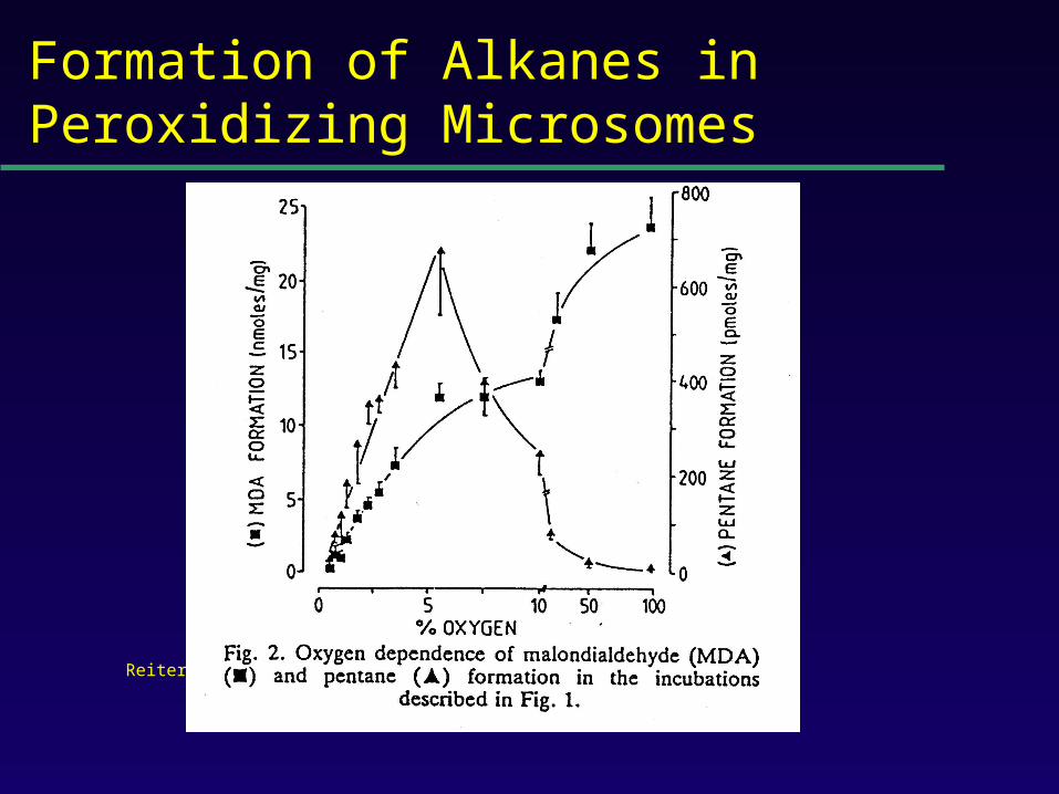
Formation of Alkanes in Peroxidizing Microsomes
Reiter R et al. Biochem. Pharm. 36: 925-9, 1987.

F2-Isoprostanes
Arachidonyl-containing lipids are peroxidized to PGF2-like compounds,
termed F2-isoprostanes.
Formed independent of the cyclooxygenase by peroxidation of arachidonate.
Generated in large amounts in vivo.
Exert potent biological activity.

Pathway of Isoprostane Formation
5-series 12-series 8-series 15-series

Analysis of F2-Isoprostanes
Measured either free or after liberation from tissue lipids.
Purified by Sep-Pak extraction and TLC and derivatized to PFB ester, TMS ethers.
Analyzed using stable isotope dilution techniques employing a deuteriated standard by gas chromatography/mass spectrometry.

Analysis of F2-Isoprostanes in Human Plasma

Formation of F2-Isoprostanes in Peroxidizing Microsomes
F2-IsoP formation correlates with disappearance of
arachidonate and generation of MDA.

CCl4-Mediated Oxidant Injury

F2-Isoprostanes Are a Reliable Marker of Lipid Peroxidation in Vivo
Comparison of formation of MDA and F2-IsoPs with
hepatic injury in CCl4-treated rats.

F2-IsoPs as a Measure of Oxidant Stress
BOSS study (2000)-IsoPs most accurate measure of oxidant stress in CCl4-treated rats.
Deficiencies in antioxidants in vivo are associated with increased IsoP formation.
Antioxidants decrease IsoP levels in animals and humans.
IsoP levels are increased in animal models of human diseases and human disorders associated with oxidant stress.

Biomarkers of Oxidative Stress Study
CCl4-induced oxidant stress in rats.
Markers quantified and compared to hepatic histology/enzyme leak:
Plasma and urine IsoPs
Plasma antioxidants
Plasma GSH and GSSG
Protein carbonyls and specific amino acid oxidation products
8-hydroxy-deoxyguanosine
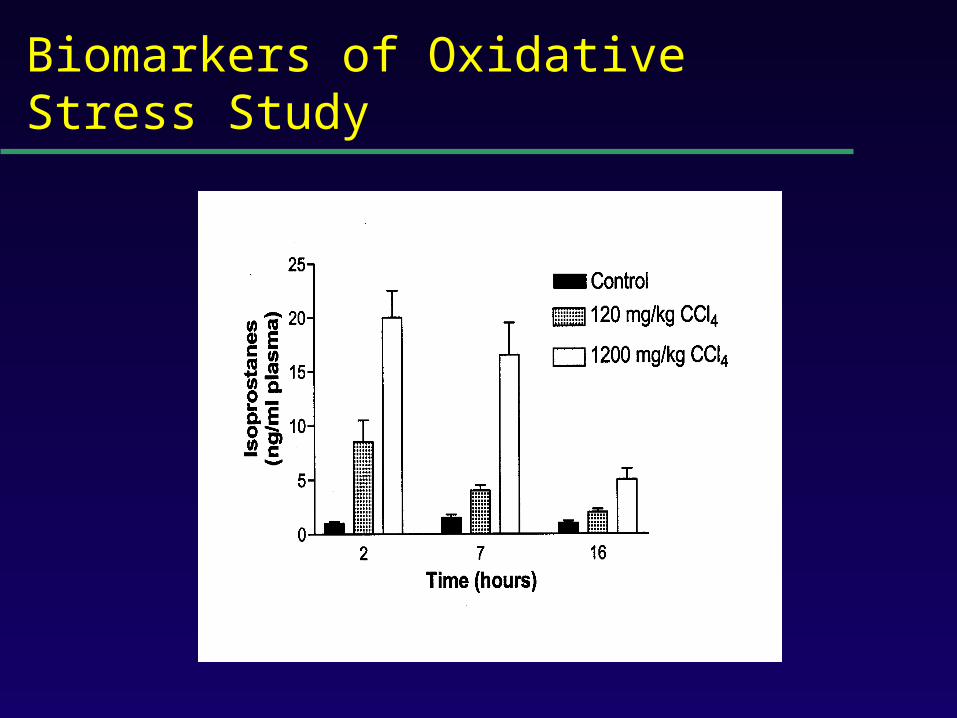
Biomarkers of Oxidative Stress Study

Isoprostanes as an Index of Oxidant Stress in Humans-Atherosclerosis
Oxidation of lipoproteins plays a pivotal role in the development of atherosclerotic lesions.
Levels of IsoPs in oxidized LDL correlate with other markers of lipid peroxidation.
Risk factors for atherosclerosis are associated with increased levels of IsoPs in vivo in humans. Chronic cigarette smoking
Hyperhomocysteinemia
Diabetes mellitus

F2-IsoPs Are Increased in Humans With Hypercholesterolemia
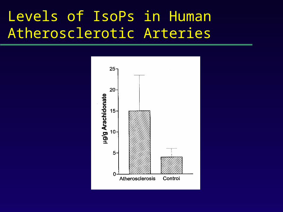
Levels of IsoPs in Human Atherosclerotic Arteries

Vitamin C Does Not Alter Isoprostane Levels in Healthy Humans

Vitamin E Decreases IsoP Levels in Hypercholesterolemic Humans
* p < 0.05
* *

Vitamin E Reduces Isoprostane Formation in Humans– Effect of Dose
P<0.001

Advantages of Isoprostane Quantification to Assess Oxidant Stress
Isoprostanes are stable molecules.
The assay is highly precise and accurate.
IsoPs can be detected in all fluids and tissues.
Normal ranges can be defined.
Allows for studies to evaluate the effects of interventions on endogenous lipid peroxidation.

Disadvantages of Isoprostane Quantification to Assess Oxidant Stress
Samples must either be analyzed immediately or stored at –70o C.
Increases in IsoPs locally in tissues or fluids aren’t detected by measuring systemic oxidant stress.

Isoprostanes Are Increased Selectively in the Central Nervous System of Humans with AD

Disadvantages of Isoprostane Quantification to Assess Oxidant Stress
Samples must either be analyzed immediately or stored at –70o C.
Increases in IsoPs locally in tissues or fluids aren’t detected by measuring systemic oxidant stress.
F2-IsoPs represents only one of a myriad of
arachidonate oxygenation products.

Classes of IsoPs

Unified Pathway of Arachidonate Oxidation

Disadvantages of Isoprostane Quantification to Assess Oxidant Stress
Samples must either be analyzed immediately or stored at –70o C.
Increases in IsoPs locally in tissues or fluids aren’t detected by measuring systemic oxidant stress.
F2-IsoPs represents only one of a myriad of
arachidonate oxygenation products.
Analysis is labor intensive and requires expensive equipment.

Immunoassay Methods to Quantify IsoPs
Immunoassays advantageous because they are more economical and less labor intensive.
Polyclonal antibodies have been made by several investigators and are commercially available.
Accurate quantification using immunoassays requires initial compound purification.
Amounts measured by immunoassays often differ from those obtained by mass spectrometry.
We are currently collaborating with Unilever Ltd. in the generation of highly specific monoclonal abs.

Disadvantages of Isoprostane Quantification to Assess Oxidant Stress
Samples must either be analyzed immediately or stored at –70o C.
Increases in IsoPs locally in tissues or fluids aren’t detected by measuring systemic oxidant stress.
F2-IsoPs represents only one of a myriad of arachidonate oxygenation products.
Analysis is labor intensive and requires expensive equipment.
Urinary IsoPs are not a valid measure of systemic oxidant stress since a major source of urinary IsoPs is likely the kidney.

Metabolic Fate of 15-F2t-IsoP (8-iso-PGF2) in Humans

Analysis of Human Urine for F2-IsoP-M

Levels of F2-IsoP-M Are Increased in Rats Administered CCl4

Levels of F2-IsoP-M in Normal and Hypercholesterolemic Humans

Vitamin E Lowers F2-IsoP-M Excretion in Hypercholesterolemic Humans
* p < 0.05
**

Summary
Quantification of F2-isoprostanes is an accurate measure of oxidant injury and lipid peroxidation in vitro and in vivo.
Current methods employ mass spectrometry and immunoassays.
The development of an assay to measure F2-IsoP-M may offer certain advantages over the quantification of parent F2-IsoPs.

Conclusions
Numerous methods exist to quantify lipid peroxidation.
Most assays currently available are accurate measures of lipid peroxidation in vitro.
Studies suggest that these assays are either less accurate measures of lipid peroxidation in vivo or too little information exists to accurately assess them.
Quantification of isoprostanes represents a significant advance in our ability to quantify lipid peroxidation in vivo.