Embed Size (px)
Citation preview
Can J Gastroenterol Vol 24 No 1 January 2010 61
Look out before polypectomy in patients with diverticular disease – a case of a large, inverted
diverticulum of the colon resembling a pedunculated polyp
Omero Alessandro Paoluzi MD, Claudio Tosti MD, Fabio Andrei MD, Italo Stroppa MD, Francesco Pallone MD
Gastrointestinal Unit, Centre of Excellence for the Study of Complex and Multifactorial Diseases, Department of Internal Medicine, Tor Vergata University, Rome, Italy
Correspondence: Dr Omero Alessandro Paoluzi, UOC di Gastroenterologia, Dipartimento di Medicina Interna, Università Tor Vergata, Viale Oxford 81, 00133 Roma, Italy. Telephone 39-06-20900969, fax 39-06-20903738, e-mail [email protected]
Received for publication April 21, 2009. Accepted May 2, 2009
Diverticular disease of the colon, a condition frequently observed in clinical practice, may be responsible for
abdominal symptoms requiring colonoscopy. Colonoscopy may reveal the presence of polyps, often adenomas concomi-tant with diverticular disease (1-3), which may be removed by endoscopic polypectomy. This procedure has been estimated to account for approximately 50% of the cases of perforation reported during colonoscopy, the incidence of which has been reported to be less than 0.05% (4-6). The risk of such a com-plication is higher in the event of an inverted colonic diver-ticulum, which may be misinterpreted as a polypoid lesion at colonoscopy. To date, fewer than 20 cases of inverted colonic diverticula, diagnosed at colonoscopy or following air contrast barium enema, have been reported in the literature (7-14).
The present report describes a patient who underwent a screening colonoscopy, which revealed a voluminous, pedun-culated polyp recognized to be an inverted giant colonic diver-ticulum before endoscopic polypectomy.
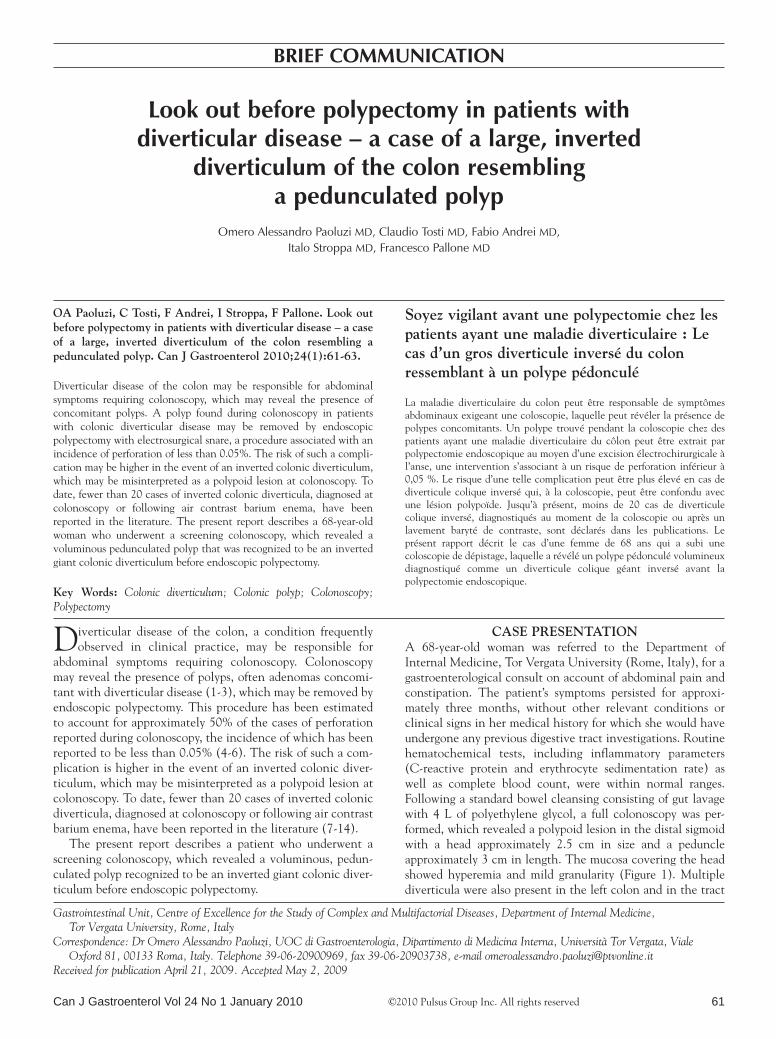
CASE PRESENTATIONA 68-year-old woman was referred to the Department of Internal Medicine, Tor Vergata University (Rome, Italy), for a gastroenterological consult on account of abdominal pain and constipation. The patient’s symptoms persisted for approxi-mately three months, without other relevant conditions or clinical signs in her medical history for which she would have undergone any previous digestive tract investigations. Routine hematochemical tests, including inflammatory parameters (C-reactive protein and erythrocyte sedimentation rate) as well as complete blood count, were within normal ranges. Following a standard bowel cleansing consisting of gut lavage with 4 L of polyethylene glycol, a full colonoscopy was per-formed, which revealed a polypoid lesion in the distal sigmoid with a head approximately 2.5 cm in size and a peduncle approximately 3 cm in length. The mucosa covering the head showed hyperemia and mild granularity (Figure 1). Multiple diverticula were also present in the left colon and in the tract
Brief communication
©2010 Pulsus Group Inc. All rights reserved
OA Paoluzi, C Tosti, F Andrei, I Stroppa, F Pallone. Look out before polypectomy in patients with diverticular disease – a case of a large, inverted diverticulum of the colon resembling a pedunculated polyp. Can J Gastroenterol 2010;24(1):61-63.
Diverticular disease of the colon may be responsible for abdominal symptoms requiring colonoscopy, which may reveal the presence of concomitant polyps. A polyp found during colonoscopy in patients with colonic diverticular disease may be removed by endoscopic polypectomy with electrosurgical snare, a procedure associated with an incidence of perforation of less than 0.05%. The risk of such a compli-cation may be higher in the event of an inverted colonic diverticulum, which may be misinterpreted as a polypoid lesion at colonoscopy. To date, fewer than 20 cases of inverted colonic diverticula, diagnosed at colonoscopy or following air contrast barium enema, have been reported in the literature. The present report describes a 68-year-old woman who underwent a screening colonoscopy, which revealed a voluminous pedunculated polyp that was recognized to be an inverted giant colonic diverticulum before endoscopic polypectomy.
Key Words: Colonic diverticulum; Colonic polyp; Colonoscopy; Polypectomy
Soyez vigilant avant une polypectomie chez les patients ayant une maladie diverticulaire : Le cas d’un gros diverticule inversé du colon ressemblant à un polype pédonculé
La maladie diverticulaire du colon peut être responsable de symptômes abdominaux exigeant une coloscopie, laquelle peut révéler la présence de polypes concomitants. Un polype trouvé pendant la coloscopie chez des patients ayant une maladie diverticulaire du côlon peut être extrait par polypectomie endoscopique au moyen d’une excision électrochirurgicale à l’anse, une intervention s’associant à un risque de perforation inférieur à 0,05 %. Le risque d’une telle complication peut être plus élevé en cas de diverticule colique inversé qui, à la coloscopie, peut être confondu avec une lésion polypoïde. Jusqu’à présent, moins de 20 cas de diverticule colique inversé, diagnostiqués au moment de la coloscopie ou après un lavement baryté de contraste, sont déclarés dans les publications. Le présent rapport décrit le cas d’une femme de 68 ans qui a subi une coloscopie de dépistage, laquelle a révélé un polype pédonculé volumineux diagnostiqué comme un diverticule colique géant inversé avant la polypectomie endoscopique.
Paoluzi et al
Can J Gastroenterol Vol 24 No 1 January 201062
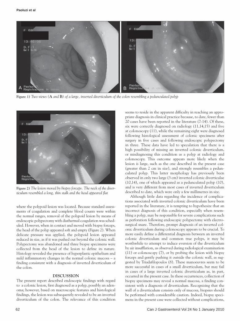
where the polypoid lesion was located. Because standard assess-ments of coagulation and complete blood counts were within the normal ranges, removal of the polypoid lesion by means of endoscopic polypectomy with diathermal coagulation was sched-uled. However, when in contact and moved with biopsy forceps, the head of the polyp appeared soft and empty (Figure 2). When delicate pressure was applied, the polypoid lesion appeared reduced in size, as if it was pushed out beyond the colonic wall. Polypectomy was abandoned and three biopsy specimens were collected from the head of the lesion to define its nature. Histology revealed the presence of hyperplastic epithelium and mild inflammatory changes in the normal colonic mucosa – a finding consistent with a diagnosis of inverted diverticulum of the colon.
DISCUSSIONThe present report described endoscopic findings with regard to a colonic lesion, first diagnosed as a polyp, possibly an aden-oma; however, based on macroscopic features and histological findings, the lesion was subsequently revealed to be an inverted diverticulum of the colon. The relevance of this condition
seems to reside in the apparent difficulty in reaching an appro-priate diagnosis in clinical practice because, to date, fewer than 20 cases have been reported in the literature (7-14). Of these, six were correctly diagnosed on radiology (11,14,15) and five at colonoscopy (11), while the remaining eight were diagnosed following histological assessment of colonic specimens after surgery in five cases and following endoscopic polypectomy in three. These data have led to speculation that there is a high possibility of missing an inverted colonic diverticulum, or misdiagnosing this condition as a polyp at radiology and colonoscopy. This outcome appears more likely when the lesion is large, such as the one described in the present case (greater than 2 cm in size), and strongly resembles a pedun-culated polyp. This latter morphology has previously been observed in only two large (3 cm) inverted colonic diverticulae (10,14), one of which appeared as a pedunculated polyp (10), and is very different from most cases of inverted diverticulum described to date, which were only a few millimetres in size.
Although little data regarding the incidence of complica-tions associated with inverted colonic diverticulum have been reported in the literature, it is tempting to hypothesize that an incorrect diagnosis of this condition, especially when resem-bling a polyp, may be responsible for severe complications such as perforation following endoscopic polypectomy with electro-surgical snare. Therefore, prompt diagnosis of an inverted col-onic diverticulum during colonoscopy appears to be crucial. To more easily define a differential diagnosis between an inverted colonic diverticulum and common true polyps, it may be worthwhile to attempt to induce eversion of the diverticulum by air insufflation, as observed during radiological examination (11) or colonoscopy (7), or by probing the lesion with biopsy forceps and gently pushing it outside the colonic wall, as sug-gested by Triadafilopoulos (8). These manoeuvres seem to be more successful in cases of a small diverticulum, but may fail in cases of a large inverted colonic diverticulum as, in part, occurred in the present case. In these occurrences, collection of biopsy specimens may reveal a normal mucosa, a finding con-sistent with a diagnosis of diverticulum. Recognizing that the wall of a diverticulum consists only of mucosa, biopsies should be performed with considerable caution. Indeed, biopsy speci-mens in the present case were collected without complications,
Figure 1) Two views (A and B) of a large, inverted diverticulum of the colon resembling a pedunculated polyp
Figure 2) The lesion moved by biopsy forceps. The neck of the diver-ticulum resembled a long, thin stalk and the head appeared flat
Inverted giant diverticulum resembling a polyp
Can J Gastroenterol Vol 24 No 1 January 2010 63
REFERENCES1. Stefansson T, Ekbom A, Sparen P, Pahlman L. Increased risk of left
sided colon cancer in patients with diverticular disease. Gut 1993;34:499-502.
2. Morini S, Hassan C, Zullo A, et al. Diverticular disease as a risk factor for sigmoid colon adenomas. Dig Liver Dis 2002;34:635-9.
3. Kieff BJ, Eckert GJ, Imperiale TF. Is diverticulosis associated with colorectal neoplasia? A cross-sectional colonoscopic study. Am J Gastroenterol 2004;99:2007-11.
4. Araghizadeh FY, Timmcke AE, Opelka FG, Hicks TC, Beck DE. Colonoscopic perforations. Dis Colon Rectum 2001;44:713-6.
a finding consistent with other reports (7). Colonic perforation following biopsy collection from an inverted colonic diverticu-lum has been described in only one case (9). Thus, the risk of colonic perforation following biopsy collection from a large, inverted colonic diverticulum appears to be low.
CONCLUSIONThe present report highlights the possibility of finding an inverted colonic diverticulum during colonoscopy. This possi-bility must be considered carefully to reach a correct diagnosis and avoid potentially dangerous procedures such as endoscopic polypectomy.
5. Korman LY, Overholt BF, Box T, Winker CK. Perforation during colonoscopy in endoscopic ambulatory surgical centers. Gastrointest Endosc 2003;58:554-7.
6. Cobb WS, Heniford BT, Sigmon LB, et al. Colonoscopic perforations: Incidence, management, and outcomes. Am Surg 2004;70:750-7.
7. Yusuf SI, Grant C. Inverted colonic diverticulum: A rare finding in a common condition? Gastrointest Endosc 2000;52:111-5.
8. Triadafilopoulos G. Images in clinical medicine. Inverted colonic diverticulum. N Engl J Med 1999;341:1508.
9. Hollander E, David G. Inverted sigmoid diverticulum simulating polyps. Orv Hetil 1993;134:639-40.
10. Dumas O, Jouffre C, Desportes R, Etaix JP, Barthelemy C, Audigier JC. Inverted sigmoid diverticulum: A misleading polyp. Gastrointest Endosc 1991;37:587-8.
11. Glick SN. Inverted colonic diverticulum: Air contrast barium enema findings in six cases. Am J Roentgenol 1991;156:961-4.
12. Ladas SD, Prigouris SP, Pantelidaki C, Raptis A. Endoscopic removal of inverted sigmoid diverticulum – is it a dangerous procedure? Endoscopy 1989;21:243-4.
13. Shah AN, Mazza BR. The detection of an inverted diverticulum by colonoscopy. Gastrointest Endosc 1982;28:188-9.
14. Freeny PC, Walker JH. Inverted diverticula of the gastrointestinal tract. Gastrointest Radiol 1979;4:57-9.
15. Posner R, Solomon A. Dilemma of an inverted cecal diverticulum simulating a pedunculated polyp: CT appearance. Abdom Imaging 1995;20:440-1.

Submit your manuscripts athttp://www.hindawi.com
Stem CellsInternational
Hindawi Publishing Corporationhttp://www.hindawi.com Volume 2014
Hindawi Publishing Corporationhttp://www.hindawi.com Volume 2014
MEDIATORSINFLAMMATION
of
Hindawi Publishing Corporationhttp://www.hindawi.com Volume 2014
Behavioural Neurology
EndocrinologyInternational Journal of
Hindawi Publishing Corporationhttp://www.hindawi.com Volume 2014
Hindawi Publishing Corporationhttp://www.hindawi.com Volume 2014
Disease Markers
Hindawi Publishing Corporationhttp://www.hindawi.com Volume 2014
BioMed Research International
OncologyJournal of
Hindawi Publishing Corporationhttp://www.hindawi.com Volume 2014
Hindawi Publishing Corporationhttp://www.hindawi.com Volume 2014
Oxidative Medicine and Cellular Longevity
Hindawi Publishing Corporationhttp://www.hindawi.com Volume 2014
PPAR Research
The Scientific World JournalHindawi Publishing Corporation http://www.hindawi.com Volume 2014
Immunology ResearchHindawi Publishing Corporationhttp://www.hindawi.com Volume 2014
Journal of
ObesityJournal of
Hindawi Publishing Corporationhttp://www.hindawi.com Volume 2014
Hindawi Publishing Corporationhttp://www.hindawi.com Volume 2014
Computational and Mathematical Methods in Medicine
OphthalmologyJournal of
Hindawi Publishing Corporationhttp://www.hindawi.com Volume 2014
Diabetes ResearchJournal of
Hindawi Publishing Corporationhttp://www.hindawi.com Volume 2014
Hindawi Publishing Corporationhttp://www.hindawi.com Volume 2014
Research and TreatmentAIDS
Hindawi Publishing Corporationhttp://www.hindawi.com Volume 2014
Gastroenterology Research and Practice
Hindawi Publishing Corporationhttp://www.hindawi.com Volume 2014
Parkinson’s Disease
Evidence-Based Complementary and Alternative Medicine
Volume 2014Hindawi Publishing Corporationhttp://www.hindawi.com