Embed Size (px)
Citation preview
Central Annals of Sports Medicine and Research
Cite this article: Knight JE, Brown G (2017) Longitudinal Patella Stress Fracture in an Ultra-Marathon Runner. Ann Sports Med Res 4(2): 1104.
*Corresponding authorJill Eggers Knight, Department of Orthopedics and Sports Medicine, Franciscan Orthopedic Associates, 1608 S. “J” ST. Tacoma, WA 98405, USA, Tel: 206-499-9654; Email:
Submitted: 22 February 2017
Accepted: 19 April 2017
Published: 20 April 2017
ISSN: 2379-0571
Copyright© 2017 Knight
OPEN ACCESS
Keywords•Patella stress fracture•Patellectomy•Contralateral knee
Abstract
This is a case report of a female ultra-marathon runner who presented with recalcitrant anterior knee pain. Initial radiographs revealed a longitudinal patellar sclerotic fracture versus a bipartite patella. Further investigation via MRI revealed a lateral longitudinal patella stress fracture. Due to the small size of the sclerotic fragment, a partial patellectomy was performed. A lateral release was performed because of a tight iliotibial band/patellar retinaculum. The patient was able to resume running two months post-operatively.
Case reports of patella stress fractures are reviewed describing etiology, biomechanics, diagnosis and treatment of longitudinal and transverse patella stress fractures.
Stress fractures of the lower extremity are common among athletes, although stress fractures of the patella are a rare entity. The incidence of patella stress fractures in lower extremity stress fractures is approximately 1%. A stress fracture may occur when there is repeated stress to bone via trauma or overuse with insufficient time to allow healing and remodeling to occur. This results in micro fractures which ultimately lead to macroscopic fracture lines and possible displacement.
In part due to the rarity of patellar stress fractures, diagnosis may be challenging. Frequently, plain radiographs may be misinterpreted as bipartite patella or Sinding-Larsen-Johansson disease. In many cases, scrutiny or further investigation does not ensue until protracted conservative therapy has failed. This can lead to an extensive delay in treatment and therefore a high index of suspicion is needed to treat these athletes in a timely fashion.
We report on a 45-year-old female ultra-marathon runner with a longitudinal lateral patellar stress fracture. Pietu and Hauet report two different mechanisms by which patellar stress fractures may occur: running which involves a lower force but greater frequency, and jumping/kicking motions which equate to repetitive forceful contractions. These mechanisms are thought to result in different patterns of patellar stress fractures; longitudinal and transverse.
Case Report
Longitudinal Patella Stress Fracture in an Ultra-Marathon RunnerJill Eggers Knight* and Gregory BrownDepartment of Orthopedics and Sports Medicine, Franciscan Orthopedic Associates, USA
CASE PRESENTATIONThe subject is a healthy 45-year-old who began running ultra-
marathons approximately two years ago. Five months prior to presentation, as she progressively increased her miles, the patient developed diffuse anterior knee pain accompanied by an effusion and a nodular lesion in the anterior/superior aspect of her right knee that she rated a 5/10 on the Visual Analog Scale [1]. The patient denied any direct trauma to her knee at any time. There was no instance where she was unable to bear weight or not able to extend her knee. She denied any “pop” or “crack” in the anterior aspect of her knee. After the patient completed the marathon, the patient discontinued running. When the diffuse anterior knee pain did not abate after two months, she presented for evaluation.
Exam at the time of presentation revealed a mild effusion, with extension to 20 and flexion to 130° without pain. She had
no ligamentous laxity and the Q angle was 14° [2]. She also had a firm anterolateral painful non- mobile lesion that measured 1 x 1.5 cm. The consistency was firm but not contiguous to the patella. She had minimal pain to palpation at the medial patellar facet, negative apprehension and inhibition signs, and positive grind and “J” tracking signs. She had no evidence of pes planus or femoral ante version. No neurological deficits were demonstrated. The remainder of the exam was normal.
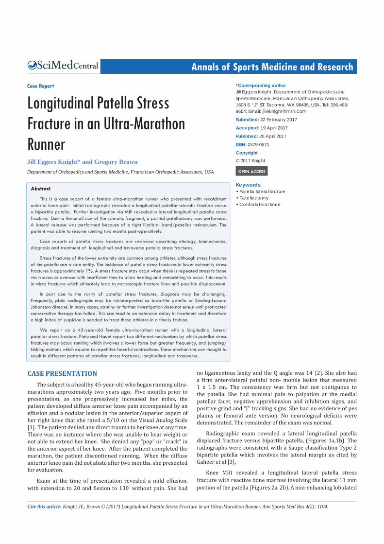
Radiographic exam revealed a lateral longitudinal patella displaced fracture versus bipartite patella, (Figures 1a,1b). The radiographs were consistent with a Saupe classification Type 2 bipartite patella which involves the lateral margin as cited by Gaheer et al [3].
Knee MRI revealed a longitudinal lateral patella stress fracture with reactive bone marrow involving the lateral 11 mm portion of the patella (Figures 2a, 2b). A non-enhancing lobulated

Central
Knight (2017)Email:
Ann Sports Med Res 4(2): 1104 (2017) 2/3
longitudinal cystic mass having the appearance of a chronic ganglion was noted extending superiorly from the stress fracture and anterior to the quadriceps tendon. (Figures 3a, 3b) show the anterior/posterior radiographs post-operatively.
The patient underwent operative treatment for her longitudinal lateral patellar stress fracture. The stress fracture was sclerotic and the lateral fragment was relatively small so excision was recommended. Exam under anesthesia revealed decreased medial patellar mobility (<25% compared to >50% on the contralateral knee) consistent with iliotibial band tightness.
An open partial patellectomy (lateral fragment excision) was performed. The lateral retinaculum was not repaired, resulting in a lateral release (the medial patellar mobility increased to 50%). The ganglion excision was excised. There was moderate lateral swelling immediately after surgery because the lateral retinaculum was not repaired and fluid extravasated into the subcutaneous tissues. The swelling resolved with compression. The subject did well and was able to resume running eight weeks postoperatively [4].
DISCUSSIONA stress fracture may occur when repeated stress is applied
to the bone from trauma or overuse. The muscles cannot absorb the energy and the energy is absorbed by the underlying bone causing microfractures. The microfractures progress to a “fatigue” fracture or stress fracture. There are two types of stress fractures: fatigue and insufficiency. Fatigue fractures occur when repetitive stresses are applied to normal bone. Insufficiency fractures occur when normal stresses are applied to weaken bone. There are two types of patellar stress fractures described in the literature; longitudinal and transverse.
Again, patellar stress fractures are a rare injury. They have a higher incidence in males compared to females [5] and are usually unilateral unlike a bipartite patella. The transverse pattern is much more common than the longitudinal [6]. The earliest documented patellar stress fracture case in the English language literature was in 1960 by Devas [7]. Since that time, 39 cases in total to date have been documented. Thirty-five cases are tabulated in Brown et al [8] and four additional cases reports are noted [8,9]. The sports associated with longitudinal patellar stress fracture group are running (5), gymnastics (1), Japanese fencing (1), floor hockey/football (1), and weightlifting (1). Long distance running was the predominant sport associated with the longitudinal type (p = 0.007 [Brown]). The sports associated with the transverse fractures were in descending order, basketball (8), soccer(5), running(3), high jump(2), weightlifting (2), hockey(1), skiing/sail boarding (1), belly dancing (1), palm wine tapping (1), gymnastics(1), volleyball (1), orienteering (1), Kendo/sprinting (1), and tennis (1). Of the transverse fractures, 5 were located in the middle one third, 24 were located in the distal one third, and 2 were located at the junction of the middle/distal one thirds. Of all patellar stress fractures, 13 occurred in patients ranging from 9 to 18 years.
The typical presentation of a transverse patellar stress fracture is in an athlete participating in jumping/kicking sports. Acute onset of anterior knee pain is often accompanied by a loud “pop” with an effusion or hemarthrosis. The athlete is usually unable to continue to participate in the sport. There is usually no overt history of previous trauma to the knee. The patient typically has tenderness surrounding the patella and with transverse fractures they may lose the ability to extend the leg. Longitudinal patellar stress fractures have a more insidious presentation of anterior knee pain that progresses over time and is associated with running, particularly long distance running [7]. Worsening pain or failure to respond to conservative therapies should prompt further investigation. Insufficiency fracture risk factors should be assessed, such as female athlete triad syndrome.
Figure 1 (a): Pre-operative anterior posterior (AP) knee radiograph demonstrates a lateral longitudinal displaced patellar fracture fragment versus Type 2 bipartite patella. 1(b): Pre-operative lateral knee radiograph.
Figure 2 (a): MRI axial image demonstrating a lateral stress fracture with reactive bone marrow and chronic ganglion extending superiorly from the stress fracture. 2(b): MRI coronal demonstrating reactive marrow.
Figure 3 (a): Post-operative AP radiograph after fragment excision. 3(b): LAT Post-operative sunrise radiograph after fragment excision.

Central
Knight (2017)Email:
Ann Sports Med Res 4(2): 1104 (2017) 3/3
Radiographs should include anterior/posterior, lateral and sunrise views. Contralateral knee radiographs can help differentiate bipartite patella diagnoses.
There are radiographic similarities between longitudinal patellar stress fractures and bipartite patellae. A bipartite patella is the result of failure of the secondary ossification center. There are three types of bipartite patella categorized by Saupe in which Type 1 is the inferior pole, Type II is the lateral margin, and Type III is the superior lateral pole. The type II configuration is similar to a longitudinal stress fracture. However, bipartite patellae have a more inferolateral oblique configuration as opposed to a straight vertical orientation. A bipartite patella has more rounded edges.
MRI is the test of choice as opposed to bone scan for differentiation and this typically reveals bone edema and a separation line for both bipartite patella and patellar stress fractures. Chronicity is elucidated by sclerotic edges, marrow edema and cystic changes. The configuration of the secondary ossification center in bipartite patellae typically has intact cartilaginous continuity whereas the stress fracture has disrupted continuity. Diagnosis of a chronic symptomatic bipartite patella may be more difficult and therefore history and physical exam may further assist in the correct diagnosis.
The biomechanical mechanisms of transverse and longitudinal patellar stress fractures have been described [Brown]. Transverse patellar stress fractures are the result of direct repetitive bending and tensile forces generated by the quadriceps mechanism and they are initiated on the anterior surface of the patella, Boden and Osborne [3,4]. Sudden vigorous contractions are more closely linked to the transverse type. Longitudinal stress fractures are thought to occur due to tight lateral structures such as the lateral retinaculum and iliotibial band, which creates an increased bending moment in the lateral facet axial plane. Longitudinal stress fractures result from lower stress/higher frequency as seen in running, particularly with long distances. Their presentation appears to be more chronic than their transverse counterpart.
The treatment of patellar stress fractures depends on three factors: (1) longitudinal or transverse, (2) acute or chronic, and (3) non displaced or displaced. If the stress fracture is non-displaced, treatment consists of casting, immobilization, and/or activity modification.
Various methods have been used to surgically treat patellar stress fractures. Longitudinal patellar stress fractures that are largely of the lateral facet and comprise less than 25% of the patella are treated with excision and consideration of lateral retinacular release or lengthening [4] or iliotibial band release [Keeley].
Both techniques decrease bending stresses of the lateral patella facet over the lateral trochlear flare. Larger fragments that are not sclerotic have been fixed with suture tension band [Devas], absorbable cross-pins [Sillampaa], and two interfragmentary compression screws [9]. For displaced transverse fractures, operative intervention has included fragment excision with patellar tendon repair, suture tension band, open reduction and internal fixation (ORIF) with K-wire tension band, ORIF with cannulated screws, or ORIF a single cortical screw [4].
Although patellar stress fractures are rare, clinicians must have a high index of suspicion when anterior knee pain fails conservative therapy. Reassessment of the history and physical examination may assist in diagnosis of patellar stress fractures. However, obtaining further imaging studies are crucial to making the correct diagnosis. Timing is of essence, particularly for high-level athletes, so appropriate treatment can be initiated and athletes can return to their sport.
ACKNOWLEDGMENTSThe authors express their appreciation and thanks to Brynn
Beals, CHI Franciscan Lead Librarian for her extensive assistance with the research and to Loni Sears for her technical support.
REFERENCES1. Arendt EA, Griffiths HJ. The use of MR imaging in the assessment and
clinical management of stress reactions of bone in high-performance athletes. Clin Sports Med. 1997; 16: 291-306.
2. Atsumi S, Arai Y, Kato K, Nishimura A, Nakazora S, Nakagawa S , et al. Transverse Stress Fracture of the Proximal Patella: A Case Report. Medicine (Baltimore). 2016; 95: 2649.
3. Boden BP, Osbahr DC. High-risk stress fractures: evaluation and treatment. J Am Acad Orthop Surg. 2000; 8: 344-353.
4. Brown GA, Stringer MA, Arendt EA. Stress Fractures in Athletes: Diagnosis and Management Stress Fractures of the Patella. 2015; 125-135.
5. Gaheer RS, Kapoor S, Rysavy M. Contemporary management of symptomatic bipartite patella. Orthopedics. 2009; 32.
6. Keeley A, Bloomfield P, Cairns P, Molnar R. Iliotibial band release as an adjunct to the surgical management of patellar stress fracture in the athlete: a case report and review of the litera. Sports Med Arthrosc Rehabil Ther Technol. 2009; 1:15.
7. Mason RW, Moore TE, Walker CW, Kathol MH. Patellar fatigue fractures. Skeletal Radiol. 1996; 25: 329-332.
8. Park CJ, Suh KT, Lee SM, Cho HJ. Longitudinal stress fracture of the patella in a female weightlifter. J Orthop Sci. 2016; 21: 241-244.
9. Sillanpää PJ, Paakkala A, Paakkala T, Mäenpää H, Toivanen J. Displaced longitudinal stress fracture of the patella: a case report. J Bone Joint Surg Am. 2010; 92: 2344-2347.
Knight JE, Brown G (2017) Longitudinal Patella Stress Fracture in an Ultra-Marathon Runner. Ann Sports Med Res 4(2): 1104.
Cite this article


![Patellar fractures in children · 2019-11-25 · wiring of patella. Ndeleva et al. [20] has reported a case of transverse fracture patella in a 10 year old boy 2 months after a fall](https://img.pdfslide.us/doc/110x75/5e91a45dec2da26adc61c4a0/patellar-fractures-in-children-2019-11-25-wiring-of-patella-ndeleva-et-al-20.jpg)


![Management of comminuted patellar fracture fixation using ... … · effect on simple transverse patellar fracture [13], and the curative effect on the comminuted patella remains](https://img.pdfslide.us/doc/110x75/60a273c826934d09c56642c1/management-of-comminuted-patellar-fracture-fixation-using-effect-on-simple.jpg)


![Percutaneous crossing screws, a novel technique for ... · A high percentage of patella fractures have a transverse fracture pattern [1,2]. Although several stabilization techniques](https://img.pdfslide.us/doc/110x75/60a274543596cf77221cc56a/percutaneous-crossing-screws-a-novel-technique-for-a-high-percentage-of-patella.jpg)