Embed Size (px)
Citation preview
Long-Term RenalConsequences of Preterm
BirthMegan Sutherland, BBiomedSci (Hons), PhDa, Dana Ryan, BSc (Hons)a,M. Jane Black, BSc (Hons), PhDa, Alison L. Kent, BMBS, FRACP, MDb,c,*
KEYWORDS
� Glomeruli � Preterm � Chorioamnionitis � Diabetes � Preeclampsia� Growth restriction � Antenatal steroids
KEY POINTS
� Several antenatal factors have the potential to impair kidney development, including fetalgrowth restriction, maternal hypertension, and diabetes.
� Preterm birth is associated with several postnatal risk factors for kidney development,including increased physiologic requirements related to ex utero life, nephrotoxic medica-tions, acute kidney injury, and postnatal growth failure.
� Children and adults born preterm may have reduced kidney size and increased bloodpressure (BP), which likely predispose to renal disease later in life.
� The population of individuals born preterm continues to increase worldwide; it is expectedthat further evidence of renal dysfunction after preterm birth will continue to emerge in thefuture.
CLINICAL SCENARIO 1
A woman presented to the delivery suite at 2611 weeks’ gestation with rupturedmembranes and was managed with intravenous ampicillin 500 mg 6 hourly, genta-micin 120 mg daily for 48 hours and then converted to oral azithromycin 500 mg everythree days. She received antenatal steroids (betamethasone 12 mg daily for 2 doses)and was admitted to the antenatal ward. Four days later, she developed a feverand tachycardia, with an associated increase in white cell count on full bloodpicture and increased C-reactive protein levels. Labor ensued, and she delivered a
Disclosure: None.a Department of Anatomy and Developmental Biology, Monash University, Level 3, Boulevard 76,Wellington Road, Clayton, Victoria 3800, Australia; b Department of Neonatology, CentenaryHospital for Women and Children, Canberra Hospital, PO Box 11, Woden 2606, Australian CapitalTerritory, Australia; c Australian National University Medical School, Canberra 2601, AustralianCapital Territory, Australia* Corresponding author.E-mail address: [email protected]
Clin Perinatol - (2014) -–-http://dx.doi.org/10.1016/j.clp.2014.05.006 perinatology.theclinics.com0095-5108/14/$ – see front matter Crown Copyright � 2014 Published by Elsevier Inc. All rights reserved.
Sutherland et al2
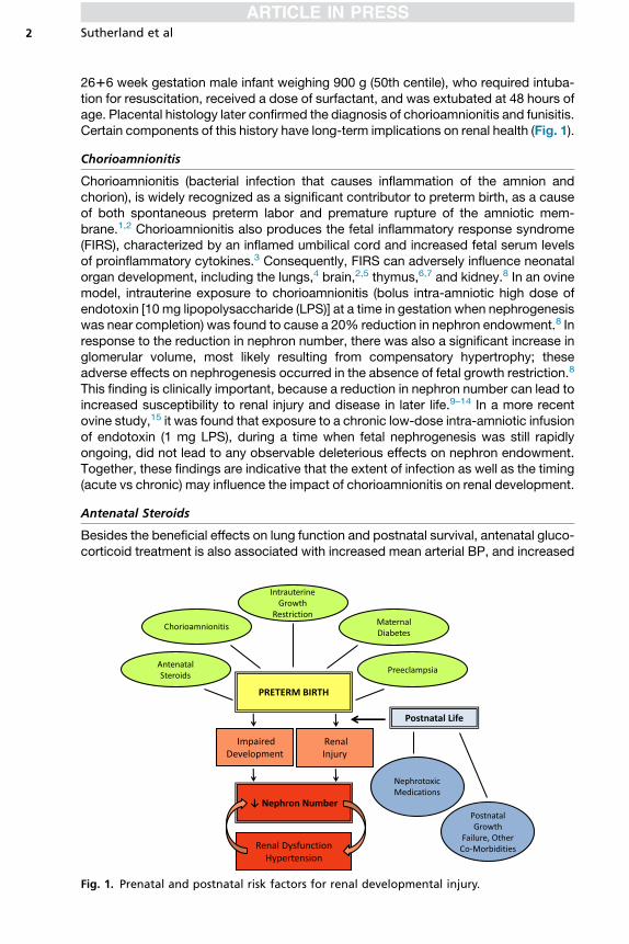
2616 week gestation male infant weighing 900 g (50th centile), who required intuba-tion for resuscitation, received a dose of surfactant, and was extubated at 48 hours ofage. Placental histology later confirmed the diagnosis of chorioamnionitis and funisitis.Certain components of this history have long-term implications on renal health (Fig. 1).
Chorioamnionitis
Chorioamnionitis (bacterial infection that causes inflammation of the amnion andchorion), is widely recognized as a significant contributor to preterm birth, as a causeof both spontaneous preterm labor and premature rupture of the amniotic mem-brane.1,2 Chorioamnionitis also produces the fetal inflammatory response syndrome(FIRS), characterized by an inflamed umbilical cord and increased fetal serum levelsof proinflammatory cytokines.3 Consequently, FIRS can adversely influence neonatalorgan development, including the lungs,4 brain,2,5 thymus,6,7 and kidney.8 In an ovinemodel, intrauterine exposure to chorioamnionitis (bolus intra-amniotic high dose ofendotoxin [10 mg lipopolysaccharide (LPS)] at a time in gestation when nephrogenesiswas near completion) was found to cause a 20% reduction in nephron endowment.8 Inresponse to the reduction in nephron number, there was also a significant increase inglomerular volume, most likely resulting from compensatory hypertrophy; theseadverse effects on nephrogenesis occurred in the absence of fetal growth restriction.8
This finding is clinically important, because a reduction in nephron number can lead toincreased susceptibility to renal injury and disease in later life.9–14 In a more recentovine study,15 it was found that exposure to a chronic low-dose intra-amniotic infusionof endotoxin (1 mg LPS), during a time when fetal nephrogenesis was still rapidlyongoing, did not lead to any observable deleterious effects on nephron endowment.Together, these findings are indicative that the extent of infection as well as the timing(acute vs chronic) may influence the impact of chorioamnionitis on renal development.
Antenatal Steroids
Besides the beneficial effects on lung function and postnatal survival, antenatal gluco-corticoid treatment is also associated with increased mean arterial BP, and increased
Fig. 1. Prenatal and postnatal risk factors for renal developmental injury.
Long-Term Renal Consequences of Preterm Birth 3
renal blood flow and glomerular filtration rate (GFR),16 indicating that acceleratedfunctional maturation of the kidney also occurs after treatment. Similarly, in studiesconducted in a baboon model of preterm birth,17 there was evidence of acceleratedrenal maturation with a greater number of mature nephrons present in the glucocorti-coid-exposed kidneys when examined both at the time of preterm delivery and at post-natal day 21. Although it has not been established, it is possible that this acceleratedmaturation results in an earlier cessation of nephrogenesis. In this regard, in other studiesperformed in animal models (including rodents18–20 and sheep21), antenatal glucocorti-coid administration was shown to lead to a significant reduction in nephron endowmentof the exposed offspring. A study by de Vries and colleagues22 similarly showed thatpostnatal administration of dexamethasone in the neonatal rat (at a time of ongoingpostnatal nephrogenesis) leads to a reduction in glomerular density.
Preterm Birth
The transition from an intrauterine to an extrauterine environment involves a sudden in-crease in blood oxygen concentrations, as well as increased systemic BP and organperfusion. The effects that this hemodynamic shiftmay have on the immature developingkidney (with nephrogenesis ongoing at the time of preterm birth) are poorly understood.After birth, the preterm kidney is functionally immature, with a low GFR, inability tomaintain electrolyte balance,23 and proteinuria.24 Acute kidney injury is also common.25
Some (but not all26) studies of kidney size in preterm neonates have shown significantlyreduced kidney length or volume compared with neonates born at term.27–30
Histomorphologic studies31 of renal tissue collected at autopsy have shown accel-erated postnatal maturation of the preterm kidney, with reduced nephrogenic zonewidth, reduced renal vesicle formation, and increased number of glomerular genera-tions compared with age-matched fetal controls. At the completion of nephrogenesis,a reduction in the number of glomerular generations formed (suggestive of a nephrondeficit) has also been reported.10 In addition, glomeruli that are abnormal in appear-ance (enlarged Bowman space and shrunken glomerular tuft) were present in the outerrenal cortex of preterm kidneys31; in some neonates, up to 13% of glomeruli wereaffected. These glomeruli have scant capillarization, suggestive of impaired vasculardevelopment or injury, which is likely to render them nonfunctional.17,32 In a baboonmodel of preterm birth, nephron density was found to be significantly lower in the pre-term kidneys, and nephron number was at the lower end of the normal distribution,17
and in mice delivered 1 to 2 days preterm, nephron number was significantlyreduced.33 Together, these findings suggest that preterm neonates may have botha reduced endowment of nephrons at the completion of nephrogenesis, as well a pre-disposition to nephron loss in the neonatal period, which may have important implica-tions for long-term renal health.
CLINICAL SCENARIO 1 (CONTINUED)
Because of the maternal history of clinical chorioamnionitis, the preterm infant wascommenced on ampicillin 25 mg/kg/dose 12 hourly and gentamicin 2.5 mg/kg/dose24 hourly, which were ceased at 48 hours of age after a negative blood culture. Onday 2 of life, an echocardiogram was performed, and the infant was found to have alarge patent ductus arteriosus, with an increased left atrium/aorta ratio. He wascommenced on intravenous indomethacin 0.2 mg/kg first dose, 0.1 mg/kg subse-quent doses daily for 5 days; however, at the end of the third day of treatment, hedeveloped abdominal distension, bilious aspirates, and hypotension and had anabdominal radiograph, with signs consistent with necrotizing enterocolitis. He
Sutherland et al4
required reintubation and inotropes, was not fed enterally for 10 days, and received7 days of ampicillin 25 mg/kg/dose 12 hourly, gentamicin 2.5 mg/kg/dose 24 hourly,and metronidazole 7.5 mg/kg/dose 48 hourly. He was recommenced on feeds butcontinued to have significant feed intolerance until full feeds were achieved on day30. He was extubated at 28 days of age, requiring dexamethasone, and continuedto require continuous positive airway pressure (CPAP) until 35 weeks postconcep-tional age and oxygen until 38 weeks postconceptional age. His weight on dischargewas 2.2 kg, which was less than the 3rd centile, having decreased from the 50th cen-tile at birth. This relatively common series of complications of extreme prematurityhave further renal implications.
Nephrotoxic Medications
The prenatal and postnatal administration of a variety of nephrotoxic medications iscommon practice in the setting of preterm birth.34–38 Experimental studies have shownthat antenatal exposure to the most commonly administered aminoglycoside antibiotic,gentamicin, causes significant reductions in nephronendowment.39Gentamicin accumu-lates in renal proximal tubule cells, leading to cellular necrosis40; postnatally, this results inincreased sodium excretion, proteinuria, and reduced GFR in affected neonates.41–43
Exposure of the developing kidney to b-lactam antibiotics (such as ampicillin) has alsobeen shown to result in impaired nephrogenesis and cystic tubular dilation.44
The consequences of antenatal nonsteroidal antiinflammatory drugs (NSAIDs)(indomethacin, ibuprofen) exposure on the fetal kidney is well described both in exper-imental models (nephron deficits, reduced cortical renal volume,45–47 and severe renaldysplasia48) and in human neonates (glomerular cysts and renal dysfunction49,50).However, less is known about the effects of postnatal NSAID exposure on the kidney.In general, the administration of NSAIDs causes systemic vasoconstriction, which inturn leads to significant reductions in renal blood flow and urine output.51–53 A studyin human preterm neonates54 recently showed significantly increased number ofpodocytes in the urine, and increased urine albumin excretion in those treated withindomethacin after birth, which indicates glomerular injury. Furthermore, an experi-mental study conducted in a neonatal rodent model55 showed that postnatal admin-istration of NSAIDs or gentamicin led to proximal tubule vacuolization, interstitialedema, and podocyte foot process effacement; the most severe effects wereobserved in animals that received combined NSAID and gentamicin treatment. In anonhuman primate (baboon) model,56 animals administered ibuprofen after pretermbirth had a significantly reduced nephrogenic zone width. This finding suggests thatprostaglandin inhibition may impair nephrogenesis. Kent and colleagues57 in theirearly study reported that there was no effect of early postnatal NSAID and gentamicinexposure on nephron endowment in a rat model at 14 days but have now shown thatthe adult rat has a significantly reduced nephron number after early administration ofindomethacin but not ibuprofen (Kent AL, Koina M, Gubhaju L, et al: Indomethacinadministered early in the postnatal period results in reduced glomeruli in the adultrat. Submitted for publication).
Growth Restriction
Over the past decade, it has been well established that intrauterine growth restriction(IUGR) results in a reduced nephron endowment58–62 and is linked to subsequent renaldysfunction.9,58,63–66 The number of nephrons formed during nephrogenesis in earlylife determines the lifelong functional capacity of the kidney; there have been a multi-tude of experimental studies (in many species) showing that the number of nephronsformed within the kidney is directly proportional to kidney size at birth but may not be
Long-Term Renal Consequences of Preterm Birth 5
consistent in fetuses that have been growth-restricted late in gestation (when nephro-genesis is completed).17,62,67,68 IUGR is often a comorbidity of preterm birth; it islinked to the cause of spontaneous preterm birth, and is also a common cause of inter-ventional preterm delivery, which is required to facilitate infant survival.69 Of greatestconcern, newborns who are born both prematurely and IUGR are at a high risk of infantmorbidity and mortality.70–73 Long-term studies in children and adults have found thatindividuals born both preterm and IUGR have an increased propensity for impairedkidney growth28,59,74 and renal dysfunction,75,76 compared with preterm individualswho were non-IUGR at birth. Besides impaired growth in utero, extrauterine growthrestriction is a common sequela of preterm birth and has been shown to have a simi-larly adverse effect on renal function.76
CLINICAL SCENARIO 2
A woman presented to the antenatal clinic at 2610 weeks’ gestation and was found tohave preeclampsia with BP of 140/95, an increased urate level, but at this stage,normal renal function and normal umbilical arterial Doppler results. She had a bodymass index (calculated as weight in kilograms divided by the square of height in me-ters) of 40 and had insulin-requiring gestational diabetes. The estimated fetal weightwas 660 g, which is on the 5th centile, with the fetal size at her 20-week morphologyscan being on the 78th centile. She was admitted to the antenatal ward andcommenced on oral antihypertensives (Labetalol 200 mg 8 hourly) and received ante-natal steroids. Over the next 2 weeks, her BP became more difficult to control and shewas on 2 antihypertensive medications on maximal doses (Labetalol, Methyldopa250 mg 6 hourly). Her renal function began to deteriorate, and the umbilical arterialDoppler results were increased. At 2814 weeks, she was given magnesium sulfatefor fetal neuroprotection, and the female neonate was delivered by cesarean sectionwith a birth weight of 720 g (10th centile). The placental histology confirmed uteropla-cental insufficiency, with accelerated maturation, increased syncytial knot formation,and areas of infarction and chorangiosis.
Maternal Disease
DiabetesThis clinical scenario has become increasingly more frequent in infants born tomothers with maternal diseases.77,78 With the increasing prevalence of obesity andassociated type 2 diabetes in the United States and other developed countries, theincidence of maternal diabetes is increasing.79 Intrauterine exposure to maternal dia-betes can lead to either macrosomia or microsomia of the fetus.80 Fetal macrosomia,specifically asymmetric macrosomia, results in exaggerated fetal growth in responseto the increased supply of glucose and other nutrients across the placenta.81 How-ever, conversely, in the case of severe maternal diabetes, maternal complicationssuch as vasculopathy and nephropathy may occur, which subsequently lead toIUGR of the developing fetus.In addition, maternal diabetes has been shown to have other adverse effects on
embryogenesis, which may further increase the risk of IUGR82 and consequently in-crease the susceptibility of impaired nephrogenesis and renal injury in the neonatalperiod. For example, in a study of Pima Indians with type 2 diabetes,83 there was foundto be a strong association with renal dysfunction after birth; 58% of infants who wereexposed to diabetes in utero showed 4 times higher urinary albumin excretion (albu-min/creatinine ratio >30 mg/g), compared with unexposed infants. Other studies84
have clearly shown a risk of renal malformations in infants born to diabetic mothers;
Sutherland et al6
the estimated risk of delivering a child with renal agenesis/dysgenesis is more than 3times greater for mothers with diabetes compared with nondiabetic mothers. Animalstudies have further highlighted the adverse effects of intrauterine exposure tomaternal diabetes on kidney development and function. In the mouse model,82 ahigh-glucose intrauterine milieu led to significantly smaller body length, kidney size,and glomerular size, and a 40% reduction in nephron endowment compared with con-trol offspring; the level of maternal hyperglycemia predicted the severity of the adverseeffects on kidney size and on glomerular endowment. There was also evidence ofglomerular collapse as a result of an increase in glomerular and tubular apoptoticevents.82 In follow-up studies,85 20-week-old mice offspring exposed to maternal dia-betes were 20% lighter, with significantly increased urinary albumin excretion, glomer-ular hypertrophy, renal fibrosis, and an increased propensity for the development ofhypertension.
PreeclampsiaPreeclampsia is known to be a major risk factor for fetal and neonatal mortality,IUGR, and preterm birth; maternal hypertension alone also increases risk, but toa lesser extent than preeclampsia.86 Besides inducing growth restriction viaimpaired uteroplacental perfusion, preeclampsia also stimulates a proinflammatory,prooxidant and antiangiogenic intrauterine environment.87 In animal models, thissetting is known to lead to IUGR, decreased kidney size, hypertension, andimpaired vascularization of the developing lung88,89; however, potential effects onthe vascular development of other organs are unknown. Among children born pre-term, maternal preeclampsia is an independent risk factor for the development ofhypertension.90
CLINICAL SCENARIO 2 (CONTINUED)
This 28-week gestation baby required CPAP for 3 weeks, took 3 weeks to achieve fullfeeds, and went home at 40 weeks postconceptional age formula feeding weighing3.1 kg (10th centile). Over the next 12 months, her weight gain increased significantly,such that her weight at 12 months of age was 11.4 kg (95th centile).
Obesity After Growth Restriction
The increasing epidemic of obesity worldwide has also seen a resultant increase inchronic renal failure,91 and there is mounting evidence of the adverse effects of obesityon renal hemodynamics and structure.92 The preterm infant has several early renalhits, which may compromise long-term renal function, and this is further compoundedlater in life by insults such as the induction of obesity; this supports the multihit natureof chronic renal disease.93 Preterm birth and the associated renal risk factors outlinedso far have the potential to significantly reduce nephron number. With the increasedfunctional demands (as seen with obesity), there is likely to be accelerated onsetand increased severity of renal disease when glomerular number (functional reserve)is low as a result of growth restriction early in life. A reduced nephron endowment sub-sequently leads to glomerular hypertrophy, and this is likely to be greatly accentuatedwhen the functional demands on the kidneys are increased. Furthermore, the sus-tained glomerular hyperfiltration may lead to glomerular dysfunction and subsequentglomerular demise.93 Supporting this theory, Abitbol and colleagues94 have shownthat obesity and preterm birth are additive risks in the progression of kidney diseasein children.
Long-Term Renal Consequences of Preterm Birth 7
Long-Term Consequences of Preterm Birth on the Kidney
As described, there are a multitude of factors associated with preterm birth that havethe potential to impair renal development in the fetus or neonate. Impaired growth anda reduced nephron endowment are strongly linked to the later development of hyper-tension, glomerular injury, and renal dysfunction.95,96 In this regard, there is now anincreasing body of evidence supporting the hypothesis that preterm birth has long-term consequences for health; in particular, preterm birth has been strongly linkedto increased hypertension risk in both children and adults.97 In children born preterm,most studies have reported a reduction in kidney size compared with age-matchedindividuals born at term.98–100 Zaffanello and colleagues99 further reported a signifi-cantly decreased kidney size in children born at 26 to 28 weeks’ gestation, comparedwith those born at 30 to 31 weeks’ gestation, indicating that the severity of prematurityhas an important effect on kidney growth. Only 1 study has examined the effect of pre-term birth on kidney size in adulthood. In this study, Keijzer-Veen and colleagues101
found that at 20 years of age, after preterm birth at less than 32 weeks’ gestation, fe-male adults had a significantly decreased kidney length and volume (both absoluteand relative) compared with individuals born at term.However, findings from the few studies to have examined long-term renal function
after preterm birth are less conclusive. Rodriguez-Soriano and colleagues102 andIacobelli and colleagues103 showed that GFR was significantly reduced in preterm-born children compared with term controls, with impairments in electrolyte excretion
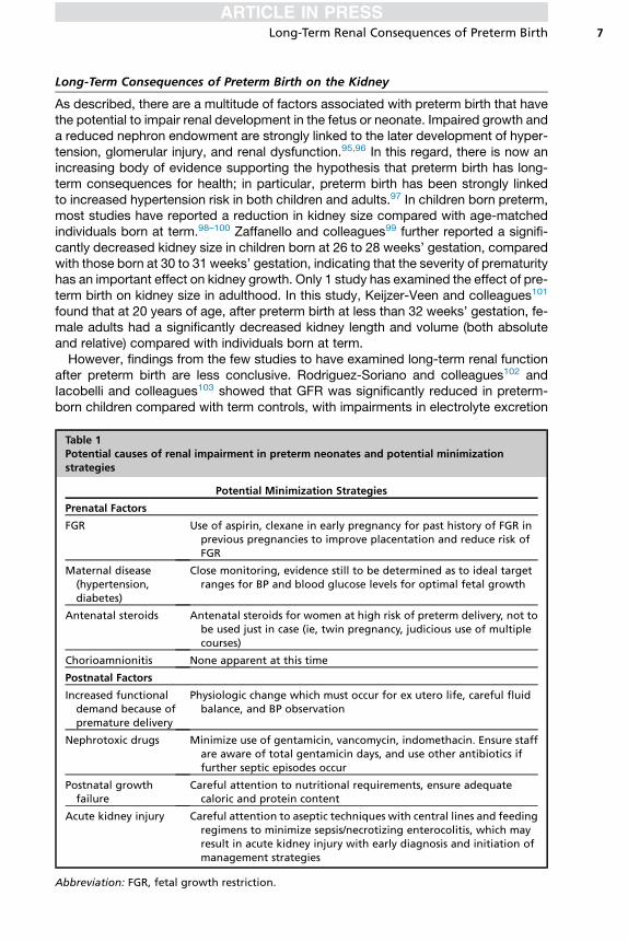
Table 1Potential causes of renal impairment in preterm neonates and potential minimizationstrategies
Potential Minimization Strategies
Prenatal Factors
FGR Use of aspirin, clexane in early pregnancy for past history of FGR inprevious pregnancies to improve placentation and reduce risk ofFGR
Maternal disease(hypertension,diabetes)
Close monitoring, evidence still to be determined as to ideal targetranges for BP and blood glucose levels for optimal fetal growth
Antenatal steroids Antenatal steroids for women at high risk of preterm delivery, not tobe used just in case (ie, twin pregnancy, judicious use of multiplecourses)
Chorioamnionitis None apparent at this time
Postnatal Factors
Increased functionaldemand because ofpremature delivery
Physiologic change which must occur for ex utero life, careful fluidbalance, and BP observation
Nephrotoxic drugs Minimize use of gentamicin, vancomycin, indomethacin. Ensure staffare aware of total gentamicin days, and use other antibiotics iffurther septic episodes occur
Postnatal growthfailure
Careful attention to nutritional requirements, ensure adequatecaloric and protein content
Acute kidney injury Careful attention to aseptic techniques with central lines and feedingregimens to minimize sepsis/necrotizing enterocolitis, which mayresult in acute kidney injury with early diagnosis and initiation ofmanagement strategies
Abbreviation: FGR, fetal growth restriction.
Sutherland et al8
also evident. In children examined at 6 to 8 years of age, Iacobelli and colleagues103
also found microalbuminuria in 8.3% of the preterm children. In contrast, other studiesin children98,105 and young adults104,106 born preterm reported no difference in renalfunction compared with term controls. However, survival after extremely preterm birthis a relatively recent phenomenon. Hence, it is likely that as the increasing populationworldwide of survivors of very and extremely preterm birth reach middle and olderage, the adverse long-term consequences of preterm birth will become increasinglyevident. We need to continue to look carefully at the evidence available and try to mini-mize known potential adverse effects on the developing kidney (Table 1) as well aspursuing further research in this arena to prevent long-term health sequelae.
SUMMARY
The early life environment, as first postulated by Barker,107 has the potential to in-fluence future cardiovascular health risks. The developing kidney of the preterm infantmay be affected by several in utero and neonatal insults that may influence nephro-genesis, resulting in reduced functional nephron number at the beginning of life.This reduction in nephron number may lead to vulnerability to hypertension in adult-hood, increasing cardiovascular risks for myocardial infarction and stroke, as wellas impaired renal function. Ongoing research is required into the potential risks tonephrogenesis, along with ways to minimize harm and maximize nephron numberand function in preterm neonates to reduce the risk of long-term renal and cardiovas-cular sequelae.
REFERENCES
1. Mueller-Heubach E, Rubinstein DN, Schwarz SS. Histologic chorioamnionitisand preterm delivery in different patient populations. Obstet Gynecol 1990;75(4):622–6.
2. Goldenberg RL, Hauth JC, Andrews WW. Intrauterine infection and pretermdelivery. N Engl J Med 2000;342(20):1500–7.
3. Gantert M, Been JV, Gavilanes AW, et al. Chorioamnionitis: a multiorgan diseaseof the fetus. J Perinatol 2010;30(Suppl 1):s21–30.
4. Kallapur SJ, Jobe AH. Contribution of inflammation to lung injury and develop-ment. Arch Dis Child Fetal Neonatal Ed 2006;91:F132–5.
5. De Felice C, Toti P, Laurini RN, et al. Early neonatal brain injury in histologic cho-rioamnionitis. J Pediatr 2001;138(1):101–4.
6. Toti P, De Felice C, Stumpo M, et al. Acute thymic involution in fetuses and ne-onates with chorioamnionitis. Hum Pathol 2000;31(9):1121–8.
7. Kunzmann S, Glogger K, Been JV, et al. Thymic changes after chorioamnionitisinduced by intraamniotic lipopolysaccharide in fetal sheep. Am J Obstet Gyne-col 2010;190:e471–9.
8. Galinsky R, Moss TJ, Gubhaju L, et al. Effect of intra-amniotic lipopolysaccha-ride on nephron number in preterm fetal sheep. Am J Physiol Renal Physiol2011;301(2):F280–5.
9. Brenner B, Garcia D, Anderson S. Glomeruli and blood pressure. Less of one,more the other? Am J Hypertens 1988;1(4 Pt 1):335–47.
10. Rodrıguez MM, Gomez AH, Abitbol CL, et al. Histomorphometric analysis ofpostnatal glomerulogenesis in extremely preterm infants. Pediatr Dev Pathol2004;7(1):17–25.
11. Hoy WE, Hughson MD, Bertram JF, et al. Nephron number, hypertension, renaldisease, and renal failure. J Am Soc Nephrol 2005;16(9):2557–64.
Long-Term Renal Consequences of Preterm Birth 9
12. Zandi-Nejad K, Luyckx V, Brenner B. Adult hypertension and kidney disease:the role of fetal programming. Hypertension 2006;47(3):502–8.
13. Nehiri T, Duong Van Huyen JP, Viltard M, et al. Exposure to maternal diabetesinduces salt-sensitive hypertension and impairs renal function in adult ratoffspring. Diabetes 2008;57(8):2167–75.
14. Gray IP, Cooper PA, Cory BJ, et al. The intrauterine environment is a strongdeterminant of glucose tolerance during the neonatal period, even in prematu-rity. J Clin Endocrinol Metab 2002;87(9):4252–6.
15. Ryan D, Atik A, De Matteo R, et al. Chronic intrauterine exposure to endotoxindoes not alter fetal nephron number or glomerular size. Clin Exp PharmacolPhysiol 2013;40(11):789–94.
16. Jahnukainen T, Chen M, Berg U, et al. Antenatal glucocorticoids and renal func-tion after birth. Semin Neonatol 2001;6(4):351–5.
17. Gubhaju L, Sutherland MR, Yoder BA, et al. Is nephrogenesis affected by pre-term birth? Studies in a non-human primate model. Am J Physiol Renal Physiol2009;297(6):F1668–77.
18. Celsi G, Kistner A, Aizman R, et al. Prenatal dexamethasone causes oligoneph-ronia, sodium retention, and higher blood pressure in offspring. Pediatr Res1998;44:317–22.
19. Ortiz LA, Quan A, Weinberg A, et al. Effect of prenatal dexamethasone on ratrenal development. Kidney Int 2001;59(5):1663–9.
20. Ortiz LA, Quan A, Zarzar F, et al. Prenatal dexamethasone programs hyperten-sion and renal injury in the rat. Hypertension 2003;41(2):328–34.
21. Wintour EM, Moritz KM, Johnson K, et al. Reduced nephron number in adultsheep, hypertensive as a result of prenatal glucocorticoid treatment. J Physiol2003;549(Pt 3):929–35.
22. de Vries WB, van den Borne P, Goldschmeding R, et al. Neonatal dexametha-sone treatment in the rat leads to kidney damage in adulthood. Pediatr Res2010;67(1):72–6.
23. Aperia A, Broberger O, Elinder G, et al. Postnatal development of renal functionin pre-term and full-term infants. Acta Paediatr Scand 1981;70(2):183–7.
24. Tsukahara H, Fujii Y, Tsuchida S, et al. Renal handling of albumin and beta-2-microglobulin in neonates. Nephron 1994;68(2):212–6.
25. Walker MW, Clark RH, Spitzer AR. Elevation in plasma creatinine and renal fail-ure in premature neonates without major anomalies: terminology, occurrenceand factors associated with increased risk. J Perinatol 2011;31(3):199–205.
26. Kent AL, Jyoti R, Robertson C, et al. Does extreme prematurity affect kidney vol-ume at term corrected age? J Matern Fetal Neonatal Med 2009;22(5):435–8.
27. Schmidt IM, Chellakooty M, Boisen KA, et al. Impaired kidney growth in low-birth-weight children: distinct effects of maturity and weight for gestationalage. Kidney Int 2005;68(2):731–40.
28. Drougia A, Giapros V, Hotoura E, et al. The effects of gestational age and growthrestriction on compensatory kidney growth. Nephrol Dial Transplant 2009;24(1):142–8.
29. Huang HP, Tsai IJ, Lai YC, et al. Early postnatal renal growth in premature in-fants. Nephrology (Carlton) 2007;12(6):572–5.
30. Kandasamy Y, Smith R, Wright IM, et al. Extra-uterine renal growth in preterminfants: oligonephropathy and prematurity. Pediatr Nephrol 2013;28(9):1791–6.
31. Sutherland MR, Gubhaju L, Moore L, et al. Accelerated maturation and ab-normal morphology in the preterm neonatal kidney. J Am Soc Nephrol 2011;22(7):1365–74.
Sutherland et al10
32. Gubhaju L, Sutherland MR, Black MJ. Preterm birth and the kidney: implicationsfor long-term renal health. Reprod Sci 2011;18(4):322–33.
33. Stelloh C, Allen KP, Mattson DL, et al. Prematurity in mice leads to reductionin nephron number, hypertension, and proteinuria. Transl Res 2012;159(2):80–9.
34. Schreuder MF, Bueters RR, Huigen MC, et al. Effect of drugs on renal develop-ment. Clin J Am Soc Nephrol 2011;6(1):212–7.
35. Cullen L, Young R, Bertram J. Studies on the effects of gentamicin on rat meta-nephric development in vitro. Nephrology 2000;5:115–23.
36. Gilbert T, Gaonach S, Moreau E, et al. Defect of nephrogenesis induced bygentamicin in rat metanephric organ culture. Lab Invest 1994;70:656–66.
37. Gilbert T, Lelievre-Pegorier M, Merlet-Benichou C. Immediate and long-termrenal effects of fetal exposure to gentamicin. Pediatr Nephrol 1990;4:445–50.
38. Gilbert T, Lelievre-Pegorier M, Malienou R, et al. Effects of prenatal and post-natal exposure to gentamicin on renal differentiation in the rat. Toxicology1987;43(3):301–13.
39. Mallie JP, Gerard H, Gerard A. In-utero gentamicin-induced nephrotoxicity inrats. Pediatr Pharmacol (New York) 1986;5(4):229–39.
40. Nagai J, Takano M. Molecular aspects of renal handling of aminoglycosides andstrategies for preventing the nephrotoxicity. Drug Metab Pharmacokinet 2004;19(3):159–70.
41. Giapros V, Andronikou SK, Cholevas VI, et al. Renal function and effect of ami-noglycoside therapy during the first ten days of life. Pediatr Nephrol 2003;18:46–52.
42. Giapros VI, Andronikou SK, Cholevas VI, et al. Acute effects of gentamicin onglomerular and tubular functions in preterm neonates. Pediatr Nephrol 2006;21(10):1389–92.
43. Langhendries JP, Battisti O, Bertrand J. Aminoglycoside nephrotoxicity andurinary excretion of N-acetyl-beta-D-glucosaminidase. Biol Neonate 1988;53:253–9.
44. Nathanson S, Moreau E, Merlet-Benichou C, et al. In utero and in vitro exposureto beta-lactams impair kidney development in the rat. J Am Soc Nephrol 2000;11(5):874–84.
45. Saez F, Reverte V, Salazar F, et al. Hypertension and sex differences in the age-related renal changes when cyclooxygenase-2 activity is reduced during neph-rogenesis. Hypertension 2009;53(2):331–7.
46. Komhoff M, Wang JL, Cheng HF, et al. Cyclooxygenase-2-selective inhibitorsimpair glomerulogenesis and renal cortical development. Kidney Int 2000;57(2):414–22.
47. Olliges A, Wimmer S, Nusing RM. Defects in mouse nephrogenesis induced byselective and non-selective cyclooxygenase-2 inhibitors. Br J Pharmacol 2011;163(5):927–36.
48. Norwood VF, Morham SG, Smithies O. Postnatal development and progressionof renal dysplasia in cyclooxygenase-2 null mice. Kidney Int 2000;58(6):2291–300.
49. Kaplan BS, Restaino I, Raval DS, et al. Renal failure in the neonate associatedwith in utero exposure to non-steroidal anti-inflammatory agents. Pediatr Neph-rol 1994;8(6):700–4.
50. van der Heijden BJ, Carlus C, Narcy F, et al. Persistent anuria, neonatal death,and renal microcystic lesions after prenatal exposure to indomethacin. Am JObstet Gynecol 1994;171(3):617–23.
Long-Term Renal Consequences of Preterm Birth 11
51. Sener A, Smith FG. Glomerular and tubular responses to N(G)-nitro-L-argininemethyl ester are age dependent in conscious lambs. Am J Physiol Regul IntegrComp Physiol 2002;282(5):R1512–20.
52. Keller RL, Tacy TA, Fields S, et al. Combined treatment with a nonselectivenitric oxide synthase inhibitor (l-NMMA) and indomethacin increases ductusconstriction in extremely premature newborns. Pediatr Res 2005;58(6):1216–21.
53. Kang NS, Yoo KH, Cheon H, et al. Indomethacin treatment decreases renalblood flow velocity in human neonates. Biol Neonate 1999;76(5):261–5.
54. Kent AL, Brown L, Broom M, et al. Increased urinary podocytes following indo-methacin suggests drug-induced glomerular injury. Pediatr Nephrol 2012;27(7):1111–7.
55. Kent AL, Maxwell LE, Koina ME, et al. Renal glomeruli and tubular injuryfollowing indomethacin, ibuprofen, and gentamicin exposure in a neonatal ratmodel. Pediatr Res 2007;62(3):307–12.
56. Sutherland MR, Yoder BA, McCurnin D, et al. Effects of ibuprofen treatment onthe developing preterm baboon kidney. Am J Physiol Renal Physiol 2012;302(10):F1286–92.
57. Kent AL, Douglas-Denton R, Shadbolt B, et al. Indomethacin, ibuprofen andgentamicin administered during late stages of glomerulogenesis do not reduceglomerular number at 14 days of age in the neonatal rat. Pediatr Nephrol 2009;24(6):1143–9.
58. Hinchliffe SA, Lynch MR, Sargent PH, et al. The effect of intrauterine growthretardation on the development of renal nephrons. Br J Obstet Gynaecol1992;99:296–301.
59. Manalich R, Reyes L, Herrera M, et al. Relationship between weight at birth andthe number and size of renal glomeruli in humans: a histomorphometric study.Kidney Int 2000;58:770–3.
60. Zimanyi MA, Denton KM, Forbes JM, et al. A developmental nephron deficit inrats is associated with increased susceptibility to a secondary renal injury dueto advanced glycation end-products. Diabetologia 2006;49(4):801–10.
61. Makrakis J, Zimanyi MA, Black MJ. Retinoic acid enhances nephron endowmentin rats exposed to maternal protein restriction. Pediatr Nephrol 2007;22(11):1861–7.
62. Zohdi V, Moritz KM, Bubb KJ, et al. Nephrogenesis and the renal renin-angiotensin system in fetal sheep: effects of intrauterine growth restriction dur-ing late gestation. Am J Physiol Regul Integr Comp Physiol 2007;293(3):R1267–73.
63. Brenner BM, Chertow GM. Congenital oligonephropathy and the etiology ofadult hypertension and progressive renal injury. Am J Kidney Dis 1994;23(2):171–5.
64. Merlet-Benichou C, Gilbert T, Muffat-Joly M, et al. Intrauterine growth retardationleads to a permanent nephron deficit in the rat. Pediatr Nephrol 1994;8(2):175–80.
65. Bassan H, Trejo LL, Kariv N, et al. Experimental intrauterine growth retardationalters renal development. Pediatr Nephrol 2000;15:192–5.
66. White SL, Perkovic V, Cass A, et al. Is low birth weight an antecedent of CKD inlater life? A systematic review of observational studies. Am J Kidney Dis 2009;54(2):248–61.
67. Gubhaju L, Black MJ. The baboon as a good model for studies of human kidneydevelopment. Pediatr Res 2005;58(3):505–9.
Sutherland et al12
68. Sutherland MR, Gubhaju L, Yoder BA, et al. The effects of postnatal retinoic acidadministration on nephron endowment in the preterm baboon kidney. PediatrRes 2009;65(4):397–402.
69. Diderholm B. Perintatal energy metabolism with reference to IUGR and SGA:studies in pregnant women and newborn infants. Indian J Med Res 2009;130:612–7.
70. Behrman RE, Lees MH, Peterson EN, et al. Distribution of the circulation in thenormal and asphyxiated fetal primate. Am J Obstet Gynecol 1970;108(6):956–69.
71. Lang U, Baker RS, Braems G, et al. Uterine blood flow–a determinant of fetalgrowth. Eur J Obstet Gynecol Reprod Biol 2003;110(Suppl 1):S55–61.
72. Kiserud T. Physiology of the fetal circulation. Semin Fetal Neonatal Med 2005;10(6):493–503.
73. Cox B, Kotlyar M, Evangelou AI, et al. Comparative systems biology of humanand mouse as a tool to guide the modeling of human placental pathology.Mol Syst Biol 2009;5:279.
74. Woods L, Weeks D, Rasch R. Programming of adult blood pressure by maternalprotein restriction: role of nephrogenesis. Kidney Int 2004;65(4):1339–48.
75. Keijzer-Veen MG, Schrevel M, Finken MJ, et al, Dutch POPS-19 CollaborativeStudy Group. Microalbuminuria and lower glomerular filtration rate at youngadult age in subjects born very premature and after intrauterine growth retarda-tion. J Am Soc Nephrol 2005;16(9):2762–8.
76. Bacchetta J, Harambat J, Dubourg L, et al. Both extrauterine and intrauterinegrowth restriction impair renal function in children born very preterm. KidneyInt 2009;76(4):445–52.
77. Poston L. Intergenerational transmission of insulin resistance and type 2 dia-betes. Prog Biophys Mol Biol 2011;106(1):315–22.
78. Boney CM, Verma A, Tucker R, et al. Metabolic syndrome in childhood: associ-ation with birth weight, maternal obesity, and gestational diabetes mellitus. Pe-diatrics 2005;115(3):e290–6.
79. Kim SY, England JL, Sharma JA, et al. Gestational diabetes mellitus and riskof childhood overweight and obesity in offspring: a systematic review. ExpDiabetes Res 2011;2011:541308.
80. Aerts L, Holemans K, Van Assche FA. Maternal diabetes during pregnancy:consequences for the offspring. Diabetes Metab Rev 1990;6(3):147–67.
81. Van Assche FA, Holemans K, Aerts L. Long-term consequences for offspring ofdiabetes during pregnancy. Br Med Bull 2001;60:173–82.
82. Tran S, Chen YW, Chenier I, et al. Maternal diabetes modulates renal morpho-genesis in offspring. J Am Soc Nephrol 2008;19(5):943–52.
83. Nelson RG, Morgenstern H, Bennet PH. Intrauterine diabetes exposure and therisk of renal disease in diabetic Pima Indians. Diabetes 1998;47:1489–93.
84. Davis EM, Peck JD, Thompson D, et al. Maternal diabetes and renal agenesis/dysgenesis. Birth Defects Res 2010;88:722–7.
85. Chen YW, Chenier I, Tran S, et al. Maternal diabetes programs hypertension andkidney injury in offspring. Pediatr Nephrol 2010;25(7):1319–29.
86. Villar J, Carroli G, Wojdyla D, et al, World Health Organization Antenatal CareTrial Research Group. Preeclampsia, gestational hypertension and intrauterinegrowth restriction, related or independent conditions? Am J Obstet Gynecol2006;194(4):921–31.
87. Suppo de Souza Rugolo LM, Bentlin MR, Trindade CE. Preeclampsia: effect onthe fetus and newborn. Neo Reviews 2011;12:e198–206.
Long-Term Renal Consequences of Preterm Birth 13
88. Alexander BT. Placental insufficiency leads to development of hypertension ingrowth-restricted offspring. Hypertension 2003;41(3):457–62.
89. Tang JR, Karumanchi SA, Seedorf G, et al. Excess soluble vascular endothelialgrowth factor receptor-1 in amniotic fluid impairs lung growth in rats: linking pre-eclampsia with bronchopulmonary dysplasia. Am J Physiol Lung Cell Mol Phys-iol 2012;302(1):L36–46.
90. Vohr BR, Allan W, Katz KH, et al. Early predictors of hypertension in prematurelyborn adolescents. Acta Paediatr 2010;99(12):1812–8.
91. Ejerblad E, Fored CM, Lindblad P, et al. Obesity and risk for chronic renal failure.J Am Soc Nephrol 2006;17:1695–702.
92. Mallamaci F, Tripepi G. Obesity and CKD progression: hard facts on fat CKD pa-tients. Nephrol Dial Transplant 2013;28(Suppl 4):iv105–8.
93. Nenov VD, Taal MW, Sakharova OV, et al. Multi-hit nature of chronic renal dis-ease. Curr Opin Nephrol Hypertens 2000;9:85–97.
94. Abitbol CL, Chandar J, Rodriguez MM, et al. Obesity and preterm birth: additiverisks in the progression of kidney disease in children. Pediatr Nephrol 2009;24(7):1363–70.
95. Hoy WE, Bertram JF, Denton RD, et al. Nephron number, glomerular volume,renal disease and hypertension. Curr Opin Nephrol Hypertens 2008;17(3):258–65.
96. Luyckx VA, Bertram JF, Brenner BM, et al. Effect of fetal and child health onkidney development and long-term risk of hypertension and kidney disease.Lancet 2013;382(9888):273–83.
97. Sutherland MR, Bertagnolli M, Lukaszewski MA, et al. Preterm birth and hyper-tension risk: the oxidative stress paradigm. Hypertension 2014;63(1):12–8.
98. Rakow A, Johansson S, Legnevall L, et al. Renal volume and function in school-age children born preterm or small for gestational age. Pediatr Nephrol 2008;23(8):1309–15.
99. Zaffanello M, Brugnara M, Bruno C, et al. Renal function and volume of infantsborn with a very low birth-weight: a preliminary cross-sectional study. Acta Pae-diatr 2010;99(8):1192–8.
100. Kwinta P, Klimek M, Drozdz D, et al. Assessment of long-term renal complica-tions in extremely low birth weight children. Pediatr Nephrol 2011;26(7):1095–103.
101. Keijzer-Veen MG, Devos AS, Meradji M, et al. Reduced renal length and volume20 years after very preterm birth. Pediatr Nephrol 2010;25:499–507.
102. Rodrıguez-Soriano J, Aguirre M, Oliveros R, et al. Long-term renal follow-up ofextremely low birth weight infants. Pediatr Nephrol 2005;20:579–84.
103. Iacobelli S, Loprieno S, Bonsante F, et al. Renal function in early childhood invery low birthweight infants. Am J Perinatol 2007;24(10):587–92.
104. Keijzer-Veen MG, Kleinveld HA, Lequin MH, et al. Renal function and size atyoung adult age after intrauterine growth restriction and very premature birth.Am J Kidney Dis 2007;50(4):542–51.
105. Vanpee M, Blennow M, Linne T, et al. Renal function in very low birth weightinfants: normal maturity reached during early childhood. J Pediatr 1992;121(5 Pt 1):784–8.
106. Kistner A, Celsi G, Vanpee M, et al. Increased blood pressure but normal renalfunction in adult women born preterm. Pediatr Nephrol 2000;15(3–4):215–20.
107. Barker DJP, Osmond C. Infant mortality, childhood, nutrition, and ischaemicheart disease in England and Wales. Lancet 1986;1(8489):1077–81.