Embed Size (px)
Citation preview

Eur Aesplr J, 1993, 8, 599-606 Printed In UK • all rtghte reaerved
EXPERT GROUP REPORT
Copyright CERS Journala Ltd 1993 European Respiratory Journal
ISSN 0903 • 1938
Long-term nasal ventilation In neuromuscular disorders: report of a Consensus Conference
D. Robert*, T.N. Willig**, J. Paulus**
Long-term nasal ventilation in neuromuscular disorders: report of ta Consensus Con· ference. D. Robert, T.N. Willig, J. Paulus. @ERS Journals Ltd. 1993.
"HOpital de la Croix Rousse, Lyon, France, • • Association Fran~aise contre les Myopathies ABSTRACf: Following the colloquium entitled "Nasal Ventilation and Neuromus·
cular Disease", which took place In Lyon, France, October 24, 1991, a group or authorities on neuromuscular disorders met to establish a consensus concerning the application or this technique, In the clinical setting, for patients with Duchenne's muscular dystrophy, non-Duchenne myopathJes and the spinal muscular atrophies. This report summari.zes recommendations Issuing from this conference. The conclusions drawn from this work should not be applied to patients with other dlag· noses.
Correspondence: D. Robert Service de R~aninlation et Assistance Respiratoire HOpitalla Croix • Rousse 93 Grande rue de la Croix·Rousse 69317 Lyon Cedex 4, France
Eur Respir J., 1993, 6, 599-606
Respiratory muscle involvement occurs in many neuro· muscular disorders and, unless treated, respiratory failure is usually the cause of death. This is particularly the case for individuals with Duchenne's muscular dystrophy, for whom rapid clinical progression almost invariably results in respiratory failure during late adolescence. Likewise, for individuals with spinal muscular atrophy, congenital muscular dystrophy, or other congenital myopathy, respiratory muscle involvement can be severe and can necessitate ventilatory support in early childhood.
A number of techniques of respiratory assistance can now be used. Non-invasive respiratory aids including positive pressure methods by mouthpiece or nasal mask have been thoroughly explored during the last few years. There is now a need to establish indications for their use and to understand their limitations in the various clinical settings involving neuromuscular patients. The multi· plicity of techniques, both invasive and non-invasive, currently available makes it difficult for centres with lit· tie experience to know which to use, and under what circumstances. At the same time, current research is providing better tolerated and less costly non-invasive methods. Furthermore, although it is firmly established that ventilatory assistance can both prolong and improve quality of life, the use of many non-invasive techniques is not included in the education of health professionals, nor is their use understood or appreciated in many health centres.
This conference was held in October 25, 1991 in Lyon
Keywords: Nasal ventilation neuromuscular disorders
Received: December 16 1992 Accepted after revision February 17 1993
and brought together experience from many centres. The objective was to defme the role of nasal ventilation in the treatment of neuromuscular ventilatory insufficiency incorporating all available methods, and to focus and coordinate related research efforts.
Publications elaborating the topics presented at this conference appear in the recent issue of the European Respiratory Review [1].
Use of nasal ventilation
Early, informed and detailed counselling of the patient and family includes the setting of therapeutic goals, and overcoming practical concerns in order to ensure longterm co-operation. Among the fundamental concerns to be addressed are: the wishes and motivation of the patient, the concerns of the patient's care-givers, the capacity to manage acute intercurrent illnesses, the ability to preserve an effective assisted cough, and the presence of impaired swallowing (not including the iatrogenic impairment resulting from intubation).
Nasal ventilation [2] can be introduced to the patient in a variety of clinical settings, including intensive care, rehabilitation, or ambulatory care. When introduction is problematical, the patient should be referred to experi· enced clinicians and specialized centres, equipped to monitor physiological parameters and the patient's progress using nasal ventilation during sleep. There was a clear consensus on the need for a multidisciplinary
Expert Participants: J.R. Bach, (UMD·The New Jersey Medical School, Newark, NJ, USA), A Barois, (HOpital Raymond Poincar~. Garches, France), J.C. Chevrolet, (HOphaJ Cantont.l Universitaire, Geneva, Switzerland), A Durocber, (HOpital Calmctte, Lille, France), B. Echenne, (Centre Gui de Chauliac, MootpeWer, France), D. Floret, (HOpital Edouard Heniot, Lyon, France), P. Gajdos, (HOpital Raymood Poincar~. Garcbes, France). P. Leger, (HOpital de la Croix Rousse, Lyon, France), J.M. Potu, (HOpitaux de Brabois, Vandoeuvre Les Nancy, France).

600 D. ROBERT, T.N. Wll.lJG, J. PAULUS
approach for the successful institution of nasal ventilation. The need for a specifically trained and dedicated treatment team, with specialized equipment and supplies, must also be appreciated by medical administrators. It should be noted that the institution of nocturnal nasal ventilation permits the patient and his family to request benefits from the third Complement d'Allocation d'Education Speciale, for those living in France.
Technical aspects
Ventilators and accessory respiratory equipment and sup· plies [3]
Masks. Nasal masks [4], which optimize efficacy and comfort, must form an airtight seal around the nose, apply minimal pressure to skin contact surfaces, add little dead space to the ventilator circuitry, be light, quickly and easily fabricated, durable, easily put on, and positioned firmly and non-traumatically. Except during emergency situations, the use of custom-moulded masks is preferred. These may include masks made from facial impressions or moulages, a technique which requires technical skill, but generally results in a better adapted and tolerated mask. A firm outer shell ensures a firmer fixation over the nose. Such an outer shell might be fabricated individually for each nasal mask, or it may be commercially available in different sizes. The gasket between the shell and skin surface may be constructed in soft silicone-based material.
Masks are difficult to construct from a facial moulage; however, for adults, they can, at least in theory, be conveniently reconstructed from the same moulage. This potential advantage does not exist for the paediatric or adolescent patient, with rapidly changing dentition.
Industrially prepared stock nasal masks include models which cover the nose (Respironics, Monroeville, P A, USA; Healthdyne, Cedar Grove, NJ, USA), or systems which involve cannulation of the nostrils (Adam circuit by Puritan-Bennett Boulder, CO, USA). These readily obtainable models appear to be most useful in the emergency setting. Occasionally, however, they can be readily tolerated by patients for long-term use.
There was no consensus concerning the use of custom prepared oral masks or oronasal masks with bite plate retention, since there is little in the literature concerning these appliances, and fabrication is both effort-intensive and time-consuming. However, because of the potential for aspiration of vomit, the nocturnal use of oronasal masks, with or without strap retention, is contraindicated in the paediatric population, and use of strap-retained appliances is also contraindicated for any individual without upper extremity function. These precautions do not pertain to adults using oral or oronasal masks with bite plate retention, since these appliances can be quickly extruded by tongue thrust in the event of need for immediate removal.
Respirators. Taking into account that experience with recently released pressure-cycled ventilators continues to
be limited, and that only volume-cycled ventilators are readily available for home mechanical ventilation, there was consensus on generally recommending the use of volume-cycled ventilators in the assist control or control mode. Recommended ventilator settings are: 1) frequency usually near that of spontaneous breathing at 15-20 cycles·min·', or greater for small children; 2) an inspiratory/expiratory (liE) ratio of 1/2 to 1/1; and 3) a relatively large delivered tidal volume of 15-20 ml·kg-1, since an undetermined volume will leak around the mask or out of the mouth.
Assisted ventilation should usually be provided without the use of supplemental oxygen. With these modes of assisted ventilation, airway pressures are a function of ventilator settings and volitional respiratory muscle efforts. The use of newer modes of assisted ventilation such as pressure assist methods (BiP AP®, Respironics Inc., Monroeville, P A, USA), which have begun to be used in the home setting, should be further evaluated for use by neuromuscular patients.
Circuits. Ventilator circuits should be light weight. There should be an expiratory valve placed in line as near as possible to the nasal mask, to minimize dead space ventilation. Supplemental humidification is recommended for paediatric use, but does not appear to be needed for adults, except when treating inspissated mucus. Temperature and humidification should be adjusted to avoid excessive condensation in the circuit. Distilled water should be used, except when it provokes reactive bronchospasm. Changing the water daily and using disposable tubing helps to maintain adequate hygiene. Heating the humidifiers consumes too much electricity for their use to be practical during active wheelchair use. The necessity to maintain the humidifier and the water trap below the level of the patient's proximal tubing, in order to avoid aspiration of condensate or water directly from the humidifier, also deters use whilst in a wheelchair, and argues for humidifier placement under or on the floor beside the patient's bed. Use of a regenerative humidifier has not been sufficiently documented in conjunction with nasal ventilation to permit any valid conclusions on its utility.
The introduction of nasal ventilation in the clinical setting
The first sessions of nasal ventilation [2] are held under the direction of experienced personnel, during waking hours, with the patient alert. The initial objective is to familiarize the patient with the technique, and to establish ventilator settings. This is accomplished by clinical observation and oximetry monitoring. Patient comfort and familiarity with nasal ventilation are critical for determining appropriate ventilator setting. For small children, it appears to be advantageous to introduce nasal ventilation during sleep.
The effectiveness of nocturnal nasal ventilation is achieved by clinical evaluation, nocturnal oximetry monitoring (indispensable), and taking of arterial blood gases
LONG-TERM NASAL VENTilATION IN NEUROMUSCULAR DISORDERS 601
at the tennination of assisted ventilation in the morning [5]. Except when specifically indicated during acute illnesses, it is apparently not useful to resort to repeated arterial blood gas sampling. The use of transcutaneous carbon dioxide tension (Ptcco2), or end-tidal carbon dioxide tension ~) monitoring remains controversial, and necessitates patient referral to the relatively few centres which use these techniques. The monitoring of mechanical parameters, such as expired volumes and ventilator circuit pressures, which can be correlated with alveolar ventilation and oxyhaemoglobin saturation, are also very useful.
The use of polysomnography is only indicated for patients presenting with difficult diagnosis, those having difficulty adapting to nasal ventilation, or taking sedative medications, or in a research protocol [5]. Polysomnography equipment is expensive, of limited access to many patients, and found only in specialized centres. Polysomnography does not appear to be particularly useful for the paediatric population, except when there is inconsistency between ventilatory function (vital capacity supine and sitting), and capillary blood gases obtained during unassisted ventilation.
There was a general consensus that one definite area of research interest lies in understanding the use of nasal ventilation during sleep. Polysomnographic monitoring during nasal ventilation should shed light on sleeprelated phenomena, which are similar in appearance to central and obstructive apnoeas in individuals with sleep disordered breathing.
Ongoing monitoring
Nasal ventilation is most often used by patients maintained at home. Routine monitoring of its efficacy is recommended every 3-6 months by: 1) clinical evaluation; 2) continuous nocturnal pulse oximetry, which can be perfonned with the patient at home or in a hospital setting; 3) when possible, nocturnal monitoring of inspiratory pressures and/or expiratory volumes; and 4) arterial blood gas sampling at the end of a session of nasal ventilation.
Blood gas samplings of small children can be taken from an ear lobe, both before and after a session of assisted ventilation. One must be careful to only compare samplings of the same child. When the sample can be withdrawn with minimal difficulty, the measurement of total venous blood col content can gauge the extent of chronic hypercapnia.
Regular pulmonary function testing, and an echocardiographic evaluation, complements and completes the comprehensive yearly evaluations of these individuals.
Depending on each particular case, the evaluation of the patient's adaptation to his nasal masks and modification of them as necessary can be undertaken in the home environment or in specialized hospital centres. For the adult, refabrication of a moulded nasal mask becomes necessary at least every ~12 months. For patients using prolonged periods of nasal ventilation (>12 h·day1), it is often useful to have more than one
type of nasal mask, in order to vary the areas of contact with the surface of the skin.
Occasional, difficult to resolve problems with the use of nasal masks include skin irritation, conjunctivitis, drying out of mucous membranes, air leaks, aerophagia and, possibly, colic when abdominal distention is severe [6, 7]. Air leaks out of the mouth and around the mask are inherent in this fonn of assisted ventilation, and can limit its efficacy. They can become significant well after onset, thus, necessitating periodic re-evaluations. Reevaluation should be performed at any time when a patient becomes symptomatic, or when indicated by clinical evaluation. Air leaks around the mask can be unpleasant and responsible for morning conjunctival injection. Digestive symptoms from abdominal distention and gastric dilatation, indicating excessive aerophagia, may be alleviated with the use of a custom-prepared abdominal binder.
During intercurrent upper respiratory tract infections, management should be adapted to each clinical situation: when nocturnal nasal ventilation is prescribed because of the appearance of isolated oxyhaemoglobin desaturations, it may be possible, with close clinical observation, to discontinue assisted ventilation for a few days.
For other cases it may be necessary to increase the daily nasal ventilation periods, and often the delivered volumes as well. Alternative, preferably non-invasive, fonns of ventilatory assistance may also be indicated during these periods. These measures can help to prevent a worsening of respiratory symptomatology and pulmonary complications. When the patient can effectively use assisted ventilation via a mouthpiece, the use of this method can conveniently free the nose.
Application in the treatment of Duchenne's muscular dystrophy
In this disease, the objectives of using nasal ventilation are to improve alveolar ventilation, to diminish the shunt effect, to decrease the risk of pulmonary complications and, in doing so, to decrease the number of hospitalizations, and postpone the need for a tracheostomy.
The appearance of certain clinical signs indicate the need for instituting nasal ventilation. These include daytime somnolence, nocturnal sleep difficulties, morning headaches, decreased attention span, fatigue, shortness of breath, or the appearance of complications including repeated respiratory congestion, atelectasis or pneumonias.
In the absence of clinical symptoms, the institution of nocturnal nasal ventilation is indicated when arterial carbon dioxide tension (Pacoz) is greater than 45 mmHg (6 kPa) and/or arterial oxygen tension (Paoz) is less than 60 mmHg (8 kPa) in arterial blood gas samples taken just upon waking in the morning, later during the day, or when significant nocturnal oxyhaemoglobin desaturation occurs [8]. For the latter, there are no widely accepted Criteria indicating the use of nocturnal nasal ventilation. However, a reasonable indication may be that of nocturnal oxyhaemoglobin desaturation below 90% for 20% or more of the monitoring period.
602
Re-evaluation at least annually
Re-evaluation at least biannually
D. ROBERT, T.N. wn.IJO, I. PAULUS
No
No
Blood gas evaluation whilst awake
Nocturnal oximetry or transcutaneous Po2 or Pco2 monitoring
Mechanical ventilatory assistance
Yes
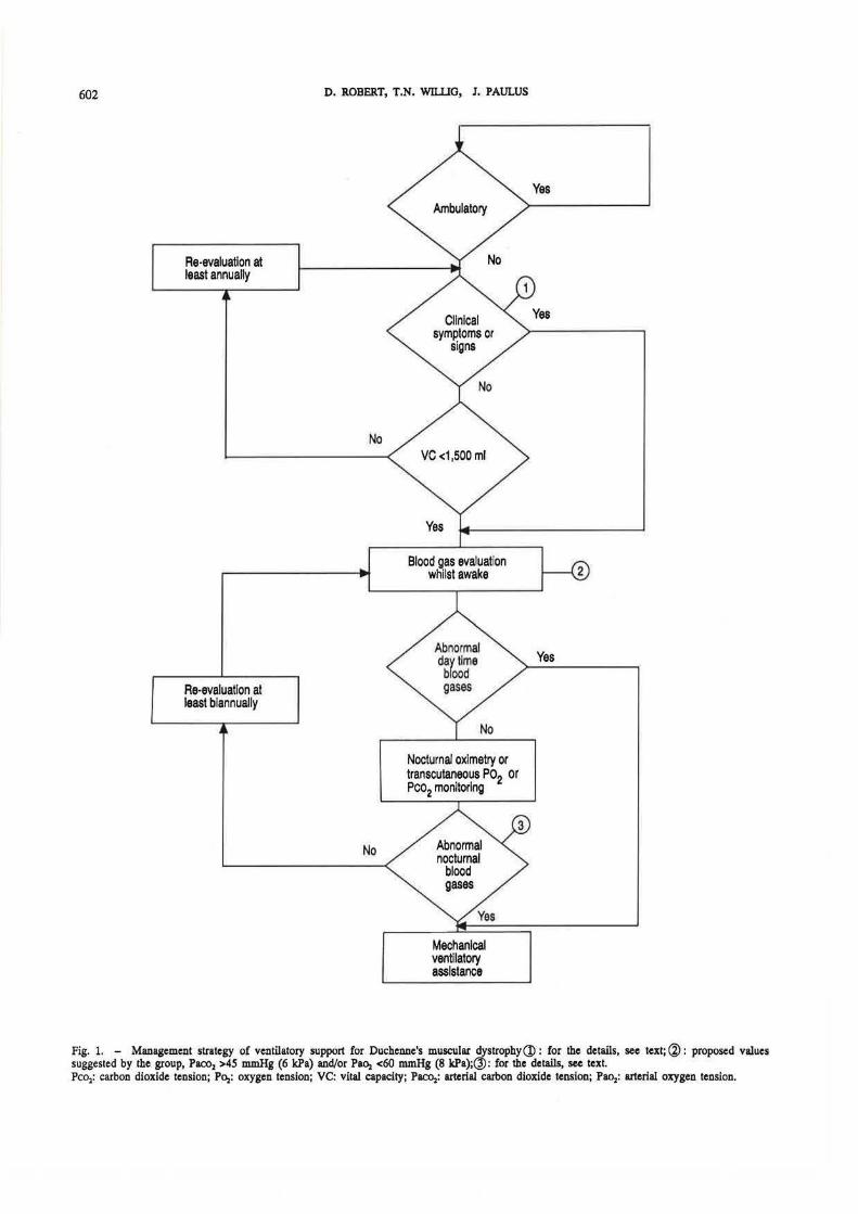
Fig. 1. - Management strategy of ventilatory support for Duchenne's muscular dystrophy(j) : for the details, see text;@ : proposed values suggested by the group, Paco2 >45 mmHg (6 kPa) and/or P&Oz <60 mmHg (8 kPa);@: for the details, see text. Pco2: carbon dioxide tension; P<>z: oxygen tension; VC: vital capacity; P&COz: arterial carbon dioxide tension; Pao2: arterial oxygen tension.

LONG-TERM NASAL VENTILATION IN NEUROMUSCUU\R DISORDERS 603
If nocturnal blood gases are normal, or if the use of nocturnal ventilatory assistance had been refused by the patient early on, its use must become virtually mandatory if blood gases are found to be abnormal during waking hours, or if clinical signs of chronic alveolar hypoventilation appear. The choice of the method of assisted ventilation to be used depends in large part on the experience of the prescribing physician with the various alternatives, as well as on the wishes of the patient. Whenever possible, the use of noninvasive means of ventilatory support are preferred, and should usually be instituted early. For Duchenne's muscular dystrophy patients the simple schema in figure 1 summarizes management strategy.
During episodes of airway congestion, the elimination of airway secretions can be facilitated by hyperinsufflations, followed by the use of techniques to increase the patient's cough expiratory flows and, when beneficial, by prolonging the periods of ventilatory assistance [9].
It does not seem to be necessary to systematically use nasal ventilation in the perioperative time of scoliosis surgery for young patients {12-13 yrs of age). On the other hand, when surgical intervention is delayed until 14-16 yrs of age, it may be necessary to use nasal ventilation, sometimes over a period of weeks in the postoperative period, until the vital capacity returns to preoperative levels. During this time, nasal ventilation can be an effective alternative to intubation for ventilatory assistance. Nasal masks, therefore, should be obtained and possibly fabricated, and nasal ventilation introduced prior to surgery.
Despite the initially encouraging results reported from one centre [10], the outcome of a more recent multicentre study on the prophylactic use of nasal ventilation failed . to support the contention that the use of nasal ventilation could have any effect on the progression of the restrictive pulmonary syndrome, or on the rate of loss of vital capacity for individuals with Duchenne's muscular dystrophy [11].
Contraindications to the use of nasal ventilation for Duchenne's muscular dystrophy patients are similar to those encountered for other conditions, that is, essentially, the concurrence of severe difficulties with swallowing. This only becomes a problem, however, in the very late stage. There does not appear to be any contraindication to the use of this method in the presence of cardiac dysfunction.
The progressive nature of muscular weakness for Duchenne's muscular dystrophy patients can eventually create the need to resort to alternative methods of ventilatory assistance, once the patient is using over 12-15 h of nasal ventilation daily.
Application In the treatment of the spinal muscular atrophies and congenitaJ myopathies
The indications for assisting the ventilation of these patients (12] are based on the determination of blood gas
abnormalities and/or the appearance of clinical signs of ventilatory insufficiency. Respiratory insufficiency, however, can exist from birth. The use of nasal ventilation is not applicable at this early age. Nasal ventilation can be proposed for very young children with the following considerations: 1) it must be used in conjunction with a custom-prepared abdominal binder; 2) supplemental humidification appears to be indispensable, and heating of the air necessary for all very small children; 3) the nasal masks must be changed frequently, to accommodate growth of facial structures; and 4) effectiveness can be improved by simple methods to decrease oral air leakage, such as with the use of nipple pacifiers.
Finally, patients using nasal ventilation who are to have thoracolumbar orthoses cast, should undergo casting whilst using nasal ventilation, so that the ultimate orthotic fit is sufficient to obtain optimal lung expansion during assisted ventilation. These patients must also use nasal ventilation promptly following any surgical procedures, if the risk of resorting to intubation is to be minimized.
The use of nasal ventilation is limited: 1) in the presence of severe difficulties with swallowing which accompany some forms of congenital myopathy and spinal muscular atrophies with bulbar involvement; and 2) for newboms, for whom nasal passages are inadequate. Furthermore, nasal ventilation may be contraindicated until a baby has acquired the capacity to breathe as much through the nose as through the mouth. These factors explain why the technique has not been used successfully for children younger than 7 months of age.
Airflow turbulence and decremental pressures in the lung periphery in small children necessitate careful choice of the ventilator to be used, and precise setting and monitoring of ventilator parameters. Furthermore, the alarms of certain portable ventilators have not been designed for use with small children .
The follow-up of the use of nasal ventilation in these patients is similar to that proposed for individuals with Duchenne's muscular dystrophy. Although the reliability of Pt~ measurements is greater in small children, as previously noted, this technique is not utilized in all centres because of the lengthy set-up time, inconvenience of requiring frequent changes of skin electrode placement, and expense.
For congenital muscular dystrophy patients, ventilatory assistance may become necessary at any time during early childhood or after puberty, even though ambulation may still be possible.
The congenital myopathies include conditions of greatly varying severity. For some of these conditions, long-term ventilatory assistance is rarely, if ever, needed. With other conditions, it might become necessary at any age. Respiratory complications are not always entirely due to respiratory muscle weakness, but may be due, at least in part, to concurrent central or obstructive apnoeas or hypopnoeas in these conditions.
Except for patients with prenatal, or neonatal onset before 3 months of age, nasal ventilation appears to be a very useful technique for patients with spinal muscular atrophy. Early use of respiratory therapy and chest physical therapeutic techniques may be useful for permitting

604 D. ROBERT, T.N. WIUJG, 1. PAULUS
normal development of the respiratory exchange membrane and prolonging survival.
Institutional and home use of nasal ventilation and its limitations
The various potential obstacles to the successful introduction and use of nasal ventilation can be, for the most part, avoided or resolved. Thorough explanations and patient interaction with others already benefiting from nasal ventilation can be helpful. It is also necessary to give considerable attention to the masks to be used. Several may need to be tried in order to find the best one for minimizing air leaks and maximizing comfort. Careful and meticulous modification of custom-prepared masks may be necessary.
When air leakage during nasal ventilation causes disruptive sounding of ventilator alarms, these alarms can be muffled and oxyhaemoglobin desaturation alarms substituted. No good solution has been proposed to resolve the difficulties associated with impaired swallowing or aerophagia, except, perhaps, for a possible trial using an abdominal binder for the latter condition. It should also be noted that nasal ventilation is often not well-tolerated when it is used by asymptomatic patients and indicated by the presence of nocturnal blood gas abnormalities alone. Symptoms may, however, appear insidiously, and progress without the patient or his care-givers realizing the possible benefit which could be derived from the use of assisted ventilation.
As soon as a respiratory tract infection occurs, it is necessary to assure effective airway secretion clearance by manually-assisted coughing, postural drainage, etc. [9]. The effectiveness of the many devices being proposed to facilitate the expectoration of airway secretions should be conf1Ill1ed by additional studies. Patient surveillance must be particularly intensive during these episodes. Continuous oximetry monitoring is especially helpful, and is equally convenient for use at home or in the hospital. Hospitalization may become necessary when the patient's care-givers are unable to manage the situation, or when early signs of acute respiratory failure occur despite continuous nasal ventilation.
Nasal ventilation can be considered ineffective or insufficient [7] when there is: 1) persistence or reappearance of symptoms; or 2) adequate ventilation but the need for assisted ventilation exceeds 16 h·day-1• Certain centres report the effective use of nasal ventilation alone or in combination with other non-invasive aids, such as mouthpiece ventilation 24 h·day·1 by patients with Duchenne's muscular dystrophy [9].
It is, therefore, possible to optimize the use of nasal ventilation by adjusting or modifying the masks and/or the ventilator settings, decreasing air leaks, increasing periods of daily use, or by switching to, or combining its use with other effective non-invasive methods. The choice depends on the availability of alternative methods, the experience of the treatment centres with the various alternatives, the motivation of the patient, and the capabilities of his care-givers.
Complementary techniques and alternatives
Complementary measures
Four techniques can complement the use of nasal ventilation: 1. Periodic hyperinsufflations for the purpose of facilitating normal alveolar development are used in some centres for 5-20 min periods several times a day preferably with pressure-cycled ventilators [13]. Two particular situations make the use of periodic hyperinsufflations mandatory: i) children with paradoxical thoracoabdominal breathing, or whose thoracic and, therefore, pulmonary growth is compromised; and ii) and children with radiographic evidence of rib cage asymmetry, who are at risk of developing ventilatory insufficiency at a pace greater than would be predicted by the extent of their respiratory muscle weakness. The efficacy of the use of periodic hyperinsufflations in patients with Duchenne's muscular dystrophy needs to be studied further. 2. Apparatus facilitating the elimination of airway secretions deserve re-evaluation, on the basis of results from one centre [9]. These mechanical insufflatorexsufflators provide a hyperinsuffiation followed by a sudden decompression or exsufflation. 3. Glossopharyngeal breathing (frog breathing) has been known, for 40 years, to be able to provide deep breaths and time free of ventilatory support. It has been effective for patients with relatively stable or slowly progressive conditions, such as post-poliomyelitis, traumatic high level quadriplegia, or spinal muscular atrophies. The use of this technique is much less frequently effective for patients with rapidly progressive myopathies, such as Duchenne's muscular dystrophy and is difficult to teach to small children. 4. Certain respiratory and chest therapy techniques are useful for treating episodes of airway restriction in certain patient populations, when employed by experienced practitioners.
Complementary techniques or alternative methods of ventilatory assistance
Ventilatory assistance via a mouthpiece can be used by patients with relatively stable ventilatory failure, such as those post-poliomyelitis, traumatic high level quadriplegia, etc. up to 24 h·day-1• For rapidly progressive neuromuscular patients, however, its use may be considered to be indicated for ventilatory assistance not exceeding 16-20 h·day-1 [7]. One centre has reported 24 h use of mouthpiece ventilation for numerous patients with rapidly progressive neuromuscular ventilatory failure, including those with Duchenne's muscular dystrophy [9]. For nocturnal use, a Bennett lip seal (Puritan-Bennett, Boulder, CO, USA) minimizes leakage around the mouthpiece and assures firm retention. Custom moulded mouthpieces have also been described to enhance comfort and seal of the lips. For daytime use, the mouthpiece should be set up near the patient's mouth, so that it be can grasped and released in accordance with the patient's need for ventilatory assistance. It provides better appearance than

LONG-TERM NASAL VENTilATION IN NEUROMUSCUlAR DISORDERS 605
other positive pressure methods, and facilitates more normal speech production. Thus, this technique can provide effective ventilatory support during sleeping and waking hours, and the absence of measurable vital capacity or ventilator-free time need not be considered a contra-indication to its use.
Difficulties with mouthpiece ventilation occur when oral and facial muscles become extremely weak. Long-term use, particularly during the years of skeletal growth, can also cause dental deformations. The risk of aerophagia is identical to that during nasal ventilation and, as with the former, severe weakness of the swallowing muscles is a contraindication.
The choice between assisting ventilation via a mouthpiece or a nasal mask depends on the experience of the treatment centre, and on patient choice after trying both techniques. Mouthpiece ventilation can be offered as an alternative to nasal ventilation for periods during the day, or it can substitute for it entirely in some patients. Assisted ventilation via an oronasal mask may also be used, particularly for nocturnal support.
Several types of negative pressure body ventilators can assist or support ventilation [14]. These include iron lungs, chest shell ventilators, and pulmowraps. These devices create subatmospheric pressure around the chest and abdomen, causing air to enter the mouth and nose and, thus, assist alveolar ventilation. The iron lung is the most efficacious and comfortable of these ventilators, but it is heavy and bulky. Furthermore, except for the chest shell ventilator, the time required to set up patients to use these ventilators is relatively long, and includes the time needed to eliminate air leakage around the iron lung cervical collar or pulmowrap seals. Several centres recommend alternating the use of negative pressure body ventilators with non-invasive positive pressure techniques or the intermittent abdominal pressure ventilator, in order to optimize the ventilatory support regimen. There have been concerns, however, that the use of negative pressure body ventilators can be associated with obstructive apnoea and oxyhaemoglobin desaturation during sleep, justifying the use of periodic evaluation of nocturnal oximetry or polysomnography in order to more completely evaluate these nocturnal events. Because of decreased efficacy with time, several centres have reported converting patients ventilated by these methods to other, more effective means of ventilatory aid, including mouthpiece and nasal ventilation techniques.
The use of negative pressure body ventilators is not recommended for children, because of possible unfortunate effects on skeletal growth, and possible complications from immobility. Furthermore, the enclosure and continuous airflow exposure to the body can cause wide temperature variations for small patients, particularly those with spinal muscular atrophy and impaired temperature regulation. Despite long-term use of negative pressure ventilatory support in several centres, there is no consensus on the use of these methods by patients with Duchenne's muscular dystrophy, in view of the alternatives now available.
Tracheostomy ventilation is most effective and the most frequently used method for long-term ventilatory support
[15]. However, disadvantages of using this technique have been described [9]. The tracheostomy tube can damage the trachea, cause haemorrhage, fistulae, and obstruction by granulation overgrowth or stenosis. Fortunately, these complications do not appear to be frequent in the experience of the centres represented in this report. It would, however, be useful to study the possible benefits to be obtained from tracheostomy tube modifications, including the use of other materials in their manufacture. Disadvantages with the use of a tracheostomy include the need to always have someone present, whether it be medical personnel, a family member, or attendant, to aspirate tracheal secretions when necessary, as well as to clean and change the tubes themselves. There appears to be relatively few intervening infections with the use of tracheostomy ventilation in the community setting. There is, however, rapid and permanent bacterial colonization of the tracheostomy site and airway, without apparent pulmonary complications for the majority of cases. When one considers both the effectiveness and disadvantages inherent in using this technique, the patient's wishes should be strongly considered when the decision of which type of ventilatory aid to be used is being made.
The intermittent abdominal pressure ventilator is effective for some patients [16]. This ventilator must be used with the patient in the seated or semi-seated position. This makes its use inconvenient for nocturnal aid. Furthermore, it is usually inadequate for patients with severe spinal deformity, obesity, or poor pulmonary compliance. This technique appears to be most useful in a regimen of non-invasive ventilatory aids, but can also be used to supplement periods of tracheostomy intermittent positive pressure ventilation (IPPV).
Conclusion
Nasal ventilation can clearly be effective in the treatment of neuromuscular ventilatory insufficiency. It is most often the initial method of choice for the adolescent or adult. However, in the treatment of Duchenne's muscular dystrophy, its utilization does not appear to be recommended when disease progression has advanced to the need for ventilator support of over 18 h·day·•. In this case, one can either complement periods of nasal ventilation with other methods of non-invasive ventilatory support, or propose a tracheostomy. Proper management during this transition period to up to 24 h ventilatory assistance is critical, and necessitates frequent and careful surveillance by specialized medical teams.
Nasal ventilation is also applicable to young paediatric neuromuscular patients, including those with spinal muscular atrophy, or congenital muscular dystrophy or other myopathy, provided that bulbar muscular involvement is not severe. The application masks must, however, be changed or modified more often due to the growth of facial structures.
For all cases, it is necessary to repeat spirometric evaluation and blood gases and, in particular, oxyhaemoglobin saturation, on a regular basis, in order to initiate

606 D. ROBERT, T.N. WILUG, 1. PAULUS
treatment before the occurrence of acute respiratory failure or other serious respiratory complications. Since the first signs of blood gas disturbance and alveolar hypoventilation occur during sleep, both nocturnal noninvasive blood gas monitoring and, when indicated, ventilatory assistance should be introduced.
AclawwUdpunls: The authors gratefully aclcnowledge M. Francois for typing the successive versions of the consensus report and the help of 1. Bach for the translation of the report into English.
References
1. Willig TN, Robert D, Paulus J, Leger P, eds. - Nasal ventilation in neuromuscular disorders. Eur Respir Rev 1993; 3(12): 243-307. 2. Delguste P, Rodenstein DO. - Implementation and monitoring of mechanical ventilation via nasal access. Eur Respir Rev 1993; 3(12): 266-269. 3. Bach JR, Sortor SM, Saporito LR. - Interfaces for noninvasive intermittent positive pressure ventilatory support in North America. Eur Respir Rev 1993; 3(12): 254-259. 4. Comette A, Mougel D. - Ventilatory assistance via the nasal route: masks and fittings. Eur Respir Rev 1993; 3(12): 250-253. 5. Langevin B, Leger R, Gerard M, Sukkar F, Guez A, Robert D. - Monitoring nasal ventilation. Eur Respir Rev 1993; 3(12): 260-265. 6. Paulus J, Willig TN. - Nasal ventilation in neuromuscular disorders: respiratory management and patient's experience. Eur Respir Rev 1993; 3(12): 245-249. 7. Leger P, Langevin B, Guez A. Robert D. - What to do
when nasal ventilation fails for neuromuscular patients? Eur Respir Rev 1993; 3(12): 27~283. 8. Raphael J-C, Chevret S, Chastang Cl, Bouvet F. - Home mechanical ventilation in Duchenne's muscular dystrophy: in search of a therapeutic strategy. Eur Respir Rev 1992; 3(12): 270-274. 9. Bach JR. - Management of neuromuscular ventilatory failure by 24 hour non-invasive intermittent positive pressure ventilation. Eur Respir Rev 1993; 3(12): 284-291. 10. Rideau Y. - The Duchenne's muscular dystrophy child. Care of wheelchair dependent patient-death prevention. VI International Congress on Neuromuscular Diseases. (Abstract): Muscle Nerve 1986; 9 (Suppl. SS): 86. 11. Raphael JC, Chevret S, Chastang Cl, Bouvet F. -Groupe multicentrique sur !'evaluation de la ventilation nasale de prevention dans la dystrophic musculaire de Duchenne de Boulogne (DMDB). Resultats intermediaires d'une etude randomisee. XIX Congr~s de la Societe de Reanimation de Langue Francaise. Paris, 22-25 novembre 1990. Rtan Soins Intens MM. Urg 1990; 6: 516. 12. Barois A. Estoumet-Mathiaud B. - Nasal ventilation in congenital myopathies and spinal muscular atrophies. Eur Respir Rev 1993; 3(12): 275-278. 13. Barois A. Bataille J, Estournet B. - La ventilation A domicile par voie buccale chez !'enfant dans les maladies neuromusculaires. Agresso/ogie 1985; 26: 645-649. 14. Schiavina M, Fabiani A. - Intermittent negative pressure ventilation in neuromuscular diseases. Eur Respir Rev 1993; 3(12): 292-299. 15. Soudon P. - Mechanical ventilation by tracheostomy in neuromuscular diseases: experience and evaluation. Eur Respir Rev 1993; 3(12): 300-304. 16. Milane J, Jonquet 0, Bertrand P. - Ventilatory assistance by pneumatic belt. Eur Respir Rev 1993; 3(12): 305-307.