Embed Size (px)
Citation preview

Long-Term Effectiveness of PhotodynamicTherapy by Using a Hydrophilic
Photosensitizer ATX-S10(Na) AgainstExperimental Choroidal Neovascularization
in RatsMasakazu Kanai, MB,1* Akira Obana, MD,1 Yuko Gohto, MD,1 Satoshi Nagata, MB,1
Tokuhiko Miki, MD,1 Kenji Kaneda, MD,2 and Susumu Nakajima, MD3
1Department of Ophthalmology, Osaka City University Medical School, Osaka 545-8585, Japan2Department of Anatomy, Osaka City University Medical School, Osaka 545-8585, Japan
3Division of Surgical Operation, Asahikawa Medical College, Asahikawa 078, Japan
Background and Objective: We previously demonstrated that a hydro-philic photosensitizer ATX-S10 had a potent photodynamic effect. Thisstudy was designed to reveal the long-term effectiveness of photody-namic therapy (PDT) with this agent in occluding choroidal neovascu-larization (CNV) and its selectivity in the neovascular tissue.Study Design/Materials and Methods: Experimental CNV was inducedby intense photocoagulation in rat eyes. Immediately or 2 hours afterintravenous injection of 8 mg/kg body weight of ATX-S10(Na), a cis iso-mer of ATX-S10, eyes were irradiated by a diode laser at the radiance of3.25–65.3 J/cm2 Vascular occlusion was identified by fundus photogra-phy, fluorescein angiography, and histology at 1, 3, 7, 14, and 28 daysafter PDT. As controls, non-neovascular eyes were subjected to PDTand similarly analyzed.Results: By using the following treatment parameters, PDT with ATX-S10(Na) successfully occluded CNV without causing occlusion of reti-nal capillaries for 28 days; 7.4 and 19.6 J/cm2 immediately after dyeinjection and 36.7 and 65.3 J/cm2 2 hours after injection. Although theseconditions also caused occlusion of normal choriocapillaries and mildinjuries of retinal vessels, retinal pigment epithelium, and photorecep-tors at 1 day, retinal vessels and pigment epithelial cells recoveredfrom damages by 28 days. No injuries were found in the inner retina.Conclusion: In optimal treatment conditions, PDT with ATX-S10(Na)can induce long-term, selective occlusion of CNV without causing irre-versible damages in the inner retina. Lasers Surg. Med. 26:48–57,2000. © 2000 Wiley-Liss, Inc.
Key words: ATX-S10(Na); choroidal neovascularization; photodynamic therapy;long-term effectiveness; retinal damage
INTRODUCTION
Laser photocoagulation has long been usedin clinic as effective treatment modality for cho-roidal neovascularization (CNV) in age-relatedmacular degeneration [1–3]. However, it oftenproduces damage to the sensory retina, incom-plete CNV closure, and frequent neovascular re-currence, resulting in a visual loss [3]. As for al-ternative therapeutic procedures such as sub-
Grant sponsor: Ministry of Health and Welfare; Grant num-ber: Health Science Research Grants H10-018.
The authors have no financial interest in the present issue.
This article was presented in part at the Annual Meeting ofthe Association for Research in Vision and Ophthalmology in1998.
*Correspondence to: Masakazu Kanai, MB, Department ofOphthalmology, Osaka City University Medical School,Asahi-machi 1-5-7, Abeno-ku, Osaka 545-8585, Japan.
Accepted 29 September 1999
Lasers in Surgery and Medicine 26:48–57 (2000)
© 2000 Wiley-Liss, Inc.

macular surgery, radiation, and interferonadministration, the efficacy remains unclear[4–9].
Photodynamic therapy (PDT), which wasoriginally developed as a treatment modality formalignant tumors [10], has aroused great interestamong ophthalmologists as a promising newmethod for CNV treatment [11,12], because of itspotential for destroying vascular endothelial cells[13]. Clinical studies on PDT have been carriedout by using liposomal benzoporphyrin (BPD) andtin-ethyl etiopurpurin (SnET2) as photosensitiz-ers [14,15]. These two agents are lipid-soluble, sothey need to be continuously infused into the vein.Recently, a water-soluble photosensitizer ATX-S10(Na) has been developed and can be adminis-tered as a bolus intravenous injection [16,17]. Wepreviously revealed that PDT with this agent waseffective for occlusion of experimental CNV andcorneal neovascularization in rats, rabbits, andmonkeys [18–20] and determined the optimaltherapeutic parameters, i.e., dye dosage, irradia-tion dose, and time intervals between dye injec-tion and laser irradiation, for the selective occlu-sion of neovascularization. Another problem inthe PDT with liposomal BPD was recanalizationand recurrence of neovascularization, which fre-quently occurred at the late periods after PDT[14,15]. We here examined the optimal conditionsof PDT by using ATX-S10(Na) that produce long-term effectiveness with regard to selective occlu-sion of CNV and low incidence of recanalization orrecurrence.
MATERIALS AND METHODS
Animals
Thirty-four eyes from 17 Brown-Norway rats(200–250 g body weight) were used. Animals werealways treated in accordance with the Associationfor Research in Vision and Ophthalmology reso-lution on the use of animals in research.
Induction of Experimental CNV
Rats were anesthetized by the intraperitone-al injection of 10 mg/kg body weight of Nembutal.The pupils were mydriatic with 0.5% tropicamideand 0.5% phenylephrine hydrochloride (Midrin P,Santen Co., Osaka, Japan). For topical anesthe-sia, 0.4% oxybuprocaine hydrochloride (Benoxil,Santen Co.) was used. Intense photocoagulationwas induced at four sites in the fundus, sparing
large-sized retinal vessels located near the opticdisc, with an argon green laser (Coherent, CA; l4 514 nm). A 100-mm spot was irradiated for 0.1seconds with a power of 150 mW. At 20–25 daysafter photocoagulation, induction of CNV wasconfirmed by fundus photography and fluoresceinangiography by using a fundus camera (JenesisKowa, Nagoya, Japan). Fluorescein angiographywas conducted at various time intervals after in-travenous injection of 0.2 ml of 5% fluorescein so-dium.
Photodynamic Therapy
One day after confirming the presence ofCNV, 8 mg/kg body weight of ATX-S10(Na) (Pho-tochemical, Inc., Okayama, Japan) dissolved inwater at the concentration of 10 mg/ml was in-jected into the tail vein. Immediately or 2 hoursafter dye injection, the site of CNV was irradiatedby a 670-nm diode laser (Hamamatsu Photonics,Hamamatsu, Japan) as previously reported [18].The spot-size was set large enough to cover theentire lesion of CNV. Irradiation at the retinalsurface was calculated from the values at the cor-neal surface according to our previous study [18].To evaluate the influence of radiant exposure onthe photodynamic effect, radiant exposure wasvaried from 3.2 to 65.3 J/cm2, by keeping the timeof treatment constant (either 120 or 240 seconds)and changing irradiance. To assess the influenceof irradiance on the photodynamic effect, irradi-ance was varied from 27.3 to 272 mW/cm2, keep-ing the radiant exposure constant (10 J/cm2 whenirradiated immediately after injection and 50J/cm2 when irradiated 2 hours after injection) andchanging the time of treatment. Table 1 summa-rizes the treatment parameters and numbers oftested lesions. As controls, two animals receivedeither dye injection or laser irradiation alone.
For the analysis of photodynamic effect onthe normal retina, laser irradiation was applied tothe normal fundus by using the therapeutic pa-rameters that had been demonstrated to induceselective occlusion of CNV. The treatment param-eters and numbers of tested eyes are shown inTable 2.
Assessment of Therapeutic Efficacy
Therapeutic efficacy of PDT was evaluatedat 1, 3, 7, 14, and 28 days after PDT based onfluorescein angiographic and histologic observa-tions. For histology, at 28 days after PDT, eyeswere enucleated under anesthesia. The CNV le-
Long-Term Effectiveness of Photodynamic Therapy 49
sions were excised and fixed in Karnovsky’s fixa-tive overnight at 4°C. They were post-fixed in 2%OsO4 in 0.1 M phosphate buffer, pH 7.4, dehy-drated in ethanol and embedded in Polybed (Poly-science, Inc., Warrington, PA). Semithin sectionswere stained with toluidine blue and observed bylight microscopy. Thin sections were stained withuranyl acetate and lead citrate and observed un-der a JEM-1200EX electron microscope (JEOL,Tokyo, Japan).
RESULTSPhotodynamic Effect of ATX-S10(Na) on CNV
Before PDT, CNV induction was identifiedby fundus photography as subretinal whitish le-sions and by fluorescein angiography as a net-work appearance in the early phase and fluores-cein leakage in the late phase (Fig. 1A). AfterPDT, the CNV closure and retinal capillary injurywere identified by the fluorescein angiographicfindings of no fluorescein leakage and poor perfu-sion, respectively. The results of PDT with regardto the effectiveness and selectivity were dividedinto three categories: (1) “not effective,” both CNVand retinal capillaries were patent; (2) “effective,”CNV was closed, whereas retinal capillaries were
open (Fig. 1B); and (3) “excessive,” both CNV andretinal capillaries were closed (Fig. 2A). However,CNV lesions that received “effective” PDT oftendisplayed reappearance of fluorescein leakagerepresentative of recanalization within 28 days(Fig. 1C). Retinal capillaries that had closed after“excessive” PDT, as well, often underwent recana-lization within 28 days (Fig. 2B).
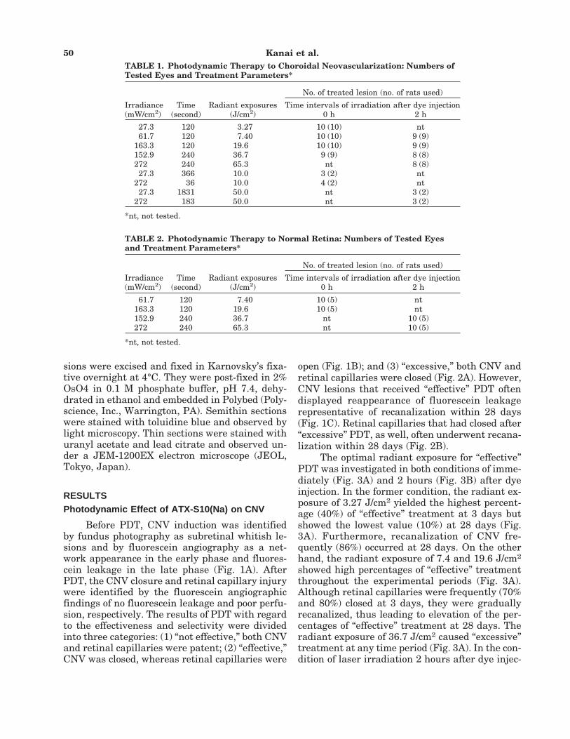
The optimal radiant exposure for “effective”PDT was investigated in both conditions of imme-diately (Fig. 3A) and 2 hours (Fig. 3B) after dyeinjection. In the former condition, the radiant ex-posure of 3.27 J/cm2 yielded the highest percent-age (40%) of “effective” treatment at 3 days butshowed the lowest value (10%) at 28 days (Fig.3A). Furthermore, recanalization of CNV fre-quently (86%) occurred at 28 days. On the otherhand, the radiant exposure of 7.4 and 19.6 J/cm2
showed high percentages of “effective” treatmentthroughout the experimental periods (Fig. 3A).Although retinal capillaries were frequently (70%and 80%) closed at 3 days, they were graduallyrecanalized, thus leading to elevation of the per-centages of “effective” treatment at 28 days. Theradiant exposure of 36.7 J/cm2 caused “excessive”treatment at any time period (Fig. 3A). In the con-dition of laser irradiation 2 hours after dye injec-
TABLE 1. Photodynamic Therapy to Choroidal Neovascularization: Numbers ofTested Eyes and Treatment Parameters*
Irradiance(mW/cm2)
Time(second)
Radiant exposures(J/cm2)
No. of treated lesion (no. of rats used)
Time intervals of irradiation after dye injection0 h 2 h
27.3 120 3.27 10 (10) nt61.7 120 7.40 10 (10) 9 (9)
163.3 120 19.6 10 (10) 9 (9)152.9 240 36.7 9 (9) 8 (8)272 240 65.3 nt 8 (8)27.3 366 10.0 3 (2) nt
272 36 10.0 4 (2) nt27.3 1831 50.0 nt 3 (2)
272 183 50.0 nt 3 (2)
*nt, not tested.
TABLE 2. Photodynamic Therapy to Normal Retina: Numbers of Tested Eyesand Treatment Parameters*
Irradiance(mW/cm2)
Time(second)
Radiant exposures(J/cm2)
No. of treated lesion (no. of rats used)
Time intervals of irradiation after dye injection0 h 2 h
61.7 120 7.40 10 (5) nt163.3 120 19.6 10 (5) nt152.9 240 36.7 nt 10 (5)272 240 65.3 nt 10 (5)
*nt, not tested.
50 Kanai et al.

tion, photodynamic effects were generally milderthan in the condition of irradiation immediatelyafter injection. The radiant exposure of 36.7 and65.3 J/cm2 gave higher percentages (63% and75%, respectively) of “effective” treatment thanthat of 7.4 and 19.6 J/cm2 (22% and 44%, respec-tively) at 28 days (Fig. 3B).
We further examined the effectiveness ofPDT by different irradiances, keeping radiant ex-posure constant. In both conditions of the radiantexposure of 10 J/cm2 irradiated immediately afterinjection (Fig. 4) and 50 J/cm2 2 hours after injec-tion, CNV closure and retinal capillary damages
Fig. 1. Fluorescein angiograph of choroidal neovasculariza-tion (CNV) before (A), and 3 days (B) and 28 days (C) afterphotodynamic therapy. Irradiation was done 2 hours afterATX-S10(Na) injection with the radiant exposure of 7.40J/cm2. A: Fluorescein leakage is found in the lesion of CNV inthe late phase of angiography. B: Fluorescein leakage is nolonger seen. Retinal capillaries in the irradiated portion re-main intact. C: Fluorescein leakage takes place in the neo-vascular lesion, indicating the recanalization.
Fig. 2. Fluorescein angiograph of choroidal neovasculariza-tion (CNV) 3 days (A) and 28 days (B) after photodynamictherapy. Irradiation was done 2 hours after ATX-S10(Na) in-jection with the radiant exposure of 36.7 J/cm2. A: Both CNVand retinal capillaries are occluded, giving rise to a fluores-cein defect. B: There is no fluorescein leakage in the neovas-cular lesion. Retinal capillaries are well perfused.
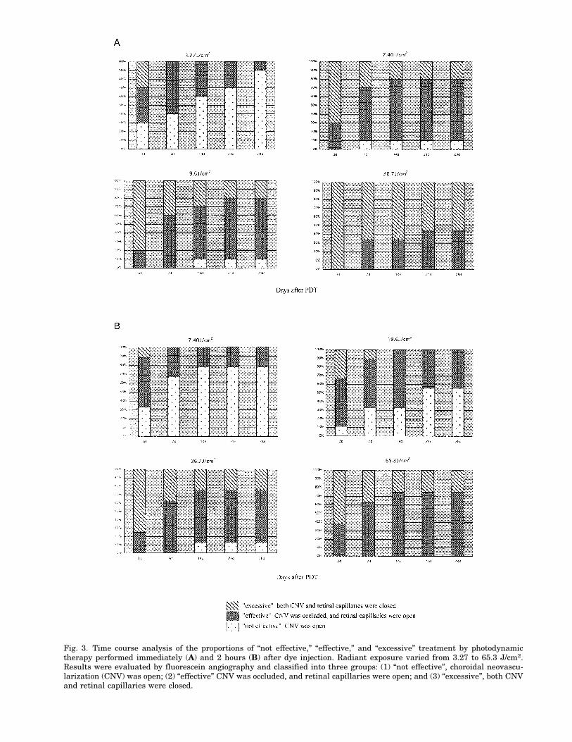
Fig. 3. Time course analysis of the proportions of “not effective,” “effective,” and “excessive” treatment by photodynamictherapy performed immediately (A) and 2 hours (B) after dye injection. Radiant exposure varied from 3.27 to 65.3 J/cm2.Results were evaluated by fluorescein angiography and classified into three groups: (1) “not effective”, choroidal neovascu-larization (CNV) was open; (2) “effective” CNV was occluded, and retinal capillaries were open; and (3) “excessive”, both CNVand retinal capillaries were closed.

were similar between the irradiance of 27.3 mW/cm2 (Fig. 4A) and 272 mW/cm2 (Fig. 4B).
When lesions were “effectively” treated byPDT, no exudative retinal detachment occurredas demonstrated by fundus photography, al-though there remained subretinal whitish prolif-erative tissue and pigmentation as the conse-quence of photocoagulation (Fig. 5A). On the otherhand, when they were “excessively” treated, reti-nal pigment epithelium in the irradiated area be-came atrophic (Fig. 5B).
Light and electron microscopy confirmed thecomplete occlusion of CNV in the subretinal pro-liferative tissue at 28 days after “effective” PDT(Fig. 6A,B). Basal lamina-like structures re-mained around the remnant of CNV (Fig. 6B).Retinal pigment epithelial cells and surroundingfibroblasts showed prominent proliferation.
Photodynamic Effects of ATX-S10(Na) on theNormal Retina
Because the present neovascularizationmodel included photocoagulation-induced retinaland choroidal damages, to evaluate the injuringeffect of PDT on the normal tissue, we irradiatednon-neovascularized eyes with the parametersthat gave “effective” CNV occlusion, i.e., 7.4 and19.6 J/cm2 just after dye injection and 36.7 and65.3 J/cm2 2 hours after injection. Results weresimilar among these parameters. At 1 day afterPDT, the irradiation portion of the retina looked
Fig. 4. Fluorescein angiograph of choroidal neovasculariza-tion (CNV) treated with different irradiances (A: 27.3 mW/cm2; B: 272 mW/cm2), keeping the radiant exposure constant(10 J/cm2), at 1 day after photodynamic therapy. CNV wassimilarly closed in both lesions. The areas of damaged retinalpigment epithelium and occlusion of choriocapillaries are alsosimilar.
Fig. 5. Fundus photographs of “effective” (A) and “excessive”(B) treatment by photodynamic therapy at 28 days. Irradia-tion was done 2 hours after ATX-S10(Na) injection. A: Theradiant exposure of 36.7 J/cm2. There is subretinal fibroustissue and pigmentation at the center and margin, respec-tively, of the irradiation field. B: The radiant exposure of 65.3J/cm2. Retinal pigment epithelium is atrophic around the sub-retinal fibrous tissue.
Long-Term Effectiveness of Photodynamic Therapy 53
whitish by fundus photography and displayed byfluorescence angiography central hypofluores-cence indicative of choriocapillary occlusion in theearly phase and marginal fluorescein leakage in-dicative of disruption of retinal pigment epitheliallayer in the late phase. Retinal capillary occlusionwas observed in three of 10 lesions, whereas reti-nal arteriole/venule occlusion was not detected.At 3 days, retinal capillary occlusion decreased in
incidence to one of eight lesions. Fluorescein leak-age also decreased. At 7 days, all the retinal cap-illaries were recanalized. At 14 and 28 days, reti-nal whitening disappeared by fundus photogra-phy, making the boundary of lesions obscure andleaving pigmentation (Fig. 7A). By fluorescein an-giography, fluorescein leakage was no longerseen, although hypofluorescence in the earlyphase remained (Fig. 7B).
Light microscopically, choriocapillaries re-mained occluded from day 1 (Fig. 8A) to day 28(Fig. 8B), whereas retinal capillaries were patentat 28 days. Electron microscopically, the lumens
Fig. 6. Light micrograph (A) and electron micrograph (B) of“effective” treatment of choroidal neovascularization (CNV)by photodynamic therapy (PDT) at 28 days. PDT was donewith the radiant exposure of 7.4 J/cm2 just after ATX-S10(Na)injection. A: Neovascularization in the subretinal prolifera-tive tissue (arrow) is occluded, whereas retinal capillaries areintact (arrowheads). Original magnification, ×66. B: Neovas-cularization is diminished in the subretinal proliferative tis-sue, but basal lamina-like structures (arrowheads) remain.Scale bar 4 1 mm.
Fig. 7. Fundus photograph (A) and fluorescein angiograph(B) of the non-neovascular funds at 28 days after PDT. Irra-diation was done at the radiant exposure of 7.4 J/cm2 justafter dye injection. A: Retinal whitening disappears, leavingpigmentation. The boundary between treated and nontreatedarea becomes obscure. B: There is only slight hypofluores-cence at the center of irradiation field. Fluorescein leakage isnot seen.
54 Kanai et al.

of retinal capillaries were often occupied withthrombus at 1 day, but no necrotic changes weredetected in the endothelial cells. Retinal and cho-roidal arterioles/venules showed no morphologicchanges. Although retinal pigment epithelial cellsand photoreceptor cells were injured at 1 day, theformer cells were repaired by day 7 through re-generation, but the latter cells remained damageduntil 28 days. The inner nuclear layer of theretina was not injured.
DISCUSSION
We previously reported that PDT with ATX-S10 effectively occluded CNV without closing theretinal vessels one day after irradiation in the fol-lowing conditions, i.e., the radiance exposure of7.4 J/cm2 immediately after injection of 16 mg/kgbody weight ATX-S10 and 22.0 J/cm2 2–4 hoursafter injection [18]. The present study has dem-onstrated that this procedure yields a long-term,selective occlusion of CNV until 28 days withoutcausing irreversible damages to the inner retina.(We used here 8 mg/kg body weight ATX-S10[Na],because ATX-S10[Na] contains only a cis-isomerand has more potent photochemical effects thanconventional ATX-S10 [21]).
The optimal parameters of irradiation forthe long-term occlusion of CNV were (1) radiantexposure of 7.4 and 19.6 J/cm2 immediately afterdye injection, and (2) 36.7 and 65.3 J/cm2 2 hoursafter injection. Although these conditions of PDTalso induced retinal capillary occlusion to someextent at 1 day, the occlusion was reversible andrecanalization developed until 7 days, thus in-creasing the rate of “effective” treatment at 28days. Although the rate of “effective” treatmentwas higher in radiant exposure of 3.27 J/cm2 com-pared with these optimal conditions at 1 day,many CNV underwent recanalization at 28 days.The mechanism of recanalization after PDT is notwell understood. The present histologic findingthat thrombus formation but not endothelial de-struction was responsible for retinal capillary clo-sure suggests that recanalization may be the phe-nomenon of recovery of the blood flow from im-pairment. In our previous experiment that usedthe monkey [19], retinal capillaries were not oc-cluded in the optimal conditions of PDT whichinduced CNV closure. Therefore, there remainsthe possibility that dye dosages smaller than 8mg/kg body weight may induce more selective oc-clusion of CNV in rats.
It has been demonstrated for other photosen-sitizers that lower irradiance improves the PDTeffect even in the same radiant exposures [22–24].In particular, Sitnik and Henderson [24] sug-gested that photodynamic therapy carried out atlow irradiance may enhance tumor response andselectivity. However, in the present study on thePDT with ATX-S10(Na), CNV was similarly oc-cluded by different irradiances at the same radi-ant exposures.
In the PDT-treated non-neovascular eyes, al-though the choriocapillaries, retinal pigment epi-
Fig. 8. Light micrographs of non-neovascular fundus at 1 day(A) and 28 days (B) after photodynamic therapy. Irradiationwas done at the radiant exposure of 7.4 J/cm2 immediatelyafter dye injection. A: Choriocapillaries at the center of irra-diated area are occluded (arrows). Retinal pigment epithelialcells undergo necrosis in a wider area (arrowheads) than cho-riocapillary closure. B: Although the inner nuclear layer ofthe retina (INL) shows no alterations, the outer nuclear layer(ONL) shows a decreased number of cells with disappearanceof outer and inner segments of photoreceptor cells. Retinalpigment epithelial cells (RPE) are partially stratified. Chorio-capillaries are occluded in the irradiation area (arrows), butretinal vessels and choroidal arteries and veins are intact.Original magnification, ×100.
Long-Term Effectiveness of Photodynamic Therapy 55

thelial cells, and photoreceptor cells were dam-aged, the inner nuclear layer, ganglion cell layer,nerve fiber layer, and choroidal arteries/veinswere free from injuries, indicating that PDT in-duced much less damage to the normal tissuecompared with laser photocoagulation, which af-fects all layers of the retina [25,26]. Our previousstudy demonstrated that intravenously injectedATX-S10 distributed in the choriocapillaries andretinal pigment epithelial cells but not in the pho-toreceptor cells [18]. By taking this data into ac-count, injuries in the choriocapillaries and retinalpigment epithelial cells are considered to resultfrom the primary action of PDT, whereas those inphotoreceptor cells might be secondary to primaryinjuries. Consistent with this view, injuries ofphotoreceptor cells did not appear soon after PDT[18] but gradually advanced at later than 1 day asshown here. Furthermore, different from the in-juries of photoreceptor cells that persisted until28 days, those of retinal pigment epithelial cellswere repaired by means of epithelial regenerationand, as the consequence of recovery of barrierfunction, edema in the outer layer of retina wasimproved at 28 days.
In conclusion, PDT with ATX-S10(Na), withcarefully optimized delivery and treatment pa-rameters, can be shown to induce the long-term,selective occlusion of CNV without causing irre-versible damages to retinal vessels and innerlayer of the retina. Considering the inevitable in-jury of photoreceptor cells by PDT, unnecessarilybroad irradiation should be avoided.
ACKNOWLEDGMENTS
The authors thank Isao Sakata, PhD, Photo-chemical, Inc., Okayama, and Ryosuke Machida,PhD, Wyeth-Lederle Japan, Tokyo, for providingATX-S10(Na) and Mr. Toru Hirano, HamamatsuPhotonics, Inc., Hamamatsu, for providing the di-ode laser.
REFERENCES
1. Macular Photocoagulation Study Group. Argon laser pho-tocoagulation for neovascular maculopathy. Three-yearresults from randomized clinical trials. Arch Ophthalmol1986;104:694–701.
2. Macular Photocoagulation Study Group. Krypton laserphotocoagulation for neovascular lesions of age-relatedmacular degeneration. Results of a randomized clinicaltrial. Arch Ophthalmol 1990;108:816–824.
3. Macular Photocoagulation Study Group. Laser photoco-agulation of subfoveal neovascular lesions of age-related
macular degeneration. Updated findings from two clini-cal trials. Arch Ophthalmol 1993;111:1200–1209.
4. Hudson HL, Frambach DA, Lopez PF. Relation of thefunctional and structural fundus changes after submacu-lar surgery for neovascular age-related macular degen-eration. Br J Ophthalmol 1995;79:417–423.
5. Roth DB, Downie AA, Charles ST. Visual results aftersubmacular surgery for neovascularization in age-relatedmacular degeneration. Ophthalmic Surg Lasers 1997;28:920–925.
6. Postgens H, Bodanowitz S, Kroll P. Low-dose radiationtherapy for age-related macular degeneration. GraefesArch Clin Exp Ophthalmol 1997;235:656–661.
7. Stalmans P, Leys A, Van Limbergen E. External beamradiotherapy (20 Gy, 2 Gy fractions) fails to control thegrowth of choroidal neovascularization in age-relatedmacular degeneration: a review of 111 cases. Retina1997;17:481–92
8. Pharmacological Therapy for Macular DegenerationStudy Group. Interferon alfa-2a is ineffective for patientswith choroidal neovascularization secondary to age-related macular degeneration. Results of a prospectiverandomized placebo-controlled clinical trial. Arch Oph-thalmol. 1997;115:865–872.
9. Chan CK, Kempin SJ, Noble SK, Palmer GA. The treat-ment of choroidal neovascular membranes by alpha in-terferon. An efficacy and toxicity study. Ophthalmology1994;101:289–300
10. Dougherty TJ, Kaufman JE, Goldfarb A, Weihaupt KR,Boyle D, Mittleman A. Photoradiation therapy for thetreatment of malignant tumors. Cancer Res 1978;38:2628–2635
11. Miller JW, Walsh AW, Kramer M, Hasan T, Michaud N,Flotte TJ, Haimovici R, Gragoudas ES. Photodynamictherapy of experimental choroidal neovascularization us-ing lipoprotein-derived benzoporphyrin. Arch Ophthal-mol 1995;113:810–818.
12. Kramer M, Miller JW, Michaud N, Moulton RS, Hasan T,Flotte TJ, Gragoudas ES. Liposomal benzoporphyrin de-rivative verteporfin photodynamic therapy. Ophthalmol-ogy 1996;103:427–438.
13. Berenbaum MC, Hall GW, Hoyes AD. Cerebral photosen-sitization by haematoporphyrin derivative. Evidence foran endothelial site of action. Br J Cancer 1986;53:81–89.
14. Gragoudas ES, Schmidt-Erfurth U, Sickenberg M, Pour-naras CJ, Bressler MN, Strong A, Hoehne U, Fsadni M,Lane AM, Lagua H, Birngruber R, Donati G, Zografos L,van den Bergh H, Piguet B, Miller JW. Results and pre-liminary dosimetry of photodynamic therapy for choroi-dal neovascularization in age-related macular degenera-tion in a phase I/II study. Invest Ophthalmol Vis Sci1997;38:S17.
15. Thomas EL, Rosen R, Murphy R, Puliafito C, SeyedsadrM, Jonsson P, Crean D, Purlytin TM. (SnET2)-photodynamic therapy produces closure of subfoveal cho-roidal neovascularization in humans. Invest OphthalmolVis Sci 1998;39:S242.
16. Nakajima S, Sakata I, Takemura T, Hayashi H, Kubo Y,Samejima N, Koshimizu K. Tumor-localizing and photo-sensitizing of photochlorin ATX-S10. In: Spinelli S, FanteD, Marchesanin R, editors. Photodynamic therapy andbiomedical lasers. Amsterdam: Elsevier Science; 1992. p531–534.
56 Kanai et al.

17. Nakajima S, Sakata I, Hirano T, Takemura T. Therapeu-tic effect of interstitial photodynamic therapy using ATX-S10(Na) and a diode laser on radio-resistant SCCVII tu-mors of C3H/He mice. Anticancer Drugs 1998;9:539–543.
18. Obana A, Goto Y, Kaneda K, Nakajima S, Takemura T,Miki T. Selective occlusion of choroidal neovasculariza-tion by photodynamic therapy with a water-soluble pho-tosensitizer, ATX-S10. Lasers Surg Med 1999;24:209–222
19. Obana A, Gohto Y, Kanai M, Nagata S, Miki T, KanedaK. Photodynamic occlusion of the choroidal neovascular-ization of primate eyes using a new hydrophilic photo-sensitizer, ATX-S10. Invest Ophthalmol Vis Sci 1998;39:S389.
20. Gohto Y, Obana A, Kaneda K, Miki T: Photodynamic ef-fect of a new photosensitizer ATX-S10 on corneal neovas-cularization. Exp Eye Res. 1998;67:313–322.
21. Nakajima S, Sakata I. Hirano T. Takemura T. Therapeu-tic effect of interstitial photodynamic therapy using ATX-S10(Na) and a diode laser on radio-resistant SCCVII tu-
mors of C3H/He mice. Anticancer Drugs 1998;9:539–543.
22. Foster TH, Murant RS, Bryant RG, Knox RS, Gibson SL,Hilf R. Oxygen consumption and diffusion effects in pho-todynamic therapy. Radiat Res 1991;126:296–303.
23. Sitnik TM, Hampton JA, Henderson BW. Reduction oftumour oxygenation during and after photodynamictherapy in vivo: effects of fluence rate. Br J Cancer 1998;77:1386–1394.
24. Sitnik TM, Henderson BW. The effect of fluence rate ontumor and normal tissue response to photodynamictherapy. Photochem Photobiol 1998;67:462–466.
25. Guyer DR, Fine SL, Murphy RP, Green WR. Clinicopath-ologic correlation of Krypton and Argon Laser photoco-agulation in a patient with a subfoveal choroidal neovas-cular membrane. Retina 1986;6:157–163.
26. Wu JS, Lewis H, Fine SL, Grover DA, Green WR. Clini-copathologic findings in a patient with serpiginous cho-roiditis and treated choroidal neovascularization. Retina1989;9:292–301.
Long-Term Effectiveness of Photodynamic Therapy 57


![Unilateral Choroidal Osteoma with Choroidal Neovascularization...Surgical evacuation of the choroidal neovascular membrane has been reported [12] but the visual outcome was not favorable](https://img.pdfslide.us/doc/110x75/6053732923e31173be575e28/unilateral-choroidal-osteoma-with-choroidal-neovascularization-surgical-evacuation.jpg)