Embed Size (px)
Citation preview

RESEARCH ARTICLE Open Access
Long-term changes in renal function aftertreatment initiation and the importance ofearly diagnosis in maintaining renalfunction among IgG4-relatedtubulointerstitial nephritis patients in JapanHaruna Arai1, Soshiro Ogata1,2, Takaya Ozeki3, Kazuo Takahashi1, Naotake Tsuboi1, Shoichi Maruyama3,Daijo Inaguma1, Midori Hasegawa1, Yukio Yuzawa1 and Hiroki Hayashi1*
Abstract
Background: The present study aimed to investigate associations between long-term renal function, whether IgG4-related tubulointerstitial nephritis (TIN) was diagnosed by renal biopsy at initial examination, chronic kidney disease(CKD) stage, and histological stage in patients with IgG4-related TIN.
Methods: This study used a retrospective cohort design including almost all patients who underwent renal biopsyat Fujita Health University Hospital and Nagoya University or its affiliated hospitals in Aichi between April 2003 andMarch 2015 (n = 6977 renal biopsies). The primary outcome was longitudinal changes in eGFR. Main exposures werewhether IgG4-related TIN was diagnosed by renal biopsy at the initial examination, CKD stage, and its histologicalstage. Linear mixed models were performed to examine associations.
Results: Of the 6977 samples, there were 24 patients (with 201 records due to repeated measures) with IgG4-related TIN (20 men, mean age, 68.7 ± 9.7 years). They were followed up 6.6 ± 2.8 years after the renal biopsy andunderwent glucocorticoid treatment. We found significant increase in eGFR from the baseline to 2 and 6 monthsafter treatment initiation, which was maintained until 60 months. Patients initially diagnosed with IgG4-related TINhad higher eGFR from the baseline (at the start of treatment) to 60 months than those who were not. Comparedwith patients with CKD stage 3, patients with CKD stages 4 and 5 had lower eGFR at the baseline and other timepoints. Patients with histological stage B had comparatively lower eGFR at each point than stage A patients. Thosemean differences of eGFR were stable from the baseline to 60 months.
Conclusions: After the treatment initiation, renal function rapidly improved and maintained for a long period, evenwith advanced CKD stage. We showed importance of early diagnosis of IgG4-related TIN in maintaining eGFR.
Keywords: Chronic kidney disease, Glucocorticoid, IgG4-related disease, IgG4-related tubulointerstitial nephritis,Renal biopsy, Renal pathology
© The Author(s). 2020 Open Access This article is licensed under a Creative Commons Attribution 4.0 International License,which permits use, sharing, adaptation, distribution and reproduction in any medium or format, as long as you giveappropriate credit to the original author(s) and the source, provide a link to the Creative Commons licence, and indicate ifchanges were made. The images or other third party material in this article are included in the article's Creative Commonslicence, unless indicated otherwise in a credit line to the material. If material is not included in the article's Creative Commonslicence and your intended use is not permitted by statutory regulation or exceeds the permitted use, you will need to obtainpermission directly from the copyright holder. To view a copy of this licence, visit http://creativecommons.org/licenses/by/4.0/.The Creative Commons Public Domain Dedication waiver (http://creativecommons.org/publicdomain/zero/1.0/) applies to thedata made available in this article, unless otherwise stated in a credit line to the data.
* Correspondence: [email protected] of Nephrology, Fujita Health University School of Medicine,1-98 Dengakugakubo, Kutsukake-cho, Toyoake, Aichi 470-1192, JapanFull list of author information is available at the end of the article
Arai et al. Arthritis Research & Therapy (2020) 22:261 https://doi.org/10.1186/s13075-020-02320-x

IntroductionIgG4-related disease (IgG4-RD) is a systemic fibroin-flammatory condition characterized by the infiltrationof IgG4-bearing plasma cells into affected organs andtissues [1–3]. In 2001, Hamano et al. demonstratedthat the serum level of IgG4 was significantly elevatedin patients with autoimmune pancreatitis (AIP) [4, 5].In 2003, Kamisawa et al. proposed a new clinicopath-ological entity: IgG4-related autoimmune disease [6].On the other hand, Mikulicz’s disease is associatedwith a high serum IgG4 level and infiltration of nu-merous IgG4-positive plasma cells in the affectedglands [7]. These conditions have come to be under-stood as a systemic disease related to IgG4, and thename “IgG4-RD” was proposed at an internationalsymposium in Boston in 2011 [8].Renal disorder is a characteristic condition associ-
ated with IgG4-RD. IgG4-related kidney disease(IgG4-RKD) is pathologically characterized by IgG4-positive lymphoplasmacyte-rich tubulointerstitialnephritis (TIN) with fibrosclerosis [2, 9–13]. Patientswith IgG4-RKD were only 0.67% (47 of 6978) of pa-tients who underwent renal biopsies in the JapanRenal Biopsy Registry (J-RBR) between 2012 and 2013as a nationwide registry. The reason why we pay at-tention to IgG4-RKD is that this occasionally pro-gresses to chronic renal failure [14, 15]. In 2011, aworking group in the Japanese Society of Nephrology(JSN) proposed diagnostic criteria for IgG4-RKD thatcovers renal parenchymal lesions and renal pelvic le-sions [10], and Mayo Clinic proposed diagnostic cri-teria for IgG4-related TIN [11]. Previous studiesshowed characteristics of IgG4-RKD that were clinical,serological, radiographic, and histopathologic whenIgG4-RKD was diagnosed [9–12].Glucocorticoid treatment for IgG4-RKD has been
shown to improve renal dysfunction as well as radiologicaland serological abnormalities. However, most previousstudies were limited to relatively short observational pe-riods [9, 11, 16–18] except for one, which showed a long-term clinical course of renal function in patients withIgG4-RKD [14]. Additionally, it has been uncertainwhether long-term clinical courses of renal function areassociated with how and whether IgG4-related TIN wasdiagnosed by renal biopsy at the initial examination or not(because there were patients who had renal biopsy beforeIgG4-related TIN became a recognized entity), chronickidney disease (CKD) stage, and histological stage in pa-tients with IgG4-related TIN.The present study aimed to investigate the associations be-
tween longitudinal changes in eGFR, whether IgG4-relatedTIN was diagnosed by renal biopsy at the initial examinationor not (initial diagnose with IgG4-related TIN), CKD stage,and histological stage with eGFR values at each time point.
Materials and methodsStudy design and participantsThis study used a retrospective cohort design using aFujita Health University cohort and the Nagoya KidneyDisease Registry (N-KDR) cohort, which was based on al-most all of the patients who underwent renal biopsy atthose hospitals in Aichi prefecture between 2003 and 2015(n = 6977 renal biopsies). Of the total 6977 renal biopsies,24 patients (with 201 records due to repeated measures)were diagnosed with IgG4-related TIN by the authors ofthe present study. There were 6 patients who had notbeen diagnosed and 18 patients who had been diagnosedwith IgG4-related TIN by renal biopsy at the initial exam-ination. The details of this cohort were described in Add-itional file 1: Supplementary Data 1, Fig. S1.Inclusion criteria of the present study were as follows:
patients (1) who underwent renal biopsy between April2003 and March 2015 and (2) who were diagnosed withIgG4-related TIN by the authors of the present studyfrom medical records and renal biopsy specimen. IgG4-related TIN was diagnosed as follows. When serumIgG4 of the patients were measured, we used the diag-nostic criteria for IgG4-RKD proposed by Japan [10]:They fulfilled the following: (1) elevation of serum IgG4level (IgG4 > 135 mg/dl) and (2) the TIN features charac-teristic of IgG4-RKD, namely, dense lymphoplasmacyticinfiltration with infiltrating IgG4-positive plasma cells >10/high power field (HPF) and/or IgG4/IgG-positiveplasma cells > 40% with fibrosis, and characteristic fibro-sis surrounding nests of lymphocytes and/or plasmacells. When serum IgG4 of the patients were not mea-sured, the diagnostic criteria for IgG4-TIN proposed byNorth America [11] were used in the present study. Thisis because serum IgG4 were rarely measured before theconcept of the IgG4-related TIN was proposed. Theyfulfilled the following: (1) the histologic feature ofplasma cell-rich TIN with increased IgG4-positiveplasma cells and (2) at least one other feature from thecategories of imaging, serology (elevated serum IgG4 ortotal IgG level), or other organ involvement. Note thatthere were 6 patients who had not been diagnosed and18 patients who had been diagnosed with IgG4-relatedTIN by renal biopsy at the initial examination.
Measurement of clinical dataTo show characteristics of the present patients, we de-rived the following information when they underwentrenal biopsy from their medical records: age, gender,serum creatinine (Cr), IgG, IgG4, C3, C4 levels, protein-uria, and hematuria. We also collected the following in-formation between the renal biopsy and the last date ofobservation: prednisolone (PSL) dose, combined othernon-steroidal immunosuppressive agents, kidney events(temporary hemodialysis (HD), maintenance HD), and
Arai et al. Arthritis Research & Therapy (2020) 22:261 Page 2 of 12

relapse records from their medical records. For relapses,glucocorticoid dose was increased according to the dis-cretion of the physician in charge to control the emer-ging or worsening symptoms of IgG4-RD. [19]
eGFRWe retrospectively collected eGFR at the start of treat-ment (i.e., baseline) and at 1, 2, 6, 12, 24, 36, and 60months from medical records. The eGFR was calculatedbased on an equation defined by the Japanese Society ofNephrology. The equation is as follows: eGFR (mL/min/1.73 m2) = 194 × serum creatinine−1.094 × Age−0.287 ×0.739 (if female). The equation has regularly been usedin Japanese clinical settings [20].
Definition of CKD stage and histological stageAll patients were categorized into the following CKDstages: stage 3 (eGFR 30–59 mL/min/1.73m2), stage 4(eGFR 15–29 mL/min/1.73m2), and stage 5 (eGFR < 15mL/min/1.73m2) referring to KDIGO CKD guideline2012. Interstitial inflammation and fibrosis in IgG4-related TIN were classified according to the stage [2,11]. Stages were defined as follows: stage A, active cellu-lar infiltration with little fibrosis; stage B, active cellularinfiltration with mild but distinct interstitial fibrosis;stage C, interstitial fibrosis dominant with mild cellularinfiltration; and stage D, advanced interstitial fibrosiswith little cellular infiltration. We categorized patientsinto the most relevant histological stage, as conflictinghistological stages were present in the same specimen.
Statistical analysesBaseline characteristics were summarized by means(standard deviations [sd]) for continuous variables and N(%) for categorical variables.To investigate the associations of diagnosis of IgG4-
related TIN, CKD stage, and histological stage witheGFR values at each time point and longitudinal changesin eGFR, linear mixed models (LMM) were performedwith random intercept and random slopes. In theLMMs, we modeled eGFR at the start of treatment andat 1, 2, 6, 12, 24, 36, and 60months after as the primaryoutcome. Time after treatment initiation and variablesrepresenting pre- and post-treatment initiation were in-cluded in the models to investigate changes in eGFR,which were also used as the random slopes (i.e., individ-ual changes in eGFR). Diagnosis of IgG4-related TIN(whether or not diagnosis was confirmed at the initialexamination), CKD stage (3 [reference], 4, and 5), andhistological stage (A [reference] and B) were modeled asexposures of interest. We included interaction terms be-tween the time after the treatment initiation and eachexposure of interest to compare changes in eGFR be-tween groups of each exposure of interest. In the LMMs,
we obtained regression coefficients and their 95% confi-dence intervals (CI) representing mean differences (95%CI) in eGFR and changes in eGFR between the groupsof each exposure of interest. These were adjusted for ageat the renal biopsy, sex, and hemodialysis in model 1.Model 2 was adjusted for the covariates in model 1, dif-ference in the number of days between the renal biopsyand the treatment initiation, CKD stage when the inter-ested exposure was diagnosis of IgG4-related TIN, anddiagnosis of IgG4-related TIN when the interested ex-posure was CKD stage. Additionally, when the interestedexposure was histological stage, model 2 was adjustedfor covariates in model 1 and diagnosis of IgG4-relatedTIN. In model 2 for histological stage as the interestedexposure, CKD stage and difference in the number ofdays between the renal biopsy and treatment initiationwere not adjusted because we considered these variablesas intermediate variables. Note that intermediate vari-ables should not be adjusted because they are viewed asa form of over-adjustment when analyzing associationsof independent and dependent variables [21]. We alsoinvestigated mean differences in eGFR between treat-ment initiation and each time point adjusted for age,sex, hemodialysis, diagnosis of IgG4-related TIN, differ-ence in the number of days between the renal biopsyand treatment initiation, and CKD stage. The linearmixed models were performed by lcmm [22] package ofR statistical software [23].
ResultsCharacteristics of the present patientsWe summarized the baseline (at the start of treatment)characteristics of the 24 patients (with 201 records dueto repeated measures) with IgG4-related TIN (20 menand 4 women; mean age, 68.7 ± 9.7 years) who followedup 6.6 ± 2.8 years (range 2.2–13.7 years) after the renalbiopsy in Table 1. There were 6 patients who had notbeen diagnosed and 18 patients who had been diagnosedwith IgG4-related TIN by renal biopsy at the initialexamination. The mean values (SD) of serum creatininelevel and eGFR were 3.21 (2.47) mg/dl and 26.0 (16.6)mL/min/1.73m2, respectively. For the CKD stage, 10(41.7%), 7 (29.2%), and 7 (29.2%) patients were stages 3,4, and 5, respectively. For the histological main stage, 6(25.0%), 15 (62.5%), and 3 (12.5%) patients were stagesA, B, and C, respectively. The reasons for performingrenal biopsy were decreased kidney function (50.0%) anddecreased kidney function with radiographic abnormal-ities (50.0%). Additionally, we described other character-istics of the present patients during the clinical courses(Table 2). All patients were treated with glucocorticoid(PSL initial dose: mean ± SD 36.7 ± 9.1 mg/day; 0.6 ± 0.1mg/kg/day). At the last review, 5 patients were weanedfrom glucocorticoid, and 19 patients still on
Arai et al. Arthritis Research & Therapy (2020) 22:261 Page 3 of 12
glucocorticoid (mean ± SD 3.7 ± 2.8 mg/day). Two pa-tients (8.3%) required temporary hemodialysis (HD) inthe early stage of the disease. There were no patientswho progressed to end-stage renal disease, which de-mands maintenance HD. Two patients died of duodenalcarcinoma and interstitial lung disease.
Changes of eGFRMean differences of eGFR between the baseline andeach time point were estimated by LMM and aresummarized in Fig. 1. Mean values of eGFR be-tween the baseline and each time point were signifi-cantly different. We especially found large changesin eGFR from the baseline to 2 and 6 months aftertreatment initiation. The mean differences of eGFRbetween the baseline and from 6 to 60 were rela-tively stable (Fig. 1).
Changes of eGFR by diagnosis of IgG4-related TINWe investigated mean differences in eGFR between pa-tients who had not been diagnosed with IgG4-relatedTIN before it became a recognized entity (n = 6) and pa-tients who had been initially diagnosed with IgG4-related TIN at the renal biopsy (n = 18). We used LMMswith adjustment for age, sex, and hemodialysis in model1 and with additional adjustment for the CKD stage anddifference in the number of days between the renal bi-opsy and the initiation of glucocorticoid treatment inmodel 2 (Table 3, Additional file 1: Table S1). Signifi-cantly higher values of eGFR were observed in patientswho had been diagnosed with IgG4-related TIN at therenal biopsy compared to patients who had not been di-agnosed (the mean difference of eGFR [95% CI] 6.1[0.7,11.5] at the baseline, 6.1[0.8, 11.3] at 2 months, 6.0[1.3,10.6] at 12 months, 5.7[0.9, 10.4] at 36 months). On theother hand, there were no significant difference in
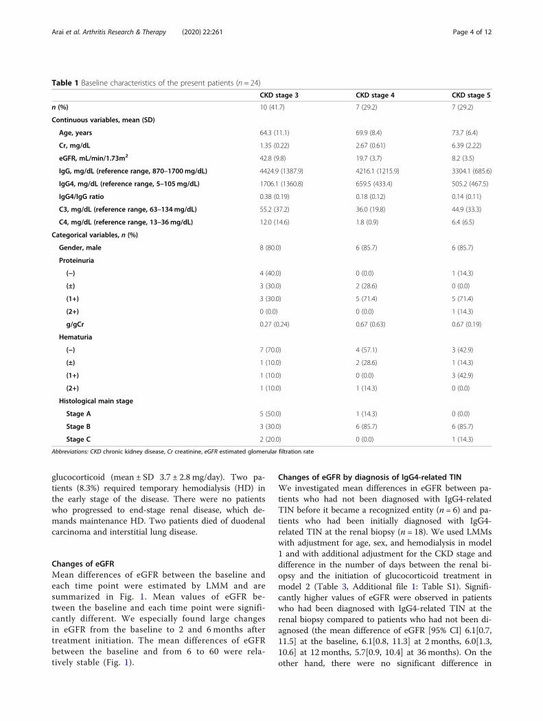
Table 1 Baseline characteristics of the present patients (n = 24)
CKD stage 3 CKD stage 4 CKD stage 5
n (%) 10 (41.7) 7 (29.2) 7 (29.2)
Continuous variables, mean (SD)
Age, years 64.3 (11.1) 69.9 (8.4) 73.7 (6.4)
Cr, mg/dL 1.35 (0.22) 2.67 (0.61) 6.39 (2.22)
eGFR, mL/min/1.73m2 42.8 (9.8) 19.7 (3.7) 8.2 (3.5)
IgG, mg/dL (reference range, 870–1700mg/dL) 4424.9 (1387.9) 4216.1 (1215.9) 3304.1 (685.6)
IgG4, mg/dL (reference range, 5–105mg/dL) 1706.1 (1360.8) 659.5 (433.4) 505.2 (467.5)
IgG4/IgG ratio 0.38 (0.19) 0.18 (0.12) 0.14 (0.11)
C3, mg/dL (reference range, 63–134mg/dL) 55.2 (37.2) 36.0 (19.8) 44.9 (33.3)
C4, mg/dL (reference range, 13–36mg/dL) 12.0 (14.6) 1.8 (0.9) 6.4 (6.5)
Categorical variables, n (%)
Gender, male 8 (80.0) 6 (85.7) 6 (85.7)
Proteinuria
(−) 4 (40.0) 0 (0.0) 1 (14.3)
(±) 3 (30.0) 2 (28.6) 0 (0.0)
(1+) 3 (30.0) 5 (71.4) 5 (71.4)
(2+) 0 (0.0) 0 (0.0) 1 (14.3)
g/gCr 0.27 (0.24) 0.67 (0.63) 0.67 (0.19)
Hematuria
(−) 7 (70.0) 4 (57.1) 3 (42.9)
(±) 1 (10.0) 2 (28.6) 1 (14.3)
(1+) 1 (10.0) 0 (0.0) 3 (42.9)
(2+) 1 (10.0) 1 (14.3) 0 (0.0)
Histological main stage
Stage A 5 (50.0) 1 (14.3) 0 (0.0)
Stage B 3 (30.0) 6 (85.7) 6 (85.7)
Stage C 2 (20.0) 0 (0.0) 1 (14.3)
Abbreviations: CKD chronic kidney disease, Cr creatinine, eGFR estimated glomerular filtration rate
Arai et al. Arthritis Research & Therapy (2020) 22:261 Page 4 of 12

Table
2BaselinecharacteristicsandeG
FR(Cr)at
thetim
eof
renalb
iopsy,thetim
eof
treatm
entinitiation,andeach
timepo
intaftertreatm
entinitiation
eGFR
,mL/min/1.73m
2(Cr,mg/dL)
No.
Sex
Follo
w-
up (yr)a
Prim
ary
diagno
sis
Extraren
allesion
sPS
LTx (m
g/
day)
Duration
ofPS
Ltrea
tmen
t(yr)
Atthe
timeof
rena
lbiopsy
Baselineb
1mon
thfrom
the
baseline
2 mon
ths
from
the
baseline
6 mon
ths
from
the
baseline
12 mon
ths
from
the
baseline
24 mon
ths
from
the
baseline
36 mon
ths
from
the
baseline
60 mon
ths
from
the
baseline
Thenu
mber
ofdays
betwee
nrena
lbiopsy
andtheinitiation
ofPS
Ltrea
tmen
t
Cases
notdiagn
osed
byrena
lbiopsy
attheinitiale
xaminationd
1M
13.7
TIN
associated
with
immun
edisorder
Thr,Ca,
2513.8
15.4
(3.32)
12.8
(3.95)
19.8
(2.64)
21.6
(2.44)
23.6
(2.25)
26.9
(1.99)
28.0
(1.91)
26.7
(1.99)
27.8(1.9)
32
2M
7.8
TINof
Sjog
ren’s
synd
rome
Thr
302.5c
11.6
(3.24)
15.8
(3.22)
25.4
(2.08)
30.5
(1.76)
37.4
(1.46)
28.6
(1.86)
11.7
(4.19)
38.1
(1.42)
NA
−17
3F
12.2
TINof
Sjog
ren’s
synd
rome
Ly,sinusitis,
extratho
racic
lesion
307.5
37.4(1.2)
46.3(1.3)
55.6(1.1)
55.6(1.1)
55.6(1.1)
63.5
(0.97)
78.0(0.8)
68.2(0.9)
64.4
(0.94)
−96
4M
9.3
TIN
associated
with
immun
edisorder
Sa,Ly,Lu,Pa,
409.3
15.7(3.5)
16.8
(3.29)
16.8
(3.29)
38.0
(1.56)
42.1
(1.42)
45.3
(1.32)
44.7
(1.33)
46.4
(1.28)
34.3
(1.67)
−7
5M
11.0
Low-grade
lymph
oma
Sa,Ly,Thr
600.5c
34.3(1.6)
34.3(1.6)
NA
NA
NA
51.4(1.1)
51.2(1.1)
51.0(1.1)
46.0
(1.21)
−90
6M
7.8
TINwith
immun
ecomplexes
Ca
407.8
16.6(3.1)
16.6(3.1)
37.3
(1.48)
37.9
(1.46)
37.1
(1.49)
34.6
(1.58)
33.3
(1.63)
35.1
(1.55)
23.3
(2.24)
−14
Cases
diagno
sedbyrena
lbiopsy
attheinitiale
xamination
7M
4.7
IgG4-
relatedTIN
Sa,Ly,Lu,
Thy,Pa
504.6
45.9
(1.25)
45.9
(1.25)
56.1
(1.04)
49.8
(1.16)
53.8
(1.08)
57.7
(1.01)
52.8
(1.09)
48.7
(1.17)
NA
−23
8M
7.7
IgG4-
relatedTIN
NA
401.9c
5.4(9.0)
5.4(9.0)
9.2(5.51)
12.9
(4.04)
19.1
(2.82)
19.0
(2.82)
21.7
(2.49)
21.5(2.5)
20.5
(2.59)
−1
9M
6.9
IgG4-
relatedTIN
NA
406.9
18.3
(2.86)
19.5
(2.82)
24.6
(2.18)
27.1(2.0)
24.9
(2.16)
39.5
(1.41)
36.3
(1.52)
38.9
(1.42)
36.9
(1.48)
2
10M
7.3
IgG4-
relatedTIN
Sa,Ly,La
507.0
54.0
(1.11)
59.0
(1.05)
59.0
(1.05)
68.0
(0.92)
73.0
(0.86)
54.0
(1.13)
62.0
(0.99)
59.0
(1.03)
56.0
(1.07)
−36
11M
6.6
IgG4-
relatedTIN
Ly,RPF
406.6
11.4
(4.35)
9.3(5.25)
19.6
(2.65)
21.7
(2.41)
24.5
(2.16)
23.7
(2.21)
22.4
(2.33)
21.5
(2.41)
24.8(2.1)
0
12M
4.6
IgG4-
relatedTIN
Ly50
4.6
7.5(6.2)
7.5(6.2)
22.0
(2.28)
29.0
(1.79)
29.0
(1.82)
25.0
(2.04)
27.0
(1.89)
24.0
(2.08)
26.0
(1.97)
−2
13M
5.4
IgG4-
relatedTIN
Sa,La,RPF
404.4c
12.4
(3.96)
12.4
(3.96)
28.7
(1.84)
28.4
(1.86)
29.8
(1.89)
32.2
(1.65)
36.3
(1.48)
32.4
(1.63)
29.3
(1.78)
−12
Arai et al. Arthritis Research & Therapy (2020) 22:261 Page 5 of 12

Table
2BaselinecharacteristicsandeG
FR(Cr)at
thetim
eof
renalb
iopsy,thetim
eof
treatm
entinitiation,andeach
timepo
intaftertreatm
entinitiation(Con
tinued)
eGFR
,mL/min/1.73m
2(Cr,mg/dL)
No.
Sex
Follo
w-
up (yr)a
Prim
ary
diagno
sis
Extraren
allesion
sPS
LTx (m
g/
day)
Duration
ofPS
Ltrea
tmen
t(yr)
Atthe
timeof
rena
lbiopsy
Baselineb
1mon
thfrom
the
baseline
2 mon
ths
from
the
baseline
6 mon
ths
from
the
baseline
12 mon
ths
from
the
baseline
24 mon
ths
from
the
baseline
36 mon
ths
from
the
baseline
60 mon
ths
from
the
baseline
Thenu
mber
ofdays
betwee
nrena
lbiopsy
andtheinitiation
ofPS
Ltrea
tmen
t
14M
4.6
IgG4-
relatedTIN
Ly30
4.6
26.1
(2.06)
26.1
(2.06)
35.9
(1.54)
35.9
(1.54)
34.5
(1.59)
37.6
(1.47)
39.8
(1.39)
42.7(1.3)
NA
−10
15M
3.8
IgG4-
relatedTIN
RPF,Ao
303.2
25.2
(2.08)
34.1
(1.58)
28.0
(1.89)
24.7
(2.12)
25.3
(2.07)
33.2
(1.61)
38.0
(1.42)
32.8
(1.62)
NA
−14
16F
2.8
IgG4-
relatedTIN
Ly,Lu
252.8
22.7(1.7)
22.5(1.7)
27.5
(1.42)
33.3
(1.19)
33.0(1.2)
34.3
(1.16)
33.5
(1.18)
33.7
(1.17)
NA
−13
17M
2.7
IgG4-
relatedTIN
NA
302.7
9.3(5.18)
6.7(6.94)
12.1
(4.06)
16.6
(3.03)
15.7
(3.19)
19.0
(2.68)
16.6
(3.02)
NA
NA
−5
18F
2.2
IgG4-
relatedTIN
NA
300.8
4.3(7.81)
3.5(9.44)
12.1
(2.85)
13.0
(2.83)
11.6
(3.13)
12.3
(2.98)
11.6
(3.13)
NA
NA
18
19M
3.8
IgG4-
relatedTIN
RPF,Ao
303.8
21.8
(2.39)
20.7
(2.51)
32.5
(1.66)
39.5
(1.39)
34.3
(1.58)
34.9
(1.55)
36.1(1.5)
38.4
(1.41)
NA
5
20F
8.1
IgG4-
relatedTIN
Sa,La
308.0
27.4
(1.53)
30.4
(1.39)
41.0
(1.06)
43.2
(1.01)
49.4
(0.89)
43.5(1.0)
66.0
(0.68)
55.0(0.8)
51.0
(0.85)
−19
21M
7.8
IgG4-
relatedTIN
Sa,Lu,Pa
457.7
49.5(1.3)
49.5(1.3)
87.7
(0.77)
70.5
(0.94)
84.7
(0.79)
80.3
(0.83)
83.1(0.8)
92.6
(0.72)
80.4
(0.81)
−13
22M
7.4
IgG4-
relatedTIN
Sa,Ly
307.4
44.7
(1.27)
44.7
(1.27)
51.1
(1.12)
61.1
(0.95)
62.6
(0.93)
58.4
(0.99)
63.0
(0.92)
61.8
(0.93)
55.6
(1.02)
−4
23M
6.7
IgG4-
relatedTIN
Sa,Ly,RPF,
Ca
306.2
55.8
(1.01)
52.7
(1.06)
63.1(0.9)
52.7
(1.06)
65.4
(0.87)
52.5
(1.06)
53.4
(1.04)
52.1
(1.06)
NA
−181
24M
4.9
IgG4-
relatedTIN
Sa,RPF
354.4
31.4
(1.69)
31.4
(1.69)
55.2
(1.01)
63.3
(0.89)
45.1
(1.21)
48.1
(1.14)
51.9
(1.06)
47(1.15)
NA
−13
Abb
reviations:M
men
,Ffemale,
Yryear,TIN
tubu
lointerstitialn
ephritis,PSLTx
initial
dose
ofpred
nisolone
,eGFR
estim
ated
glom
erular
filtrationrate,C
rcreatin
ine,
NAno
tavailable,
Aope
riaortitis,C
ape
ricarditis,La
dacryo
aden
itis,Lu
lung
lesion
,Lylymph
aden
itis,Pa
type
1au
toim
mun
epa
ncreatitis,RP
Fretrop
erito
neal
fibrosis,Sa
sialad
enitis,Thrthrombo
cytope
nia,Thythyroiditis
a Len
gthof
timebe
tweentherena
lbiopsyan
dthelast
date
ofob
servation
bTh
eba
selin
ewas
thetim
eof
treatm
entinitiation.
IfeG
FRat
thetim
eof
treatm
entinitiationwas
notavailable,
eGFR
attherena
lbiopsywas
used
c The
sepa
tientswerere-treated
becauseof
relapseafterdiscon
tinua
tionof
glucocorticoid
dTh
iswas
becausetherewerepa
tientswho
hadrena
lbiopsybe
fore
IgG4-relatedTINbe
camearecogn
ized
entity.Pa
tientsin
thisgrou
pwas
notdiag
nosedwith
IgG4-relatedTINdu
ringtheob
servationpe
riodof
the
presen
tstud
y
Arai et al. Arthritis Research & Therapy (2020) 22:261 Page 6 of 12

change rates of eGFR (i.e., eGFR slope) between the pa-tients who had been diagnosed with IgG4-related TINand patients who had not been diagnosed (p = 0.87). Themean differences of eGFR were stable during the clinicalcourses (baseline to 60 months), meaning cases that wereable to be diagnosed maintained high eGFR throughouta long period of after treatment.
Changes of eGFR by CKD stageWe investigated mean differences in eGFR betweenpatients with CKD stage 3, CKD stage 4, and CKD
stage 5, using LMMs with adjustment for age, sex,and hemodialysis in model 1 and with additional ad-justment for whether or not they were diagnosed withIgG4-related TIN at the initial examination and dif-ference in the number of days between the renal bi-opsy and the initiation of glucocorticoid treatment inmodel 2 (Table 4, Fig. 2). Significantly lower values ofeGFR were observed in patients with CKD stage 4compared to patients with CKD stage 3. Patients withCKD stage 5 had comparatively lower eGFR than pa-tients with CKD stage 3. On the other hand, there
Fig. 1 Mean differences (95% CI) of eGFR between the baseline and each time point were estimated by linear mixed models. Mean values ofeGFR between the baseline and each time point were significantly different, which meant that eGFR at each time point was significantly highercompared to that at the baseline. We especially found large changes in eGFR from the baseline to 2 and 6months after treatment initiation. Themean differences of eGFR between the baseline and from 6 to 60 were relatively stable. Abbreviations: CI, confidence interval; eGFR, estimatedglomerular filtration rate
Table 3 Mean differences (95% CI) in eGFR at the baseline and each time point according to whether or not patients had beendiagnosed at the initial examination (n = 24), obtained by linear mixed models
Months after treatment Baselinec 1 2 6 12 24 36 60
Mean differences in eGFR (ml/min/1.73 m2) in model 1a
Cases not diagnosed by renal biopsy at theinitial examinationd
Ref. Ref. Ref. Ref. Ref. Ref. Ref. Ref.
Cases diagnosed by renal biopsy at theinitial examination
9.7 (− 1.5,20.9)
9.7 (− 1.2,20.6)
9.7 (− 1.5,20.9)
9.7 (− 0.9,20.4)
9.7 (− 0.2,19.6)
9.7 (0.5,18.9)
9.7 (1.1,18.3)
9.7 (1.6,17.8)
Mean differences in eGFR (ml/min/1.73 m2) in model 2b
Cases not diagnosed by renal biopsy at theinitial examinationd
Ref. Ref. Ref. Ref. Ref. Ref. Ref. Ref.
Cases diagnosed by renal biopsy at theinitial examination
6.1 (0.7,11.5)
6.1 (0.7,11.4)
6.1 (0.8,11.3)
6.0 (1,11.0)
6.0 (1.3,10.6)
5.8 (1.4,10.3)
5.7 (0.9,10.4)
5.4 (− 1.2,12.0)
Abbreviations: CI confidence interval, eGFR estimated glomerular filtration rate, Ref. reference group, CKD chronic kidney diseaseaModel 1 was adjusted for age at the renal biopsy, sex, and hemodialysisbModel 2 was the model 1 with additional adjustment for CKD stage and difference in the number of days between the renal biopsy and the initiation ofglucocorticoid treatmentcThe baseline was the time at treatment initiationdThis was because there were patients who had renal biopsy before IgG4-related TIN became a recognized entity. Patients in this group were not diagnosed withIgG4-related TIN during the observation period of the present study
Arai et al. Arthritis Research & Therapy (2020) 22:261 Page 7 of 12

were no significant difference in change rates of eGFR(i.e., eGFR slope) among the patients with CKD stage3 (reference), CKD stage 4 (p = 0.62), and CKD stage5 (p = 0.43).
Changes of eGFR in histological stagesWe investigated mean differences in eGFR between pa-tients with stage A and stage B, using LMMs with ad-justment for age, sex, and hemodialysis in model 1 andwith additional adjustment for whether or not they werediagnosed with IgG4-related TIN at the initial examin-ation in model 2 (Table 4, Fig. 3). Significantly lowervalues of eGFR were observed in patients with stage Bcompared to patients with stage A. On the other hand,there were no significant difference in change rates ofeGFR between the patients with stage A (reference) andpatients with stage B (p = 0.79).
DiscussionRenal function rapidly improved by the two-month markfrom the initial glucocorticoid therapy and was sustainedfor a long period by maintenance therapy. Patients whowere initially diagnosed with IgG4-related TIN by renal
biopsy had higher eGFR during clinical courses com-pared to those that were not. Compared to CKD stage 3,eGFR in CKD stage 4 and 5 was significantly lowerthroughout the entire period, but there was no signifi-cant difference in its change rate. Patients with histo-logical stage B had significantly lower eGFR comparedto those with histological stage A throughout the entireperiod, but there was no significant difference in itschange rate.Renal function rapidly improved by the 2-month mark
from the initial glucocorticoid therapy and was sustainedfor a long period by maintenance therapy, which wassupported by the following previous studies. After initi-ation of glucocorticoid therapy for IgG4-RKD, renalfunction, hypocomplementemia, and radiological abnor-malities were reported to rapidly improve in 1–2 monthsin previous studies that had a short observational period[9, 11, 16–18]. In the retrospective study of Saeki et al.which observed the clinical course of eGFR after treat-ment in patients with IgG4-RKD, patients’ renal functionimproved and their eGFR was maintained during thetime that they received maintenance glucocorticoid ther-apy within median 34 months [14].
Table 4 Mean differences (95% CI) in eGFR at each time point between CKD stage and histological stage obtained by linear mixedmodels
Months aftertreatment
Baselined 1 2 6 12 24 36 60
Mean differences (ml/min/1.73 m2)
Model 1a
CKD stage 3 Ref. Ref. Ref. Ref. Ref. Ref. Ref. Ref.
CKD stage 4 − 19.2 (− 25.2,− 13.1)
− 19.2 (− 25.1,− 13.2)
− 19.1 (− 25, −13.2)
− 19.0 (− 24.6,− 13.4)
− 18.8 (− 24.1,− 13.6)
− 18.5 (− 23.5,− 13.5)
− 18.2 (− 23.5,− 12.9)
− 17.5 (− 24.7,− 10.3)
CKD stage 5 − 26.2 (− 33.3,− 19.1)
− 26.2 (− 33.2,− 19.1)
− 26.1 (− 33.1,− 19.1)
− 25.9 (− 32.7,− 19.1)
− 25.6 (− 32.1,− 19.1)
− 25.0 (− 31.2,− 18.7)
− 24.3 (− 30.8,− 17.8)
− 23.1 (− 31.2,− 15)
Histologicalstage Ac
Ref. Ref. Ref. Ref. Ref. Ref. Ref. Ref.
Histologicalstage B
− 13.9 (− 23.7,− 4.1)
− 13.9 (− 23.6,− 4.2)
− 13.9 (− 23.4,− 4.3)
− 13.8 (− 23.2,− 4.4)
− 13.6 (− 22.8,− 4.5)
− 13.4 (− 21.9,− 4.9)
− 13.1 (− 21.4,− 4.8)
− 12.6 (− 21.4,− 3.8)
Model 2b
CKD stage 3 Ref. Ref. Ref. Ref. Ref. Ref. Ref. Ref.
CKD stage 4 − 15.6 (− 21.6,− 9.5)
− 15.6 (− 21.5,− 9.6)
− 15.5 (− 21.4,− 9.6)
− 15.4 (− 21.1,− 9.7)
− 15.2 (− 20.6,− 9.7)
− 14.7 (− 20.1,− 9.3)
− 14.3 (− 20.2,− 8.4)
− 13.4 (− 21.3,− 5.5)
CKD stage 5 − 22.9 (− 29.9,− 16.0)
− 22.9 (− 29.8,− 16.0)
− 22.8 (− 29.7,− 16.0)
− 22.6 (− 29.3,− 15.9)
− 22.3 (− 28.7,− 15.8)
− 21.6 (− 28.0,− 15.2)
− 20.9 (− 27.6,− 14.1)
− 19.5 (− 28.0,− 11.0)
Histologicalstage Ac
Ref. Ref. Ref. Ref. Ref. Ref. Ref. Ref.
Histologicalstage B
− 13.5 (− 23.1,− 4.0)
− 13.5 (− 23, −4.1)
− 13.5 (− 22.9,− 4.1)
− 13.4 (− 22.5,− 4.4)
− 13.3 (− 21.9,− 4.7)
− 13.1 (− 21,− 5.1)
− 12.8 (− 20.4,− 5.3)
− 12.4 (− 20.3,− 4.4)
Abbreviations: CI confidence interval, eGFR estimated glomerular filtration rate, CKD chronic kidney disease, Ref. reference groupaModel 1 was adjusted for age at the renal biopsy, sex, and hemodialysisbModel 2 was model 1 with additional adjustment for diagnosis of IgG4-related TIN and difference in the number of days between the renal biopsy and theinitiation of glucocorticoid treatment when the interested exposure was CKD stage. Model 2 was model 1 with additional adjustment for whether or not theywere diagnosed with IgG4-related TIN at the initial examination when the interested exposure was histological stagecInterstitial fibrosis is more clearly seen in stage B than in stage AdThe baseline was the time of treatment initiation
Arai et al. Arthritis Research & Therapy (2020) 22:261 Page 8 of 12

In the present study, patients who were initially diag-nosed with IgG4-related TIN at renal biopsy had highereGFR during the clinical courses compared to those thatwere not. By appropriately diagnosing at an early stage ofthe disease, it can be said that early treatment can be initi-ated and renal function maintained compared to when itis not properly diagnosed. In the 2000s, conditions such asAIP and Mikulicz’s disease were recognized as systemicdiseases related to IgG4, and the number of reports ofIgG4-RD rapidly increased since around the year 2010.Moreover, before IgG4-RD became a recognized entityand spread as a disease concept, it may have been misdiag-nosed as malignancy or another immune-mediated condi-tion such as Sjögren’s syndrome.In the present patients with CKD stages 3, 4, and 5, their
eGFR were improved after the treatment. Additionally,eGFR in CKD stages 4 and 5 was significantly lowerthroughout the entire period than that in CKD stage 3,but there was no significant difference in its change rate.Similar results were obtained in a previous study showingthat eGFR in patients whose eGFR had been less than 60ml/min/1.73m2 before treatment was improved aftertreatment, but did not exceed 60ml/min/1.73m2 [14]. Thereason for this may be that patients with lower pre-treatment eGFR already have progressing fibrosis [14, 24].
In past reports, patients with eGFR<60mL/min/1.73m2
before treatment had developed renal atrophy after treat-ment [14]. In reports evaluating histopathological findingsin IgG4-related TIN from repeat renal biopsy while receiv-ing glucocorticoid therapy, regional fibrosis developed inthe interstitium, even though the area of cell infiltrationdecreased [16, 17]. These data might suggest that thehistological fibrotic lesions correspond to the atrophic le-sions demonstrated by imaging [16]. Patients with lowerpre-treatment eGFR had already developed more lesionswith advanced fibrosis and such lesions resulted in irre-versible atrophic changes despite glucocorticoid therapy[14, 24]. Furthermore, in the present study in investigatinglongitudinal changes in eGFR by histological stage, histo-logical stage B had significantly lower eGFR compared tohistological stage A throughout the entire period, butthere was no significant difference in its change rate.There have been no reports concerning long-term renaloutcome in histological stage. This result supports past re-ports, which suggests improvement of renal function waslimited due to the already advanced histological fibrosisand irreversible renal atrophy.The international consensus guidance statement on
the management and treatment of IgG4-RD recom-mends urgent treatment of IgG4-related TIN to prevent
Fig. 2 Least squares means (95% CI) of eGFR (mL/min/1.73m2) at baseline (i.e., at treatment initiation) and months after treatment initiationaccording to CKD stage 3, stage 4, and stage 5 in patients with IgG4-related TIN (n = 24) based on linear mixed models. Significantly lower valuesof eGFR were observed in patients with CKD stage 4 compared to patients with CKD stage 3. Patients with CKD stage 5 had comparatively lowereGFR than patients with CKD stage 3. Those mean differences in eGFR correspond to Table 4. On the other hand, there were no significantdifference in change rates of eGFR (i.e., eGFR slope) among the patients with CKD stage 3, CKD stage 4, and CKD stage 5 (the mean difference ofthe monthly slope of the eGFR [95% CI], 0.0 [− 0.1 to 0.2] in CKD stage 4 and 0.1 [− 0.1 to 0.2] in CKD stage 5 compared with CKD stage 3 inmodel 2), meaning the mean differences of eGFR were stable during the clinical courses (baseline to 60months). Abbreviations: CI, confidenceinterval; CKD, chronic kidney disease; TIN, tubulointerstitial nephritis; eGFR, estimated glomerular filtration rate
Arai et al. Arthritis Research & Therapy (2020) 22:261 Page 9 of 12

irreversible renal failure [15]. Therefore, it is important tostart glucocorticoid therapy at early CKD stage and earlyhistological stage in maintaining eGFR for a long period.Note that even in IgG4-related TIN with advanced CKDstage, eGFR has improved after glucocorticoid therapy, soit can be thought that treatment is effective.In the present study, glucocorticoid therapy was used for
all patients. Glucocorticoid therapy is common in Japan;however, other countries in North America and Europe donot perform a long-term low-dose maintenance glucocortic-oid therapy [25–27], and instead, B cell depletion with rituxi-mab and immunosuppressive agent is used with IgG4-RDpatients [15, 28–30]. Therefore, further studies similar to thisstudy are necessary in other countries.This present study has the following strengths. First, it
was based on almost all renal biopsy results of which pa-tients with IgG4-related TIN were treated in Aichi,which could reduce selection bias. Second, we followedup on the longitudinal changes in eGFR of the IgG4-related TIN cases for a long period. Additionally, weevaluated the longitudinal changes in eGFR in CKDstage more advanced than stage 3 and in histologicalstage, which were not evaluated in previous studies well.This enabled us to show an even more detailed changerate of eGFR compared to previous reports.
This study has some limitations. First, the numberof patients is small. This is because IgG4-RD is arare disease, which was also showed in J-RBR as anationwide registry with high representability inJapan. Note that the present study was based on al-most all renal biopsy results in Aichi. The presentstudy warrants further studies investigating long-term clinical course of renal function in IgG4-relatedTIN. Additionally, findings from the study may notbe generalizable outside of Japan because treatmentprocedures of IgG4-RD in Japan are different fromNorth America and Europe. Second, the treatmentregimen and follow-up protocols were inconsistentamong patients because of this retrospective multi-center cohort study. In this instance, 1 patient wastreated after a diagnosis of non-Hodgkin’s lymph-oma with cyclophosphamide, doxorubicin, vincris-tine, and prednisone (CHOP) chemotherapy plusrituximab. For 4 patients, an immunosuppressant(azathioprine 50 mg or mizoribine 50 mg) was addedto the glucocorticoid therapy. However, as there isno consensus regarding treatment of IgG4-relatedTIN, further investigation is necessary. Third, re-lapse could not be statistically analyzed because ofthe small sample size, and because it is a
Fig. 3 Least squares means (95% CI) of eGFR (mL/min/1.73m2) at baseline (i.e., at treatment initiation) and months after the treatment initiationaccording to histological stage A and stage B in patients with IgG4-related TIN (n = 24) based on linear mixed models. Significantly lower valuesof eGFR were observed in patients with stage B compared to patients with stage A. Those mean differences in eGFR correspond to Table 4. Onthe other hand, there were no significant difference in change rates of eGFR (i.e., eGFR slope) between patients with stage A and stage B (themean difference of the monthly slope of the eGFR [95% CI] 0.0 [− 0.1 to 0.2] in model 2), meaning the mean differences of eGFR were stableduring the clinical course (baseline to 60 months). Abbreviations: CI, confidence interval; eGFR, estimated glomerular filtration rate; TIN,tubulointerstitial nephritis
Arai et al. Arthritis Research & Therapy (2020) 22:261 Page 10 of 12

retrospective cohort design, the definition of relapsediffered per patient. However, as relapse is a signifi-cant clinical outcome, we will indicate the relapseoccurrence percentage: 8 (33.3%) of 24 treated pa-tients experienced relapse, of which 4 patients re-lapsed during maintenance therapy.
ConclusionsIn conclusion, renal function of patients with IgG4-related TIN was improved and maintained for a longperiod after glucocorticoid therapy was implemented.Compared to patients with IgG4-related TIN and CKDstage 3 or histological stage A, those with CKD stage 4and 5 or histological stage B had lower eGFR values dur-ing the entire observational period, which suggested thatearly diagnosis and treatment are important in maintain-ing renal function. Additionally, as renal function im-proved after glucocorticoid therapy in all CKD stages,this can be expected even in an advanced CKD stage.
Supplementary informationSupplementary information accompanies this paper at https://doi.org/10.1186/s13075-020-02320-x.
Additional file 1: Supplementary data 1. Description of the cohortused in the present study. Figure S1. Frequency and proportion ofpathological diagnoses. Supplementary data 2. Diagnosis rate of IgG4-related TIN (2003-2008 and 2009-2015). Table S1. Baseline characteristicsof the present patients by diagnosis or non-diagnosis of IgG4-related TINat the initial examination.
AbbreviationsAIP: Autoimmune pancreatitis; CHOP: Cyclophosphamide, doxorubicin,vincristine, and prednisone; CI: Confidence intervals; CKD: Chronic kidneydisease; Cr: Creatinine; eGFR: Estimated glomerular filtration rate;HD: Temporary hemodialysis; IgG4-RD: IgG4-related disease; IgG4-RKD: IgG4-related kidney disease; LMM: Linear mixed models; PSL: Prednisolone;SD: Standard deviations; TIN: Tubulointerstitial nephritis
AcknowledgementsNone.
Authors’ contributionsH.A., H.H., and Y.Y. contributed to the research idea and study design. H.A.,T.O., K.T., and S.M. contributed to the data acquisition. H.A., N.T., D.I., M.H.,and H.H. contributed to the data interpretation. S.O. performed the statisticalanalysis. H.A., S.O., and H.H. prepared the manuscript. N.T., S.M., D.I., M.H., andY.Y. provided intellectual content of critical importance to the work. Allauthors approved the final submission.
FundingThis work was supported in part by Health and Labour Sciences ResearchGrants for research on intractable diseases (The Research Team forAutoimmune Diseases) from the Ministry of Health, Labour and Welfare ofJapan (H29-nanchitou(nan)-ippan-008). This work was also supported by theAichi Kidney Foundation.
Availability of data and materialsNot applicable.
Ethics approval and consent to participateThis study was approved by the Ethics Committee of Fujita Health Universityand Nagoya University. The need for patient consent was waived due to the
retrospective nature of the study and the absence of personally identifiabledata in the report.
Consent for publicationNot applicable.
Competing interestsThe authors declare that they have no competing interests.
Author details1Department of Nephrology, Fujita Health University School of Medicine,1-98 Dengakugakubo, Kutsukake-cho, Toyoake, Aichi 470-1192, Japan.2Preventive Medicine and Epidemiology, National Cerebral andCardiovascular Center, 6-1 Kishibe-Shimmachi, Suita, Osaka 564-8565, Japan.3Department of Nephrology, Nagoya University Graduate School ofMedicine, 65 Tsurumai-cho, Showa-ku, Nagoya, Aichi 466-8550, Japan.
Received: 3 June 2020 Accepted: 17 September 2020
References1. Rheumatology M, Ky AS. Comprehensive diagnostic criteria for IgG4-related
disease ( IgG4-RD ), 2011. Mod Rheumatol. 2012;22:21–30.2. Yamaguchi Y, Kanetsuna Y, Honda K, Yamanaka N, Kawano M, Nagata M,
et al. Characteristic tubulointerstitial nephritis in IgG4-related disease. HumPathol. 2012;43:536–49.
3. Stone JH, Brito-Zerón P, Bosch X, Ramos-Casals M. Diagnostic approach tothe complexity of IgG4-related disease. Mayo Clin Proc. 2015;90:927–39.
4. Hamano H, Kawa S, Horiuchi A, Unno H, Furuya N, Akamatsu T, et al. Highserum IgG4 concentrations in patients with sclerosing pancreatitis. N Engl JMed. 2001;344:732–8.
5. Saeki T, Kawano M. IgG4-related kidney disease. Kidney Int. 2013;85:251–7.6. Kamisawa T, Funata N, Hayashi Y, Eishi Y, Koike M, Tsuruta K, et al. A new
clinicopathological entity of IgG4-related autoimmune disease. JGastroenterol. 2003;38:982–4.
7. Yamamoto M, Ohara M, Suzuki C, Naishiro Y, Yamamoto H, Takahashi H,et al. Elevated IgG4 concentrations in serum of patients with Mikulicz’sdisease. Scand J Rheumatol. 2004;33:432–3.
8. Deshpande V, Zen Y, Chan JKC, Yi EE, Sato Y, Yoshino T, et al. Consensus statementon the pathology of IgG4-related disease. Mod Pathol. 2012;25:1181–92.
9. Saeki T, Nishi S, Imai N, Ito T, Yamazaki H, Kawano M, et al.Clinicopathological characteristics of patients with IgG4-relatedtubulointerstitial nephritis. Kidney Int. 2010;78:1016–23.
10. Kawano M, Saeki T, Nakashima H, Nishi S, Yamaguchi Y, Hisano S, et al.Proposal for diagnostic criteria for IgG4-related kidney disease. Clin ExpNephrol. 2011;15:615–26.
11. Raissian Y, Nasr SH, Larsen CP, Colvin RB, Smyrk TC, Takahashi N, et al.Diagnosis of IgG4-related tubulointerstitial nephritis. J Am Soc Nephrol.2011;22:1343–52.
12. Nishi S, Imai N, Yoshida K, Ito Y, Saeki T. Clinicopathological findings ofimmunoglobulin G4-related kidney disease. Clin Exp Nephrol. 2011;15:810–9.
13. Cornell LD. IgG4-related kidney disease. Semin Diagn Pathol. 2012;29:245–50.14. Saeki T, Kawano M, Mizushima I, Yamamoto M, Wada Y, Nakashima H, et al.
The clinical course of patients with IgG4-related kidney disease. Kidney IntNature Publishing Group. 2013;84:826–33.
15. Nishi S, Imai N, Yoshida K, Ito Y, Saeki T. Clinicopathological findings ofimmunoglobulin G4-related kidney disease. Clin. Exp. Nephrol.2011;15:810–819.
16. Mizushima I, Yamada K, Fujii H, Inoue D, Umehara H, Yamagishi M, et al.Clinical and histological changes associated with corticosteroid therapy inIgG4-related tubulointerstitial nephritis. Mod Rheumatol. 2012;22:859–70.
17. Arai H, Hayashi H, Takahashi K, Koide S, Sato W, Hasegawa M, et al.Tubulointerstitial fibrosis in patients with IgG4-related kidney disease:pathological findings on repeat renal biopsy. Rheumatol Int. 2015;35:1093–101.
18. Saeki T, Kawano M, Mizushima I, Yamamoto M, Wada Y, Ubara Y, et al.Recovery of renal function after glucocorticoid therapy for IgG4-relatedkidney disease with renal dysfunction. Clin Exp Nephrol Springer Tokyo.2016;20:87–93.
19. Shirakashi M, Yoshifuji H, Kodama Y, Chiba T, Yamamoto M, Takahashi H,et al. Factors in glucocorticoid regimens associated with treatment
Arai et al. Arthritis Research & Therapy (2020) 22:261 Page 11 of 12

response and relapses of IgG4-related disease: a multicentre study. Sci RepNature. 2018;8.
20. Matsuo S, Imai E, Horio M, Yasuda Y, Tomita K, Nitta K, et al. Revisedequations for estimated GFR from serum creatinine in Japan. Am J KidneyDis WB Saunders. 2009;53:982–92.
21. Schisterman EF, Cole SR, Platt RW. Overadjustment bias and unnecessaryadjustment in epidemiologic studies. Epidemiology. 2009;20:488–95.
22. Proust-Lima C, Philipps V, Liquet B. Estimation of extended mixed modelsusing latent classes and latent processes: the {R} package {lcmm}. J StatSoftw. 2017;78:1–56.
23. core Team R. R: a language and environment for statistical computing. RFound Stat Comput Vienna, Austria. 2018;.
24. Mizushima I, Yamamoto M, Inoue D, Nishi S, Taniguchi Y, Ubara Y, et al.Factors related to renal cortical atrophy development after glucocorticoidtherapy in IgG4-related kidney disease: a retrospective multicenter study.Arthritis Res Ther; 2016;18:1–9.
25. Ghazale A, Chari ST, Zhang L, Smyrk TC, Takahashi N, Levy MJ, et al.Immunoglobulin G4-associated cholangitis: clinical profile and response totherapy. Gastroenterology. 2008;134:706–15.
26. Raina A, Yadav D, Krasinskas AM, McGrath KM, Khalid A, Sanders M, et al.Evaluation and management of autoimmune pancreatitis: experience at alarge us center. Am J Gastroenterol. 2009;104:2295–306.
27. Fernández-Codina A, Martínez-Valle F, Pinilla B, López C, De Torres I, Solans-Laqué R, et al. IgG4-related disease: results from a multicenter Spanishregistry. Med (United States). Lippincott Williams and Wilkins; 2015;94.
28. Hart PA, Topazian MD, Witzig TE, Clain JE, Gleeson FC, Klebig RR, et al.Treatment of relapsing autoimmune pancreatitis with immunomodulatorsand rituximab: the Mayo Clinic experience. Gut. 2013;62:1607–15.
29. Carruthers MN, Topazian MD, Khosroshahi A, Witzig TE, Wallace ZS, Hart PA,et al. Rituximab for IgG4-related disease: a prospective, open-label trial. AnnRheum Dis. 2015;74:1171–7.
30. Quattrocchio G, Barreca A, Demarchi A, Solfietti L, Beltrame G, Fenoglio R,et al. IgG4-related kidney disease: the effects of a rituximab-basedimmunosuppressive therapy. Oncotarget. 2018;9:21337–47.
Publisher’s NoteSpringer Nature remains neutral with regard to jurisdictional claims inpublished maps and institutional affiliations.
Arai et al. Arthritis Research & Therapy (2020) 22:261 Page 12 of 12