Embed Size (px)
Citation preview
Long-Lasting Immune Responses 4 Years after
GAD-Alum Treatment in Children with Type 1
Diabetes
Stina Axelsson, Mikael Chéramy, Maria Hjorth, Mikael Pihl, Linda Åkerman,
Emanuela Martinuzzi, Roberto Mallone, Johnny Ludvigsson and Rosaura Casas
Linköping University Post Print
N.B.: When citing this work, cite the original article.
Original Publication:
Stina Axelsson, Mikael Chéramy, Maria Hjorth, Mikael Pihl, Linda Åkerman, Emanuela
Martinuzzi, Roberto Mallone, Johnny Ludvigsson and Rosaura Casas, Long-Lasting Immune
Responses 4 Years after GAD-Alum Treatment in Children with Type 1 Diabetes, 2011,
PLoS ONE, (6), 12.
http://dx.doi.org/10.1371/journal.pone.0029008
Licensee: Public Library of Science
http://www.plos.org/
Postprint available at: Linköping University Electronic Press
http://urn.kb.se/resolve?urn=urn:nbn:se:liu:diva-74156
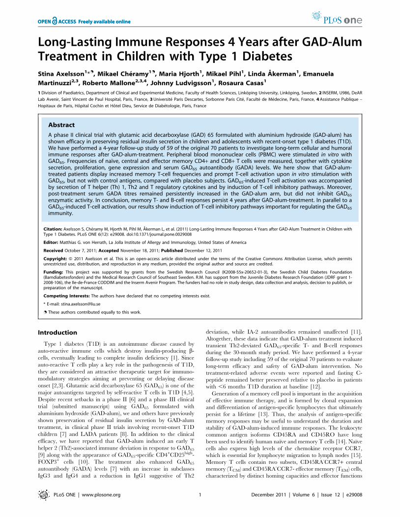
Long-Lasting Immune Responses 4 Years after GAD-AlumTreatment in Children with Type 1 DiabetesStina Axelsson1*., Mikael Cheramy1., Maria Hjorth1, Mikael Pihl1, Linda Akerman1, Emanuela
Martinuzzi2,3, Roberto Mallone2,3,4, Johnny Ludvigsson1, Rosaura Casas1
1 Division of Paediatrics, Department of Clinical and Experimental Medicine, Faculty of Health Sciences, Linkoping University, Linkoping, Sweden, 2 INSERM, U986, DeAR
Lab Avenir, Saint Vincent de Paul Hospital, Paris, France, 3 Universite Paris Descartes, Sorbonne Paris Cite, Faculte de Medecine, Paris, France, 4 Assistance Publique –
Hopitaux de Paris, Hopital Cochin et Hotel Dieu, Service de Diabetologie, Paris, France
Abstract
A phase II clinical trial with glutamic acid decarboxylase (GAD) 65 formulated with aluminium hydroxide (GAD-alum) hasshown efficacy in preserving residual insulin secretion in children and adolescents with recent-onset type 1 diabetes (T1D).We have performed a 4-year follow-up study of 59 of the original 70 patients to investigate long-term cellular and humoralimmune responses after GAD-alum-treatment. Peripheral blood mononuclear cells (PBMC) were stimulated in vitro withGAD65. Frequencies of naıve, central and effector memory CD4+ and CD8+ T cells were measured, together with cytokinesecretion, proliferation, gene expression and serum GAD65 autoantibody (GADA) levels. We here show that GAD-alum-treated patients display increased memory T-cell frequencies and prompt T-cell activation upon in vitro stimulation withGAD65, but not with control antigens, compared with placebo subjects. GAD65-induced T-cell activation was accompaniedby secretion of T helper (Th) 1, Th2 and T regulatory cytokines and by induction of T-cell inhibitory pathways. Moreover,post-treatment serum GADA titres remained persistently increased in the GAD-alum arm, but did not inhibit GAD65
enzymatic activity. In conclusion, memory T- and B-cell responses persist 4 years after GAD-alum-treatment. In parallel to aGAD65-induced T-cell activation, our results show induction of T-cell inhibitory pathways important for regulating the GAD65
immunity.
Citation: Axelsson S, Cheramy M, Hjorth M, Pihl M, Akerman L, et al. (2011) Long-Lasting Immune Responses 4 Years after GAD-Alum Treatment in Children withType 1 Diabetes. PLoS ONE 6(12): e29008. doi:10.1371/journal.pone.0029008
Editor: Matthias G. von Herrath, La Jolla Institute of Allergy and Immunology, United States of America
Received October 7, 2011; Accepted November 18, 2011; Published December 12, 2011
Copyright: � 2011 Axelsson et al. This is an open-access article distributed under the terms of the Creative Commons Attribution License, which permitsunrestricted use, distribution, and reproduction in any medium, provided the original author and source are credited.
Funding: This project was supported by grants from the Swedish Research Council (K2008-55x-20652-01-3), the Swedish Child Diabetes Foundation(Barndiabetesfonden) and the Medical Research Council of Southeast Sweden. R.M. has support from the Juvenile Diabetes Research Foundation (JDRF grant 1-2008-106), the Ile-de-France CODDIM and the Inserm Avenir Program. The funders had no role in study design, data collection and analysis, decision to publish, orpreparation of the manuscript.
Competing Interests: The authors have declared that no competing interests exist.
* E-mail: [email protected]
. These authors contributed equally to this work.
Introduction
Type 1 diabetes (T1D) is an autoimmune disease caused by
auto-reactive immune cells which destroy insulin-producing b-
cells, eventually leading to complete insulin deficiency [1]. Since
auto-reactive T cells play a key role in the pathogenesis of T1D,
they are considered an attractive therapeutic target for immuno-
modulatory strategies aiming at preventing or delaying disease
onset [2,3]. Glutamic acid decarboxylase 65 (GAD65) is one of the
major autoantigens targeted by self-reactive T cells in T1D [4,5].
Despite recent setbacks in a phase II [6] and a phase III clinical
trial (submitted manuscript) using GAD65 formulated with
aluminium hydroxide (GAD-alum), we and others have previously
shown preservation of residual insulin secretion by GAD-alum
treatment, in clinical phase II trials involving recent-onset T1D
children [7] and LADA patients [8]. In addition to the clinical
efficacy, we have reported that GAD-alum induced an early T
helper 2 (Th2)-associated immune deviation in response to GAD65
[9] along with the appearance of GAD65-specific CD4+CD25high-
FOXP3+ cells [10]. The treatment also enhanced GAD65
autoantibody (GADA) levels [7] with an increase in subclasses
IgG3 and IgG4 and a reduction in IgG1 suggestive of Th2
deviation, while IA-2 autoantibodies remained unaffected [11].
Altogether, these data indicate that GAD-alum treatment induced
transient Th2-deviated GAD65-specific T- and B-cell responses
during the 30-month study period. We have performed a 4-year
follow-up study including 59 of the original 70 patients to evaluate
long-term efficacy and safety of GAD-alum intervention. No
treatment-related adverse events were reported and fasting C-
peptide remained better preserved relative to placebo in patients
with ,6 months T1D duration at baseline [12].
Generation of a memory cell pool is important in the acquisition
of effective immune therapy, and is formed by clonal expansion
and differentiation of antigen-specific lymphocytes that ultimately
persist for a lifetime [13]. Thus, the analysis of antigen-specific
memory responses may be useful to understand the duration and
stability of GAD-alum-induced immune responses. The leukocyte
common antigen isoforms CD45RA and CD45RO have long
been used to identify human naıve and memory T cells [14]. Naıve
cells also express high levels of the chemokine receptor CCR7,
which is essential for lymphocyte migration to lymph nodes [15].
Memory T cells contain two subsets, CD45RA-CCR7+ central
memory (TCM) and CD45RA-CCR7- effector memory (TEM) cells,
characterized by distinct homing capacities and effector functions
PLoS ONE | www.plosone.org 1 December 2011 | Volume 6 | Issue 12 | e29008

[15]. Upon re-stimulation, TEM show a low threshold for
activation and produce cytokines with rapid kinetics. Antigen re-
challenge also initiates a memory Th-controlled memory B-cell
response that promotes robust antibody production and enhance-
ment of the antigen-specific memory B-cell compartment [16].
The aim of this study was to evaluate the long-term antigen-
specific memory T- and B-cell responses in T1D children treated
with GAD-alum. We here show that treated patients display
sustained GADA levels, increased memory T-cell frequencies and
prompt T-cell activation upon in vitro stimulation with GAD65, 4
years after GAD-alum intervention. In parallel to a GAD65-induced
T-cell activation, our results show induction of T-cell inhibitory
pathways important for regulating the GAD65 immunity.
Materials and Methods
Ethics StatementThis study was approved by the Research Ethics Committee at
the Faculty of Health Sciences, Linkoping University, Sweden.
Written informed consent was obtained from all patients, and for
those ,18 years old also their parents, in accordance with the
Declaration of Helsinki.
SubjectsThe design and characteristics of the trial have previously been
described [7]. Briefly, 70 T1D children between 10 and 18 years of
age with less than 18 months of disease duration were recruited at 8
Swedish paediatric centres. All participants had a fasting serum C-
peptide level above 0.1 nmol/l and detectable GADA at inclusion.
Patients were randomized to subcutaneous injections of 20 mg
GAD-alum (DiamydH, Diamyd Medical; n = 35) or placebo (alum
only; n = 35) at day 0 and a booster injection 4 weeks later in a
double blind setting. After 4 years, patients and their parents were
asked whether they were willing to participate in a follow-up study.
Fifty-nine patients agreed to participate, of whom 29 had been
treated with GAD-alum and 30 had received placebo.
Isolation of PBMCPBMC were isolated from sodium-heparinised venous fasting
blood samples as described previously [9], and immediately
stimulated in vitro for Luminex cytokine assay, PCR array and flow
cytometry analyse. Remaining PBMC were cryopreserved in
aliquots and used for T-cell enzyme-linked immunospot (ELISpot)
and proliferation assays. It was not possible to perform all the
different laboratory analyses on each study participant, due to the
limited sample size. All laboratory work was performed in a
blinded manner.
Flow cytometry analysisStaining of PBMC from GAD-alum- (n = 20) and placebo-
treated patients (n = 23) was performed as previously described
[17]. Briefly, PBMC were cultured for 7 days in AIM-V medium
(Invitrogen) with or without 5 mg/ml of GAD65 (Diamyd Medical).
After incubation, 106 PBMC were stained with Alexa-700-
conjugated anti-CD3, allophycocyanin (APC)-Cy7-conjugated
anti-CD4, phycoerythrin (PE)-Cy7-conjugated anti-CD8, PE-
Cy5-conjugated anti-CD45RA (BD Biosciences) and PE-conju-
gated anti-CCR7 (R&D Systems). Isotype controls (BD Bioscienc-
es) were included to estimate the amount of non-specific binding.
Flow cytometry was performed with a BD FACSAria, and data
analyzed in blind using Kaluza version 1.1 (Beckman Coulter).
Lymphocytes were gated by forward (FSC) and side scatter (SSC)
and the CD3+ events were plotted against side scatter to identify T
cells.
Gene expression analysis by quantitative Real-Time PCRarray
Expression of 15 selected genes (Table 1) was analyzed using a
customized Human Gene RT2 profilerTM PCR array (SABios-
ciences). PBMC were cultured for 24 h in AIM-V medium with or
without 5 mg/ml of GAD65, and total RNA was isolated according
to the RNeasy 96 vacuum/spin protocol (Qiagen) and quantified
by optical density (OD) measurements at 260 nm. The purity of
the RNA was ensured with an OD 260/280 ratio above 1.8, and
RNA integrity was confirmed using Agilent 2100 bioanalyzer
(Agilent Technologies). Each RNA sample (0.12 mg) was tran-
scribed into PCR template with the RT2 First Strand Kit
(SABiosciences). Templates were then combined with RT2
SYBRH Green/ROXTM qPCR Master Mix, and aliquots of
25 ml were loaded into each well containing the pre-dispensed
gene-specific primer sets. ABI Prism 7900HT was employed for
sequence detection, and sequence detection systems (SDS) version
2.3 (Applied Biosystems) was used to calculate the threshold cycle
(Ct) values. An evaluation of the quality controls provided the
relative levels of genomic DNA contamination and inhibition of
either the reverse transcription or the PCR itself.
Relative gene expression was calculated with the delta-delta Ct
(DDCt) method, using the normalized DCt value of each sample,
calculated by subtracting the average Ct value of two housekeep-
ing genes (GAPDH and HPRT1) from the Ct value of the gene of
interest. The spontaneous Ct value was thereafter subtracted from
Table 1. Target genes included in the quantitative Real-TimePCR array.
Symbol Accession Description
Cytokines
IL-2 NM_000586 Interleukin 2
IL-7 NM_000880 Interleukin 7
IL-15 NM_000585 Interleukin 15
TGF-b1 NM_000660 Transformig growthfactor, beta 1
Cytokine receptors
IL-2RA (CD25) NM_000417 Interleukin 2 receptor, alpha
IL-15RA (CD122) NM_002189 Interleukin 15 receptor, alpha
JAK/STATsignalling pathway
JAK3 NM_000215 Janus kinase 3
STAT5A NM_003152 Signal Transducer andActivator of Transcription 5a
STAT5B NM_012448 Signal Transducer andActivator of Transcription 5b
T-cell regulators
FOXP3 NM_014009 Forkhead box P3
PDCD1 NM_005018 Programmed cell death 1
PD-L1 (CD274) NM_014143 Programmed Death Ligand-1
BCL-2 NM_000633 B-cell lymphoma 2
T-cell activation
CD69 NM_001781 CD69 molecule
B-cell regulator
PRDM1 NM_182907 PR domain containing 1,with ZNF domain
doi:10.1371/journal.pone.0029008.t001
Immunological Memory and GAD-Alum Treatment
PLoS ONE | www.plosone.org 2 December 2011 | Volume 6 | Issue 12 | e29008

the Ct value of the GAD65-stimulated sample. To calculate the
DDCt, the average DCt value of each gene in the placebo group
was subtracted from the average Ct value of the corresponding
gene in the GAD-alum group. The fold-change for each gene was
calculated as 2(2DDCt).
Lymphocyte proliferation assaysPBMC were re-suspended at 106 cells/ml in AIM-V medium
and incubated in triplicates (26105 cells/well) in round-bottom
96-well plates with 5 mg/ml GAD65, 10 mg/ml insulinoma antigen
2 (IA-2)853-872 peptide (ProImmune), 5 mg/ml tetanus toxoid
(TTX; Statens Serum Institut), 5 mg/ml phytohaemagglutinin
(PHA; Sigma) or no antigen. After 3 days, cells were pulsed for
18 h with 0.2 mCi of [3H] thymidine/well (Perkin Elmer), and
thereafter harvested. Proliferation was recorded using a 1450
Wallac MicroBeta counter and expressed as stimulation index (SI),
calculated as the median of triplicates in presence of stimulus
divided by the median of triplicates with medium alone.
Cytokine secretion assaysOne million PBMC diluted in 1 ml AIM-V medium supple-
mented with 20 mM b-mercaptoethanol (Sigma) were cultured for
72 h in the presence of 5 mg/ml GAD65, 10 mg/ml IA-2853–872,
100 ng/ml TTX (Calbiochem) or in medium alone at 37uC in 5%
CO2. The cytokines interleukin (IL)-1b, IL-2, IL-5, IL-7, IL-10,
IL-13, IL-15, IL-17, tumour necrosis factor (TNF)-a and
interferon (IFN)- c were measured in cell culture supernatants
using a Bio-PlexTM Human Cytokine Panel (Bio-Rad) according
to the manufacturer’s instructions as previously described [9]. The
specific antigen-induced cytokine secretion was calculated by
subtracting the spontaneous secretion (i.e. secretion from PBMC
cultured in medium alone).
Detection of antigen-specific T-cell responses by ELISpotDetection of antigen-specific T-cell responses was performed
with an accelerated co-cultured dendritic cell (acDC)-amplified
ELISpot assay, as described [18]. Briefly, cryopreserved PBMC
were thawed, washed twice in AIM-V medium and re-suspended
at 106106/ml in AIM-V medium containing 1000 U/ml GM-
CSF and 500 U/ml IL-4 (both from R&D). Cells were seeded at
106/100 ml/well in flat-bottom 96-well plates and stimulated with
10 mg/ml GAD65, 40 mg/ml TTX (Statens Serum Institut) or no
antigen at 37uC in 5% CO2. After 24 h, 100 ml AIM-V medium
containing 100 U/ml TNF-a, 10 ng/ml IL-1b, 1 mM prostaglan-
din E2 and 0.5 ng/ml IL-7 was added to each well and cultured
for another 24 h. Following this 48 h stimulation, non-adherent
cells were washed, re-suspended in fresh AIM-V medium, seeded
in quadruplicates at 16105 cells/well and incubated for 6 h in 96-
well PVDF plates (Millipore) precoated with anti-IFN-c or anti-IL-
4 Abs (U-CyTech). Secretion of IFN-c and IL-4 was visualized
with a biotin-conjugated anti-IFN-c or -IL-4 Ab (U-CyTech),
alkaline phosphatase-conjugated ExtrAvidin and Sigmafast 5-
bromo-4-chloro-3-indolyl phosphate/nitro blue tetrazolium
(BCIP/NBT) tablets (both from Sigma), as described [19]. Spots
were counted using a Bioreader 5000 Pro-SF (Bio-Sys). Means of
quadruplicate wells were calculated and the results expressed as
spot-forming cells (SFC)/106 PBMC after background subtraction.
The cut-off for a positive response was set at 3 SD above the
average basal reactivity [19].
Autoantibody and GADA IgG subclass analysesSerum GADA and IA-2A titres were determined using a radio-
binding assay employing 35S-labeled recombinant human GAD65
and IA-2, as previously described [11]. The GADA IgG1, 2, 3 and
4 subclasses were measured using a modification of the conven-
tional GADA assay [11].
GAD65 enzymatic activity assayRecombinant human GAD65 enzymatic activity was measured
in the presence of patient serum by a 14CO2-trapping method
based on the enzymatic conversion of glutamate to GABA as
previously described [11], and expressed as a percentage of the
maximum GAD65 enzymatic activity. As GADA-positive serum
from Stiff person syndrome (SPS) patients has been shown to
inhibit this reaction [20], serum from one SPS patient was
included in each assay as a positive control for inhibition.
C-peptideC-peptide levels were measured in serum samples with a time-
resolved fluoroimmunoassay (AutoDELFIATM C-peptide kit,
Wallac), described previously [7]. Stimulated C-peptide was
measured during a mixed meal tolerance test (MMTT) in patients
who had a maximal C-peptide response of .0.20 nmol/l at the
30-month follow-up, i.e. 21 GAD-alum-treated patients and 10
patients in the placebo group. Clinical effect of treatment was
defined by changes in stimulated C-peptide measured as area
under the curve (AUC) from baseline to 48 months.
Statistical analysisAs the immunological markers were not normally distributed,
non-parametric tests corrected for ties were used. Unpaired analyses
were performed using the Mann-Whitney U-test, and correlations
were analysed with Spearman’s rank correlation coefficient test.
Differences within groups were calculated by Wilcoxon signed rank
test. A probability level of ,0.05 was considered statistically
significant. Calculations were performed using PASW statistics
version 18 for Windows (SPSS Inc).
Results
GAD-alum-treated patients display increased memoryCD4+ T-cell frequencies after in vitro GAD65 stimulation
To test whether their frequencies were altered after GAD-alum-
treatment, naıve and memory CD4+ and CD8+ subsets were
analyzed in resting (Fig. 1A) and GAD65-stimulated (Fig. 1B)
PBMC. The frequency of naıve (CD45RA+CCR7+), TCM
(CD45RA-CCR7+) and TEM (CD45RA-CCR7-) CD4+ cells in
resting cultures did not differ between the placebo and GAD-alum
groups (Fig. 1C). When stimulated with GAD65, frequencies of
TCM (p = 0.040) and TEM (p,0.001) increased whereas naıve cells
decreased (p,0.001) in GAD-alum-treated patients, while the
placebo group remained unaffected. The frequency of TCM
(p = 0.041) and TEM (p = 0.006) was also significantly higher, and
naıve cells lower (p,0.001) in the GAD-alum group compared to
the placebo group.
Similarly, the frequency of naıve, TCM and TEM CD8+ cells in
resting cultures did not differ between the placebo- and GAD-
alum-treated patients (Fig. 1D). However, after GAD65-stimula-
tion, the frequency of TCM (p = 0.010) and TEM (p,0.001)
increased in GAD-alum-treated patients, whereas naıve CD8+decreased (p,0.001). The proportion of TEM was also significantly
higher (p = 0.004), and naıve cells lower (p,0.001) compared to
the placebo group, which remained unaffected upon GAD65-
stimulation.
Induction of a cell subset with higher SSC and FSC was evident
upon GAD65-stimulation only in GAD-alum treated patients
Immunological Memory and GAD-Alum Treatment
PLoS ONE | www.plosone.org 3 December 2011 | Volume 6 | Issue 12 | e29008
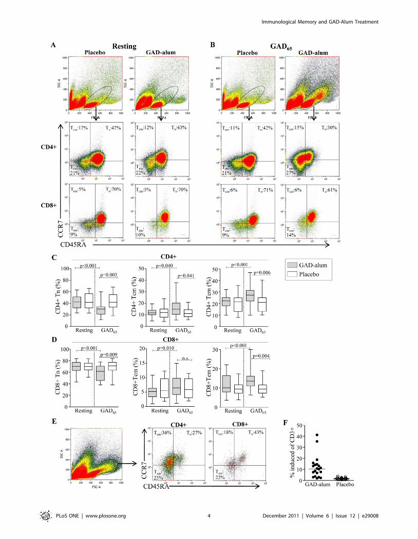
Immunological Memory and GAD-Alum Treatment
PLoS ONE | www.plosone.org 4 December 2011 | Volume 6 | Issue 12 | e29008

(Fig. 1E–F). The majority of cells within this population were
CD4+ with a memory phenotype.
In vitro stimulation with GAD65 induces T-cell activationand proliferation in GAD-alum-treated patients
As memory T cells are characterized by a low activation
threshold, we next analyzed the effect of antigen challenge on the
induction of T-cell activation markers and proliferative responses.
GAD65-induced gene expression of CD69, CD25 and PD-1 was
up-regulated in patients treated with GAD-alum (n = 18) com-
pared to placebo (n = 19) (2-, 4.5- and 1.5-fold, respectively;
p,0.001; Fig. 2A). In addition, up-regulation of components of
the IL-2 signalling pathway including IL-2 (3.2-fold; p,0.001),
JAK3 (1.5-fold; p = 0.027) and STAT5a (1.4-fold; p = 0.006),
together with the transcription factors FOXP3 (2.7-fold; p,0.001)
and PRDM1 (1.6-fold; p,0.001), and the anti-apoptotic molecule
BCL-2 (1.4-fold; p = 0.011) was detected upon GAD65-stimulation
in GAD-alum- vs. placebo-treated patients. Furthermore, the PD-
1 ligand (PD–L1) was also markedly up-regulated (3.5-fold;
p,0.001) in GAD-alum-treated patients. Other up-regulated
markers included IL-7 (2.0-fold; p = 0.004), IL-15 (1.5-fold;
p = 0.005), IL-15 receptor (3.2-fold: p,0.001) and TGF-b (1.4-
fold; p = 0.015), while STAT5b and the housekeeping genes
GAPDH and HPRT1, to which all expression were normalized,
were not significantly different between the two treatment arms.
Proliferative responses to GAD65 were also significantly higher
in the GAD-alum-treated patients compared to placebo
(p = 0.011; Fig. 2B). In contrast, proliferative responses to the
control antigens IA-2853-872 (T1D-associated antigen), TTX
(irrelevant control) and PHA (positive control) did not differ
between GAD-alum- and placebo-treated patients.
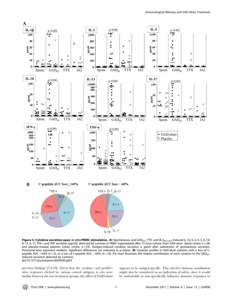
GAD65-stimulation of PBMC induces cytokine secretion inGAD-alum-treated patients
Since memory T cells are capable of immediate effector
cytokine production when stimulated in vitro, we sought to study
the cytokine profile in PBMC supernatants after antigen challenge
using a multiplex Luminex assay. When stimulated with GAD65,
secretion of IL-1b, IL-2, IL-5, IL-10, IL-13, IL-17, IFN-c and
TNF-a was higher in PBMC from GAD-alum-treated patients
compared to the placebo group (Fig. 3A). In contrast, spontaneous
as well as TTX and IA-2853–872-induced secretion were similar in
the two groups. Although the secreted levels of IL-7 and IL-15
were below the detection limit in the Luminex assay and although
TGF-b was not available for multiplex testing, mRNA expression
of these cytokines was up-regulated in PBMC from GAD-alum-
treated patients re-challenged with GAD65, as shown in Fig. 2A.
In order to search for immune surrogate markers of clinical
efficacy, we analyzed the association between cytokine secretion
and b-cell function, as measured by stimulated C-peptide. No
statistical significant associations were observed between cytokine
production, or any other immune marker included in this study,
and stimulated C-peptide. Still, to graphically illustrate the
cytokine profile in relation to clinical efficacy, GAD-alum-treated
patients were divided in two subgroups; patients with a loss of C-
peptide AUC #60% (n = 5), and patients with a loss of AUC
.60% (n = 16; Fig. 3B). The cytokine profile in patients with a loss
of AUC #60% was characterized by a more pronounced GAD65-
induced IL-5, IL-10, IL-13 and IL-2 secretion, whereas patients
with a loss of AUC .60% had a more pronounced inflammatory
profile characterized by IFN-c, IL-1b and IL-17 secretion.
GAD-alum-treated patients display higher numbers ofIFN-c- and IL-4-secreting T cells upon in vitro GAD65-stimulation
Quantification of antigen-specific T-cell responses was per-
formed using an acDC-amplified ELISpot readout [18]. The
GAD65-induced IFN-c (p = 0.016) and IL-4 (p = 0.001) spot
forming cells (SFC) were significantly increased in GAD-alum-
treated patients compared to the placebo group (Fig. 4A-B),
whereas TTX-induced SFC was similar in the two groups.
Furthermore, GAD65-induced IFN-c SFC correlated with GAD65-
induced IFN-c secretion (r = 0.74, p = 0.002; Fig. 4C), and IL-4
SPC with secretion of the Th2 cytokine IL-13 (r = 0.84, p,0.001;
Fig. 4D), in the GAD-alum-treated patients.
Sustained high serum GADA titres in GAD-alum-treatedpatients
Since autoantibody determination may be useful in assessing the
long-lasting immunological impact of autoantigen treatment, we
next analysed serum GADA titres. Our results show higher GADA
levels in GAD-alum-treated patients compared to placebo, 4 years
after treatment (p = 0.034; Fig. 5A). The GADA titres were also
higher compared to baseline levels in the GAD-alum- (p = 0.007)
but not in the placebo-treated group. In addition, IA-2A levels
were determined in order to confirm that the persistent humoral
response was antigen-specific. No difference between the two
groups was observed (not shown). Further, the GADA IgG 1-4
subclass distribution was determined, as Th1- and Th2-cell
cytokine production influence IgG-subclass switching [21,22].
However, the subclass distribution did not differ between groups
(Fig. 5B), nor did it differ compared to baseline (not shown).
High GADA titres are often found in SPS patients, raising
concerns that therapies boosting GADA may have deleterious
neurological effects. However, SPS is characterized by GADA
which inhibit the GAD65 enzymatic activity [20], which is not the
case in T1D. Therefore, we investigated whether serum containing
high GADA titres generated by GAD-alum treatment, was
inhibitory. The enzymatic activity did not differ when rhGAD65
was incubated with serum from GAD-alum- (median 90 %, range
42-100) and placebo-treated patients (median 91 %, range 65-
100), but was significantly higher compared to GAD65 incubated
with control serum from a SPS patient (median 20 %, range 19-
24; p,0.001; Fig. 5C).
Discussion
We have shown significant preservation of residual insulin
secretion 4 years after GAD-alum treatment in T1D children and
Figure 1. Frequencies of naıve, central memory and effector memory CD4+ and CD8+ T cells. (A-B) Representative flow cytometryanalysis from one placebo- and one GAD-alum-treated-patient assessed in resting and GAD65-stimulated PBMC cultures. The gate was set to includeboth small and large lymphocytes. Median percentages of naıve (CD45RA+CCR7+;Tn), central memory (CD45RA-CCR7+;TCM) and effector memory(CD45RA-CCR7 -;TEM) CD4+ and CD8+ T cells are indicated in each plot. (C–D) Frequencies of naıve (Tn), central memory (TCM) and effector memory(TEM) CD4+ and CD8+ T cells, assessed in resting and GAD65-stimulated PBMC cultures from GAD-alum- (n = 20) and placebo-treated patients (n = 23).Box plots represents median and range, significant differences are indicated as p-values. (E) Representative dot plot of the GAD65-induced cell subsetwith higher SSC and FSC evident only in GAD-alum-treated patients. Median percentages of naıve and memory CD4+ and CD8+ subsets are indicatedin each plot. (F) Frequency of CD3+ cells that occupied the induced cell subset. Horizontal line represent median.doi:10.1371/journal.pone.0029008.g001
Immunological Memory and GAD-Alum Treatment
PLoS ONE | www.plosone.org 5 December 2011 | Volume 6 | Issue 12 | e29008

adolescents with ,6 months T1D duration at inclusion,
compared to placebo [12]. In the present study, we aimed to
characterize the long-term antigen-specific memory T- and B-cell
responses. Detection of antigen-specific memory cells ex vivo is a
great challenge due to low frequencies. A previous study has
demonstrated that only one in 30,000 or less CD4+ T-cells in
peripheral blood from patients with recent-onset T1D is GAD65-
specific [23], and activation and in vitro amplification of the
GAD65-specific T-cells is crucial for detection. Our results show
induction of T-cell subsets with a predominant memory
phenotype upon in vitro GAD65-stimulation in PBMC from
GAD-alum-treated patients. This suggests clonal expansion of the
memory T-cell compartment upon antigen re-challenge, in
parallel to the observed proportional reduction in naıve T-cell
percentage.
When stimulated in vitro, memory T cells display low activation
thresholds, immediate cytokine production and vigorous prolifer-
ation [13]. Our results show that the T-cell activation markers
CD69, CD25 and PD-1 were all up-regulated in PBMC from
GAD-alum-treated patients, and that proliferative T-cell responses
to GAD65 together with GAD65-induced cytokine secretion were
significantly higher compared to placebo, the latter confirming our
Figure 2. GAD65-induced gene expression and PBMC proliferation. (A) Fold change for 15 target genes between placebo (n = 19) and GAD-alum-treated (n = 18) patients. The DDCt was calculated by subtraction of the average DCt value of each gene in the placebo group from the averageCt value of the corresponding gene in the GAD-alum group. The fold-change for each gene was calculated as 2(2DDCt). Raw threshold cycle (Ct) values(median and range) for the two groups are listed. (B) Proliferative responses (median and range) to GAD65, IA-2853–872, TTX and PHA in GAD-alum-(n = 10) and placebo-treated patients (n = 7). Proliferation is expressed as stimulation index (SI) calculated from the median of triplicates in thepresence of stimulus divided by the median of triplicates with medium alone. Significant differences are indicated as p-values.doi:10.1371/journal.pone.0029008.g002
Immunological Memory and GAD-Alum Treatment
PLoS ONE | www.plosone.org 6 December 2011 | Volume 6 | Issue 12 | e29008
previous findings [7,9,10]. Given that the cytokine- and prolifer-
ative responses elicited by various control antigens in vitro were
similar between the two treatment groups, the effect of GAD-alum
appears to be antigen-specific. This selective immune modulation
might also be considered as an indication of safety, since it would
be undesirable to non-specifically influence immune responses to
Figure 3. Cytokine secretion upon in vitro PBMC stimulation. (A) Spontaneous, and GAD65-, TTX- and IA-2853–872-induced IL-1b, IL-2, IL-5, IL-10,IL-13, IL-17, IFN-c and TNF secretion (pg/ml), detected by Luminex in PBMC supernatants after 72 hour culture, from GAD-alum- (black circles; n = 28)and placebo-treated patients (white circles; n = 29). Antigen-induced cytokine secretion is given after subtraction of spontaneous secretion.Horizontal lines represent medians. Significant differences are indicated as p-values. (B) Cytokine profiles in GAD-alum patients with a loss of C-peptide AUC ,60% (n = 5), or a loss of C-peptide AUC .60% (n = 16). Pie chart illustrates the relative contribution of each cytokine to the GAD65-induced secretion detected by Luminex.doi:10.1371/journal.pone.0029008.g003
Immunological Memory and GAD-Alum Treatment
PLoS ONE | www.plosone.org 7 December 2011 | Volume 6 | Issue 12 | e29008

unrelated antigens. Besides detecting cytokine secretion in PBMC,
we quantified antigen-specific IFN-c and IL-4 T-cell responses
using an acDC assay. PBMC were selected on the basis of
availability and thereby a limiting factor for including additional
cytokines. In the assay, antigen and DC-activating agents rapidly
induce, pulse and mature DCs, thus lining up the sequential steps
of T-cell activation within 48 h and amplifying antigen-specific
responses. The utility of these acDC-based assays for immune
monitoring of vaccination trials has been previously demonstrated
[18,24]. The number of GAD65-induced IL-4 SFC, a cytokine
difficult to detect with the Luminex assay, was significantly
increased in the GAD-alum group compared to placebo. Further,
IL-4 SFC correlated with IL-13 secretion, two Th2 cytokines with
overlapping biological effects that share receptor components [25].
In parallel, the GAD65-induced IFN-c SFC were also increased in
the GAD-alum-treated patients, and correlated with the GAD65-
induced IFN-c secretion, supporting the reliability of the cytokine
observations.
Proliferation of memory T cells can be driven not only by
antigenic stimulation but also by cytokines. Here we show that
gene expression of IL-7 and IL-15, two cytokines that are
constitutively produced by a variety of cells and play an essential
role for maintenance of both CD4+ and CD8+ T cells [13], was
higher after GAD65-stimulation in the GAD-alum-treated group.
In addition, IL-2, which is involved in long-term survival of
antigen experienced CD4+ and regulatory T cells [26,27], was
also induced by GAD65-stimulation. Receptors for IL-2, IL-7
and IL-15 transmit signals mainly through STAT5, which is a
critical factor for inducing and maintaining the expression of
FOXP3 [28], and of the anti-apoptotic molecule Bcl-2 [29]. Up-
regulation of the aforementioned cytokines and their receptors
upon GAD65-stimulation, together with that of their associated
signalling pathways and transcription factors suggests their
involvement in the maintenance of a long-lasting GAD65-specific
T-cell memory population. B-cell memory is characterized by
persistent elevated specific antibody titres and generation of
long-lived memory B cells [30]. Elevated GADA levels 4 years
after GAD-alum-treatment, together with up-regulated PRDM1,
a transcription factor essential for development of Ig-secreting
cells and maintenance of long-lived plasma cells [31], suggests an
induction of plasma cells continuously secreting GADA.
PRDM1 is also expressed in effector and memory T cells
[32,33], and appears to have a role in Th2 cells by repressing
Th1 genes [34].
The outcome of a T-cell response is shaped by the balance
between co-stimulatory and co-inhibitory signals, which are often
simultaneously provided to T cells by their surrounding cells. PD-1
is a member of the CD28 superfamily of immunoreceptors that is
up-regulated following TCR stimulation [35], and interaction with
its ligand PD-L1 inhibits T-cell effector functions [36]. Up-
regulation of PD-1/PD-L1 in parallel to GAD65-induced T-cell
activation and proliferation in the GAD-alum group demonstrates
activation of co-inhibitory pathways important for regulating the
immune balance.
Figure 4. Quantification of antigen-specific T-cell responses by an acDC-amplified ELISpot assay. GAD65- and TTX-induced (A) IFN-c and(B) IL-4 spot forming cells (SFC) in GAD-alum- (filled symbols, n = 15) and placebo-treated patients (open symbols, n = 12), expressed as SFC/106 PBMCcalculated from the mean of quadruplicates after background subtraction. Median and interquartile range is indicated. (C) Correlation betweenGAD65-induced IFN-c SFC, detected by acDC ELISpot and IFN-c secretion, detected by Luminex, in GAD-alum treated patients. (D) Correlationbetween GAD65-induced IL-4 SFC, detected by acDC ELISpot and IL-13 secretion, detected by Luminex, in GAD-alum-treated patients. Significantdifferences are indicated as p-values and correlation coefficient as r.doi:10.1371/journal.pone.0029008.g004
Immunological Memory and GAD-Alum Treatment
PLoS ONE | www.plosone.org 8 December 2011 | Volume 6 | Issue 12 | e29008

Reliable biomarkers associated with therapeutic success
following vaccination with b-cell antigens are still lacking. We
have previously shown that, although GAD-alum-treatment
induced a GAD65-specific cell population characterised by a
broad cytokine profile [7], the response was preceded by an early
Th2 immune deviation [9]. The cytokine profile observed in
patients with better preserved C-peptide after 4 years, even
though not statistically assured, may suggest that a beneficial
clinical response might be associated with a persistent Th2/Treg-
skewed GAD65-specific immune response. In a vaccination trial
by Harrison and co-workers using intranasal insulin, immune
responses were characterized by IFN-c ELISpot and autoanti-
body measurements [24]. In contrast to our findings, antigen-
specific IFN-c and antibody responses decreased following
treatment, suggesting that the therapeutic effect (or lack thereof)
may be linked to different immunological mechanisms. The
administration route and the use of alum adjuvant may be
important factors in triggering these different mechanisms.
Recently a phase II trial [6] and a European phase III trial
(submitted manuscript) using GAD-alum have failed to reach
their primary outcome. However, it cannot yet be excluded that
treatment might be beneficial in certain patient subgroups. Thus,
in order to make improvements in b-cell antigen treatment, alone
or in combination with other therapies, it is of utmost importance
to learn more about the immunological effects.
In conclusion, we here show persistent GAD65-specific cellular-
and humoral immune responses 4 years after GAD-alum
intervention in T1D children. Prompt re-activation of GAD65-
reactive T cells upon in vitro antigen challenge was accompanied by
secretion of Th1, Th2 and Treg cytokines and by induction of co-
inhibitory pathways that may be of importance for regulating the
GAD65 immunity.
Acknowledgments
We would like to thank Ingela Johansson, Gosia Smolinska-Konefal, Lena
Berglert and Emma Ong-Palsson for their skilful laboratory work.
Author Contributions
Conceived and designed the experiments: SA MC MH RC. Performed the
experiments: SA MC MH MP LA. Analyzed the data: SA MC LA EM
RC. Contributed reagents/materials/analysis tools: RM EM JL RC.
Wrote the paper: SA MC RC. Critical revision of the paper: RM.
References
1. Atkinson MA, Maclaren NK (1994) The pathogenesis of insulin-dependent
diabetes mellitus. N Engl J Med 331: 1428–1436.
2. Mallone R, van Endert P (2008) T cells in the pathogenesis of type 1 diabetes.
Curr Diab Rep 8: 101–106.
Figure 5. Serum GADA levels, GADA IgG1-4 subclass distribution and GAD65 enzymatic activity. (A) Serum GADA titres (U/ml; medianand interquartile range) from baseline to 4 years, in GAD-alum treated patients (filled symbols, n = 29) and in placebo (open symbols, n = 30). (B)Serum GADA IgG 1–4 subclass distribution (median and range), expressed as a percentage of total GADA IgG in GAD-alum- and placebo groups. (C)Recombinant human GAD65 enzymatic activity measured in the presence of serum from GAD-alum- and placebo-treated patients. One SPS patientwas included in each assay as a positive control for enzymatic inhibition. Results are reported as a percentage (median and range) of maximumGAD65 enzymatic activity. Significant differences are indicated as p-values.doi:10.1371/journal.pone.0029008.g005
Immunological Memory and GAD-Alum Treatment
PLoS ONE | www.plosone.org 9 December 2011 | Volume 6 | Issue 12 | e29008

3. Ludvigsson J (2009) Adequate doses of autoantigen administered using the
appropriate route may create tolerance and stop autoimmunity. Diabetologia
52: 175–176.
4. Baekkeskov S, Aanstoot H-J, Christgau S, Reetz A, Solimena M, et al. (1990)
Identificaion of the 64K autoantigen in insulin-dependent diabetes as the
GABA-synthesizing enzyme glutamic acid decarboxylase. Nature 347:
151–156.
5. Kaufman D, Clare-Salzler M, Tian J, Forsthuber T, Ting G, et al. (1993)
Spontaneous loss of T-cell tolerance to glutamic acid decarboxylase in murine
insulin-dependent diabetes. Nature 366: 15–17.
6. Wherrett DK, Bundy B, Becker DJ, DiMeglio LA, Gitelman SE, et al. (2011)
Antigen-based therapy with glutamic acid decarboxylase (GAD) vaccine in
patients with recent-onset type 1 diabetes: a randomised double-blind trial.
Lancet 378: 319–327.
7. Ludvigsson J, Faresjo M, Hjorth M, Axelsson S, Cheramy M, et al. (2008) GAD
treatment and insulin secretion in recent-onset type 1 diabetes. N Engl J Med
359: 1909–1920.
8. Agardh CD, Cilio CM, Lethagen A, Lynch K, Leslie RD, et al. (2005) Clinical
evidence for the safety of GAD65 immunomodulation in adult-onset
autoimmune diabetes. J Diabetes Complications 19: 238–246.
9. Axelsson S, Hjorth M, Akerman L, Ludvigsson J, Casas R (2010) Early induction
of GAD(65)-reactive Th2 response in type 1 diabetic children treated with alum-
formulated GAD(65). Diabetes Metab Res Rev 26: 559–568.
10. Hjorth M, Axelsson S, Ryden A, Faresjo M, Ludvigsson J, et al. (2011) GAD-
alum treatment induces GAD65-specific CD4+CD25highFOXP3+ cells in type
1 diabetic patients. Clin Immunol 138: 117–126.
11. Cheramy M, Skoglund C, Johansson I, Ludvigsson J, Hampe CS, et al. (2010)
GAD-alum treatment in patients with type 1 diabetes and the subsequent effect
on GADA IgG subclass distribution, GAD65 enzyme activity and humoral
response. Clin Immunol 137: 31–40.
12. Ludvigsson J, Hjorth M, Cheramy M, Axelsson S, Pihl M, et al. (2011) Extended
evaluation of the safety and efficacy of GAD treatment of children and
adolescents with recent-onset type 1 diabetes: a randomised controlled trial.
Diabetologia 54: 634–640.
13. Sallusto F, Geginat J, Lanzavecchia A (2004) Central memory and effector
memory T cell subsets: function, generation, and maintenance. Annu Rev
Immunol 22: 745–763.
14. Michie CA, McLean A, Alcock C, Beverley PC (1992) Lifespan of human
lymphocyte subsets defined by CD45 isoforms. Nature 360: 264–265.
15. Sallusto F, Lenig D, Forster R, Lipp M, Lanzavecchia A (1999) Two subsets of
memory T lymphocytes with distinct homing potentials and effector functions.
Nature 401: 708–712.
16. McHeyzer-Williams LJ, Malherbe LP, McHeyzer-Williams MG (2006)
Checkpoints in memory B-cell evolution. Immunol Rev 211: 255–268.
17. Hedman M, Faresjo M, Axelsson S, Ludvigsson J, Casas R (2008) Impaired
CD4 and CD8 T cell phenotype and reduced chemokine secretion in recent-
onset type 1 diabetic children. Clin Exp Immunol 153: 360–368.
18. Martinuzzi E, Afonso G, Gagnerault MC, Naselli G, Mittag D, et al. (2011)
Accelerated co-cultured dendritic cells (acDCs) enhance human antigen-specific
T-cell responses. Blood.
19. Martinuzzi E, Novelli G, Scotto M, Blancou P, Bach JM, et al. (2008) The
frequency and immunodominance of islet-specific CD8+ T-cell responseschange after type 1 diabetes diagnosis and treatment. Diabetes 57: 1312–1320.
20. Raju R, Foote J, Banga JP, Hall TR, Padoa CJ, et al. (2005) Analysis of GAD65
autoantibodies in Stiff-Person syndrome patients. J Immunol 175: 7755–7762.21. Gascan H, Gauchat JF, Roncarolo MG, Yssel H, Spits H, et al. (1991) Human B
cell clones can be induced to proliferate and to switch to IgE and IgG4 synthesisby interleukin 4 and a signal provided by activated CD4+ T cell clones. J Exp
Med 173: 747–750.
22. Briere F, Servet-Delprat C, Bridon JM, Saint-Remy JM, Banchereau J (1994)Human interleukin 10 induces naive surface immunoglobulin D+ (sIgD+) B cells
to secrete IgG1 and IgG3. J Exp Med 179: 757–762.23. Reijonen H, Novak EJ, Kochik S, Heninger A, Liu AW, et al. (2002) Detection
of GAD65-specific T-cells by major histocompatibility complex class II tetramersin type 1 diabetic patients and at-risk subjects. Diabetes 51: 1375–1382.
24. Fourlanos S, Perry C, Gellert SA, Martinuzzi E, Mallone R, et al. (2011)
Evidence that nasal insulin induces immune tolerance to insulin in adults withautoimmune diabetes. Diabetes 60: 1237–1245.
25. Zurawski SM, Chomarat P, Djossou O, Bidaud C, McKenzie AN, et al. (1995)The primary binding subunit of the human interleukin-4 receptor is also a
component of the interleukin-13 receptor. J Biol Chem 270: 13869–13878.
26. Dooms H, Abbas AK (2006) Control of CD4+ T-cell memory by cytokines andcostimulators. Immunol Rev 211: 23–38.
27. Dooms H, Wolslegel K, Lin P, Abbas AK (2007) Interleukin-2 enhances CD4+T cell memory by promoting the generation of IL-7R alpha-expressing cells.
J Exp Med 204: 547–557.28. Passerini L, Allan SE, Battaglia M, Di Nunzio S, Alstad AN, et al. (2008)
STAT5-signaling cytokines regulate the expression of FOXP3 in CD4+CD25+regulatory T cells and CD4+CD25- effector T cells. Int Immunol 20: 421–431.
29. Lord JD, McIntosh BC, Greenberg PD, Nelson BH (2000) The IL-2 receptor
promotes lymphocyte proliferation and induction of the c-myc, bcl-2, and bcl-xgenes through the trans-activation domain of Stat5. J Immunol 164: 2533–2541.
30. Schittek B, Rajewsky K (1990) Maintenance of B-cell memory by long-lived cells
generated from proliferating precursors. Nature 346: 749–751.31. Martins G, Calame K (2008) Regulation and functions of Blimp-1 in T and B
lymphocytes. Annu Rev Immunol 26: 133–169.32. Martins GA, Cimmino L, Shapiro-Shelef M, Szabolcs M, Herron A, et al. (2006)
Transcriptional repressor Blimp-1 regulates T cell homeostasis and function. NatImmunol 7: 457–465.
33. Kallies A, Hawkins ED, Belz GT, Metcalf D, Hommel M, et al. (2006)
Transcriptional repressor Blimp-1 is essential for T cell homeostasis and self-tolerance. Nat Immunol 7: 466–474.
34. Cimmino L, Martins GA, Liao J, Magnusdottir E, Grunig G, et al. (2008) Blimp-1 attenuates Th1 differentiation by repression of ifng, tbx21, and bcl6 gene
expression. J Immunol 181: 2338–2347.
35. Vibhakar R, Juan G, Traganos F, Darzynkiewicz Z, Finger LR (1997)Activation-induced expression of human programmed death-1 gene in T-
lymphocytes. Exp Cell Res 232: 25–28.36. Freeman GJ, Long AJ, Iwai Y, Bourque K, Chernova T, et al. (2000)
Engagement of the PD-1 immunoinhibitory receptor by a novel B7 familymember leads to negative regulation of lymphocyte activation. J Exp Med 192:
1027–1034.
Immunological Memory and GAD-Alum Treatment
PLoS ONE | www.plosone.org 10 December 2011 | Volume 6 | Issue 12 | e29008