Embed Size (px)
Citation preview

Long cases in paediatrics

Acute fever
Presenting complaint
Fever
State the duration
History of the presenting complaint
Description of the fever
Describe the following details of the fever in a chronological order
Onset and any preceding symptoms
Progression
Height of the fever – documented or not
Resolution of the fever and state of the child in between episodes
Recurrence (comment on the fever pattern if possible)
What the mother did at home? Especially important is the dose of paracetamol
Associated factors
Associated features
Ask for symptoms related to the important symptoms to try to identify a focus of infection and
to think of a differential diagnosis
Disease Symptoms
Dengue fever Headache, retro –orbital pain, arthralgia and myalgia, anorexia, nausea and vomiting Warning signs Abdominal pain, mucosal bleeding and other bleeding manifestations, lethargy and restlessness
Respiratory tract infection Ask for Cough, sputum (if sputum is associated state the color and amount), rhinorrhoea, chest pain associated with breathing and difficulty in breathing
Ear infection Ear pain and discharge
Pharyngitis Ask for sore throat, pain on swallowing
CNS infection (Meningitis and encephalitis)
Headache, photophobia, altered behavior and loss of consciousness, seizures
GI infection Ask for passage of loose stools
Hepatitis Yellowish discoloration of the eyes, darkening of the urine
Leptospirosis Exposure to muddy water/ possible contaminated water

Septic arthritis and osteomyelitis Bone pain, joint pain and swelling
Urinary tract infection Crying on passage of urine, frequency, hematuria
History of exposure and epidemiological history of the fever
Ask for history of contact with infected or otherwise ill persons
Travel history if relevant
History of cases of fever especially dengue fever in the community
Past medical history and surgical history
Other components of the history
Social history
Environment
Describe the surrounding environment of the house especially with regard to possible mosquito
breeding sites
Ask if the garbage sites are cleaned regularly and ask if mosquito spraying is done regularly in
the area
Ask for the involvement of the MOH, PHI and other staff for dengue prevention in the area
Ask for possible breeding sites within the house
If the child is attending montessori or school inquire about the environment
Impact of the disease on the child and on the parents
Inquire about the amount of school missed by the child
Impact on the parents as the child has to stay in the hospital
Concern of the parents
Other general factors on the family background
Occupation of the parents
Social circumstances of the family
Economic status of the family
Extended family support

Examination
General impression of the patient
Look at the appearance of the patient
Look at the alertness and activity of the child
Examine the vital signs of the patient
Pulse rate and volume
Capillary refill time
Blood pressure and pulse pressure
Respiratory rate and signs of respiratory distress
General examination
Do a complete examination from head to toe
Look for skin rashes
Eyes
Conjuctivae for pallor and the sclera for icterus
Photophobia and neck stiffness
Ear discharge
Sinus tenderness
Lymphadenopathy
Open the mouth and look at the general oral hygiene
Examine the throat and inspect the pharynx and tonsils
Respiratory system
Look for evidence of pneumonia
Look for pleural effusion – pneumonia, dengue
CVS
Abdomen
Examine for hepatosplenomegaly
Other masses
Free fluid in the abdomen
Nervous system
Should be assessed completely in a patient with suspected CNS infection
Musculoskeletal – Look for joint swelling and other features of acute inflammation of the joint

Discussion
Dengue fever
What is the diagnosis?
The first step in a long case of acute fever is to make a diagnosis and classify the severity
The most common case of acute fever given for the exam is dengue fever
How would you perform the tourniquet test?
Tourniquet test
The technique may be asked during the discussion
First measure the systolic and diastolic blood pressures
Maintain the blood pressure at a point midway between the systolic and diastolic blood
pressure for a duration of 5 minutes
Observe for petechial hemorrhages and draw a 1”x1” square in the area of maximum petechial
hemorrhages
Probable dengue Dengue with warning signs Severe dengue
Living in an endemic area Fever Two of the following Nausea, vomiting Arthralgia and myalgia Rash Positive tourniquet test Leucopenia
Persistent vomiting Abdominal pain or tenderness Lethargy, restlessness Mucosal bleeding Clinical evidence of fluid accumulation Liver enlargement >2cm Dropping platelets and rising hematocrit
Severe plasma leakage Shock Fluid accumulation with respiratory distress Severe bleeding Severe organ involvement Hepatitis Myocarditis Encephalitis

The tourniquet test is positive when the number of petechial hemorrhages within this square
exceeds 10
What is the natural history of dengue?
What is the pathogenesis of severe dengue? /DHF
There are 4 serotypes of the dengue virus
Primary infection from 1 serotype causes lifelong immunity to that particular serotype
However during a secondary infection from another serotype these antibodies promote viral
replication and increase the viral load
There is also an exaggerated immune response and a cytokine storm which causes endothelial
dysfunction, plasma leakage and platelet destruction and dysfunction
How would you manage a patient with dengue?
The management of dengue is based on the natural history of the disease. This is shown in the
following diagram
Therefore the management will differ according to the stage of the disease

The management of dengue will be discussed based on three clinical stages
Discuss the management of a case of probable dengue in the febrile phase
This can be managed on an outpatient basis
Educate the parents about the warning signs of dengue and when to admit to the hospital
Ensure good diet and hydration. If food is refused advise the parents to give fluids such as ORS,
fruit juice and milk
Advise them not to give the child any colored substances to eat or drink
Prescribe paracetamol for the fever (10-15mg/kg, 6 hourly. Maximum daily dose is 60mg/kg)
Domperidone may be prescribed for severe vomiting
How would you manage a patient in the critical phase of dengue?
Admit the patient
Remember that the most intensive management should be done in the critical phase of dengue
as this can be complicated by shock and major bleeding manifestations
Monitoring
Start the dengue monitoring process
Monitor the following parameters – Pulse rate and volume, blood pressure and pulse pressure,
cold peripheries, CRFT
Frequency of monitoring varies according to the clinical condition
Laboratory parameters – hematocrit, platelet count (hematocrit may be monitored in ward
every 6h)
Febrile phase Critical phase Recovery phase
Lasts 2-7 days Fever Flushed Arthralgia and myalgia
Is a period of 48h usually from the 3rd day of fever Fever subsides Leakage of fluid occurs Leucopenia Rise in hematocrit with drop in platelets Can have complications
General improvement of well being Good appetite Diuresis

Other investigations – ALT (hepatic dysfunction), PT/INR, serum electrolytes, serum albumin
(can be low due to the plasma leakage)
Blood should also be taken for grouping and cross matching
Antipyretics
Give paracetamol for the fever (dose stated above). A regular dose is not given as this may alter
the fever pattern
Fluid management
The maximum amount of fluid which can be given in the critical phase is calculated by the
following equation
Maximum amount of fluid = Maintenance + 5% Deficit (50ml/kg) over 48 hours. This is known
as the fluid quota
Maintenance is calculated as follows
This is used as a guide and care is taken not to exceed this. There is also no rule to complete the
quota. Fluid is given according to the clinical condition of the patient. Therefore the rate of fluid
administration should be reduced as time progresses
Fluid may be given as oral fluid (ORS, milk, fruit juice) or IV fluids (0.9% saline) or as a
combination of the two
The patient is admitted with drowsiness and weak pulses with cold extremities. How would you manage?
The earliest manifestations of shock would be prolonged CRFT, cold peripheries, rising diastolic
blood pressure with normal systolic blood pressure (reduction in the pulse pressure) and
tachycardia. This is termed compensated shock
With time a drop in the systolic blood pressure is noted. This is termed hypotensive shock
Fluid boluses should be administered to a patient in shock. The amount and rate is as given
below
Bolus = 10ml/kg over 1 hour. (Remember that this fluid volume should be deducted from the
total fluid quota)
If the patient does not improve give up to 3 repeat boluses with the last being a colloid
(hetastarch)

The fluid quota is calculated as shown above but is given over 24 hours due to the assumption
that the patient has already been in the critical phase for 24 hours.
If the patient does not recover after this management consider the possibility of an internal
bleed
Management of the convalescent phase
The most important aspect of this phase is that there is a risk of fluid overload. Therefore the
patient should be assessed for features of fluid overload and pulmonary edema
Proper fluid management in the critical phase of dengue should prevent severe fluid overload.
But if this occurs discontinue fluid supplementation and frusemide 0.1mg/kg may be given IV or
oral
What are the other complications of dengue? What is the management?
Hemorrhagic complications
A major bleed may be suspected in the following clinical scenarios
Persistent and/ or severe bleeding in an unstable patient regardless of the hematocrit
Refractory shock
Hypotensive shock with low hematocrit prior to fluid resuscitation
Management
The definitive life saving procedure would be to transfuse blood. Fresh packed cells or fresh
whole blood are the preparations of choice
Give packed cells as 10ml/kg
Continue monitoring the patient
Hepatic encephalopathy
This can be due to the virus itself or due to paracetamol overdose
A,B,C
Investigations – AST/ALT, PT/INR, RBS, renal function tests
Avoid hypoglycaemia
Lactulose (check dose)
IV antibiotics – IV metranidazole or IV cefotaxime
IV N-Acetyl cysteine 75mg/kg 6 hourly if available
IV vitamin K for 3 consecutive days
IV ranitidine for gastrointestinal bleeding

As a house officer how would you assess the patient on your daily ward round?
History
Ask for the general condition of the child
Look for any warning signs
Ask about the appetite of the child
Look for the intake of fluid by the child
Look for the urine output – should be more than 0.5ml/kg/h
Examination
PR and volume, CRFT, blood pressure
Signs of fluid overload
Management
Check the adequacy of fluids
Look for the latest reports which are available
When would you decide to discharge the patient?
Fever free for 48 hours
Improvement of the clinical status (General well being, appetite, hemodynamic parameters and
urine output)
Out of shock for at least 2 days
Rising trend in the platelet count (>50,000) with hematocrit responding to IV fluids
Other aspects of management
Notification
Education of the parents on elimination of mosquito breeding sites in the immediate vicinity and
community

Prolonged fever
Presenting complaint
Fever
State the duration
History of the presenting complaint
Description of the fever
Remember that the details should be stated in a definite chronological order
Describe the onset of the fever and state if there are any specific preceding events
Describe how the fever was assessed and the value of the height of the fever
The exact duration of the fever
Describe the response of the fever to antipyretics and the duration taken for the resolution of
the fever
Describe the dose, route of administration and frequency that the child was given paracetamol
If there is a recurrence of the fever state the time at which the fever comes back
Describe the state of the child in between episodes of fever
Are there associated chills and rigors
Describe the pattern of fever as intermittent, remittent or continuous (however this is
unreliable with the use of antipyretics)
Fever pattern Description Clinical examples
Intermittent High spiking fever which reach the baseline
Pyogenic infections TB, lymphoma, systemic onset JIA
Remittent Fluctuating fever which does not reach the baseline
Viral infections, IE, lymphoma
Continuous Sustained fever with little or no fluctuation
Typhoid, typhus
Relapsing Febrile episodes separated by one or more days without fever
Malaria, lymphoma
The next step is to make a probable diagnosis. The list of differential diagnosis in a patient with
prolonged fever is extensive but the common causes should be excluded in the history.
The main categories of causes of prolonged fever should be dealt with. These are,
Infective
Inflammatory
Connective tissue diseases
Neoplasms
Other rare causes

Category Diseases Specific points in the history
Infective Localized
Respiratory tract infections
Cough, sputum, nasal or ear discharge, sore throat
Gastrointestinal infections and localized intra abdominal abscesses
Ask for alteration of bowel habits, recurrent episodes of abdominal pain
Urinary tract infections Dysuria, frequency, hematuria and other urinary tract symptoms
Infections of the bones and joints
Ask for joint pain and swelling, limping,
Generalized Infective endocarditis Past history of heart disease, rheumatic fever with evidence of a predisposing event for bacteraemia
IMN Associated sore throat
TB Contact history of TB, chronic cough
Typhoid fever Ask for possible exposure to unhygienic food Initially presents with a slowly rising fever. Then during the 2nd week of illness classically they have high fever, abdominal distension, “pea soup” diarrhoea, constipation. The 3rd week of illness is characterized by complications – intestinal perforation
Malaria Visit to a malarial endemic area
Other zoonotic infections
Contact history with animals
Inflammatory JIA Ask for a evanescent salmon pink maculopapular rash, associated joint pain and early morning joint stiffness
SLE History of facial rashes and joint pain
Kawasaki disease Ask for history of bilateral non purulent conjunctivitis, reddish oral mucosa, erythematous rash and peeling off of the skin, edema of the limbs
Neoplastic Hematological malignancy Other malignancies
Evidence of bleeding, ask for the features of anaemia, history of bone pain, past history of recurrent infections
Other Drugs Factitious fever
Drug history

Ask for general associated symptoms such as loss of appetite and loss of weight and general
malaise
Dietary history
This is extremely important as children with prolonged fever tend to lose weight and should
have a high protein and calorie diet
Past medical history
Past surgical history
Other components of the history
Social history
Describe the following factors in the social history
General introduction to the family
Impact of the disease on the child
Impact of the disease on the parents
Environmental factors – This is especially relevant if a diagnosis of typhoid fever is suspected
Support available
Education of the parents regarding the condition and future expectations
Psychological state of the parents

Examination
General assessment of the patient
General appearance of the child, activity and growth parameters. Plot the growth parameters in
a centile chart
General examination
Skin
Examine the skin for lesions. The important skin lesions and their associations are given below
Skin lesions Associated diseases
Malar rash SLE
Salmon pink rash JIA
Petichial rash, janeway lesions, osler’s nodes IE
Palpable purpuric rashes Vasculitis
Erythema nodosum TB, IBD, SLE
Eschar +/- erythematous rash Typhus
Erythematous rash and desquamation Kawasaki disease
Head and face and neck
Examine the eyes for conjunctivitis – non purulent conjunctivitis in Kawasaki disease
Examine the fundi – miliary tubercles in TB, other diseases can also have manifestations in the
fundi – toxoplasmosis, roth spots in leukemia, Vasculitis
Examine the throat – pharyngitis and tonsillitis
Examine the oral cavity and tongue – reddened lips, strawberry tongue in Kawasaki disease
Look for palatal petichiae, tonsillar exudates in IMN
Examine for lymphadenopathy – Generalized lymphadenopathy – IMN, miliary TB,
hematological malignancies, JIA, SLE
Asymmetrical cervical lymphadenopathy in Kawasaki disease
Examine the hands for clubbing and other stigmata of infective endocarditis
Cardiovascular system
Examine the heart for murmurs – IE
Respiratory system
Examine for the BCG scar
Examine for evidence of consolidation/ pleural effusions
Abdomen
Look for hepatosplenomegaly

Any other palpable masses in the abdomen – para aortic lymph nodes in lymphoma, Wilm’s
tumor, neuroblastoma
Musculoskeletal system
Bone tenderness – osteomyelitis, leukemia
Discussion
What is the definition of PUO?
No strict definition as in adults but should be suspected if the child has fever for more than 1
week
How would you investigate a patient with PUO?
FBC
This is an important investigation. Look for the following
Anaemia – Is associated with chronic infections and inflammatory and connective tissue
disorders
Pancytopenia – Is evidence of bone marrow suppression which may occur in leukemia
Thrombocytosis – Is known to occur in inflammatory diseases and Kawasaki disease
Look at the white cell count and the predominant cell types
Blood picture
Look for any atypical cells which may suggest leukemia
Acute phase reactants
ESR, CRP, serum ferretin
Blood culture
Other investigations – renal, hepatic
Urine full report and urine culture
CXR, mantoux test
Echocardiogram
To look for vegetations, valvular dysfunction
Other investigations may be necessary – bone marrow biopsy

Infectious diseases presenting with PUO
Infectious mononucleosis
Introduction
Is caused by EBV
Presents with prolonged fever and sore throat
Examination may reveal cervical or generalized lymphadenopathy, palatal petichiae,
membranous tonsillitis and splenomegaly
Complications – splenic rupture, GBS
Investigations
FBC and blood picture – may show an absolute lymphocytosis. The blood picture will show
atypical lymphocytes
Paul- Bunnell test and monospot test – Look for heterophile antibodies which agglutinate with
sheep or horse blood. These have a low sensitivity
Specific EBV antibody test – Done at MRI
Management
This is usually supportive
Infective endocarditis
Infective endocarditis
Diagnosis
Diagnosis is based on the modified Duke’s criteria
Major criteria
Positive blood cultures
Evidence of endocarditis on echocardiography – oscillating intracardiac mass, new valvular
regurgitation, abscess
Minor criteria
Predisposing conditions
Fever
Embolic vascular signs
Immunological phenomena
Microbiological evidence not meeting the major criteria
Organisms causing IE
Viridans group of streptococci
Staphylococcus aureus

Principles of management
After obtaining the blood cultures the next step is to start high doses of empirical antibiotic
therapy via the IV route
This is done as high blood concentrations of the drug is required for penetration into the
vegetations
IV benzyl penicillin and gentamicin is the preferred combination as initial empirical therapy
After receiving the reports of the blood culture and ABST antibiotics may need to be altered
Continue antibiotics for about 4 -6 weeks
Monitor the patient for the complications of the disease
Severe valvular dysfunction and heart failure
Myocardial abscesses
Systemic emboli
Look for the following on the daily ward round
General condition of the child
Look at the fever chart and the response to antibiotics
Auscultate the murmur and note any change in the character or intensity
Look for features of heart failure
Prophylaxis, proper dental care and oral hygiene
Typhoid fever

Diagnosis
Presents with prolonged fever and the classical pattern of symptoms mentioned above in the
section on history taking
Incubation period 10-21 days
Period of illness Clinical features
Week 1 Non specific symptoms, headache, malaise and a step ladder type fever. The child may also have constipation
Week 2 More ill looking, high temperature, relative bradycardia, rose spots on the abdomen, abdominal distension and splenomegaly
Week 3 Continuous high fever, extremely ill, pea soup diarrhoea, systemic involvement
Week 4 Begins gradual improvement. May develop a carrier state
FBC – mild leukocytosis may be seen initially but later leucopenia and neutropenia
predominates
Blood culture
SAT – tests the antibodies against the H and O antigens of Salmonella typhi. Lacks sensitivity in
endemic countries
Principles of management
Antibiotic therapy
If the child is systemically ill 3rd generation cephalosporins are the drug of choice
Other drugs
Amoxicillin
Chloramphenicol
Azithromycin
Co- trimoxazole
Monitor for and manage complications

TB in children
Diagnosis
Is based on the following aspects
History – PUO, chronic cough, FTT, contact history of TB
Examination
Tuberculin skin testing
5 TU (0.1ml) of the tuberculin PPD is injected intradermally into the skin of the forearm (exact
site)
Inspection is done after 48-72h
Interpretation
Diameter of induration of ≥5 mm is considered positive in: HIV-infected children
Severely malnourished children (with clinical evidence of marasmus or kwashiorkor).
Diameter of induration of ≥10 mm is considered positive in:
All other children (whether or not they have received BCG vaccination)
Bacteriological confirmation wherever possible
This is extremely difficult to do in children as they cannot expectorate sputum but may be
considered in children
Other investigations relevant to pulmonary or extra pulmonary TB
CXR
Persistent opacification of the lung with enlarged hilar lymph nodes
Pleural effusion
Miliary shadowing
Apical infiltrates with cavitation are rarely seen in children
Other
HIV testing
Drugs used in the treatment of TB in children
Isoniazid
Rifampicin
Pyrizinamide
Streptomycin
Ethambutol
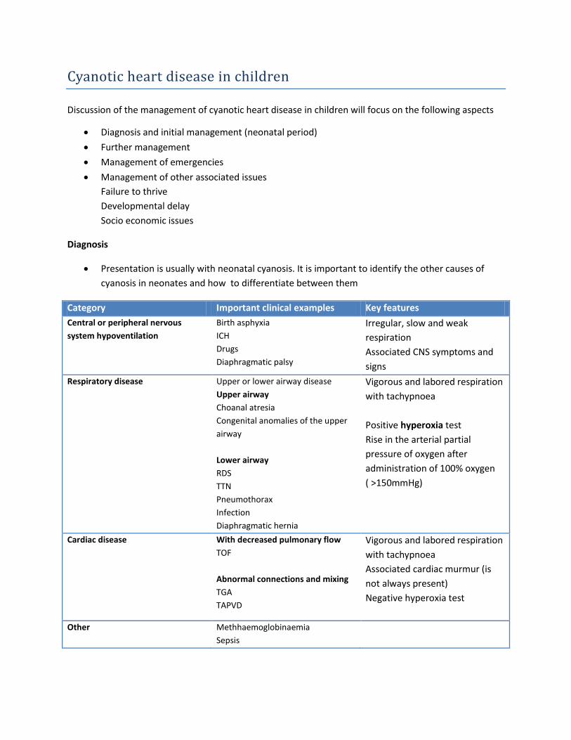
Cyanotic heart disease in children
Discussion of the management of cyanotic heart disease in children will focus on the following aspects
Diagnosis and initial management (neonatal period)
Further management
Management of emergencies
Management of other associated issues
Failure to thrive
Developmental delay
Socio economic issues
Diagnosis
Presentation is usually with neonatal cyanosis. It is important to identify the other causes of
cyanosis in neonates and how to differentiate between them
Category Important clinical examples Key features
Central or peripheral nervous
system hypoventilation
Birth asphyxia
ICH
Drugs
Diaphragmatic palsy
Irregular, slow and weak
respiration
Associated CNS symptoms and
signs
Respiratory disease Upper or lower airway disease
Upper airway
Choanal atresia
Congenital anomalies of the upper
airway
Lower airway
RDS
TTN
Pneumothorax
Infection
Diaphragmatic hernia
Vigorous and labored respiration
with tachypnoea
Positive hyperoxia test
Rise in the arterial partial
pressure of oxygen after
administration of 100% oxygen
( >150mmHg)
Cardiac disease With decreased pulmonary flow
TOF
Abnormal connections and mixing
TGA
TAPVD
Vigorous and labored respiration
with tachypnoea
Associated cardiac murmur (is
not always present)
Negative hyperoxia test
Other Methhaemoglobinaemia
Sepsis

Investigations
2D echocardiography
CXR
Initial management principles
ABC and adequate resuscitation in an optimal temperature
Proper hydration of the baby
Correction of acid base abnormalities, hypoglycaemia and electrolyte imbalance
Administration of a prostaglandin infusion
Transport the patient to a specialized center
Further management
This will depend on the diagnosis made and the associated complications
Tetralogy of fallot
Background knowledge of the anatomy and pathophysiology
Is due to abnormal deviation of the septum than separates the aortic and pulmonary outflow
tracts
Has 4 basic anatomical abnormalities. These are pulmonary infundibular stenosis, right
ventricular hypertrophy overriding aorta and VSD
The pulmonary infundibular stenosis causes right ventricular outflow tract obstruction and the
severity of this determines the symptoms
When the right ventricle contracts against the pulmonary stenosis blood is shunted across the
VSD into the aorta
Diagnosis
Clinical – See short case on TOF
Usually cyanosis is not present at birth unless the pulmonary stenosis is very severe
With age there is increased RVOT obstruction and increasing cyanosis
Central cyanosis, clubbing, ejection systolic murmur in the left mid sternal edge and soft P2
Investigations
CXR -“Boot” shaped heart with pulmonary oligaemia
ECG – Features of right ventricular hypertrophy
Echo – For the confirmation of the diagnosis
Cardiac catheterization – This will show the anatomy of the lesion and the state of the
pulmonary arteries which is important in surgical intervention

Management
As stated above TOF usually does not present with cyanosis in the neonatal period unless the
degree of pulmonary stenosis is severe
If there is neonatal cyanosis manage as stated above
There are two options in the further management of these babies. These are,
Creation of a shunt from the subclavian artery to the pulmonary artery (modified Blalock –
Tassing shunt)
Total correction
Others should be carefully followed up and a date given for corrective surgery
Complications may occur in these children
Hypercyanotic spells
Place the child in the knee chest position
Administer high flow oxygen
Administer IV morphine – maximum dose 0.2mg/kg
Correction of metabolic acidosis with IV sodium bicarbonate
For resistant spells
IV propranolol 0.1 mg/kg
After management a date for early surgery should be given. The child is also given oral
propranolol 0.5 – 1 mg/kg 6 hourly for prevention of hypercyanotic spells
Cerebral thrombosis
Cerebral abscess
Infective endocarditis
Transposition of great vessels
Background anatomy and pathophysiology
In this lesion the aorta arises from the right ventricle and the pulmonary artery arises from the
left ventricle
Therefore unsaturated blood from the right ventricle reaches the systemic circulation via the
aorta
In order for these newborns to survive there should be a connection between the two sides of
the heart. This may be via a PFO, PDA or VSD
Diagnosis
Cyanosis and tachypnoea are observed in the first few hours of life
Clinical signs are minimal on auscultation but may have a single, loud second sound. A murmur
may also be audible if there is an associated VSD

Echocardiogram is the investigation of choice for the diagnosis
Management
Is an emergency
Manage as given above. Especially the infusion of PG E1 is a critical component in the
management as it keeps the ductus arteriosus open
If there is poor response to the PG infusion an emergency balloon atrial septostomy should be
performed
Definitive surgery is by the arterial switch operation which should be performed within 14 days
of life
The situation in Sri Lanka regarding the management of congenital heart disease
Limited resources and long waiting lists
Check social support available and funding

Recurrent wheezing in childhood – Asthma
Key points in the history – 5 key points to describe
1. Describe the present episode in detail
Describe the onset, duration and progression of the symptoms
Ask for any preceding triggering factors
Describe what the mother did at home
Assess the clinical severity of the episode and what was done in hospital
2. Describe the past history and the progression up to now
Highlight the following points and use a time line for the important events
The first episode
Acute exacerbations and hospital admissions
Treatment given and the compliance
Side effects of the medication given and follow up
3. Describe the present state of the disease
4. Exclude D/D’s of recurrent wheeze and establish the probable diagnosis of asthma
Cause Important points in the history
Bronchial asthma Symptom pattern (most of these will have been described
above)
Intermittent symptoms (the child will be well in between
episodes
Diurnal variation of symptoms may be present
Definite trigger factors for the episodes and good response to
medication
Ask for family history of atopy and asthma
Structural anomalies/ congenital lesions of the respiratory
tract
This will be excluded as the onset of symptoms is later on in
life
Tuberculosis Ask for a contact history of TB
Interstitial lung disease Long standing history of symptoms, failure to thrive
Heart failure Ask for past history of cardiac disease, reduced exercise
tolerance, orthopnoea (in an older child)
Gastro esophageal reflux disease Ask if the symptoms are associated with meals and if there is
associated regurgitation
Recurrent aspiration Risk factors for aspiration
Foreign body inhalation
Rare causes – cystic fibrosis, cilliary dyskinesia,
immunodeficiency
Recurrent lower respiratory tract infections, chronic sinus
infections, failure to thrive
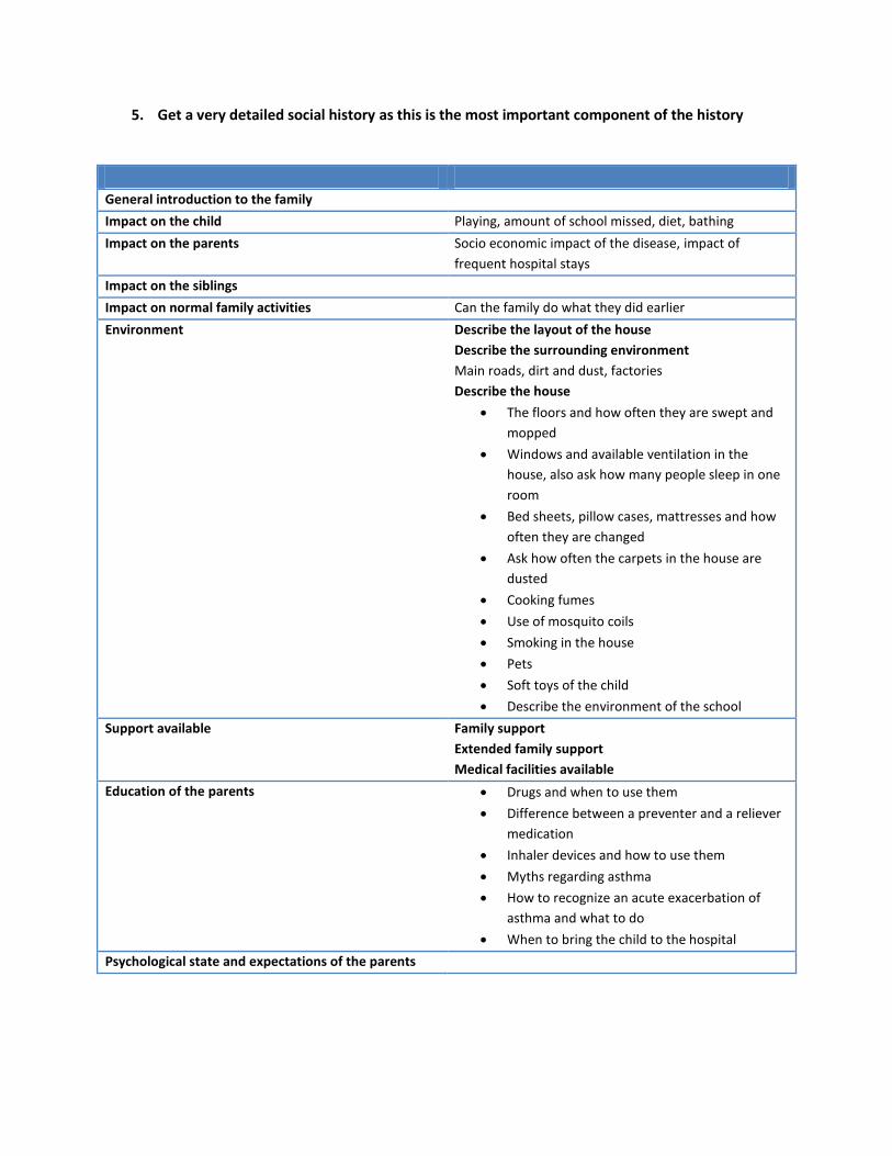
5. Get a very detailed social history as this is the most important component of the history
General introduction to the family
Impact on the child Playing, amount of school missed, diet, bathing
Impact on the parents Socio economic impact of the disease, impact of
frequent hospital stays
Impact on the siblings
Impact on normal family activities Can the family do what they did earlier
Environment Describe the layout of the house
Describe the surrounding environment
Main roads, dirt and dust, factories
Describe the house
The floors and how often they are swept and
mopped
Windows and available ventilation in the
house, also ask how many people sleep in one
room
Bed sheets, pillow cases, mattresses and how
often they are changed
Ask how often the carpets in the house are
dusted
Cooking fumes
Use of mosquito coils
Smoking in the house
Pets
Soft toys of the child
Describe the environment of the school
Support available Family support
Extended family support
Medical facilities available
Education of the parents Drugs and when to use them
Difference between a preventer and a reliever
medication
Inhaler devices and how to use them
Myths regarding asthma
How to recognize an acute exacerbation of
asthma and what to do
When to bring the child to the hospital
Psychological state and expectations of the parents

Examination
General examination
Anthropometry
Plot the weight and height of the child on a centile chart. Ask the mother for the CHDR of the
child
This is important as FTT could indicate an alternate diagnosis to bronchial asthma. Also look for
growth faltering as this could indicate steroid toxicity
Look for other evidence of steroid toxicity - Cushingnoid features
Look for cataract
Examine nose – nasal polyps
Examine the mouth and throat – oral thrush
Examine for cervical lymphadenopathy
Look for clubbing – if present could indicate an alternate diagnosis
Look for the BCG scar
Ankle edema
Skin for rashes – atopic eczema
Respiratory system
Look for evidence of respiratory distress
Look for the evidence for chronic hyperinflation of the lungs
Increased antero posterior diameter – barrel shaped chest
Harrison sulcus
Impaired liver and cardiac dullness
Liver pushed down
Auscultate for ronchi and crepitations
Never forget to examine the inhaler technique of the child

Bronchial asthma
Discussion
What is bronchial asthma?
Definition
Asthma is a chronic inflammatory condition of
the airways which is characterized by episodic
reversible airway obstruction and airway
hyper-responsiveness
The diagnosis of bronchial asthma is a primarily clinical diagnosis in children based on the typical
clinical features and the good response of these symptoms to bronchodilators
Exclude other alternate diagnoses
Other objective tests can also be carried out if the diagnosis is uncertain – FEV1/FVC ratio and
reversibility of PEFR to bronchodilators
How severe is this child’s asthma?
Category Days with symptoms Nights with symptoms
Mild intermittent 2 or less per week Less than 2 per month
Mild persistent > 2 per week but < 1 per day > 2 per month
Moderate persistent Daily > 1 per week
Severe persistent Continual Frequent
If you were the house officer on admission how would you manage an acute
exacerbation of asthma?
Management in the hospital
Acute severe asthma
Focused history and examination
Recognition of acute severe asthma and life threatening asthma is the most important point
as a house officer

Acute severe asthma Life threatening asthma
History
Breathless at rest, cannot
complete sentences in one
breath
Drowsy or confused patient
Examination Features of respiratory distress
(Tachypnoea, use of accessory
muscles of respiration, decreased
saturation)
Ronchi and crepts on
auscultation
Tachycardia
May have poor respiratory effort
Silent chest
Bradycardia
Place the child in the most comfortable position
Give high flow oxygen via face mask at 6-8 litres/min
Oxygen driven nebulization with salbutamol (0.5ml in children less than 5 years and 1ml in
children more than 5 years) with 1.5 ml of normal saline
An equivalent effect may be achieved with 10 puffs via the spacer device
Reassess in 20 minutes
Combine with ipratropium bromide 0.25mg
Give IV hydrocortisone 4mg/kg or oral prednisilone 2mg/kg at this stage
If the child is improving continue the nebulizations every 1-4 hours and oral steroids for 3-5 days
If there is no response
Continue nebulization every 20 to 30 minutes.
IV aminophylline
Give a bolus if not already on oral theophylline 5mg/kg in 2ml/kg normal saline over 30 minutes
and follow up with an infusion of 1mg/kg/h
Connect the child to a cardiac monitor as aminophylline can cause SVT
Contact your seniors
Other drugs
IV salbutamol – requires potassium monitoring and continuous cardiac monitoring
IV Magnesium sulphate
IM/SC adrenaline – 0.01ml/kg of 1:1000
Other aspects of management
IV fluids at 2/3 of maintenance
Antibiotics

With improvement
Wean the child off the nebulizations and recommence the usual inhaler medication. Consider
the cause for the acute exacerbation
Once the child has recovered from the acute episode what will be the
subsequent management?
This includes the following themes
Control of factors contributing to asthma severity
Patient education
Asthma pharmacotherapy
Regular assessment and follow up
Education of the parents
Basic facts about asthma
Importance of compliance to the medication and roles of the various medication
Skills development in the use of the various devices and their care (revise the technique of use
of these devices as it will be asked in the exam)
Monitoring response by the use of a symptom diary
Environmental modifications of asthma
How to recognize an acute exacerbation of asthma and when to seek treatment
Control of factors which contribute to asthma severity
Factors Control measures
Animal dander Keep pets away from the child
Dust mite Do frequent wet mopping of the floors and
try to avoid dry sweeping while the child is
in the house
Change pillow cases and bed sheets
regularly
Sun drying the mattresses
Clean carpets and curtains regularly
Do not give the child any soft toys
Clean the fans frequently
Indoor mold Adequate ventilation, avoid seepage of water
through the roofs and walls
Cockroaches Control
Chemical irritants Stop smoking
Avoid lighting of mosquito coils

Keep cooking fumes to a minimum
Food No restriction of the diet is made including cool
drinks and ice cream, but asthma is known to be
precipitated by some food colouring
Asthma pharmacotherapy
This has 2 aspects. These are
Long term management
Management of exacerbations of asthma
The goals of pharmacotherapy are as follows
Minimal or no chronic symptoms at day or night
Minimal or no exacerbations
No limitations on activities
Minimal adverse effects of medication
There are two categories of drugs which are used in the management of asthma. These are
preventer medication and reliever medication
Available drugs
Drug class Name of the drug
Beta 2 agonists
Short acting
Long acting
Salbutamol, terbutaline
Salmeterol
Corticosteroids Beclomethasone, fluticasone, budesonide
LTRA Montelukast
Stepwise therapy
Step Drugs used
Step 1
Mild intermittent BA
Short acting inhaled beta -2 agonists – Salbutamol
No daily medication
Step 2
Mild persistent BA
Preferred treatment
Low dose inhaled corticosteroids
Alternative treatment
Sustained release theophylline
LTRA
Step 3
Moderate persistent BA
Preferred treatment
Medium dose inhaled corticosteroids
OR
Low dose inhaled corticosteroids and long acting

beta-2 agonists
Alternative treatment
Low dose inhaled corticosteroids and either
theophylline or LTRA
In recurrent episodes of severe exacerbations
Medium dose inhaled corticosteroids and long
acting beta-2 agonists
Step 4
Severe persistent BA
Preferred treatment
High dose inhaled steroids and long acting beta-2
agonists
Consider oral steroids
At each step the other aspects of the management plan should be reinforced and short acting
beta -2 agonists may be used for acute episodes
Indications for reliever medications in bronchial asthma
Chronic persistent asthma
After an episode of life threatening asthma
Recent increase in the severity or frequency of acute exacerbations
Nocturnal asthma which disturbs the child from sleep
Frequent episodic asthma which interferes with normal life
Severe exercise induced asthma
Inaccessibility of medical care
Drug delivery devices in asthma
Selection of an appropriate device
Age of the child Suitable device
Less than 2 years Baby haler
2-5 years MDI with a spacer device (with a face mask up to 3
years)
More than 5 years MDI with spacer/DPI
More than 8 years MDI alone

Use of an MDI with a spacer device
Revise and practice the technique of the device. The most commonly asked will be the use of
the mask spacer device.
Remember to ask the patient to rinse the mouth after using a corticosteroid inhaler
Regular assessment and follow up
The following should be assessed at a routine asthma follow up
Signs and symptoms of asthma
Pulmonary function
Quality of life and functional status
Acute exacerbations during this period
Adequacy of the management
Pharmacotherapy
Consider step up or step down every 3 months
Environmental modifications
Assess for the side effects of the medication – especially steroids
Assessment of the weight and height
Measure the blood pressure
Encourage exercise
Adequate dietary calcium supplementation
Ophthalmological assessment
Now apply the above management principles to the problem list of the child. After the history and
examination ask yourself the following questions
Is this asthma?
How is the control of asthma?
Is there any indication to alter the medication?
Are there any side effects?
Are there any environmental risk factors?
If the child’s asthma is poorly controlled what will you do?
Are the drug and the dose adequate?
Is there proper compliance?
Are there any triggering factors in the environment which have not been corrected?
Is diagnosis correct?

Pneumonia in children
Discussion
The first important point which will be asked in the discussion is how the clinical diagnosis of pneumonia
was reached. This is based on the history and examination. Follow the points given below
How do you make a clinical diagnosis?
History
Presents with fever and respiratory tract symptoms
Classification of pneumonia should also be made based on the history into
Community acquired pneumonia
Hospital acquired pneumonia
Pneumonia in the immunocompromised
Examination
Febrile
Tachypnoea – This is the most sensitive and specific sign of pneumonia in children
Definition (WHO)
Age Respiratory rate
< 2 months Over 60 breaths per minute
2 month s – 12 months Over 50 breaths per minute
12 months to 5 years Over 40 breaths per minute
More than 5 years Over 20 breaths per minute
Features of respiratory distress may be present such as chest wall recessions, use of accessory
muscles of respiration, cyanosis and grunting
Examination of the chest can reveal features of a lobar consolidation, pleural effusion and other
diffuse respiratory signs
What are the investigations you would do?
Blood investigations
FBC
Acute phase reactants
Serum electrolytes
Microbiological investigations
Blood culture
Sputum culture – Difficult to obtain in most children
Pleural fluid analysis if significant pleural effusion present
Radiological investigations
CXR
How do you arrive at a possible etiological diagnosis?
This is made on the history, examination and investigations and is an important aspect to guide
the treatment
The age of the child is a good indicator to the aetiology
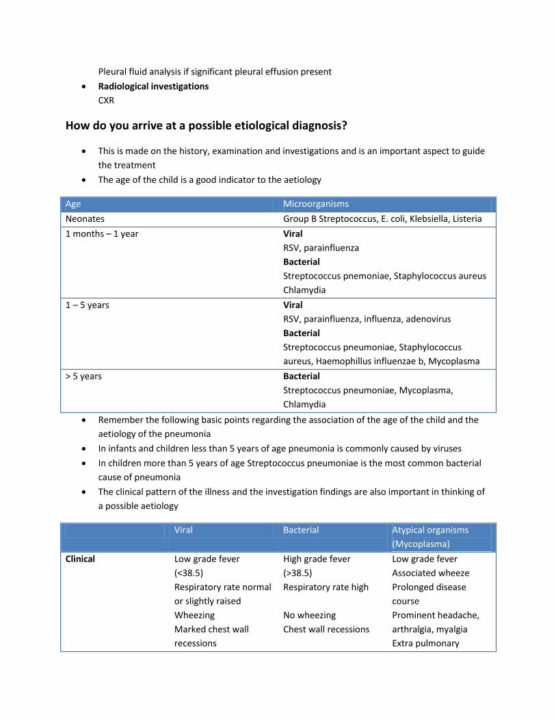
Age Microorganisms
Neonates Group B Streptococcus, E. coli, Klebsiella, Listeria
1 months – 1 year Viral
RSV, parainfluenza
Bacterial
Streptococcus pnemoniae, Staphylococcus aureus
Chlamydia
1 – 5 years Viral
RSV, parainfluenza, influenza, adenovirus
Bacterial
Streptococcus pneumoniae, Staphylococcus
aureus, Haemophillus influenzae b, Mycoplasma
> 5 years Bacterial
Streptococcus pneumoniae, Mycoplasma,
Chlamydia
Remember the following basic points regarding the association of the age of the child and the
aetiology of the pneumonia
In infants and children less than 5 years of age pneumonia is commonly caused by viruses
In children more than 5 years of age Streptococcus pneumoniae is the most common bacterial
cause of pneumonia
The clinical pattern of the illness and the investigation findings are also important in thinking of
a possible aetiology
Viral Bacterial Atypical organisms
(Mycoplasma)
Clinical Low grade fever
(<38.5)
Respiratory rate normal
or slightly raised
Wheezing
Marked chest wall
recessions
High grade fever
(>38.5)
Respiratory rate high
No wheezing
Chest wall recessions
Low grade fever
Associated wheeze
Prolonged disease
course
Prominent headache,
arthralgia, myalgia
Extra pulmonary

Hyperinflation manifestations
Investigations Usually no neutrophill
leukocytosis
CRP
Neutrophill leukocytosis
>15,000 WBC
CRP elevated > 35 to
60mg/L
Special investigations
Serology
Cold agglutinin test
Chest X ray Hyperinflation and
lobar collapse
Consolidation, pleural
effusion
Special findings may
also indicate the
probable aetiology –
Pneumatocoeles,
cavitation (S. aureus,
klebsiella)
Reticulonodular
opacification of the
lower lobe
Hilar lymphadenopathy
Interstitial infiltrates
How do you assess the severity of pneumonia?
Next you will be asked to assess the severity of the pneumonia based on the clinical assessment and the
investigations
Diagnosis
The diagnosis involves the following details
Clinical diagnosis of pneumonia
Probable aetiological diagnosis

Assessment of the severity of pneumonia
How would you manage this patient?
General management
Assess the ABC
Measure the oxygen saturation with the use of a pulse oxymeter
Oxygen therapy should be considered if the saturation is less than 92%
Obtain IV access and take blood for investigations – FBC, CRP, blood culture
Consider IV fluids if the patient cannot take orally
Correct any dehydration/ deficits
Put the child on an IV drip at 2/3 of maintenance
Manage fever and pain with paracetamol
Start a monitoring chart. Include the PR, RR, BP, oxygen saturation and the respiratory signs and
symptoms of the child
Feeding of the child – try to avoid insertion of an NG tube for feeding as this can compromise
the airway further
Antibiotic management
There are several aspects which should be considered in the antibiotic management
Whether to start antibiotics/not
In a patient with a clinical diagnosis of pneumonia empirical antibiotics should be commenced.
However antibiotics should not be used in children with mild lower respiratory tract infections
Choice of antibiotics
Age Empirical antibiotics
Children less than 5 years (excluding neonates) Oral amoxicillin (if the child is not ill and can take
orally)
IV cephalosporins (used presently in the wards)
IV ampicillin
Children > 5 years Penicillin/ cephalosporins, Macrolides -
erythromycin, clarithromycin (these can be used in
combination
Route of administration
IV should be considered if the child is extremely ill and refusing to take oral medication
Duration of treatment
Usually treatment carries on for about 7-10 days
With improvement the IV antibiotics may be switched from IV to the oral route
What would you look for in this patient on your daily ward round?
Look at the general condition of the child
Examine the respiratory system of the child
Look at the monitoring chart
Look at the latest investigations
Complications
Poor clinical response – the clinical response to treatment should take no more than 48 – 72 hours. If
the child is still unwell after this period of time consider the following factors
Is the child receiving the appropriate dose of the appropriate antibiotics?
Assess the compliance to the medication – check if the child is receiving the antibiotics or has
the child vomited the medication
Is the diagnosis correct?
Has the child developed any other complications of pneumonia – effusion, empyema, metastatic
spread
Are there any host factors predisposing to the poor response?
Other causes for the continuing fever
Assessment and further management
Do a chest x ray and assess the patient
Look for pleural effusion, worsening of the infiltrates, foreign bodies, and features suggestive of
atypical pneumonia
If the chest x ray is unremarkable a septic screen may be indicated to look for disseminated
infection
Review the antibiotic therapy
Second line investigations may be indicated if the problems persist
Management of pleural effusion


Blood and mucus diarrhoea
Key points in the history
Presenting complaint
Loose stools
State the duration
History of the presenting complaint
Describe the key features of the diarrhoea
Onset
Duration
Describe the characteristics of the stool – watery, mucoid or associated blood and
mucus
Describe the associated factors - Fever, nausea and vomiting with abdominal pain
Try to quantify the amount
Describe what the mother did at home
Describe the progression of symptoms over time
Describe what was done at the hospital
Exclude other conditions that can present with diarrhoea
Category Disease Important points in the history
Infections Dengue
Meningitis
Ask about arthralgia, myalgia,
headache and retro-orbital pain
and bleeding manifestations
Associated headache and
vomiting, photophobia,
irritability and altered behaviour
Surgical Intussusception Presents with blood and mucus
diarrhoea (classic “red currant
jelly” stools)
Ask for screaming attacks in the
child
Acute appendicitis Abdominal pain (periumbilical)
IBD Previous episodes of blood and
mucus diarrhoea

Ask for the risk factors of an infectious cause for diarrhoea
External factors Was there any consumption of food from outside?
Maternal factors Ask about the personal hygiene of the mother
Does she wash hands after going to the toilet?
Does she cut her nails?
Food preparation
Does she wash her hands before preparing food?
Does she clean the vegetables and green leaves
properly?
Are the cooking utensils cleaned regularly?
Is the food covered adequately after preparation?
Where is it stored?
Does the mother give bottle feeds? If so ask if she
boils the bottles. Ask on the preparation of the
formula milk
Do they use boiled water?
Child factors Hygiene of the child
Does the child play with sand or dirt?
Other playmates
Other family members Personal hygiene in the other family members may
also be relevant, especially of those who come in
contact with the child
Environment Give a brief account of any environmental risk
factors for diarrhoea around the house – garbage
collections, flooding, use of night soil as fertilizer
Ask for the complications of diarrhoea
Ask for the urine output of the child and for features of lethargy and drowsiness – Dehydration
Seizures
Can be due to the following causes – associated febrile convulsion, electrolyte imbalances,
hypoglycaemia, HUS, shigella encephalopathy
Past medical history
Ask for past history of episodes of diarrhoea/ dysentery
Other routine aspects of the history

Social history
Most of this has been described in the history of the presenting complaint but present the social
history in the usual order
Give special emphasis to the education level of the mother and her knowledge of diarrhoea,
preparation and administration of ORS
Examination
The key point of the examination is to look for evidence of dehydration. Look at the following
table
No dehydration Some dehydration Severe dehydration
General condition Eyes Tears Mouth and tongue Thirst
Well and alert Normal Present Moist Thirsty
Restless and irritable Sunken Absent Dry Thirsty, drinks eagerly
Lethargic, unconscious Very sunken and dry Absent Very sunken and dry Drinks poorly
Skin pinch Goes back quickly Goes back slowly Goes back very slowly
Examination of the vital signs is also extremely important as the child may present in shock
Abdomen
Look for a distended abdomen – Intestinal obstruction, paralytic ileus due to hypokalemia, gas
forming organisms
Palpable masses – Intusussception

Discussion
Dysentery
What are the causes of dysentery?
Bacillary dysentery Amoebic dysentery
Is the most common cause of dysentery More faecal matter with less amount of blood
Rare Small amounts of faecal matter with larger amounts of blood
What are the causes of bacillary dysentery?
Shigella – Shigella dysenteriae, Shigella sonnei, Shigella flexneri, Shigella boydii
Escherichia coli- Enteroinvasive and enteropathogenic
Campylobacter jejuni
What are the differences between Shigella dysenteriae and Shigella flexneri?
Compared to Shigella flexneri, shigella dysenteriae is highly infective and requires a smaller
infective dose. However it survives for only a short period of time in the environment
How would you manage this patient?
Initial
Admit the patient to the isolation room of the ward
Obtain samples for stool smear and culture
Fluid management
Follow the basic principles
Total fluid requirement = Correction of deficit + maintenance + correction of ongoing losses
Correction of deficit
Degree of dehydration Deficit Replacement Some dehydration 50 – 100ml/kg ORS 75 ml/kg over 4 hours
Severe dehydration >100ml/kg IV fluids 100ml/kg (preferred hartmann’s) Age <12 months 30ml/kg in 1hour and 70ml/kg in 5 hours Age >12months 30ml/kg in ½ hour and 70ml/kg in 2 and ½ hours
Shock >200ml/kg Give boluses at 10ml/kg over 20 minutes

Maintenance fluid calculation
This is based on the Halliday and Segar formula
Correction of ongoing losses
Give 50-100ml of ORS for each liquid stool or vomitus
Antibiotic therapy
This is based on the local antibiotic sensitivity patterns. Several antibiotic options are available
for the management. The patient should be given empirical antibiotic therapy should be based
on the local sensitivity patterns. At present the drug of choice is furozolidone 2mg/kg/dose 6
hourly for 5 days
A common side effect is darkening of the urine. The mother should be warned of this
Dietary management
Continue breastfeeding if the child is on breast feeding
A special diarrhoea diet is given in the wards
Rice kanjee and red rice kanjee – Prebiotic oligosaccharides
Anamalu – has a property of forming the stools
Yoghurt – Is a probiotic (living organisms that are colonizing organisms in the gut and prevent
the invasion of pathogenic organisms)
Rusk
Lime juice
Zn therapy
Has been shown to reduce the severity, duration and recurrences of diarrhoea. Give Zn 10-
20mg/d for 10-14 days
Monitoring of the patient
Complications of Shigella
Local
Intestinal perforation
Toxic megacolon
Proctitis and rectal prolapse
Systemic
Disseminated infection
HUS
Neurological complications
Reactive arthritis

While in the ward the patient develops seizures. What are the possible causes?
Febrile convulsion
Electrolyte imbalances – hypernatremia, hyponatremia, hypokalemia, hypocalcaemia
Hypoglycaemia
HUS
Shigella encephalopathy
How would you manage hypernatremic dehydration?
These children present with thirst out of proportion to the degree of dehydration and seizures
The main principle is not to drop the sodium rapidly as this can cause cerebral edema
Correct slowly over a period of 12 hours
How would you manage a patient with HUS?
Present 5-10 days after the onset of diarrhoea
The 3 features are
MIcroangiopathic hemolytic anaemia
Thrombocytopenia
Acute renal failure
Management is mainly supportive
Anaemia – Blood transfusion
ARF – Fluid management, management of electrolyte imbalances, mainly K+, antihypertensive
therapy and dialysis in severe cases
What is the advice you would give to the patient on discharge?
This should focus on the following themes
Prevention of further episodes of diarrhoea with proper hygienic practices of the family
What to do in an episode of diarrhoea
Give the child more fluid than usual
Teach the mother about ORS and the technique of preparation of ORS. Also tell her about other
fluids which can be used
Advise when to stop giving ORS
Continue to feed the child
Bring the child to the hospital especially if
High fever
Blood stained stools
Poor oral intake
Features of dehydration and over hydration

Edema and nephrotic syndrome
Key points in the history – 1st episode of edema
1. Describe the edema
Describe the onset of the symptoms and how the mother noticed them
Describe the location of the edema
Aggravating and relieving factors for the edema
Describe the progression of symptoms over time
What the mother did after noticing the symptoms
Describe what was done in the hospital
2. Ask specific questions based on the differential diagnosis of edema. The case which is usually
given is generalized edema
Category Disease Specific points in the history
Renal Nephrotic syndrome
Nephritic syndrome
Chronic renal failure
Usually based on the progression and
characteristics of the edema
Usually starts in the periorbital region
and then spreads downwards
Also ask for any change in the urine
Ask for associated red coloured urine
and documented elevated blood
pressure (ask the mother if she was
informed about elevated blood
pressure)
Ask for weakness, easy
fatigue(associated anaemia) and
uremic symptoms
Also ask for past history of UTI
Cardiac Heart failure Ask for past history of cardiac
disease, difficulty in breathing and
poor exercise tolerance
Gastrointestinal Cirrhosis
Protein losing enteropathy
History of yellowish discolouration of
the eyes, hematemesis, malaena,
evidence of hepatic encephalopathy
Ask for chronic diarrhoea
Other Angioedema
Drugs
Allergic history

3. After establishing that the most probable diagnosis is nephrotic syndrome try to find an
aetiology
Ask for evidence of systemic involvement – rash, joint pain and morning stiffness, fever
Infections such as hepatitis B, malaria, HIV can also cause nephrotic syndrome
4. Ask for the complications of nephrotic syndrome
Fever with abdominal pain – SBP
Flank pain with gross hematuria – Renal vein thrombosis
Calf pain +/- difficulty in breathing – DVT and pulmonary embolism
Collapse, syncope – Hypovolaemia
Abdominal pain in a patient with nephrotic syndrome
Hypovolaemia
SBP
Renal vein thrombosis
Mesenteric thrombosis
Fluid collection around the liver
Intestinal edema
Gastric irritation due to steroids
Key points in the history – Known patient with nephrotic syndrome with a relapse
1. Describe the initial episode of edema and how the diagnosis was made at the time
2. Mention what happened to the disease over time. DO NOT describe each of the relapses in
detail. Just mention the number
3. Describe the management
Mention the drugs used
Ask for the side effects of the medication
4. As given above ask for an aetiology for the condition
5. Mention the complications
6. Social history is extremely important
General introduction to the family
Impact on the child Playing, amount of school missed
Impact on the parents Socio economic impact of the disease, impact of
frequent hospital stays
Impact on the siblings
Environment Give a brief description of the environment of the
household
Support available Family support
Extended family support
Medical facilities available

Education of the parents Education of the mother on the disease
Knowledge about the drugs used and the
importance of proper compliance
Side effects of the medication
Method of urine testing
Knowledge on the diet and lifestyle
modifications
Identification of a relapse and when to
bring the child to hospital
Complications
Psychological state and expectations of the
parents
Examination
General examination
Anthropometry – Weight, height and BMI (Weight and height is used to calculate the body
surface area – this is on which the dose is calculated
Look for features of steroid toxicity
Cushingoid features
Weight gain and obesity
Hypertension
Cataract
Establish the distribution of edema
Look for vasculitic rashes in the skin – secondary cause for nephrotic syndrome
Abdomen
Look for free fluid in the abdomen
Cardiovascular
Exclude cardiac disease
Measure the blood pressure
Respiratory
Pleural effusions

Management of nephrotic syndrome
What is nephrotic syndrome?
Edema
Proteinuria (>40mg/m2/h or urine protein to creatinine ratio >200mg protein/mmol creatinine
Hypoalbuminaemia (<2.5g/dL)
Hyperlipidaemia
Diagnosis
Is based on the clinical presentation of the child and the investigations
The child will present with edema which is initially notes in the periorbital region and later
involves the dependant areas of the body and is worse towards the afternoon
Exclusion of other causes of generalized edema
What are the Investigations you will do?
Investigations to confirm the diagnosis
Urine ward test (Offers a qualitative assessment of the urinary protein) – is usually >+3
Early morning urine sample for estimation of the urine protein to creatinine ratio
24 urine collection for protein estimation
Urinanalysis is also an important investigation to look for microscopic hematuria and red cell
casts which may be found in patients with a nephrotic/ nephritic mixed picture
Serum albumin
Lipid profile may also be done (Elevated total cholesterol, LDL and triglycerides)
Note
Proteinuria in children
Transient proteinuria Orthostatic proteinuria Fixed proteinuria
Associated with fever,
dehydration, exercise
Asymptomatic
Increased protein excretion in
the upright
Indicates renal disease. Can be
due to glomerular or tubular
disease
Usually does not exceed +2 and
is normal on repeated daily
measurements
Absence of protein on an early
void sample for 3 consecutive
days
Significant proteinuria on an
early morning void sample on 3
consecutive days

Other investigations
Renal function tests and serum electrolytes
Serum complement
ESR, ANA
Hep B surface antigen
Renal biopsy
Role of renal biopsy in nephrotic syndrome
Recommendations
Age of onset less than 6 months
Initial macroscopic haematuria in the absence of infection
Persistent microscopic haematuria with hypertension
Renal failure not attributable to hypovolaemia
Persistently low C3, C4 levels
Steroid resistance
Preparation of a patient for renal biopsy
Do the initial workup of the patient. This includes the following investigations – serum
creatinine, FBC, bleeding time and clotting profile, renal ultrasound scan
Discuss with the team and arrange a date
Cross match blood
Fasting for 6 hours
Post op
Monitor vital parameters, UOP
Collect all urine samples
Complete bed rest until hematuria settles
After diagnosis
Classification
Classification of nephrotic syndrome is in to idiopathic and secondary nephrotic syndrome
Idiopathic Secondary
Minimal change disease (85%)
Focal segmental glomerulosclerosis
Membranous
Mesangioproliferative
Secondary to systemic diseases
Infections
Drugs
Connective tissue disorders and vasculitis

How would you manage the first episode of nephrotic syndrome?
General management of the child
Start daily weight chart and input/ output charts
Bed rest is not recommended
Protein restriction in the diet is also not recommended. Therefore give the child a normal
protein diet. (Salt restriction may be done until resolution of this episode). Fluid restriction is
also not recommended
Monitor the PR, volume and blood pressure
IV fluids – initially start with ½ of the maintenance over 12 hours. Then measure the urine
output and give the fluid hereafter as previous day UOP+ insensible loss
Management of gross edema. Diuretics may be used only if hypovolaemia has been corrected.
Drug of choice is frusemide 1-2 mg/kg/d. Use in conjunction with CPP. Start the CPP and give the
frusemide mid transfusion
Antibiotics – prophylactic oral
penicillin 250mg bd for 10 days
Steroid therapy
Prednisolone 60mg/m2/d given as
a single dose in the morning for
28 days. Then 40mg/m2/d on
alternate days for 28 days
Calculation of the body surface
area should be done using the
normogram which is available in
the ward
Usually respond to steroids after
2-4 weeks
Remission
Urine protein <4mg/m2/h
Urine protein negative or trace for
3 consecutive days

What is the advice you will give the parents?
The first step is to inform the parents that nephrotic syndrome is a relapsing, chronic disease
and that their support and understanding is extremely important to offer adequate
management for the child. Also give them a basic idea of what happens in nephrotic syndrome
Reassure them that progression to end stage renal failure is rare
Explain the home based management of nephrotic syndrome
Give a normal diet to the child
Should contain all the nutrients but reduce fat and refined sugar
Ensure normal activity and school attendance in the child
Explain the method of urine protein testing at home – Frequency of checking
Maintain a diary of the protein testing
Seek early medical attention for infections
Educate the parents about prednisilone
Importance of steroids and the risk of addisonian crisis if withdrawn abruptly. DO NOT stop
when the child develops an infection
Educate the parents about the side effects of medication – especially prednisolone
Behavioural changes especially irritability
Increased appetite and weight gain
Cushingoid features
Gastric irritation – therefore give with meals
With long standing steroid use other side effects may occur – Growth faltering, cataract, obesity,
hypertension, hyperglycaemia, osteopenia, recurrent infections
Advise on vaccination – avoid live vaccines for 3 months after stopping steroids
Try to avoid crowded places due to the risk of infection
Ask them to admit the child if there is edema or >+2 protein for more than 2 days at home
Method of checking for proteins at home
Collect urine to fill 2/3 of a test tube
Heat the upper part of the tube
Look for the formation of turbidity. Add a few drops of vinegar and see if the turbidity disappears (phosphates)
Quantify the amount of protein by holding the tube up against a newspaper
Nil – no turbidity, + - slight turbidity but can read the letters, ++ - cannot read the letters but can see the black color +++ - cannot see the print or the black color, ++++ - precipitate
Nil – no turbidity, +
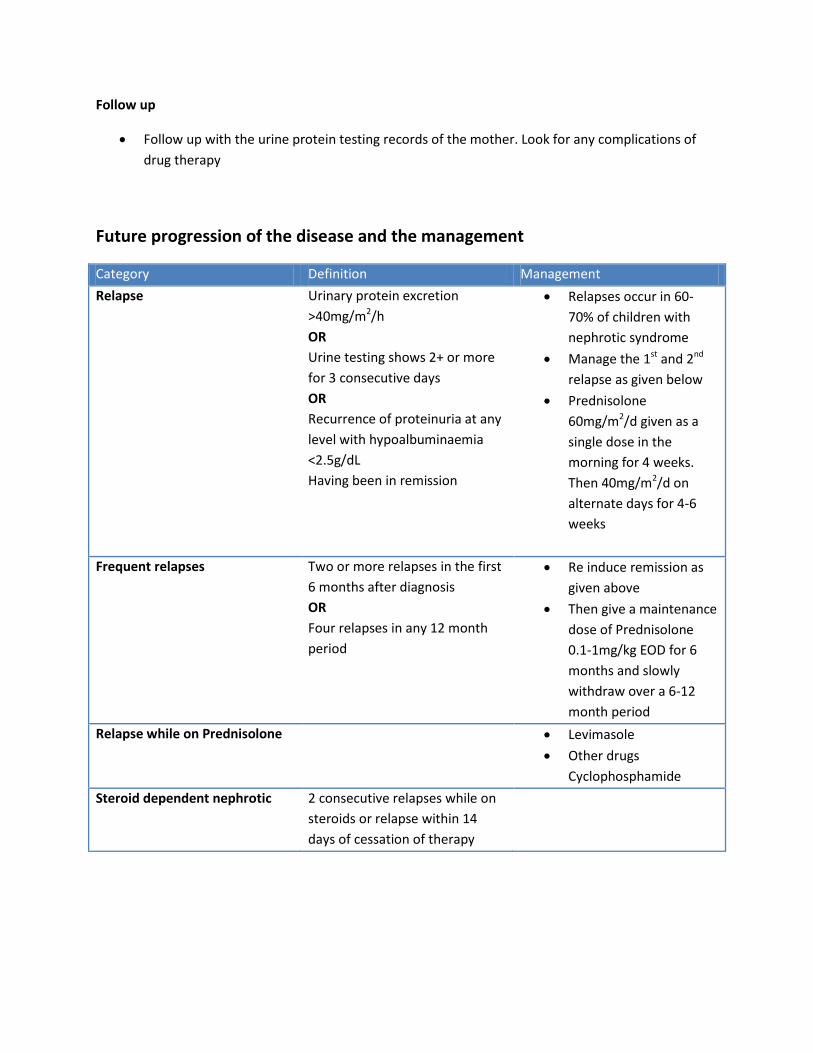
Follow up
Follow up with the urine protein testing records of the mother. Look for any complications of
drug therapy
Future progression of the disease and the management
Category Definition Management
Relapse Urinary protein excretion
>40mg/m2/h
OR
Urine testing shows 2+ or more
for 3 consecutive days
OR
Recurrence of proteinuria at any
level with hypoalbuminaemia
<2.5g/dL
Having been in remission
Relapses occur in 60-
70% of children with
nephrotic syndrome
Manage the 1st and 2nd
relapse as given below
Prednisolone
60mg/m2/d given as a
single dose in the
morning for 4 weeks.
Then 40mg/m2/d on
alternate days for 4-6
weeks
Frequent relapses Two or more relapses in the first
6 months after diagnosis
OR
Four relapses in any 12 month
period
Re induce remission as
given above
Then give a maintenance
dose of Prednisolone
0.1-1mg/kg EOD for 6
months and slowly
withdraw over a 6-12
month period
Relapse while on Prednisolone Levimasole
Other drugs
Cyclophosphamide
Steroid dependent nephrotic 2 consecutive relapses while on
steroids or relapse within 14
days of cessation of therapy
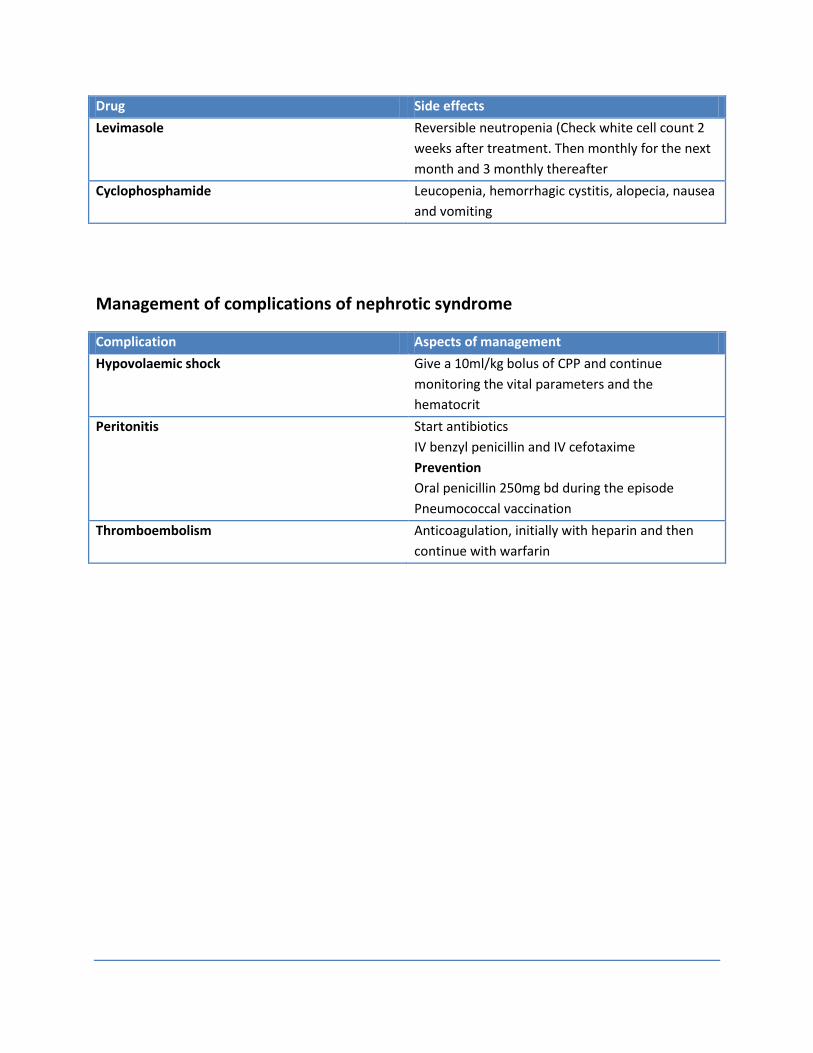
Drug Side effects
Levimasole Reversible neutropenia (Check white cell count 2
weeks after treatment. Then monthly for the next
month and 3 monthly thereafter
Cyclophosphamide Leucopenia, hemorrhagic cystitis, alopecia, nausea
and vomiting
Management of complications of nephrotic syndrome
Complication Aspects of management
Hypovolaemic shock Give a 10ml/kg bolus of CPP and continue
monitoring the vital parameters and the
hematocrit
Peritonitis Start antibiotics
IV benzyl penicillin and IV cefotaxime
Prevention
Oral penicillin 250mg bd during the episode
Pneumococcal vaccination
Thromboembolism
Anticoagulation, initially with heparin and then
continue with warfarin

Hematuria in children
History
Presenting complaint
The patient will present with red coloured urine
State the duration
History of the presenting complaint
Describe the symptom carefully based on the following points – onset, duration and progression
of the symptoms
Red coloured urine in children is not always due to hematuria but an alternative cause should be
considered only by exclusion
Describe the characteristics of the red coloured urine. This can indicate the site of bleeding
Cola coloured and mixed throughout the stream – Glomerular bleeding
Fresh blood, associated clots and more towards the end of the stream – Lower urinary tract
bleeding
Try to reach a differential diagnosis
Cause Key points in the history
Glomerular
Acute nephritic syndrome
Mixed nephrotic and nephritic syndrome
IgA nephropathy
HUS
Other rare glomerular disease
Ask for associated swelling of the body and decreased
urine output
Also ask the mother if she was told that the child had
increased blood pressure
Look for an aetiology
Ask for preceding sore throat or skin sepsis a few weeks
back (Post streptococcal glomerulonephritis)
Ask for systemic features such as fever, malaise, joint
pain and stiffness and skin rashes (Vasculitis and
connective tissue disease
(SLE, HSP)
Ask for history of recurrent gross hematuria. (Can occur
1-2 days after a URTI
Ask for preceding history of AGE (5-10days back), fever,
abdominal pain, seizures
Family history of similar disease

Extra glomerular
UTI
Stones
Trauma
Bleeding disorders
Ask for associated crying on micturition and fever
Associated abdominal pain and family history of urinary
calculi
Other sites of bleeding
Exclude other causes of red coloured urine
Consumption or red coloured food substances, drugs, associated features of jaundice and
anaemia (haemoglobinuria)
Describe what has happened to the child up to now
Ask for complications of nephritic syndrome (this will be the most probable diagnosis)
Altered level of consciousness, seizures – hypertensive encephalopathy
Acute renal failure
Dyspnoea, poor exercise tolerance – heart failure
Take the other routine aspects of the history
Examination
General examination
Anthropometry – This is extremely important in the management
Look for pallor and Icterus – could indicate hemoglobinuria
Note the distribution of edema
Look for healed skin wounds, skin rashes suggestive of SLE or other types of vasculitis
Cardiovascular examination
Measure the blood pressure
Look for features of heart failure
Abdomen
Palpate for masses – tumors of the renal tract, PCKD can present with hematuria
Neurological examination
Examine the fundus for evidence of malignant hypertension
How would you investigate a patient with nephritic syndrome?
Urine full report and microscopy
Glomerular hematuria – Red cell casts, dysmorphic red cells (special microscopy) and
proteinuria >100mg/dl
Hematuria from the tubules – White cell casts, epithelial casts
Lower urinary tract – Normal red cell morphology, proteinuria <100mg/dl
Follow up a case of glomerular hematuria with the following investigations
FBC
BU/SC and serum electrolytes
Serum complement
ASOT
DNAase B
ANA
Other investigations may be required is an extraglomerular cause is suspected – urine culture,
USS of the abdomen
Management of AGN
General management
The management of AGN is usually supportive
Start a monitoring chart
Daily weight
Input output chart
Blood pressure
Fluid management
Calculate the maintenance fluid requirement for the child and give ½ of this amount over 12
hours. Then measure the urine output over this time and continue as
Fluid input = UOP + insensible loss
Management of edema
Frusemide 1mg/kg
Diet
Give a balanced diet with restricted salt. Do not give the child foods rich in potassium
Antibiotics
Monitor for complications
Hypertensive encephalopathy
Acute renal failure
Cardiac failure

Management of acute hypertension
Diagnose hypertension
Classify the severity of hypertension as hypertensive urgency or hypertensive emergency
Diagnosis Definition Management
Hypertensive urgency Elevation of blood pressure
without severe symptoms or
evidence of target organ damage
Oral medication
Oral nifedipine
Hypertensive emergency Elevation of blood pressure with
target organ damage
ABC
IV antihypertensives – IV
hydralazine
Drop the blood pressure slowly
Management of acute renal failure
Principles of management are given below
Fluid and electrolyte balance
Give IV fluids as previous day’s urine output + insensible loss based on the body surface area of
the child
Hyperkalemia
Salbutamol nebulization
IV calcium gluconate for stabilization of the myocardium
Insulin – dextrose infusion
Potassium binding resins
Consider renal replacement therapy
When will you discharge the patient?
What is the advice you will give at discharge?
The disease process will continue for 2-3 weeks. At the end of the natural course of the disease
the child will develop polyuria. It is important to monitor the child during this period as well as
the child can get hypovolaemic

At discharge
Explain the disease to the mother
Tell them not to restrict the diet if the child
The child can be discharged on antihypertensive medication
Explain the warning signs of hypertensive encephalopathy and tell them to come to hospital
immediately
Get the blood pressure measured EOD by a GP
Review in the clinic in 1 week with a urine full report (may have microscopic hematuria)

Management of UTI in children
How would you diagnose UTI?
This is based on the clinical assessment and the findings on the urine culture and ABST
The child will present with the following symptoms based on the age. Look at the following table
as a guide
The clinical assessment should also indicate any serious underlying pathology which is
associated with the infection. The points shown below are important in this regard
Recurrent UTI
Voiding dysfunction and poor urine flow
Evidence of hydronephrosis
Palpable bladder
Evidence of chronic renal failure
The next step is to confirm the diagnosis. The investigation of choice is a urine full report and a
urine culture/ ABST
The urine culture/ ABST is considered as the gold standard of diagnosis for a UTI. Therefore the
sample collection, transport and interpretation are extremely important
These points are commonly asked at the exam

How would you collect urine for urine culture/ABST?
Methods of collection
Clean catch midstream sample
Supra pubic aspiration
Catheter samples (usually not recommended except in failed SPA
Advice to the parents on collection of CCMS urine
Ask the mother to feed the child prior to the collection of the sample
Wash hands and genitalia with soap and water. Retract the prepuce in older boys
Open the cap of the special bottle once the child has started to pass urine. Do not leave the lid
open for a long time
Discard the first part of the urine and collect the midstream urine sample directly into the
bottle. Avoid contact of the bottle with the perineum of the child
Close the cap and hand over the bottle immediately
Transportation
Fill out the request form and send immediately to the lab
If the specimen cannot be transported within 2 hours refrigerate for a maximum of 24 hours
Interpretation of the culture report
In a CCMS >105 would indicate a high probability of a UTI (80-95%). If it is between 104 and 105
an infection is likely
Any number of colonies on an SPA culture would indicate a 99% probability of an infection
Other investigations
Full blood count
CRP
Renal function tests
Serum electrolytes

Describe the initial management of UTI in children
Initial management
General management
Ensure proper hydration of the child
Manage fever and pain with paracetamol
Antibiotic therapy
Empirical antibiotic therapy
All cases of suspected febrile UTI should be started on empirical antibiotics. These should be
started after collection of urine for culture
The selected antibiotics should cover the possible organisms – E. coli, Klebsiella, Proteus and
Enterobacter
Choice of antibiotics and route of administration
Oral – Cephalexin, Co amoxyclav, Cotrimoxazole
IV (should be given if the child is extremely ill or if the child refuses to take orally
Cefotaxime, ceftriaxone, cefuroxime
Duration of treatment is usually 7 days
Follow up
Review after 48 hours
The child should show response to treatment usually within 48 hours. If the symptoms persist
check the adequacy of the antibiotic treatment and the ABST
Change or alter the dose of antibiotics if necessary
What are the further investigations necessary?
USS KUB
USS should be performed in all children with a febrile UTI within 6 weeks of the attack. Look for
Gross structural anomalies of the renal tract
Features suggestive of acute pyelonephritis
Hydronephrosis and hydroureter
Bladder

Prophylaxis and further investigations
Prophylaxis is indicated for all children < 5 years following the first attack of UTI until the USS
report is available
The continuing management is based on the following principles
In a child with a febrile UTI – If the USS is normal continue the prophylaxis until the
recommended imaging studies are available
If structural anomalies are detected or in cases or recurrent UTI continue prophylaxis till 5 years
or longer
Start prophylaxis as given below
Complete the course of antibiotics and start prophylaxis from the next day onwards. Repeat a
urine culture at the 5th day of prophylaxis
Antibiotics used for prophylaxis are given below. These drug are given as a single dose at night
Drug Use
Cephalexin Recommended in the first 1-3 months
Cotrimoxazole Avoid in infants < 1 month of age
Nitrofurantoin Avoid in less than 3 months
Nalidixic acid Avoid in less than 6 months
Investigations Indication Uses
DMSA
(Should be done after 6 months
after the UTI)
Under 1 year
All children with a febrile UTI
Under 5 years
Clinical picture suggestive of
acute pyelonephritis
Recurrent febrile UTI
Abnormal USS
Looks for renal scarring
MCUG Suspected bladder outlet
obstruction
When the USS reveals
hydronephrosis and hydroureter
Recurrent UTI
Used to diagnose posterior
urethral valves and VUR
DTPA Suspected PUJ or VUJ obstruction Used to diagnose PUJ and
VUJ obstruction
Can also give an idea about
the differential renal function
Preparation of the patient for the above investigations

Prevention of recurrent attacks of UTI – Advise the parents
Educate the parents on the condition the child is having an the prognosis
Teach them how to recognize the condition and when to bring the child to the hospital
Educate them on the importance of giving the child the recommended prophylactic medication
General
Good hydration
Avoiding constipation
Improve the hygiene of the child
Wiping of the perineum from front to back
Change the child’s nappies frequently
UTI
USS
Abnormal Normal Less than 1 year DMSA (<1year) after 6 months
1-5 years DMSA if recurrent UTI or if 1st E pyelonephritis
Relevant investigations
Surgical conditions of the urinary tract
Condition Diagnosis Management
Posterior urethral valves MCUG
Dilated, elongated posterior
urethra with a thick walled
trabeculated bladder
May have associated VUR
Cystoscopic ablation
VUR MCUG is used for the diagnosis
and the classification of VUR
Conservative medical
With antibiotic prophylaxis
Surgical intervention
Indicated in recurrent UTI in

spite of prophylaxis, patients
with impaired renal function,
recurrent new scar formation
and gross VUR
Reimplantation is performed
PUJ obstruction DTPA Pyeloplasty

Acute flaccid paralysis
Presenting complaint
Weakness of the lower limbs
State the duration
History of the presenting complaint
Remember that in a neurology case the most important aspect in the history is the chronological
order of development of the symptoms
Describe the onset of the weakness as sudden onset or gradual onset. Then go on to describe
the distribution of the weakness
Then go on to describe the progression of weakness over time
The description of the neurological impairment at each stage in the chronological order should
be described based on the activities of the child such as running and playing, walking, sitting,
standing up from a sitting position and other day to day activities
Describe the other associated neurological symptoms
Altered level of consciousness or loss of consciousness
Change in behavior
Seizures
Diplopia
Deviation of the mouth, drooling of saliva from the side of the mouth
Vertigo
Dysphagia and nasal regurgitation
Think of the possible differential diagnosis based on the site of the lesion and ask specific
questions
Site of the lesion Disease Specific questions in the history
Spinal cord Spinal cord injury
Transverse myelitis
Ask for history of trauma to the spine
Ask for associated backache,
paresthesia, bladder and bowel
incontinence
Anterior horn cell disease Poliomyelitis Ask for initial history of fever, upper
respiratory and GI symptoms. This is
followed by a symptom free period of a
few days
Then there is recurrence of the fever
and muscle tenderness, neck stiffness
and paralysis (usually asymmetric)
Peripheral nerve Guillain barre syndrome Ask for preceding history of diarrhoea
or respiratory tract illness. This is

followed a few weeks later by
paresthesia of the extremities and an
ascending paralysis
Other causes of
peripheral neuropathy
Exposure to toxins, use of long term
drugs, recent vaccines (post rabies
virus)
Neuromuscular junction Botulism Possible ingestion of contaminated
canned food
Initial cranial nerve symptoms and
descending paralysis
Myasthenia gravis Preceding history of drooping of the
eyelids or weakness which is most
prominent towards afternoon
Snake venom and toxins Ask for possible history of snakebite
and exposure to toxins
Muscle Myositis
Periodic paralysis
Muscle pain, associated skin rashes
Then go on to describe what was done to the child up to hospital admission and state the
progression of the neurological symptoms
Ask for the complications of the disease, specifically for bulbar involvement, respiratory muscle
weakness
Describe any specific management which has been done to the child
Describe the present status of the child. This is also in regard to the functional status
Past medical and surgical history
Birth history
Growth and developmental history
Immunization history
This is very important. Describe the present state of immunization
Describe clearly about the polio vaccinations of the child
Social history
This is a critical component of the history
Describe the following components of the social history
General introduction to the family
Impact of the disease on the child
Impact of the disease on the parents
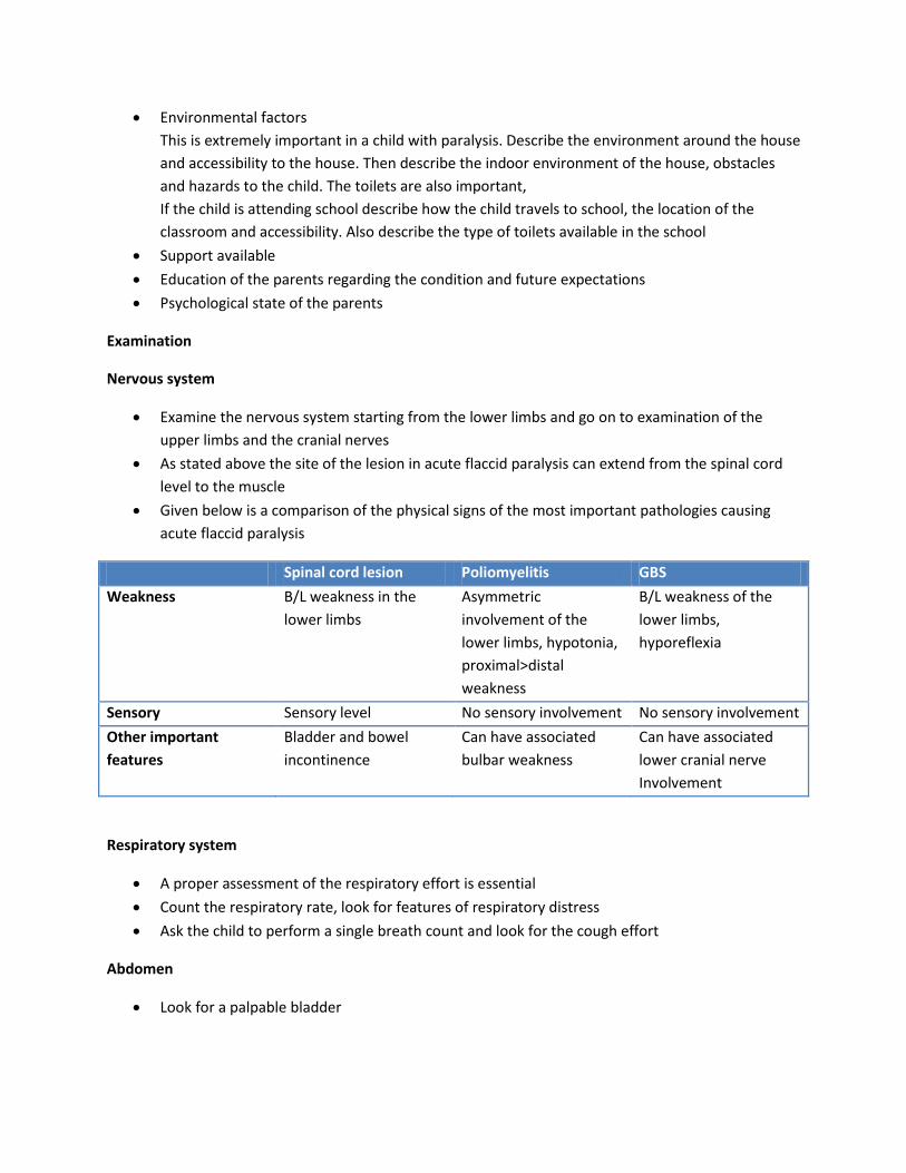
Environmental factors
This is extremely important in a child with paralysis. Describe the environment around the house
and accessibility to the house. Then describe the indoor environment of the house, obstacles
and hazards to the child. The toilets are also important,
If the child is attending school describe how the child travels to school, the location of the
classroom and accessibility. Also describe the type of toilets available in the school
Support available
Education of the parents regarding the condition and future expectations
Psychological state of the parents
Examination
Nervous system
Examine the nervous system starting from the lower limbs and go on to examination of the
upper limbs and the cranial nerves
As stated above the site of the lesion in acute flaccid paralysis can extend from the spinal cord
level to the muscle
Given below is a comparison of the physical signs of the most important pathologies causing
acute flaccid paralysis
Spinal cord lesion Poliomyelitis GBS
Weakness B/L weakness in the
lower limbs
Asymmetric
involvement of the
lower limbs, hypotonia,
proximal>distal
weakness
B/L weakness of the
lower limbs,
hyporeflexia
Sensory Sensory level No sensory involvement No sensory involvement
Other important
features
Bladder and bowel
incontinence
Can have associated
bulbar weakness
Can have associated
lower cranial nerve
Involvement
Respiratory system
A proper assessment of the respiratory effort is essential
Count the respiratory rate, look for features of respiratory distress
Ask the child to perform a single breath count and look for the cough effort
Abdomen
Look for a palpable bladder

Discussion
Guillain Barre Syndrome
Diagnosis
The diagnosis of GBS is entirely clinical
Investigations may be performed if there is any doubt about the clinical diagnosis
NCS
LP after 10 days of appearance of symptoms – shows cytoprotein dissociation
Initial management
Book an ICU bed
Assess the child and start monitoring the key clinical parameters. These are
Pulse rate and rhythm, BP – Autonomic instability
Respiratory function – Single breath count, features of respiratory distress, assessment of vital
capacity (this is often difficult in children)
Neurological parameters – State of paralysis and rate of progression with grading of the muscle
power
Admit the child to the ICU if the following are present
Rapidly progressive paralysis
Bulbar palsy
Respiratory involvement – Deteriorating SBC, features of respiratory distress, vital capacity
<20ml/kg – This is an indication for ventilation
Autonomic cardiovascular instability
Consider IVIG therapy if there is rapid progression of the neurological impairment
The recommended dose is 0.4g/kg for 5 days. The infusion rate should be slow initially as there
is risk of anaphylaxis and later increased
Continue monitoring the patient
Notification and other important investigations
Any child under 15 years of age with acute flaccid paralysis should be notified immediately. The
notification should be done to the MOH of the area and the RE by telephone
The investigation form EPID/37/1/R2004 should be completed and returned to the
epidemiological unit
Collection of stool samples
Samples should be collected within 2 weeks of the onset of paralysis into the provided special
container
Two samples of stools should be collected 24-48 hours apart
Sample should weigh 8-10g (the size of two thumbnails)

Lid should be tightly closed and packed in ice
The sample should be correctly labeled. The following details should be present
Introduction – as in any sample
Date of onset of paralysis
Date of collection of stools
Date of dispatch of stools
Last date of polio vaccination
Samples should be transported to the MRI within 72 hours of collection
The MOH should personally investigate the case of AFP and visit the community where the case
is resident
He should collect and dispatch one stool sample from 3-5 contacts and send to the MRI within
72 hours of collection
The MOH is also responsible for initiating a program of limited out break response
immunization. This includes administration of an extra dose of OPV to the children of the same
age or below living around a 2km radius of the residence of the index case
Continuing management
The patient should have supportive management while in the ward
Management of muscle pain
Bladder and bowel care
Chest physiotherapy for prevention of respiratory of chest infections
Passive physiotherapy
Rehabilitation
Patients with GBS begin spontaneous recovery after 2-3 weeks in an inverse direction to the
direction of paralysis
Physiotherapy is extremely important
Rehabilitation includes occupational therapy

Meningitis
Discussion
Diagnosis of meningitis in children
The diagnosis of meningitis is made on the history, examination and investigations
The important aspect is that the clinical presentation varies according to the age of the child
Both bacterial and viral meningitis has a similar presentation but the latter usually has a milder
course
The table given below gives the presentations in various age groups
0-2 months >2 months Older children
History Usually no specific features Fever or hypothermia Irritability and high pitched cry, lethargy, poor feeding Seizures Apnoeic attacks Altered sleep pattern
Fever Irritabilty, lethargy Seizures
Fever Irritabilty, lethargy
Examination Bulging fontanelle (Should be examined in the upright position when the child is not crying) Opisthotonus
Bulging fontanelle Brudzinki’s sign
Neck stiffness (more useful in children >3years) Kernig’s sign Photophobia
Describe the Initial management of the child
The initial management should focus on the A,B,C
Look for evidence of complications – increased intracranial pressure, sepsis, seizures
Correct any abnormalities as you find it
Obtain IV access and collect blood for investigations
Full blood count
Obtain blood for blood culture
C- reactive protein
Serum electrolytes
Renal function tests

Lumbar puncture and CSF analysis for definitive diagnosis
Remember that even though the LP is used for the definitive diagnosis of meningitis there may
be indications to delay the LP
Indications to delay the LP
Symptoms and signs of increased intracranial pressure
GCS <13 or deteriorating level of consciousness
Recent (within 30 minutes) or prolonged seizures
Focal neurological symptoms and signs
Shock
Coagulopathy
Local sepsis
The general rule should be to stabilize the patient before performing a LP
Remember that in the case that the LP should be delayed do not delay the 1st dose of IV
antibiotics
The samples for LP should be taken as follows
CSF sugar (to be interpreted with a random blood sugar taken about 30 minutes before the
procedure)
Protein
CSF culture
Full report, including gram stain
Other special investigations – bacterial antigen detection, mycobacterial, viral studies
Interpretation of the CSF report
Viral Bacterial Partially treated TB
Appearance Clear Turbid Clear Turbid, may clot on standing
Cells 15-2000 10-10000 5-10000 10-500
Differential count Lymphocytes Neutrophils Monocytes or neutrophills
Lymphocytes
Glucose >50% of BG <50% of BG Normal or decreased
<50% of BG
Protein Normal/ slightly elevated
Elevated (100-500)
Elevated (100-500)
Very high
Initial pharmacological management
Pharmacological management should be initiated after the samples are collected for blood
culture and CSF analysis
Commencement of empirical antibiotic therapy is the most important management option in
suspected acute bacterial meningitis

Do not delay the antibiotics even if the LP is delayed
Steroid therapy is also indicated in suspected acute bacterial meningitis. However there are
specific criteria for the use of steroids
Age > 3 months
Should be administered before the first dose of parenteral antibiotics
The recommended dose is 0.15/kg/dose IV every 6 hours. The first dose of steroids should be
followed by the first dose of IV empirical antibiotics
The recommended empirical antibiotics vary according to the age of the child and the antibiotic
sensitivity patterns
Age Organisms Recommended antibiotics
Neonates GBS, E. coli, listeria Ampicillin or benzyl penicillin + cefotaxime
1-2 months Neonatal organisms, haemophillus, pneumococcus, meningococcus
Ampicillin or benzyl penicillin + cefotaxime
2 months – 5 years haemophillus, pneumococcus, meningococcus
Cefotaxime/ ceftriaxone
>5 years pneumococcus, meningococcus
Cefotaxime/ ceftriaxone
The antibiotics may be altered according to the results of the cultures, based on the ABST
Continue antibiotics for 10-14 days in an uncomplicated Streptococcus pneumoniae meningitis
and 7-10 days for an uncomplicated Haemophillus influenzae b infection
As a house officer how would you assess a patient on your daily ward round?
Maintaining a monitoring chart is extremely important. This chart should include the following
data
QHT fever monitoring
Vital parameters – PR, RR, BP
Neurological assessment – GCS, pupillary reflexes, examination of the cranial nerves and limbs
Chart the OFC
Maintain an input-output chart
Keep a record of the daily investigations
IV fluids – May be given in these patients at 1/2 - 2/3rds maintenance due to the risk of SIADH in
normovolaemic and normotensive patients (But may be returned to normal if the serum
sodium is normal)
Manage dehydration and hypotension with 0.9% saline
Identify and manage complications as they arise

What are the complications of meningitis?
Early neurological
Increased intracranial pressure as a result of cerebral edema
Call and book an ICU bed
Nurse the patient with a 15-30 degree elevation of the head in the midline position
Temperature control
Appropriate fluid and electrolyte therapy – 1/2 – 2/3 maintenance after hypotension and
deficits are treated
Seizure control
Specific measures
Mannitol – 2.5 -5 ml/kg of 20% solution over 30 minutes
Frusemide – Can be used in combination with mannitol
Seizures
ABC
IV midazolam 0.15mg/kg or rectal diazepam 0.5mg/kg
For continuing seizures a bolus dose of phenytoin at 20mg/kg over 20 min may be given
Stroke
Acute hydrocephalus
Cranial nerve palsies
Hearing impairment
Subdural effusion
Other
Disseminated infection and sepsis
Electrolyte imbalance – SIADH
Nosocomial infections
Late neurological
Cognitive impairment
Chemoprophylaxis
Recommended in all household contacts irrespective of the age when at least 1 unvaccinated
contact is younger than 4 years of age
Drug of choice is Rifampicin
When index case is less than 2 years commence a full course of HIb vaccination regardless of
the vaccination status
Follow up

Brain imaging
Assessment of the hearing of the patient
Regular developmental assessment
Encephalitis
Not a common topic of discussion at the long case but the basic details given below should be known
Diagnosis
Clinical
Presents with a non specific prodromal period which is followed by CNS symptoms such as
alteration of behavior, irritability, altered level of consciousness and seizures
May be associated with meningitis
Metabolic encephalopathies and post infectious encephalomyelitis should be considered as the
differential diagnosis
Causes
Viral
HSV
Other herpes viruses – VZV, CMV, EBV
Enteroviruses
Arboviruses – JE
Investigations
LP – CSF analysis typically shows a lymphocytic predominant leucocytosis with normal CSF
glucose
EEG – Diffuse slowing or focal EEG changes
Management
This is usually supportive
Manage seizures and increased intracranial pressure

Seizures in children
History
Presenting complaint
The child will present with abnormal movements
State the duration and number of episodes over this time (latest presentation)
History of the presenting complaint
Describe the episode in detail. This includes the following details
Pre ictal phase
Ictal phase
Post ictal phase
What the mother did in response to the episode
From these details the main objective is to identify the seizure pattern and to exclude seizure
like events
Partial (focal) seizures
Are of 3 types
Simple partial, complex partial and partial seizures with 2ry generalization
The 2 important types are described below
Phase Simple partial Complex partial
Pre ictal An aura may be present An aura may be present Can start with a simple partial seizure
Ictal Consciousness is retained May present with motor symptoms – focal in origin with or without a Jacksonian march May also have features of head turning and conjugate eye movements – versive seizure Sensory presentations may also occur
Consciousness is impaired Automatisms are commonly associated – prolonged and repetitive lip smacking, chewing, swallowing and excessive salivation May also have gestural automatisms which involve alteration of behaviour May develop secondary generalization
Post ictal Child is well after the seizure Child is well after the seizure

Generalized seizures
Important seizure types which could be given at the exam are given below
Phase GTC Absence Infantile spasm
Pre ictal No preceding aura
Ictal Ictal cry Eyes rolling up Initial tonic state Subsequent clonic movements Urinary or faecal incontinence Tongue biting, frothing from the corner of the mouth
Transient loss of consciousness
Post ictal Have post ictal drowsiness

Management of seizures and epilepsy in children
Evaluation of the first seizure
In a child presenting with a seizure the first step is to make a clinical diagnosis based on the
history and examination
Look for a secondary cause (see history, examination and initial investigations)
Definition of epilepsy
Clinical condition characterized by recurrent unprovoked seizures
Diagnosis
Is a clinical diagnosis
The most important tool for the diagnosis is a firsthand witness account of the event
Classification of epilepsy in children
Can be classified based on the seizure type and also by the epileptic syndrome. A syndrome of
epilepsy is based on the age of onset, cognitive development, seizure type, findings on
examination and the EEG findings
About 50% of childhood seizures can be classified into a specific syndrome
Classification based on the seizure type
Partial (focal)
Simple partial
Complex partial
Generalized
Generalized tonic clonic
Tonic
Clonic
Myoclonic
Atonic
Absence
Infantile spasms
Unclassified
Classification of epileptic syndromes is complicated and is not asked at the long case discussion

Name Age Seizure pattern EEG pattern
Generalized epilepsies
Infantile spasms
4-6 months Flexor spasms, clusters
usually occurs on
waking
Hypsarrythmias
Lennox – Gastaut
syndrome
1-3 years Multiple seizure types,
but mostly drop attacks,
tonic seizures and
atypical absences
associated
neurodevelopmental
arrest or regression and
behaviour
Typical absence 4-12 years Absence seizures, child
is developmentally
normal. Episodes can be
induced by
hyperventilation
Generalized 3 per
second spike and wave
discharge
Juvenile myoclonic
epilepsy
Adolescence Myoclonic seizures, but
generalized tonic clonic
seizures and absence
seizures may occur
Characteristic EEG
Focal epilepsy
Benign rolandic epilepsy
4-10 years Simple partial seizures,
tonic clonic seizures in
sleep, abnormal
feelings in the tongue
and distortion of the
face
Focal sharp waves in
the centrotemporal
area
Investigations
EEG
EEG Is an important investigation in a child with epilepsy. It is usually done after the second
seizure
Uses of EEG
Determination of the seizure type
Diagnosis of epileptic syndromes
Determination if further investigations are required
Prognosis

About 40% of children with epilepsy will have a normal first EEG
Other special methods may be utilized if the EEG is not conclusive. These are sleep EEG and
video EEG (useful for evaluation of suspected pseudo seizures)
Neuroimaging
MRI
May be used in special circumstances
A complete diagnosis in a patient with seizures includes the following details (Based on the ILAE
recommendations)
Seizure semiology
Seizure type
Seizure syndrome
Impairment
Aetiology
Management
Education
The first aspect of the management is the education of the parents and caregivers of the child
Education should include the following aspects
General information on epilepsy
Information of the first aid in an attack of seizures
Lifestyle modifications
Antiepileptic drugs and their side effects
Importance of proper compliance to the medication and how to administer the drugs
Psychosocial issues and social stigma
Antiepileptic drugs
Starting of antiepileptic drugs should be done by a consultant pediatrician. It is usually initiated
only in patients with recurrent seizures
The choice of first antiepileptic drug depends on the seizure type/ syndrome, adverse effects, co
morbidity, availability and cost
Monotherapy is preferred over polytherapy
The drug should be started at a low dose and gradually increased towards the maintenance
dose

Pharmacology of antiepileptic drugs
Drug Mechanism of action Pharmacokinetics Adverse effects
Carbamazapine Blocks the voltage dependent sodium channels
Induces hepatic enzymes CNS symptoms Diplopia, blurring of vision, dizziness and ataxia Other Skin rashes, blood disorders, hepatotoxicity
Sodium valproate Decreases the breakdown of the inhibitory neurotransmitter GABA
Inhibitor of hepatic metabolism
CNS symptoms Hepatotoxicity (more in children less than 3 years) Other Weight gain, alopecia, blood disorders, pancreatitis
Phenytoin sodium Membrane stabilizing effect
Inducer of hepatic enzymes
CNS Impairment of cognitive function, Diplopia, blurring of vision, dizziness and ataxia Other Skin rashes, coarsening of facial features, hirsuitism, gum hypertrophy
Phenobarbitone Barbiturate Behavioural changes, hyperactivity, sedation
Lamotrigine Blocking of voltage dependant sodium channels
Generally well tolerated but can cause cutaneous adverse effects – TEN, Steven – Johnson syndrome (risk is higher with the concomitant use of valproate
Topiramate Blocking voltage dependent sodium channels and enhances GABA activity
Sedation, word finding problems, weight loss, acute myopia and raised intraocular pressure
Clonazepam Benzodiazepine Drowsiness, insomnia
Vigabatrin Structural analog of GABA Does not induce liver enzymes
Visual field disturbances, confusion, psychosis

Choosing an antiepileptic
The choice of a suitable antiepileptic is based on the following principles
Efficacy
Support by clinical guidance and research
Side effects
Predicted compliance to the medication
Availability
Cost
Type of seizure First choice antiepileptic drug Other options
Generalized
GTC Sodium valproate
Topiramate Lamotrigine
Absence Sodium valproate Ethosuximide
Topiramate Lamotrigine
Myoclonic Sodium valproate Lamotrigine
Clobazam Clonazepam
Infantile spasms ACTH Prednisilone
Focal Carbamazapine Sodium valproate
Lamotrigine Topiramate Clobazam Clonazepam
Follow up
Follow up of the child should be done based on the following principles
Review the last attack of seizure
If seizures are continuing rethink the diagnosis
Confirm the seizure type
Check if the dose is adequate for the age of the child
Assess the compliance for the medication
Try increasing the dose of the existing anti epileptic
Use the principles of antiepileptic drug therapy
Remember that monotherapy is preferred over polytherapy
If monotherapy in the maximal dose has failed introduce a second drug and monitor the
response. Then gradually tail off the first drug and continue monotherapy with the second
Emphasize the basic patient education on seizures
Assess the for the side effects of the medication

Assess the other parameters of the child, especially the development
When the child is seizure free for 2 years or more consider tailing off the medication
Approach to anaemia in children
This case is usually not given as a separate one but may be part of a discussion in any case
Definition of anaemia
Is a reduction in the hemoglobin concentration of the blood below the normal range
Classification of anaemias
Anaemias of inadequate production
Bone marrow failure syndromes and bone marrow
aplasia
Nutritional anaemias
Anaemia of chronic disease
Hemolytic anaemia Hereditary
Membrane defects
Enzyme defect
Disorders of the structure of hemoglobin
Acquired
Immune hemolytic anaemia
Non immune hemolytic anaemia
Morphological classification
Microcytic hypochromic anaemia Normocytic normochromic Macrocytic

Iron deficiency anaemia Hemolytic anaemia Vitamin B12 deficiency
Beta Thalassemia major and
minor
Anaemia of chronic disease Folate deficiency
Anaemia of chronic disease Bone marrow failure syndromes
Sideroblastic anaemia
Key points in the history
History of the presenting complaint
Patients will present with the features of anaemia. These include lethargy, poor exercise
tolerance, and exertional dyspnoea
Describe the onset and progression of the symptoms
Is this an isolated anaemia or part of a pancytopenia?
Ask for history of recurrent infections and bleeding manifestations which are associated with
the anaemia
Try to establish the type of anaemia – given below are the key points which should be established in
the history
Nutritional anaemia (especially
iron deficiency)
Hemolytic anaemia Anaemia of chronic disease
Get a detailed dietary history
from the mother. Include the
following
Breast feeding
Weaning
24h dietary recall of the
present diet
Present eating practices
of the child
Ask for history suggestive of
chronic blood loss
Ask for history of
malaena
Past history of worm
infestation and
treatment
Ask for history of episodes of
anaemia, yellowish
discolouration of the eyes and
darkening of the urine
Past history of recurrent blood
transfusions and jaundice and
blood transfusions at birth
Family history of recurrent blood
transfusions and anaemia
Ask for past history or symptoms
of diagnosed diseases
i.e
Cardiac disease, CRF, JIA, chronic
infections

Menstruation in older
children
Ask for drug therapy
with gastric irritant drugs
Diseases of malabsoption
Other routine components of the history
Social history
This is extremely important. Take the usual social history but pay more attention on the living
environment and socio economic status of the family
Examination
General examination
Look for pallor and Icterus
Look for the features of nutrient deficiency – especially iron deficiency
Look for the facial features of thalassemia
Look for other dysmorphic features on the general examination – these could indicate some
other rare inherited causes of anaemia (i.e. Fanconi anaemia)
Examine the skin for purpura and petichiae – pancytopenia
Look for lower limb ulcers – sickle cell anaemia and thalassemia
Abdomen
Look for hepatosplenomegaly – hemolytic anaemia
CVS
Listen for a soft systolic flow murmur over the pulmonary area
Look for evidence of heart failure in severe anaemia
Discussion
How will you investigate a child with anaemia?
Investigation Importance in the diagnosis
FBC Confirmation of anaemia by the Hb concentration Excludes a pancytopenia

Red cell indices Acts as guide to classify anaemia based on morphology
Red cell distribution width Is a quantitative assessment of the various sizes of RBC in the blood
Peripheral blood smear Establishes the morphology into microcytic hypochromic, normocytic normochromic and macrocytic Inspection of individual cells may also reveal the diagnosis
Reticulocyte count Decreased in iron deficiency anaemia and increased in hemolytic anaemia
The subsequent investigations will be based on the morphology of the anaemia


Management of iron deficiency anaemia
After the diagnosis is made the following principles are used in the management
Treat the underlying cause
Worm treatment
Management of chronic gastrointestinal bleeding
Consider blood transfusion if the anaemia is severe
Dietary management
Add iron rich foods to the child’s diet. The following foods are a good source of iron in the Sri
Lankan diet
Meat
Eggs
Fish – tuna, skip jack, hurulla, salaya, dried sprats and other dried fish
Pulses – Cowpea, mung, ulundu, bean sprouts, soya and soya based products
Dark green and other green leafy vegetables – thampala, sarana, kankun, mukunuwenna,
gotukola
Other dietary advice
Add sources of vitamin C to the diet as this increases the absorption of iron. Avoid giving tea to
children as this can decrease the absorption of iron
Consider iron supplementation
4-6mg/kg of elemental iron in 3 divided doses daily
Various preparations of iron available – ferrous sulphate, fumarate, gluconate, iron polymaltose
complex
Side effects mainly affect the gastrointestinal system – educate the mother
Follow up of the response
Initially the reticulocyte count will pick up (peak at 5-7 days)
The Hb will return to normal after 4-30d
Stores will be repleted only after 1-3 months

Thalassemia
Key points in the history
Presenting complaint
The most common reason for admission will be for routine blood transfusion
History of the presenting complaint
When was the diagnosis made?
What were the presenting features at that time?
Describe the investigations performed on the child and also state any other special
investigations such as genetic screening
Describe what has happened up to now in a chronological order
Blood transfusions
State when the child was started on regular blood transfusions
How has the frequency of blood transfusions changed over time? State the present frequency of
transfusions
Has the child developed any reactions to the blood transfusions?
Iron chelation therapy
State when this was started and the indication if possible
Describe the method of administration
Splenectomy
Complications of the disease over time
Complications due to the disease itself
State any hospital admissions where the child has been admitted with severe anaemia +/- heart
failure
History of bone pain and fractures
Recurrent infections and bleeding manifestations due to hypersplenism
Complications of iron overload
Cardiomyopathy – Ask for symptoms of heart failure, palpitations and syncopal attacks
Liver disease – Ask for history of hematemesis and malaena and hepatic encephalopathy
Diabetes mellitus – Polyuria, polydipsia
Hypothyroidism – Ask for features of hypothyroidism
Reproductive – Ask if the menstrual cycles have commenced in if the patient is a girl

Complications of iron chelation therapy
Fever, sore throat, diarrhoea
Rashes and allergic reactions
Poor vision and night blindness
Hearing impairment
Describe the follow up of the patient
State when the last of the following investigations have been done
Echo, FBS, thyroid profile, liver function tests, eye and ear referral
Family history
Consanguinity
Area of origin
Family history of similar illness
Social history
This should follow the usual format of taking a social history
Impact on the child
Impact on the parents
Impact on the siblings
Impact on the family life and social withdrawal of the family from society
Socioeconomic details of the family and the living environment
Support available
Psychological state of the child and the parents
Expectations for the future
Family planning
Examination
General examination
Anthropometric measurements – weight, height
Pubertal classification – Tanner’s staging
Face – Look for the typical thalassemic facies with frontal bossing, flat nasal bridge and maxillary
hyperplasia
Pallor and Icterus
Look for the stigmata of chronic liver disease
Look for pigmentation of the skin

Abdominal examination
Look for scars – splenectomy and desferrioxamine injection scars
Hepatosplenomegaly
Cardiovascular system
Look for evidence of cardiomyopathy and heart failure
CNS
Look for slow relaxing ankle reflexes which are associated with hypothyroidism

Neonatal sepsis
Diagnosis
History
The presentation of neonatal sepsis is usually non specific. Look for risk factors for sepsis in the history. These are
Preterm
IUGR
PROM +/- ascending infection and chorioamnionitis
Past history of GBS infection
Infections in the mother especially STD
Following are the possible presentations of neonatal sepsis
Poor feeding
Decreased level of activity and lethargy
Irritability
Seizures
Respiratory distress and apnoeic attacks
On examination
Measure the temperature of the child and plot on a temperature chart – they can have
hypothermia or hyperthermia
Measure the weight, length and OFC of the child. The initial measurements are used as a
baseline value
Look at the general condition of the child and the colour
Look at the vital parameters CRFT, heart rate and respiratory rate
Examine the fontanelles
Look for a focus of infection – eyes, ear discharge, umbilical discharge/ Erythema, rashes
Examine the abdomen for hepatosplenomegaly
How would you investigate this child?
Full blood count
Look for low platelets, high WBC with neutrophil leukocytosis (more than 25,000 total count) or
low WBC (less than 7000) with neutrophil predominance
CRP
Blood culture
Urine culture
Lumbar puncture with CSF full report and culture

Blood glucose
Swabs may be taken if there is obvious discharge but are not routinely taken. Deep ear swab
may be taken in fresh babies up to 24 hours
How would you manage this baby?
Consider admitting the child to the SCBU or NICU based on the clinical condition