Embed Size (px)
Citation preview
University of Nebraska - LincolnDigitalCommons@University of Nebraska - Lincoln
North American Crane Workshop Proceedings North American Crane Working Group
1997
LONG BONE FRACTURE MANAGEMENTIN A SANDHILL CRANE: A CASE REPORTPatrice N. KleinPatuxent Wildlife Research Center
Dorothy ThompsonPatuxent Wildlife Research Center
Follow this and additional works at: http://digitalcommons.unl.edu/nacwgproc
Part of the Behavior and Ethology Commons, Biodiversity Commons, Ornithology Commons,Population Biology Commons, and the Terrestrial and Aquatic Ecology Commons
This Article is brought to you for free and open access by the North American Crane Working Group at DigitalCommons@University of Nebraska -Lincoln. It has been accepted for inclusion in North American Crane Workshop Proceedings by an authorized administrator ofDigitalCommons@University of Nebraska - Lincoln.
Klein, Patrice N. and Thompson, Dorothy, "LONG BONE FRACTURE MANAGEMENT IN A SANDHILL CRANE: A CASEREPORT" (1997). North American Crane Workshop Proceedings. 245.http://digitalcommons.unl.edu/nacwgproc/245
LONG BONE FRACTURE MANAGEMENT IN A SANDHILL CRANE: A CASE REPORT
PATRICE N. KLEIN,l Patuxent Wildlife Research Center, 11510 American Holly Drive, Laurel, MD 20708, USA DOROTHY THOMPSON,2 Patuxent Wildlife Research Center, 11510 American Holly Drive, Laurel, MD 20708, USA
Abstract: A 65-day-old, juvenile Mississippi sandhill crane "CGrus canadensis pUlla) sustained an oblique fracture of the right middiaphyseal femur. Internal fixation by use of an intramedullary pin and full cerclage wire was performed to stabilize the fracture site. Post-operative medical management of these fractures is often unsuccessful due to secondary complications associated with long-term restraint. This report describes the successful recovery of an endangered sandhill crane with a long bone fracture of the pelvic limb through a program of incremental physical therapy beginning 2 weeks post-femoral fracture repair. Sling support, assisted walking, and hydrotherapy were methods effective in preventing muscle atrophy during bone healing.
PROC. NORTH AM. CRANE WORKSHOP 7:232-236
Key words: Gruiformes, Grus canadensis pulla, Mississippi sandhill crane, long bone fracture, internal fixation, disuse atrophy, physical therapy.
A critical goal in the medical management of long bone fractures in cranes is the rehabilitation of skeletal muscles affected by disuse atrophy. This is particularly noted in pelvic limb fractures when the recovering bird may be non-weight bearing. Carpenter (1986) reported that captive-reared juvenile cranes appeared to be predisposed to leg injuries and pelvic limb developmental abnormalities. Modifications in captive management practices and nutrition have reduced the incidence of limb deformities, but traumatic injuries continue to present a husbandry and medical management challenge. Cranes sustaining long bone fractures of the legs are often placed into support structures during the post-operative period and become debilitated in the ensuing weeks of restraint and inactivity despite fracture healing (Curro et a1. 1992, Olsen 1994). Consequently, the clinician is presented with a dilemma of a bird that cannot be returned to independ~ ent function. We therefore decided to initiate a program of physical therapy beginning 2 weeks after orthopedic surgical repair of the limb fracture to address the problems of skeletal muscle atrophy resulting from prolonged disuse.
The authors thank the Patuxent Wildlife Research Center captive propagation research group (CPRG) caretakers, volunteer staff, and G. H. Olsen for their support. Special thanks to K. Spencer for her assistance with illustrations.
CASE REPORT
A 2-month-old, male Mississippi sandhill crane sustained an oblique fracture of the right mid-diaphyse_al femur in a handling accident. The fracture site was stabilized by internal
lpresent address: The Humane Society of the U,S., 2100 L Street, N.W., Washington, DC 20037, USA.
2Present address: Wild Bird Rescue, 9058 Gorman Road, Laurel. MD 20708, USA.
232
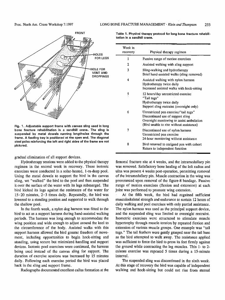
fixation with an intramedullary pin and full cerclage wire (Martin and Ritchie 1994). The bird recovered from isof1urane anesthesia and was placed in a suspended support sling. Five days after surgery, the bird escaped overnight from the support sling and sustained oblique fractures of the left distal radius and ulna. Open reduction of these fractures and internal fixation of the distal ulna were accomplished by placement of an intramedullary pin and cerclage wire. A figure-8 support bandage secured the left wing. A new canvas sling suspended in a remodeled, adjustable support frame was constructed of 2.54- x 2.54-cm, vinyl-coated wire with reinforcing diagonal steel poles (Fig. 1). The support frame was 15.8 em long x 9.5 cm wide X 14.2 em high. The canvas sling was supported in the frame by metal dowels, running lengthwise, which could be adjusted to the appropriate height. A feeding tray similarly supported by adjustable dowels was placed at the open front end of the wire frame. Cloth ties woven through an edge of the canvas sling were stretched across the top of the frame and fastened to the opposite side to secure the bird in position.
One week later, the bird presented severe bilateral leg paresis, uncoordinated movements of the leg muscles, and moderate bilateral digital flexor muscle contraction. Generalized disuse atrophy in the pelvic limbs was apparent. The hocks were hyperextended and the digits were byperflexed. Consequently, passive range of motion exercises (flexion and extension) of all leg joints from toes to hip were performed 3 - 4 times daily to stretch the contracted muscles. The bird could not support its body weight, nor ambulate, and required constant physical support. Twice daily, the bird and canvas sling were lifted from the support frame and, by holding the metal dowels, the bird was "hand-walked" for 30-minute intervals. Improved coordination but not muscle strength was noted in 7 days. Daily periods of graduated exercise and hydrotherapy were initiated to enable the bird to regain muscle strength and locomotor skills and to progress to
Proc. North Am. Crane Workshop 7:1997 LONG BONE FRACTURE MANAGEMENT, Klein alld Thompson 233
FRONT
HOLES ~~~~~nr- FOR LEGS
HOLE FOR VENT AND DROPPINGS
Fig. 1. Adjustable support frame with canvas sling used in long bone fracture rehabilitation in a sandhill crane. The sling is suspended by metal dowels running lengthwIse through the frame. A feeding tray Is positioned at the open end. The diagonal steel poles reinforcing the left and right sides of the frame are not pictured.
gradual elimination of all support devices. Hydrotherapy sessions were added to the physical therapy
regimen in the second week in recovery. These isotonic exercises were conducted in a solar-heated, I-m-deep pool. Using the metal dowels to support the bird in the canvas sling, we "walked" the bird to the pool and then suspended it over the surface of the water with its legs submerged. The bird kicked its legs against the resistance of the water for 15-20 minutes, 2-3 times daily. Repeatedly, the bird was lowered to a standing position and supported to walk through the shallow pool.
In the fourth week, a nylon dog harness was fitted to the bird to act as a support harness during hand-assisted walking periods. The harness was long enough to accommodate the wing position and wide enough to adjust around the keel to the circumference of the body. Assisted walks with this support harness al10wed the bird greater freedom of movement, including opportunities to begin hock-sitting and standing, using secure but minimized handling and support devices. Isotonic pool exercises were continued, the harness being used instead of the canvas sJing for support. The duration of exercise sessions was increased by 15 minutes daily. Following each exercise period the bird was placed back in the sling and support frame.
Radiographs documented excellent callus formation at the
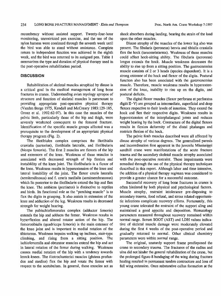
Table 1. Physical therapy protocol for long bone fracture rehablll~ tation in a sandhill crane.
Week in recovery
2
3
Physical therapy regimen
Passive range of motion exercises
Assisted walking with sling support
Sling-walking and hydrotherapy Brief hand~assisted walks (sling removed)
4 Assisted walking with nylon harness Hydrotherapy twice daily Increased assisted walks with hock-sitting
5 12 hours/day unrestricted exercise "Tail tugs"
6
7
8
Hydrotherapy twice daily Support sling restraint (overnight only)
Unrestricted pen exerciseJ"taii tugs" Discontinued use of support sling Overnight monitoring to assist ambulation (Bird unable to rise without assistance)
Discontinued use of nylon harness Unrestricted pen exercise 24~hour monitoring without assistance
Bird returned to assigned pen with cohort Return to independent function
femoral fracture site at 4 weeks, and the intramedullary pin was removed. Satisfactory bone healing of the left radius and ulna was present 4 weeks post-operation, permitting removal of the intramedullary pin. Muscle contraction in the wing was pronounced upon removal of the figure-8 bandage. Passive . range of motion exercises (flexion and extension) at each joint was performed to promote wing extension.
At the fifth week, the bird had regained sufficient musculoskeletal strength and endurance to sustain 12 hours of daily walking and pool exercises with only partial assistance. The nylon harness was used as the principal support device, and the suspended sling was limited to overnight restraint. Isometric exercises were structured to stimulate muscle hypertrophy through muscle tension by repeated flexion and extension of various muscle groups. One example was "tail tugs." The tail feathers were gently grasped near the tail base as the bird attempted to walk away. The resistance applied was sufficient to force the bird to press its feet firmly against the ground while contracting the leg muscles. This 1- to 2-minute exercise was repeated 5 times during a IS-minute interval.
The suspended sling was discontinued in the sixth week. At this stage of recovery the bird was capable of independent walking and hock-sitting but could not rise from sternal
234 LONG BONE FRACTURE MANAGEMENT, Klein and Thompson Proc. North Am. Crane Workshop 7: 1997
recumbency without assisted support. Twenty-four-hour monitoring, unrestricted pen exercise, and the use of the nylon harness were continued through the seventh week until the bird was able to stand without assistance. Complete return to independent function was achieved in the eighth week, and the bird was returned to its assigned pen. Table 1 summarizes the type and duration of physical therapy used in the post-operative rehabilitation period.
DISCUSSION
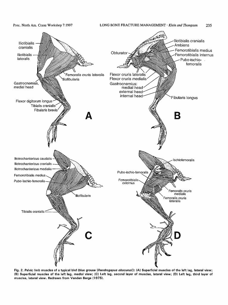
Rehabilitation of skeletal muscles atrophied by disuse is a critical goal in the medical management of long bone fractures in cranes. Understanding avian myology apropos of structure and function of skeletal muscles is paramount to providing appropriate post-operative physical therapy (Vanden Berge 1975, Kendall and McCreary 1983:129-185, Orosz et al. 1992: 62-97). In this case the muscles of the pelvic limb, particularly those of the hip and thigh, were severely weakened consequent to the femoral fracture. Identification of the specific muscle groups affected was a' prerequisite to the development of an appropriate physical therapy program (Fig. 2).
The iliotibiales complex comprises the iliotibialis cranialis (sartorius), iliotibialis lateralis, and iliofibularis (biceps femoris). The first 2 muscles are flexors of the hip and extensors of the knee. Weakness of these muscles is associated with decreased strength of hip flexion and instablility of the knee joint. The iliofibularis is a flexor of the knee. Weakness results in hyperextension of the knee and lateral instability of the joint. The flexor cruris lateralis (semitendinosus) and f. cruris medialis (semimembranosus) which lie posterior to the iliofibularis extend the hip and flex the knee. The ambiens (pectineus) is distinctive to reptiles and birds. Its functional role as the "perching muscle" is to flex the digits in grasping. It also assists in extension of the knee and adduction of the leg. Weakness results in decreased strength for weight bearing.
The pubischiofemorales complex (adductor femoris) extends the hip and adducts the femur. Weakness results in hyperflexion and altered rotator action of the hip. The femorotibialis (quadriceps femoris) is the main extensor of the knee joint and is important in medial rotation of the tibiotarsus. Weakness impairs walking up inclines, stair-type climbing, and rising from a sitting position. The ischiofemoralis and obturator muscles extend the hip and act in lateral rotation of the femur during walking. Weakness causes medial rotation of the hip with a tendency towards knock-knees. The iliotrochanterici muscles (gluteus profundus and medius) flex the hip and rotate the femur with respect to the acetabulum. In general, these muscles act as
shock absorbers during landing, bearing the strain of the load upon the other muscles.
Disuse atrophy of the muscles of the lower leg also was present. The fibularis (peroneus) brevis and tibialis cranialis flex the hock (tarsometatarsus). Weakness of these muscles could affect hock-sitting ability. The fibularis (peroneus) longus extends the hock. Muscle weakness decreases the ability to rise up from a sitting position. The gastrocnemius muscle consists of 2 or 3 heads (species dependent). It is a strong extensor of the hock and flexor of the digits. Postural function also has been associated with the gastrocnemius muscle. Therefore, muscle weakness results in hyperextension of the knee, inability to rise up on the digits, and postural deficits.
The digital flexor muscles (flexor perforans et perforatus digiti 11-V) are grouped as intermediate, superficial and deep flexors respective to their levels of insertion. They extend the hock and flex their respective digits. Weakness results in hyperextension of the interphalangeal joints and reduces weight bearing by the hock. Contracture of the digital flexors results in flexion deformity of the distal phalanges and restricts flexion of the hock.
The pelvic limb muscles described were all affected by disuse atrophy of various degrees. The generalized paresis and incoordination first apparent in the juvenile Mississippi sandhill crane were manifestations of the acute fracture trauma and the secondary skeletal muscle atrophy associated with the post-operative restraint. These impairments were remedied through the use of the physical therapy techniques described in this report. Although labor and time intensive, the addition of a physical therapy regimen was considered to provide a greater chance for a successful outcome.
Successful recovery of long bone fractures in cranes is often hindered by both physical and psychological factors. Muscle atrophy, restraint intolerance pre-disposing to secondary trauma, food refusal, and stress related opportunistic infections complicate recovery efforts. Fortunately, this young crane tolerated the restraint of the support sling and maintained a good appetite and disposition. Hematology parameters measured throughout recovery remained within normal range. Serum SGOT (AST) and LDH values indicative of skeletal muscle damage were moderately elevated during the first 4 weeks of the post-operative period and gradually returned to normal. Other clinical chemistry parameters were within normal range.
The original, unsturdy support frame predisposed the crane to secondary trauma. The fractures of the radius and ulna did not hinder the general rehabilitation of the crane, but the prolonged figure-8 bandaging of the wing during fracture healing resulted in permanent tendon contracture and loss of full wing extension. Once substantive callus formation at the
Proc. North Am. Crane Workshop 7: 1997
lliotibialis cranialis
Iliotibialis lateralis
lIiotrochantericus caudalis
lliotrochantericus cranialis
lIiotrochantericus medialis
Femorotibialis medius
Pubo-ischio-femoralis
Tibialis cranialis ---
A
c
LONG BONE FRACTURE MANAGEMENT, Klein anti Thompson 235
Gastrocnemi us: medial head
external hea internal head
Femorotibialis externus
Femorotibialis medius emorotibialis intern us
Pubo-ischiofemoralis
Fibularis longus
B
Ischiofemoralis
Fig. 2. Pelvic limb muscles of a tvplcal bird (blue grouse [Dendrogapus obscurus]): (AI Superficial muscles of the left leg, lateral view; (B) Superficial muscles of the left leg, medial view; (el left leg, second layer of muscles, lateral view; (0) left leg, third layer of muscles, lateral view. Redrawn from Vanden Berge (1975).
236 LONG BONE FRACTURE MANAGEMENT' Klein and Thompson Proc. North Am. Crane Workshop 7: 1997
fracture site was documented, physical therapy for the wing fracture and more frequent bandage changes may have prevented this disability.
The successful rehabilitation of this bird may be partly attributed to the use of environmental and sensory stimuli to support its psychological well-being. Throughout the recovery period this juvenile crane was housed in a facility that maintained adult sandhill cranes in several adjacent pens. The vocalizations and behaviors of these adults provided appropriate imprinting models. Daily walking exercises took place in an outdoor yard where the young crane could probe the soil, forage. and observe the adu1t cranes in nearby pens. A standard pelleted feed diet was provided ad Jibitum, but curiosity items such as cracked corn and mealworms were often added.
One year later, the crane exhibited normal developmental physical and behavioral characteristics (T. G. Curro, J. Langenberg, and L. Deakin, School Vet. Med., Univ. Wisconsin, Madison, unpubl. data). The bird was wen socialized in a community pen of age-matched cohorts, and demonstrated typical aggressive behavior towards penmates and caretakers. Although the physical therapy program succeeded in a captive management setting, the frequent human interaction during rehabilitation raises concerns of improper imprinting had this crane been a candidate for
release to the wild. Notwithstanding, the use of an incremental physical therapy program can be the pivotal component to the effective medical management of long bone fractures in Gruiformes.
LITERATURE CITED
CARPENTER, J. W. 1986. Cranes (Order Gruiformes). Pages 316-326 in M. E. Fowler, ed. Zoo and wild animal medicine. Second ed. W. B. Saunders Co., Philadelphia, Pa.
CURRO, T. G., J. LANGENBERG, and J. PAUL-MURPHY. 1992. A review of lameness in long-legged birds. Pmc. Annu. Conf. Assoc. Avian Vet. 1992:265-270.
KENDALL, F. P., and E. K. MCCREARY. 1983. Muscles: testing and function. Williams and Wilkins, Baltimore, Md. 451pp.
MARTIN, H., and B. W. RITCHIE. 1994. Orthopedic surgical techniques. Pages 1138-1160 in B. W. Ritchie, G. J. Harrison, and L. R. Harrison, eds. Avian medicine: principles and application. Wingers Publishing, Inc., Lake Worth, Fla.
OLSEN, G. H. 1994. Crane orthopedics: pediatrics and adults. Semin. Avian and Exotic Pet Med. 3:73-80.
OROSZ, S. E., P. K. ENSLEY, and C. J. HAYNES. 1992. Avian surgical anatomy: thoracic and pelvic limbs. W. B. Saunders Co., Philadelphia, Pa. 139pp.
VANDEN BERGE, J. C. 1975. Aves myology. Pages 1829-1843 ill R. G. Getty, ed. Sisson and Grossman's the anatomy of the domestic animals. W. B. Saunders Co., Philadelphia, Pa.