Embed Size (px)
Citation preview
Interventions for treating oral leukoplakia (Review)
Lodi G, Sardella A, Bez C, Demarosi F, Carrassi A
This is a reprint of a Cochrane review, prepared and maintained by The Cochrane Collaboration and published in The Cochrane Library2008, Issue 4
http://www.thecochranelibrary.com
Interventions for treating oral leukoplakia (Review)
Copyright © 2008 The Cochrane Collaboration. Published by John Wiley & Sons, Ltd.
T A B L E O F C O N T E N T S
1HEADER . . . . . . . . . . . . . . . . . . . . . . . . . . . . . . . . . . . . . . .
1ABSTRACT . . . . . . . . . . . . . . . . . . . . . . . . . . . . . . . . . . . . . .
2PLAIN LANGUAGE SUMMARY . . . . . . . . . . . . . . . . . . . . . . . . . . . . . .
2BACKGROUND . . . . . . . . . . . . . . . . . . . . . . . . . . . . . . . . . . . .
Figure 1. . . . . . . . . . . . . . . . . . . . . . . . . . . . . . . . . . . . . . 3
Figure 2. . . . . . . . . . . . . . . . . . . . . . . . . . . . . . . . . . . . . . 4
5OBJECTIVES . . . . . . . . . . . . . . . . . . . . . . . . . . . . . . . . . . . . .
5METHODS . . . . . . . . . . . . . . . . . . . . . . . . . . . . . . . . . . . . . .
6RESULTS . . . . . . . . . . . . . . . . . . . . . . . . . . . . . . . . . . . . . . .
10DISCUSSION . . . . . . . . . . . . . . . . . . . . . . . . . . . . . . . . . . . . .
10AUTHORS’ CONCLUSIONS . . . . . . . . . . . . . . . . . . . . . . . . . . . . . . .
11ACKNOWLEDGEMENTS . . . . . . . . . . . . . . . . . . . . . . . . . . . . . . . .
11REFERENCES . . . . . . . . . . . . . . . . . . . . . . . . . . . . . . . . . . . . .
14CHARACTERISTICS OF STUDIES . . . . . . . . . . . . . . . . . . . . . . . . . . . . .
25DATA AND ANALYSES . . . . . . . . . . . . . . . . . . . . . . . . . . . . . . . . . .
Analysis 1.1. Comparison 1 Topical or systemic treatment versus placebo, Outcome 1 Malignant transformation. . . 25
Analysis 1.2. Comparison 1 Topical or systemic treatment versus placebo, Outcome 2 Oral lesion not completely resolved. 26
Analysis 1.3. Comparison 1 Topical or systemic treatment versus placebo, Outcome 3 Histological features not improved. 27
27APPENDICES . . . . . . . . . . . . . . . . . . . . . . . . . . . . . . . . . . . . .
28WHAT’S NEW . . . . . . . . . . . . . . . . . . . . . . . . . . . . . . . . . . . . .
28HISTORY . . . . . . . . . . . . . . . . . . . . . . . . . . . . . . . . . . . . . . .
28CONTRIBUTIONS OF AUTHORS . . . . . . . . . . . . . . . . . . . . . . . . . . . . .
28DECLARATIONS OF INTEREST . . . . . . . . . . . . . . . . . . . . . . . . . . . . . .
29SOURCES OF SUPPORT . . . . . . . . . . . . . . . . . . . . . . . . . . . . . . . . .
29INDEX TERMS . . . . . . . . . . . . . . . . . . . . . . . . . . . . . . . . . . . .
iInterventions for treating oral leukoplakia (Review)
Copyright © 2008 The Cochrane Collaboration. Published by John Wiley & Sons, Ltd.
[Intervention Review]
Interventions for treating oral leukoplakia
Giovanni Lodi1, Andrea Sardella1, Cristina Bez1, Federica Demarosi1, Antonio Carrassi1
1Oral Pathology and Oral Medicine, University of Milan, Milan, Italy
Contact address: Giovanni Lodi, Oral Pathology and Oral Medicine, University of Milan, Via Beldiletto 1/3, Milan, 20142, Italy.
Editorial group: Cochrane Oral Health Group.
Publication status and date: Edited (no change to conclusions), published in Issue 4, 2008.
Review content assessed as up-to-date: 3 July 2006.
Citation: Lodi G, Sardella A, Bez C, Demarosi F, Carrassi A. Interventions for treating oral leukoplakia. Cochrane Database of SystematicReviews 2006, Issue 4. Art. No.: CD001829. DOI: 10.1002/14651858.CD001829.pub3.
Copyright © 2008 The Cochrane Collaboration. Published by John Wiley & Sons, Ltd.
A B S T R A C T
Background
Oral leukoplakia is a relatively common oral lesion that in a small but significant proportion of cases changes into cancer. Since most
leukoplakias are asymptomatic, the primary objective of treatment should be to prevent such malignant transformation.
Objectives
To assess effectiveness, safety and acceptability of treatments for leukoplakia.
Search strategy
The following databases were searched for relevant trials: Cochrane Oral Health Group’s Trials Register (to April 2006), CENTRAL
(The Cochrane Library 2006, Issue 1), MEDLINE (from 1966 to December 2005), and EMBASE (from 1980 to December 2005).
Handsearching was performed for the main oral medicine journals. References of included studies and reviews were checked. Oral
medicine experts were contacted through an European mailing list (EURORALMED).
Selection criteria
Randomised controlled trials (RCTs), enrolling patients with a diagnosis of oral leukoplakia, were included. Any surgical or medical
(topical and systemic) treatment was included. The primary outcome considered was malignant transformation of leukoplakia. Other
outcomes considered were clinical resolution, histological modification and frequency of adverse effects.
Data collection and analysis
Data were collected using a specific extraction form. Malignant transformation of leukoplakia, demonstrated by histopathological
examination, was the main outcome considered. Secondary outcomes included clinical resolution of the lesion and variation in dysplasia
severity. The validity of included studies was assessed by two review authors, on the basis of the method of allocation concealment,
blindness of the study and loss of participants. Data were analysed by calculating risk ratio. When valid and relevant data were collected,
a meta-analysis of the data was undertaken.
Main results
The possible effectiveness of surgical interventions, including laser therapy and cryotherapy, has never been studied by means of a
RCT with a no treatment/placebo arm. Twenty-five eligible RCTs of non-surgical interventions were identified: 11 were excluded for
different reasons, five were ongoing studies, leaving nine studies to be included in the review (501 patients). Two studies resulted at
low risk of bias, six at moderate risk of bias and one at high risk of bias. Vitamin A and retinoids were tested by five RCTs, two studies
1Interventions for treating oral leukoplakia (Review)
Copyright © 2008 The Cochrane Collaboration. Published by John Wiley & Sons, Ltd.
investigated beta carotene or carotenoids, the other drugs tested were bleomycin (one study), mixed tea (one study) and ketorolac (one
study). One study tested two treatments. Malignant transformation was recorded in just two studies: none of the treatments tested
showed a benefit when compared with the placebo. Treatment with beta carotene, lycopene and vitamin A or retinoids, was associated
with significant rates of clinical resolution, compared with placebo or absence of treatment. Whenever reported, a high rate of relapse
was a common finding. Side effects of variable severity were often described; however, interventions were well accepted by patients,
since drop-out rates were similar between treatment and control groups.
Authors’ conclusions
To date there is no evidence of effective treatment in preventing malignant transformation of leukoplakia. Treatments may be effective
in the resolution of lesion, however relapses and adverse effects are common.
P L A I N L A N G U A G E S U M M A R Y
Interventions for treating oral leukoplakia
No evidence from trials to show how to prevent leukoplakia in the mouth becoming malignant.
Oral leukoplakia is a thickened white patch formed in the mouth lining that cannot be rubbed off. Leukoplakia is a lesion that sometimes
becomes cancerous (a tumour that invades and destroys tissue, then spreads to other areas). Preventing this change is critical as survival
rates of more than 5 years after diagnosis with oral cancer is low. Drugs, surgery and other therapies have been tried. The review of trials
compared several drugs such as bleomycin, vitamin A and beta carotene supplements and mixed tea. There was no evidence found to
show the effects of these treatments. More research is needed.
B A C K G R O U N D
“Oral leukoplakia is a predominantly white lesion of the oral mu-
cosa that cannot be characterised as any other definable lesion” (
Axell 1996). Such a definition, also adopted by the World Health
Organization (WHO), is the result of the effort of an international
group of experts, who met in Uppsala in 1994 in order to review
leukoplakia definitions and classifications on the basis of previ-
ously published work (Axell 1984; Kramer 1978) and new scien-
tific acquisitions. Thus, leukoplakia is a clinical term used when
any other white oral lesion has been excluded. Leukoplakia is often
associated with tobacco smoking, although idiopathic forms are
not rare (Axell 1987). The role of alcohol, viruses and systemic
conditions need further investigations (Campisi 2004; Dietrich
2004).
Clinical variants of leukoplakia are classified into two groups: (1)
homogeneous leukoplakia, a lesion of uniform flat appearance that
may exhibit superficial irregularities, but with consistent texture
throughout (Figure 1); (2) non-homogeneous leukoplakia, a pre-
dominantly white or white and red lesion (erythroleukoplakia)
with an irregular texture that may be characterised by a flat, nodu-
lar or exophytic aspect (Figure 2). Histological features of both
forms of leukoplakia are quite variable and may include ortho- or
para-keratosis of various degrees, mild inflammation and dysplas-
tic changes of various degrees.
2Interventions for treating oral leukoplakia (Review)
Copyright © 2008 The Cochrane Collaboration. Published by John Wiley & Sons, Ltd.
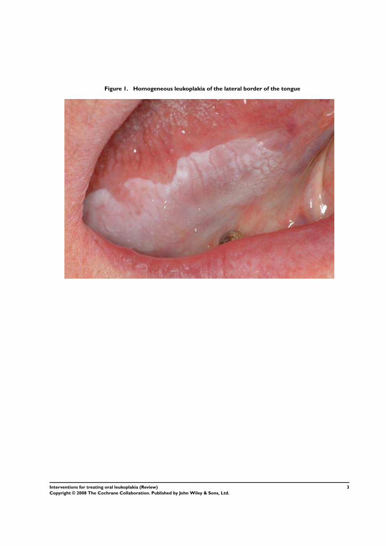
Figure 1. Homogeneous leukoplakia of the lateral border of the tongue
3Interventions for treating oral leukoplakia (Review)
Copyright © 2008 The Cochrane Collaboration. Published by John Wiley & Sons, Ltd.
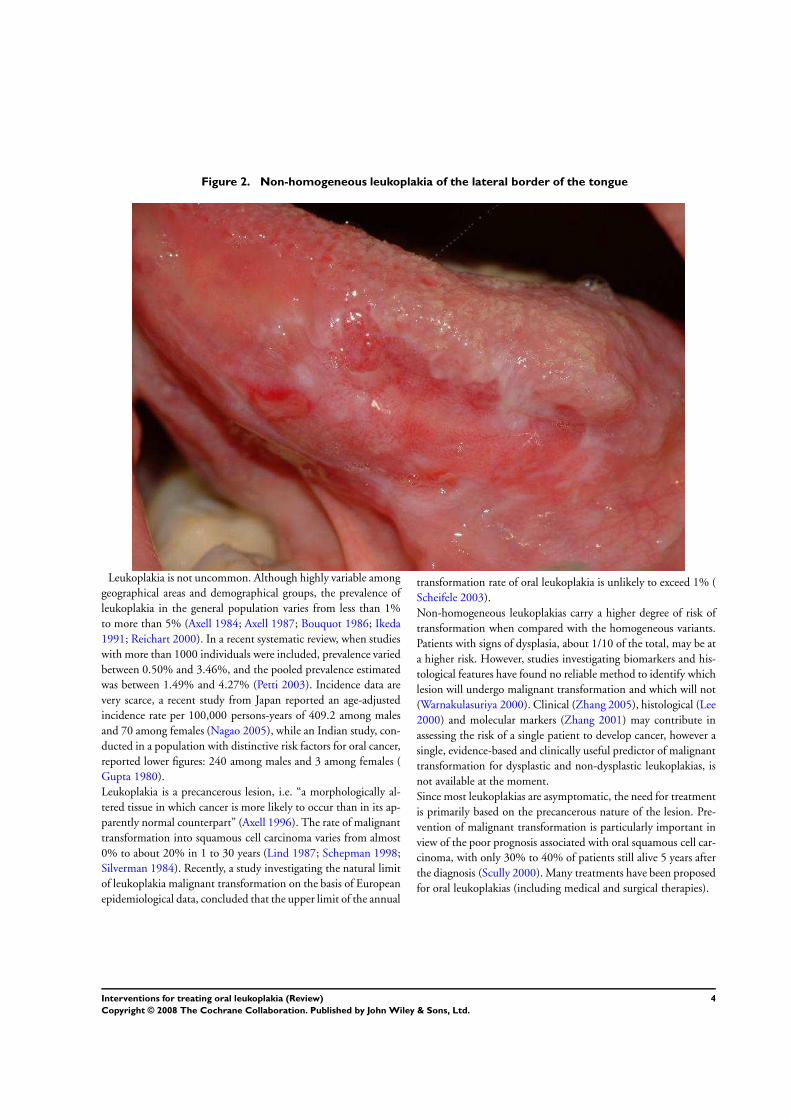
Figure 2. Non-homogeneous leukoplakia of the lateral border of the tongue
Leukoplakia is not uncommon. Although highly variable among
geographical areas and demographical groups, the prevalence of
leukoplakia in the general population varies from less than 1%
to more than 5% (Axell 1984; Axell 1987; Bouquot 1986; Ikeda
1991; Reichart 2000). In a recent systematic review, when studies
with more than 1000 individuals were included, prevalence varied
between 0.50% and 3.46%, and the pooled prevalence estimated
was between 1.49% and 4.27% (Petti 2003). Incidence data are
very scarce, a recent study from Japan reported an age-adjusted
incidence rate per 100,000 persons-years of 409.2 among males
and 70 among females (Nagao 2005), while an Indian study, con-
ducted in a population with distinctive risk factors for oral cancer,
reported lower figures: 240 among males and 3 among females (
Gupta 1980).
Leukoplakia is a precancerous lesion, i.e. “a morphologically al-
tered tissue in which cancer is more likely to occur than in its ap-
parently normal counterpart” (Axell 1996). The rate of malignant
transformation into squamous cell carcinoma varies from almost
0% to about 20% in 1 to 30 years (Lind 1987; Schepman 1998;
Silverman 1984). Recently, a study investigating the natural limit
of leukoplakia malignant transformation on the basis of European
epidemiological data, concluded that the upper limit of the annual
transformation rate of oral leukoplakia is unlikely to exceed 1% (
Scheifele 2003).
Non-homogeneous leukoplakias carry a higher degree of risk of
transformation when compared with the homogeneous variants.
Patients with signs of dysplasia, about 1/10 of the total, may be at
a higher risk. However, studies investigating biomarkers and his-
tological features have found no reliable method to identify which
lesion will undergo malignant transformation and which will not
(Warnakulasuriya 2000). Clinical (Zhang 2005), histological (Lee
2000) and molecular markers (Zhang 2001) may contribute in
assessing the risk of a single patient to develop cancer, however a
single, evidence-based and clinically useful predictor of malignant
transformation for dysplastic and non-dysplastic leukoplakias, is
not available at the moment.
Since most leukoplakias are asymptomatic, the need for treatment
is primarily based on the precancerous nature of the lesion. Pre-
vention of malignant transformation is particularly important in
view of the poor prognosis associated with oral squamous cell car-
cinoma, with only 30% to 40% of patients still alive 5 years after
the diagnosis (Scully 2000). Many treatments have been proposed
for oral leukoplakias (including medical and surgical therapies).
4Interventions for treating oral leukoplakia (Review)
Copyright © 2008 The Cochrane Collaboration. Published by John Wiley & Sons, Ltd.
O B J E C T I V E S
To compare the outcomes for patients affected by oral leukoplakia,
undergoing medical or surgical treatments or both compared with
placebo or no treatment.
M E T H O D S
Criteria for considering studies for this review
Types of studies
Randomised controlled trials (RCTs) and quasi-randomised con-
trolled trials were included.
Types of participants
Anyone with a diagnosis of oral leukoplakia as defined, at the time
of the studies, by the consensus conferences held in 1978, 1983,
1994 (Axell 1984; Axell 1996; Kramer 1978).
Types of interventions
Active
• Surgical removal of the lesion, including surgical excision,
laser surgery, cryotherapy.
• Topical medical treatment, including anti-inflammatory
agents, antimycotic agents, carotenoids and retinoids, cytotoxic
agents, etc..
• Systemic medical treatment.
• Removal of predisposing habits (e.g. tobacco, alcohol, etc.).
• Other treatment (e.g. photodynamic therapy).
• Combined treatment.
Control
• Placebo.
• No treatment.
Types of outcome measures
Primary outcomes
Because of the precancerous nature of leukoplakia, the primary ob-
jective of treatment is to prevent malignant transformation. Thus
leukoplakia morbidity, in terms of frequency of malignant trans-
formation, demonstrated by histopathological examination, was
the main outcome considered.
Secondary outcomes
As epithelial dysplasia may already be present at the time of di-
agnosis, variation in histological features was also included in the
outcome measures.
For interventions directed toward elimination of the lesion, espe-
cially by surgical methods, long term resolution and proportion
of relapsing lesions were considered.
Outcome measures included safety and acceptability of the inter-
vention, as measured by the incidence of adverse effects and pro-
portion of patients dropping out respectively.
Search methods for identification of studies
The databases searched were:
The Cochrane Oral Health Group’s Trials Register (April 2006)
The Cochrane Central Register of Controlled Trials (CENTRAL)
(The Cochrane Library 2006, Issue 1)
MEDLINE (from 1966 to December 2005)
EMBASE (from 1980 to December 2005)
The database of the National Cancer Institute (www.cancer.gov),
previously hosting CancerLit.
Sensitive search strategies were developed for each database (avail-
able from the review authors on request) using a combination of
free text and MeSH terms such as leukoplakia, oral precancer, oral
preneoplastic lesion. The search strategy for CENTRAL is given
as an example in Appendix 1.
The metaRegister of Controlled Trials was searched for relevant
trials (www.controlled-trials.com).
Language: studies in all languages were considered for translation.
Reference lists of included studies and existing reviews were
checked.
In order to detect unpublished and ongoing trials, a formal letter
requesting information was sent to Oral Medicine experts by EU-
RORALMED, an electronic mailing list reaching a vast number
of colleagues, mainly from European countries. Pharmaceutical
companies were also contacted.
The following journals were identified as being important to be
handsearched for the review:
• Cancer• Community Dentistry and Oral Epidemiology• European Journal of Oral Sciences• Journal of Dental Research• Oral Oncology• Oral Surgery, Oral Medicine, Oral Pathology, Oral Radiology
and Endodontics.
The journal issues not already covered by the Cochrane world-
wide handsearching programme (www.cochrane.org) were hand-
searched (January 1999 onwards).
5Interventions for treating oral leukoplakia (Review)
Copyright © 2008 The Cochrane Collaboration. Published by John Wiley & Sons, Ltd.
Data collection and analysis
The title and abstract of each article resulting from the different
search strategies were examined separately by two review authors.
When at least one review author considered the article relevant,
it progressed in the review process and was included in a digital
archive prepared using a dedicated software. Full reports were ob-
tained for all relevant studies.
Critical appraisal of studies
Every study reporting a randomised or quasi-randomised clinical
trial was assessed by two review authors according to the criteria for
randomised trial data suggested by Cochrane Handbook for System-atic Reviews of Interventions 4.2.5 (Higgins 2005) and Evidence-Based Medicine. How to practice and teach EBM (Sackett 1997). In
particular the study’s validity was judged on the basis of.
• Method of allocation concealment. It was considered
’adequate’ when the assignment of patients to treatments was
randomised and the randomisation schedule was kept concealed
to the researcher recruiting participants. When papers did not
report such information, the criterion was considered ’unclear’.
• Protection against performance bias (blindness of the
study). The criterion was considered ’met’ when patients and
researchers assessing outcome measures were kept ’blind’, and
’partially met’ when only one group (patients or researchers) was
kept ’blind’. When blinding was not possible (as in surgical
treatment) this criterion was not considered in the validity rating
of the study.
• Losses of participants. At least 80% of patients who entered
the trial should be included in the final analysis.
Each of these criteria was rated as ’met’, ’partially met’, ’unmet’, or
’unclear’. The global validity of the study was assessed using three
categories.
(1) Low risk of bias: all of the criteria met.
(2) Moderate risk of bias: one or more criteria partially met.
(3) High risk of bias: one or more criteria unmet or unclear.
The critical appraisal of the studies was carried out without blind-
ing the name of authors, institutions and journal. Data about
the study, its eligibility, validity, design and outcome information,
were recorded by each reviewer on an extraction form. In case of
disagreement, consensus was achieved by discussion and a new
form was completed.
Statistical analyses
When valid and relevant data were collected, a meta-analysis of
the data was undertaken. For each intervention, statistical analyses
evaluated the available data on differences among effects in terms
of morbidity (i.e. malignant changes), relapse (for interventions
directed toward elimination of the lesion), adverse effects and pa-
tients dropping out.
The primary measure of intervention effect was reduction of mor-
bidity, as indicated by the difference in incidence of malignant
changes, between the control and intervention groups.
Dichotomous data were expected for the main outcome measure
(cancerization versus absence of cancerization). In case of varia-
tions in histological features, the data were dichotomised in de-
creased severity or no change in histological features versus getting
worse. Other dichotomous data included, incidence of adverse ef-
fects and frequency of drop-out patients.
For each intervention, data on the number of patients of inter-
vention and control groups who experienced the event (outcome)
and the total number of patients, were sought and summarised.
Dichotomous data were analysed by calculating risk ratios. As we
pooled together data from studies in which true treatment effects
are likely to differ, a random-effects model has been used in the
statistical analyses.
Missing data were obtained from tables and graphs or by contact-
ing the authors.
Subgroup analysis was undertaken for class of drug (vitamin A and
retinoids). Subgroup analyses for smoking and non-smoking pa-
tients, and for lesions with or without dysplasia, were not possible
because such data were not available.
A sensitivity analysis was undertaken, excluding studies of lower
methodological quality (i.e. studies at high risk of bias).
R E S U L T S
Description of studies
See: Characteristics of included studies; Characteristics of excluded
studies; Characteristics of ongoing studies.
We found only one randomised controlled trial (RCT) evaluating
surgical interventions (Schwarz 2005), unfortunately it did not
include a no treatment/placebo group, and for this reason it was
excluded from the review (see Criteria for considering studies for
this review). Twenty-five potentially eligible RCTs were identified:
11 were excluded (see Characteristics of excluded studies table) and
five were ongoing studies (Beenken 2000; Boyle 2000; Chiang
2005; Goodin 2005; Lippman 2004), leaving nine studies to be
included in the review. Three of the included studies were a three-
arm trial (Gaeta 2000; Sankaranarayan 1997; Singh 2004), but
in two of them we pooled together the data of the active arms:
in one case the two interventions differ only in lactose content (a
putative non-active component) (Gaeta 2000), in the other the
interventions differ in dosage (Singh 2004).
Four RCTs compared topical treatment versus placebo (total-
ing 100 patients) (Epstein 1994; Gaeta 2000; Mulshine 2004;
Piattelli 1999), four RCTs compared systemic treatment ver-
sus placebo (totaling 327 patients) (Hong 1986; Sankaranarayan
6Interventions for treating oral leukoplakia (Review)
Copyright © 2008 The Cochrane Collaboration. Published by John Wiley & Sons, Ltd.
1997; Singh 2004; Stich 1988). One RCT compared an asso-
ciation of topical and systemic treatments with placebo (64 pa-
tients) (Li 1999). Vitamin A and retinoids were tested by five
RCTs (245 patients) (Gaeta 2000; Hong 1986; Piattelli 1999;
Sankaranarayan 1997; Stich 1988), two studies investigated beta
carotene (Sankaranarayan 1997) and a carotenoid (lycopene) (
Singh 2004). The other drugs tested were bleomycin (Epstein
1994), mixed tea (a mixture of whole water extract of green tea,
green tea polyphenols, and tea pigments in the ratio of 4:1:1) (
Li 1999), and ketorolac (Mulshine 2004). The total number of
patients of the included studies was 501.
In all patients enrolled in the studies a biopsy had been taken to
confirm the diagnosis of leukoplakia, however only two studies
reported the histologic criteria used (Epstein 1994; Stich 1988).
Five studies reported the percentage of dysplastic lesions that varied
from 20% to 59%. In one study dysplastic lesions were in the
treatment arms but not in the control group (Gaeta 2000).
The reported proportion of smoking and drinking patients (the
two main risk factors for oral cancer) varied from 30% to 86%
and from 18% to 86%, respectively. None of the authors reported
significant changes in these habits during the course of the trial.
In two studies (Sankaranarayan 1997; Stich 1988) the totality of
subjects recruited were chewers of tobacco-containing betel quid
(another well known risk factor for oral cancer) from the same
Indian village (Trivandrum, Kerala).
The follow-up period since the end of treatment was reported
in five RCTs (Epstein 1994; Hong 1986; Mulshine 2004;
Sankaranarayan 1997; Singh 2004), varying from 1 to 15 months.
Only two studies reported useful data on cancer development (
Epstein 1994; Sankaranarayan 1997); in Epstein’s trial only part of
the placebo group was taken into account, as 7 out of 12 patients
of this group received the active treatment at the end of the study
period, and thus were excluded from the placebo group for this
outcome.
All the included studies reported clinical changes of leukoplakias.
A complete response was defined as the complete disappearance
of the lesion in seven RCTs; six of them had also the same def-
inition for partial response (greater than 50% reduction), while
one had slightly different criteria (greater than 30% reduction) (
Li 1999). One RCT categorised the response of leukoplakias into
three groups (remission, no change, new leukoplakia) and another
used a three value descriptive clinical scale, in both cases the cri-
teria were not specified. Assessment of the histological modifica-
tions following the treatment was reported by four RCTs (Epstein
1994; Gaeta 2000; Hong 1986; Singh 2004); one RCT investi-
gated some biomarkers of DNA damage and cell proliferation (
Li 1999), but the results of this study were not included as not
comparable with the previous two. A further study reported his-
tological changes in the treatment group only (Stich 1988).
Risk of bias in included studies
On the basis of the criteria used in the critical appraisal of the
studies, two studies resulted in a low risk of bias (Epstein 1994;
Hong 1986). In both studies the methods of allocation conceal-
ment were adequate and reported in detail, and more than 80%
of the patients who entered the study were included in the fi-
nal analysis. Six randomised controlled trials (RCTs) were judged
at moderate risk of bias (Gaeta 2000; Li 1999; Mulshine 2004;
Piattelli 1999; Sankaranarayan 1997; Singh 2004), one of them
was a quasi-randomised trial, while in the other the methods of
allocation concealment were not described. The remaining study
(Stich 1988) was considered at high risk of bias because of the
unclear method of allocation concealment and the absence of pro-
tection against performance bias (blindness of the study).
Effects of interventions
Malignant transformation (Comparison 1, Outcome
1.1)
Results from two studies, including 172 patients, were available
for the analyses (as one of the studies has a three-arm design, the
control group is indicated twice in the plot). Three drugs were eval-
uated in these studies: topical bleomycin (Epstein 1994), systemic
vitamin A (Sankaranarayan 1997) and systemic beta carotene (
Sankaranarayan 1997). None of the treatments in these studies
showed a benefit when compared with the placebo.
Clinical resolution (Comparison 1, Outcome 1.2)
Data on complete resolution of the oral lesions were available
from all the nine studies included in the review (501 patients).
Three treatments (bleomycin, ketorolac and tea) were only as-
sessed in single studies and these treatments showed no bene-
fit when compared to the placebo/control group. Two studies
(Sankaranarayan 1997; Singh 2004) showed a significant ben-
efit for the systemic treatment with beta carotene or lycopene
(a carotenoid) when compared to the control (risk ratio (RR)
Random
= 0.69; 95% confidence interval (CI) 0.54 to 0.88). Five stud-
ies investigated the effectiveness of vitamin A or retinoids (
Gaeta 2000; Hong 1986; Piattelli 1999; Sankaranarayan 1997;
Stich 1988) and found a small but significant benefit (RR
Random
= 0.69; 95% CI 0.50 to 0.96). Two of these studies employed
topical retinoids (Gaeta 2000; Piattelli 1999).
Among patients treated with topical bleomycin (Epstein 1994),
two out of four patients with a complete response, for whom fol-
low-up information was available, relapsed; the same happened
for one out of two patients with a partial response and follow-up
data. Sankaranarayanan’s study reported that 14 out of 22 (64%)
7Interventions for treating oral leukoplakia (Review)
Copyright © 2008 The Cochrane Collaboration. Published by John Wiley & Sons, Ltd.
complete responders of the first arm and 8 out of 15 (54%) com-
plete responders of the second arm developed recurrent lesions (no
information was available regarding the three complete responders
of the placebo group) (Sankaranarayan 1997). Relapses were also
reported by Hong and colleagues: 9 out of 16 (56%) patients re-
sponding to treatment (partially or completely) relapsed (no in-
formation was available regarding the two partial responders of
the placebo group) (Hong 1986). In Piattelli’s study one out of
five (20%) patients responding to the experimental treatment and
one out of four (25%) patients responding to placebo relapsed (
Piattelli 1999). No data on relapses were available for the other
studies.
Histological changes
Histological changes were available from four studies (Epstein
1994; Gaeta 2000; Hong 1986; Singh 2004), but comparison
was possible only for three of them, in fact in Gaeta’s study,
the absence of dysplastic lesions in the control group made im-
possible a comparison. In the other three studies the histolog-
ical aspect of oral lesions did not improve (i.e. was stable or
got worse) more frequently with placebo than with active treat-
ment, and the difference was significant when retinoic acid (RR
Random
= 0.51; 95% CI 0.32 to 0.81) (Hong 1986) or lycopene (RR
Random
= 0.24; 95% CI 0.12 to 0.46) (Singh 2004) were employed.
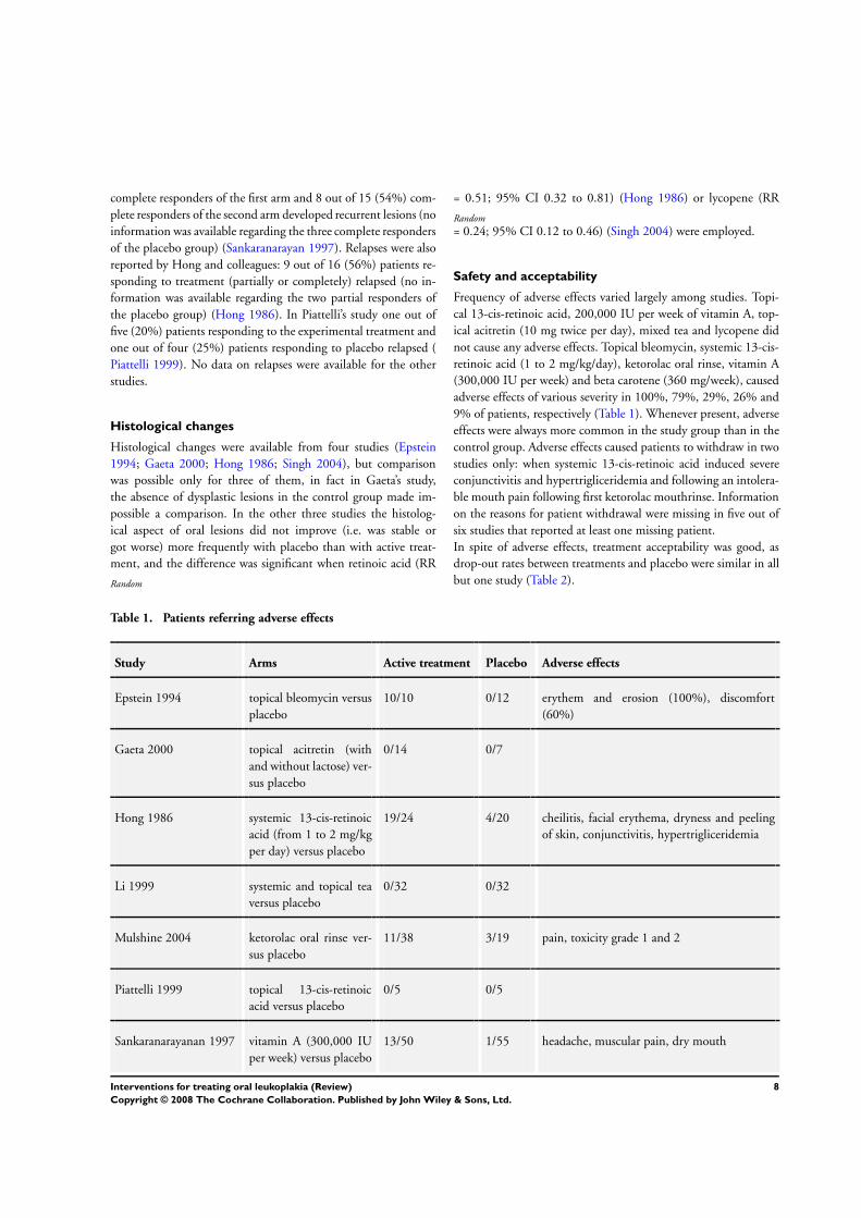
Safety and acceptability
Frequency of adverse effects varied largely among studies. Topi-
cal 13-cis-retinoic acid, 200,000 IU per week of vitamin A, top-
ical acitretin (10 mg twice per day), mixed tea and lycopene did
not cause any adverse effects. Topical bleomycin, systemic 13-cis-
retinoic acid (1 to 2 mg/kg/day), ketorolac oral rinse, vitamin A
(300,000 IU per week) and beta carotene (360 mg/week), caused
adverse effects of various severity in 100%, 79%, 29%, 26% and
9% of patients, respectively (Table 1). Whenever present, adverse
effects were always more common in the study group than in the
control group. Adverse effects caused patients to withdraw in two
studies only: when systemic 13-cis-retinoic acid induced severe
conjunctivitis and hypertrigliceridemia and following an intolera-
ble mouth pain following first ketorolac mouthrinse. Information
on the reasons for patient withdrawal were missing in five out of
six studies that reported at least one missing patient.
In spite of adverse effects, treatment acceptability was good, as
drop-out rates between treatments and placebo were similar in all
but one study (Table 2).
Table 1. Patients referring adverse effects
Study Arms Active treatment Placebo Adverse effects
Epstein 1994 topical bleomycin versus
placebo
10/10 0/12 erythem and erosion (100%), discomfort
(60%)
Gaeta 2000 topical acitretin (with
and without lactose) ver-
sus placebo
0/14 0/7
Hong 1986 systemic 13-cis-retinoic
acid (from 1 to 2 mg/kg
per day) versus placebo
19/24 4/20 cheilitis, facial erythema, dryness and peeling
of skin, conjunctivitis, hypertrigliceridemia
Li 1999 systemic and topical tea
versus placebo
0/32 0/32
Mulshine 2004 ketorolac oral rinse ver-
sus placebo
11/38 3/19 pain, toxicity grade 1 and 2
Piattelli 1999 topical 13-cis-retinoic
acid versus placebo
0/5 0/5
Sankaranarayanan 1997 vitamin A (300,000 IU
per week) versus placebo
13/50 1/55 headache, muscular pain, dry mouth
8Interventions for treating oral leukoplakia (Review)
Copyright © 2008 The Cochrane Collaboration. Published by John Wiley & Sons, Ltd.
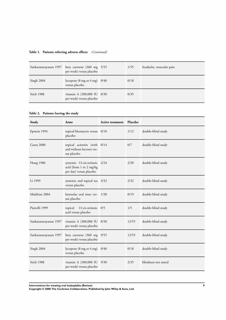
Table 1. Patients referring adverse effects (Continued)
Sankaranarayanan 1997 beta carotene (360 mg
per week) versus placebo
5/55 1/55 headache, muscular pain
Singh 2004 lycopene (8 mg or 4 mg)
versus placebo
0/40 0/18
Stich 1988 vitamin A (200,000 IU
per week) versus placebo
0/30 0/35
Table 2. Patients leaving the study
Study Arms Active treatment Placebo
Epstein 1994 topical bleomycin versus
placebo
0/10 1/12 double-blind study
Gaeta 2000 topical acitretin (with
and without lactose) ver-
sus placebo
0/14 0/7 double-blind study
Hong 1986 systemic 13-cis-retinoic
acid (from 1 to 2 mg/kg
per day) versus placebo
2/24 2/20 double-blind study
Li 1999 systemic and topical tea
versus placebo
3/32 2/32 double-blind study
Mulshine 2004 ketorolac oral rinse ver-
sus placebo
1/38 0/19 double-blind study
Piattelli 1999 topical 13-cis-retinoic
acid versus placebo
0/5 1/5 double-blind study
Sankaranarayanan 1997 vitamin A (300,000 IU
per week) versus placebo
8/50 12/55 double-blind study
Sankaranarayanan 1997 beta carotene (360 mg
per week) versus placebo
9/55 12/55 double-blind study
Singh 2004 lycopene (8 mg or 4 mg)
versus placebo
0/40 0/18 double-blind study
Stich 1988 vitamin A (200,000 IU
per week) versus placebo
9/30 2/35 blindness not stated
9Interventions for treating oral leukoplakia (Review)
Copyright © 2008 The Cochrane Collaboration. Published by John Wiley & Sons, Ltd.
D I S C U S S I O N
Leukoplakia is the most common precancerous oral lesion. Al-
though rates of malignant transformation may vary among stud-
ies, probably due to differences in diagnostic criteria and follow-
up intervals, the morbidity and mortality associated with oral can-
cer make leukoplakia a serious health problem. Nevertheless, we
found only nine studies to include in the present review, none of
these evaluated a surgical intervention. They showed little evidence
for an effective treatment in the prevention of malignant transfor-
mation. There was some evidence that vitamin A, retinoids, beta
carotene and lycopene may completely resolve the oral lesions, and
that retinoic acid and lycopene may promote histological improve-
ment, although this last result is only based on a small number of
patients.
Less than half (33% to 42%) of leukoplakias which undergo ma-
lignant change, does so within 2 years of diagnosis (Lind 1987;
Silverman 1984) and the incidence of malignant transformation
increases with the duration of follow up (Shiu 2000). Therefore,
in order to properly assess modifications in the rates of leukoplakia
malignant transformation, it would be necessary to plan studies
with large groups of patients and a long follow up: that means
multicentre randomised controlled trials (RCTs). However, the
mean follow up of the studies included in the review was no longer
than 15 months, thus the transformation rates could be underes-
timated. Besides, some researchers used outcomes different from
cancer development, in particular various cytological or histolog-
ical markers or both. Although easier to perform, studies using
such outcomes pose a double problem: first there is little evidence
of the predictive value of many of those outcomes; second, they
are hardly comparable. In addition, widespread outcomes such as
dysplasia grade, may be affected by high observer variation (Abbey
1995; Karabulut 1995). The applicability of the results of two of
the studies included (Sankaranarayan 1997; Stich 1988) should
be considered very carefully; in fact the patients included in those
studies were all betel chewers, a risk factor not common in indi-
viduals from geographical areas outside the Indian subcontinent.
Leukoplakias with different histological or molecular character-
istics may have different risks of turning into a cancer. However
the value of the prognostic factors proposed so far in the litera-
ture still need sound confirmatory data. The presence of epithe-
lial dysplasia may be predictive of a transformation to oral cancer
and the risk of cancer incidence may increase with the severity of
dysplastic changes (Lumerman 1995; Schepman 1998), although
this hypothesis has been recently challenged (Holmstrup 2006).
Unfortunately the data available did not allow us to perform a
subgroup analysis of lesions with and without dysplasia, thus it is
not possible to establish if any particular treatment may be more
indicated in the presence of dysplasia of different severity. Many
different molecular biomarkers have been proposed, still no one
seems convincing enough to be applied to clinical routine. Re-
cently, studies on a very promising prognostic marker, cited in the
previous version of this review, are being investigated for accu-
racy (Curfman 2006), following a case of scientific fraud (Horton
2006).
Leukoplakias are not morbid or lethal by themselves and have a
relatively low risk of transformation. As a consequence many sub-
jects receiving treatments have lesions that will never progress to
cancer. For this reason proposed treatments should have minimal
adverse effects in terms of incidence and severity. This is not the
case for some of the interventions evaluated. In particular, high
doses of retinoids may cause toxic effects severe enough to cause
patients to stop treatment. However, in all but one trial the num-
ber of patients leaving the study group was not much bigger than
the number of those leaving the placebo group (and in one study
it was bigger in patients taking the placebo arm than in the two
treatment arms).
It is noteworthy the absence of RCTs comparing the effects of
surgical excision versus no treatment or placebo, surgery being
the first choice in leukoplakia management for many clinicians (
Marley 1998). The only data available are from follow-up studies
comparing rates of malignant transformation in patients who did
and did not undergo surgical treatment of oral leukoplakias. Al-
though results from such studies are hardly comparable because of
differences in diagnostic and inclusion criteria, follow-up interval,
patient characteristics and surgical techniques employed (scalpel,
laser, cryotherapy), they show highly variable results and some-
times are conflicting in the conclusions (Saito 2001; Schepman
1998).
Trials evaluating interventions directed against risk factors (i.e.
smoking) are also missing.
A U T H O R S ’ C O N C L U S I O N SImplications for practice
None of the treatments studied was shown to be effective in pre-
venting malignant transformation of leukoplakias.
Some treatments were effective in healing oral leukoplakia, how-
ever they did not seem able to prevent relapses and malignant
change. For this reason the clinical healing of leukoplakia follow-
ing one of these treatments, does not release the clinician from the
duty of regular follow up.
Implications for research
Although surgery remains the first option for most clinicians, the
real effectiveness of such treatment cannot be assessed, as it was
not possible to find any randomised controlled trials of surgery
10Interventions for treating oral leukoplakia (Review)
Copyright © 2008 The Cochrane Collaboration. Published by John Wiley & Sons, Ltd.
versus no treatment (wait and see) in the prevention of malignant
transformation of leukoplakia, the only randomised controlled
trial comparing two laser techniques. Research is needed to assess
surgery in the treatment of leukoplakia.
Until definitive evidence on the predictive value of specific
biomarkers becomes available, malignant transformation should
be considered the best outcome to take into account in testing the
effectiveness of treatments for leukoplakia.
More research is needed to evaluate the effects of risk factor cessa-
tion on the malignant transformation of leukoplakia.
A C K N O W L E D G E M E N T S
The review authors wish to thank Chiara Procchio for help dur-
ing most of the work. Elena Telaro, Emma Tavender, Helen Wor-
thington and Marco Esposito for their continuous support. All the
researchers of the cited studies who have provided some of the data
used in the review and the referees for their precious suggestions.
R E F E R E N C E S
References to studies included in this review
Epstein 1994 {published and unpublished data}
Epstein JB, Wong FL, Millner A, Le ND. Topical bleomycin
treatment of oral leukoplakia: a randomized double-blind clinical
trial. Head & Neck 1994;16(6):539–44.
Gaeta 2000 {published and unpublished data}
Gaeta GM, Gombos F, Femiano F, Battista C, Minghetti P,
Montanari L, et al.Acitretin and treatment of the oral leucoplakias.
A model to have an active molecules release. Journal of the European
Academy of Dermatology and Venereology 2000;14(6):473–8.
Hong 1986 {published data only}
Hong WK, Endicott J, Itri LM, Doos W, Batsakis JG, Bell R, et
al.13-cis-retinoic acid in the treatment of oral leukoplakia. New
England Journal of Medicine 1986;315(24):1501–5.
Li 1999 {published and unpublished data}
Li N, Sun Z, Han C, Chen J. The chemopreventive effects of tea on
human oral precancerous mucosa lesions. Proceedings of the Society
for Experimental Biology and Medicine 1999;220(4):218–24.
Mulshine 2004 {published data only}
Mulshine JL, Atkinson JC, Greer RO, Papadimitrakopoulou VA,
Van Waes C, Rudy S, et al.Randomized, double-blind, placebo-
controlled phase IIb trial of the cyclooxygenase inhibitor ketorolac
as an oral rinse in oropharyngeal leukoplakia. Clinical Cancer
Research 2004;10(5):1565–73.
Piattelli 1999 {published and unpublished data}
Piattelli A, Fioroni M, Santinelli A, Rubini C. bcl-2 expression and
apoptotic bodies in 13-cis-retinoic acid (isotretinoin)-topically
treated oral leukoplakia: a pilot study. Oral Oncology 1999;35(3):
314–20.
Sankaranarayan 1997 {published data only}
Sankaranarayanan R, Mathew B, Varghese C, Sudhakaran PR,
Menon V, Jayadeep A, et al.Chemoprevention of oral leukoplakia
with vitamin A and beta carotene: an assessment. Oral Oncology
1997;33(4):231–6.
Singh 2004 {published data only}
Singh M, Krishanappa R, Bagewadi A, Keluskar V. Efficacy of oral
lycopene in the treatment of oral leukoplakia. Oral Oncology 2004;
40(6):591–6.
Stich 1988 {published data only}
Stich HF, Hornby AP, Mathew B, Sankaranarayanan R, Nair MK.
Response of oral leukoplakias to the administration of vitamin A.
Cancer Letters 1988;40(1):93–101.
References to studies excluded from this review
Bocharova 2004 {published data only}
Bocharova OA, Pozharitskaya MM, Chekalina TL, Lyzhenkova
MA, Karpova RV, Mezentseva MV, et al.Leukoplakia of oral
mucosa: pathogenesis and possible correction with phytoadaptogen.
Bulletin of Experimental Biology and Medicine 2004;138(6):578–83.
Boisnic 1994 {published data only}
Boisnic S, Branchet MC, Pascal F, Ben Slama L, Rostin M,
Szpirglas H. Topical tretinoin in the treatment of lichen planus and
leukoplakia of the mouth mucosa. A clinical evaluation [Trétinoine
topique dans le traitement des lichens plans et des leucoplasies de la
muqueuse buccale. Evaluation clinique]. [French]. Annals of
Dermatology and Venereology 1994;121(6-7):459–63.
11Interventions for treating oral leukoplakia (Review)
Copyright © 2008 The Cochrane Collaboration. Published by John Wiley & Sons, Ltd.
Chiesa 2005 {published data only}∗ Chiesa F, Tradati N, Grigolato R, Boracchi P, Biganzoli E, Crose
N, et al.Randomized trial of fenretinide (4-HPR) to prevent
recurrences, new localizations and carcinomas in patients operated
on for oral leukoplakia: long-term results. International Journal of
Cancer 2005;115(4):625–9.
Chiesa F, Tradati N, Marazza M, Rossi N, Boracchi P, Mariani L, et
al.Fenretinide (4-HPR) in chemoprevention of oral leukoplakia.
Journal of Cellular Biochemistry Supplement 1993;17F:255–61.
Chiesa F, Tradati N, Marazza M, Rossi N, Boracchi P, Mariani L, et
al.Prevention of local relapses and new localisations of oral
leukoplakias with the synthetic retinoid fenretinide (4-HPR).
Preliminary results. European Journal of Cancer. Part B, Oral
Oncology 1992;28B(2):97–102.
Costa A, Formelli F, Chiesa F, Decensi A, De Palo G, Veronesi U.
Prospects of chemoprevention of human cancers with the synthetic
retinoid fenretinide. Cancer Research 1994;54(7 Suppl):2032s–7s.
De Palo G, Veronesi U, Marubini E, Camerini T, Chiesa F, Nava
M, et al.Controlled clinical trials with fenretinide in breast cancer,
basal cell carcinoma and oral leukoplakia. Journal of Cellular
Biochemistry Supplement 1995;22:11–7.
Femiano 2001 {published data only}
Femiano F, Gombos F, Scully C, Battista C, Belnome G, Esposito
V. Oral leukoplakia: open trial of topical therapy with calcipotriol
compared with tretinoin. International Journal of Oral and
Maxillofacial Surgery 2001;30(5):402–6.
Garewal 1999 {published data only}
Garewal H, Pitcock J, Friedman S, Alberts D, Meyskens F, Ramsey
L, et al.Beta-carotene in oral leukoplakia. Proceedings, Annual
Meeting of the American Society of Clinical Oncology 1992;11:141.∗ Garewal HS, Katz RV, Meyskens F, Pitcock J, Morse D, Friedman
S, et al.Beta-carotene produces sustained remissions in patients with
oral leukoplakia: results of a multicenter prospective trial. Archives
of Otolaryngology - Head and Neck Surgery 1999;125(12):1305–10.
Krishnaswamy 1995 {published data only}
Krishnaswamy K, Prasad MP, Krishna TP, Annapurna VV, Reddy
GA. A case study of nutrient intervention of oral precancerous
lesions in India. European Journal of Cancer. Part B: Oral Oncology
1995;31B(1):41–8.
Lippman 1993 {published data only}∗ Lippman SM, Batsakis JG, Toth BB, Weber RS, Lee JJ, Martin
JW, et al.Comparison of low-dose isotretinoin with beta carotene to
prevent oral carcinogenesis. New England Journal of Medicine 1993;
328(1):15–20.
Papadimitrakopoulou VA, Lippman SM, Lee JS, Toth BB, Martin
JW, Lee JJ, et al.Long term follow-up of low-dose isotretinoin (13-
cRA) versus beta carotene to prevent oral carcinogenesis.
Proceedings, Annual Meeting of the American Society of Clinical
Oncology 1996;15:A340.
Mathew 1995 {published data only}
Mathew B, Sankaranarayanan R, Nair PP, Varghese C, Somanathan
T, Amma BP, et al.Evaluation of chemoprevention of oral cancer
with Spirulina fusiformis. Nutrition and Cancer 1995;24(2):
197–202.
Schwarz 2005 {published data only}
Schwarz F, Maraki D, Yalcinkaya S, Bieling K, Bocking A, Becker J.
Cytologic and DNA-cytometric follow-up of oral leukoplakia after
CO2- and Er:YAG-laser assisted ablation: a pilot study. Lasers in
Surgery and Medicine 2005;37(1):29–36.
Sun 1996 {published data only}
Sun Z, Shen S, Liu-X. [Treatment of oral leukoplakia with
retinamide]. [Chinese]. Chung-Hua-Kou-Chiang-Hsueh-Tsa-Chih
1996;31(3):185–7.
Zaridze 1993 {published data only}
Zaridze D, Evstifeeva T, Boyle P. Chemoprevention of oral
leukoplakia and chronic esophagitis in an area of high incidence of
oral and esophageal cancer. Annals of Epidemiology 1993;3(3):
225–34.
References to ongoing studies
Beenken 2000 {published data only}
Beenken SW. Fenretinide in treating patients with leukoplakia of
the mouth. www.controlled-trials.com/mrct/trial/NCT00004161/
1059/60505.html (2000). [: NLM identifier NCT00004161]
Boyle 2000 {unpublished data only}
Boyle J. Celecoxib in treating patients with precancerous lesions of
the mouth. www.controlled-trials.com/mrct/trial/NCT00014404/
1059/60207.html (2000). [: NLM Identifier NCT00014404]
Chiang 2005 {published data only}
Chiang C-P. Photodynamic therapy for oral leukoplakia and
erythroleukoplakia. www.controlled-trials.com/mrct/trial/
NCT00155337/1059/125975.html (2005). [: NCT00155337]
Goodin 2005 {published data only}
Goodin S. A phase II trial to assess the effects of green tea in oral
leukoplakia. www.controlled-trials.com/mrct/trial/NCT00176566/
1059/126861.html (2005).
Lippman 2004 {published data only}
Lippman SM, Sudbo J. Celecoxib and/or erlotinib in preventing
oral cancer in patients with oral leukoplakia. www.controlled-
trials.com/mrct/trial/NCT00088842/1059/59769.html (2004). [:
NCT00088842]
Additional references
Abbey 1995
Abbey LM, Kaugars GE, Gunsolley JC, Burns JC, Page DG,
Svirsky JA, et al.Intraexaminer and interexaminer reliability in the
diagnosis of oral epithelial dysplasia. Oral Surgery, Oral Medicine,
Oral Pathology, Oral Radiology and Endodontics 1995;80(2):188–91.
Axell 1984
Axell T, Holmstrup P, Kramer IRH, Pindborg JJ. International
seminar on oral leukoplakia and associated lesions related to
tobacco habits. Community Dentistry and Oral Epidemiology 1984;
12:145–54.
Axell 1987
Axell T. Occurrence of leukoplakia and some other oral white
lesions among 20,333 adult Swedish people. Community Dentistry
and Oral Epidemiology 1987;15(1):46–51.
12Interventions for treating oral leukoplakia (Review)
Copyright © 2008 The Cochrane Collaboration. Published by John Wiley & Sons, Ltd.
Axell 1996
Axell T, Pindborg JJ, Smith CJ, van der Waal I. Oral white lesions
with special reference to precancerous and tobacco- related lesions:
conclusions of an international symposium held in Uppsala,
Sweden, May 18-21 1994. International Collaborative Group on
Oral White Lesions. Journal of Oral Pathology & Medicine 1996;25
(2):49–54. [MEDLINE: 1996237276]
Bouquot 1986
Bouquot JE, Gorlin RJ. Leukoplakia, lichen planus, and other oral
keratoses in 23,616 white Americans over the age of 35 years. Oral
Surgery, Oral Medicine and Oral Pathology 1986;61(4):373–81.
Campisi 2004
Campisi G, Giovannelli L, Arico P, Lama A, Di Liberto C,
Ammatuna P, et al.HPV DNA in clinically different variants of oral
leukoplakia and lichen planus. Oral Surgery, Oral Medicine, Oral
Pathology, Oral Radiology, and Endodontics 2004;98(6):705–11.
Curfman 2006
Curfman GD, Morrissey S, Drazen JM. Expression of concern:
Sudbo J et al. DNA content as a prognostic marker in patients with
oral leukoplakia. N Engl J Med 2001;344:1270-8 and Sudbo J et
al. the influence of resection and aneuploidy on mortality in oral
leukoplakia. N Engl J Med 2004;350:1405-13. The New England
Journal of Medicine 2006;354(6):638.
Dietrich 2004
Dietrich T, Reichart PA, Scheifele C. Clinical risk factors of oral
leukoplakia in a representative sample of the US population. Oral
Oncology 2004;40(2):158–63.
Greenhalgh 1997
Greenhalgh T. How to read a paper. The Medline database. BMJ
1997;315(7101):180–3. [MEDLINE: 1997395413]
Gupta 1980
Gupta PC, Mehta FS, Daftary DK, Pindborg JJ, Bhonsle RB,
Jalnawalla PN, et al.Incidence rates of oral cancer and natural
history of oral precancerous lesions in a 10-year follow-up study of
Indian villagers. Community Dentistry and Oral Epidemiology 1980;
8(6):283–33.
Higgins 2005
Higgins JPT, Green S, editors. Assessment of study quality.
Cochrane Handbook for Systematic Reviews of Interventions 4.2.5
[updated May 2005]; Section 6. The Cochrane Library, Issue 3.
Chichester, UK: John Wiley & Sons, Ltd, 2005.
Holmstrup 2006
Holmstrup P, Vedtofte P, Reibel J, Stoltze K. Long-term treatment
outcome of oral premalignant lesions. Oral Oncology 2006;42(5):
461–74.
Horton 2006
Horton R. Retraction--Non-steroidal anti-inflammatory drugs and
the risk of oral cancer: a nested case-control study. Lancet 2006;
367(9508):382.
Ikeda 1991
Ikeda N, Ishii T, Iida S, Kawai T. Epidemiological study of oral
leukoplakia based on mass screening for oral mucosal diseases in a
selected Japanese population. Community Dentistry and Oral
Epidemiology 1991;19(3):160–3.
Karabulut 1995
Karabulut A, Reibel J, Therkildsen MH, Praetorius F, Nielesen
HW, Dabelsteen E. Observer variability in the histologic assessment
of oral premalignant lesions. Journal of Oral Pathology Medicine
1995;24(5):198–200.
Kramer 1978
Kramer IR, Lucas RB, Pindborg JJ, Sobin LH. Definition of
leukoplakia and related lesions: an aid to studies on oral precancer.
Oral Surgery, Oral Medicine and Oral Pathology 1978;46(4):518–39.
[MEDLINE: 1979053956]
Lee 2000
Lee JJ, Hong WK, Hittelman WN, Mao L, Lotan R, Shin DM, et
al.Predicting cancer development in oral leukoplakia: ten years of
translational research. Clinical Cancer Research 2000;6(5):1702–10.
Lind 1987
Lind PO. Malignant transformation in oral leukoplakia.
Scandinavian Journal of Dental Research 1987;95(6):449–55.
Lumerman 1995
Lumerman H, Freedman P, Kerpel S. Oral epithelial dysplasia and
the development of invasive squamous cell carcinoma. Oral Surgery,
Oral Medicine, Oral Pathology, Oral Radiology and Endodontics
1995;79(3):321–9.
Marley 1998
Marley JJ, Cowan CG, Lamey PJ, Linden GJ, Johnson NW,
Warnakulasuriya KA. Management of potentially malignant oral
mucosal lesions by consultant UK oral and maxillofacial surgeons.
British Journal of Oral and Maxillofacial Surgery 1998;34(1):28–36.
Nagao 2005
Nagao T, Ikeda N, Fukano H, Hashimoto S, Shimozato K,
Warnakulasuriya S. Incidence rates for oral leukoplakia and lichen
planus in a Japanese population. Journal of Oral Pathology and
Medicine 2005;34(9):532–9.
Petti 2003
Petti S. Pooled estimate of world leukoplakia prevalence: a
systematic review. Oral Oncology 2003;39:770–80.
Reichart 2000
Reichart PA. Oral mucosal lesions in a representative cross-sectional
study of aging Germans. Community Dentistry and Oral
Epidemiology 2000;28(5):390–8.
Sackett 1997
Sackett DL, Richardson WS, Rosenberg W, Haynes RB. Critically
appraising the evidence. In: Sackett DL, Richardson WS,
Rosenberg W, Haynes RB editor(s). Evidence-based medicine. How
to practice and teach EBM. Churchill Livingstone, 1997:79–156.
Saito 2001
Saito T, Sugiura C, Hirai A, Notani K, Totsuka Y, Shindoh M, et
al.Development of squamous cell carcinoma from pre-existent oral
leukoplakia: with respect to treatment modality. International
Journal of Oral and Maxillofacial Surgery 2001;30(1):49–53.
Scheifele 2003
Scheifele C, Reichart PA. Is there a natural limit of the
transformation rate of oral leukoplakia?. Oral Oncology 2003;39(5):
470–5.
13Interventions for treating oral leukoplakia (Review)
Copyright © 2008 The Cochrane Collaboration. Published by John Wiley & Sons, Ltd.
Schepman 1998
Schepman KP, van der Meij EH, Smeele LE, van der Waal I.
Malignant transformation of oral leukoplakia: a follow-up study of
a hospital-based population of 166 patients with oral leukoplakia
from The Netherlands. Oral Oncology 1998;34(4):270–5.
Scully 2000
Scully C, Porter S. ABC of oral health. Oral cancer. BMJ 2000;321
(7253):97–100.
Shiu 2000
Shiu MN, Chen TH, Chang SH, Hahn LJ. Risk factors for
leukoplakia and malignant transformation to oral carcinoma: a
leukoplakia cohort in Taiwan. British Journal of Cancer 2000;82
(11):1871–4.
Silverman 1984
Silverman S Jr, Gorsky M, Lozada F. Oral leukoplakia and
malignant transformation. A follow-up study of 257 patients.
Cancer 1984;53(3):563–8.
Warnakulasuriya 2000
Warnakulasuriya S. Lack of molecular markers to predict malignant
potential of oral precancer. Journal of Pathology 2000;190(4):
407–9.
Zhang 2001
Zhang L, Rosin MP. Loss of heterozygosity: a potential tool in
management of oral premalignant lesions?. Journal of Oral
Pathology and Medicine 2001;30(9):513–20.
Zhang 2005
Zhang L, Williams M, Poh CF, Laronde D, Epstein JB, Durham S,
et al.Toluidine blue staining identifies high-risk primary oral
premalignant lesions with poor outcome. Cancer Research 2005;65
(17):8017–21.
References to other published versions of this review
Lodi 2002
Lodi G, Sardella A, Bez C, Demarosi F, Carrassi A. Systematic
review of randomized trials for the treatment of oral leukoplakia.
Journal of Dental Education 2002;66(8):896–902.
Lodi 2004
Lodi G, Sardella A, Bez C, Demarosi F, Carrassi A. Interventions
for treating oral leukoplakia. Cochrane Database of Systematic
Reviews 2004., Issue 3. [Art. No.: CD001829. DOI: 10.1002/
14651858.CD001829.pub2]∗ Indicates the major publication for the study
14Interventions for treating oral leukoplakia (Review)
Copyright © 2008 The Cochrane Collaboration. Published by John Wiley & Sons, Ltd.
C H A R A C T E R I S T I C S O F S T U D I E S
Characteristics of included studies [ordered by study ID]
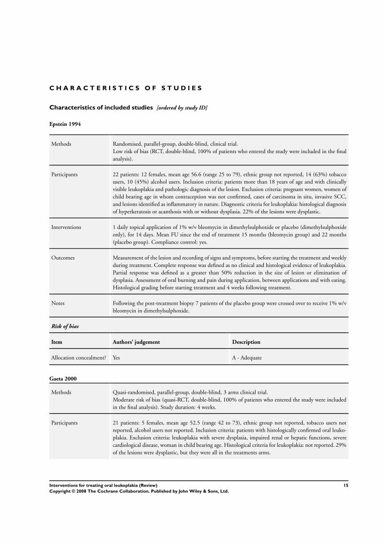
Epstein 1994
Methods Randomised, parallel-group, double-blind, clinical trial.
Low risk of bias (RCT, double-blind, 100% of patients who entered the study were included in the final
analysis).
Participants 22 patients: 12 females, mean age 56.6 (range 25 to 79), ethnic group not reported, 14 (63%) tobacco
users, 10 (45%) alcohol users. Inclusion criteria: patients more than 18 years of age and with clinically
visible leukoplakia and pathologic diagnosis of the lesion. Exclusion criteria: pregnant women, women of
child bearing age in whom contraception was not confirmed, cases of carcinoma in situ, invasive SCC,
and lesions identified as inflammatory in nature. Diagnostic criteria for leukoplakia: histological diagnosis
of hyperkeratosis or acanthosis with or without dysplasia. 22% of the lesions were dysplastic.
Interventions 1 daily topical application of 1% w/v bleomycin in dimethylsulphoxide or placebo (dimethylsulphoxide
only), for 14 days. Mean FU since the end of treatment 15 months (bleomycin group) and 22 months
(placebo group). Compliance control: yes.
Outcomes Measurement of the lesion and recording of signs and symptoms, before starting the treatment and weekly
during treatment. Complete response was defined as no clinical and histological evidence of leukoplakia.
Partial response was defined as a greater than 50% reduction in the size of lesion or elimination of
dysplasia. Assessment of oral burning and pain during application, between applications and with eating.
Histological grading before starting treatment and 4 weeks following treatment.
Notes Following the post-treatment biopsy 7 patients of the placebo group were crossed over to receive 1% w/v
bleomycin in dimethylsulphoxide.
Risk of bias
Item Authors’ judgement Description
Allocation concealment? Yes A - Adequate
Gaeta 2000
Methods Quasi-randomised, parallel-group, double-blind, 3 arms clinical trial.
Moderate risk of bias (quasi-RCT, double-blind, 100% of patients who entered the study were included
in the final analysis). Study duration: 4 weeks.
Participants 21 patients: 5 females, mean age 52.5 (range 42 to 73), ethnic group not reported, tobacco users not
reported, alcohol users not reported. Inclusion criteria: patients with histologically confirmed oral leuko-
plakia. Exclusion criteria: leukoplakia with severe dysplasia, impaired renal or hepatic functions, severe
cardiological disease, woman in child bearing age. Histological criteria for leukoplakia: not reported. 29%
of the lesions were dysplastic, but they were all in the treatments arms.
15Interventions for treating oral leukoplakia (Review)
Copyright © 2008 The Cochrane Collaboration. Published by John Wiley & Sons, Ltd.
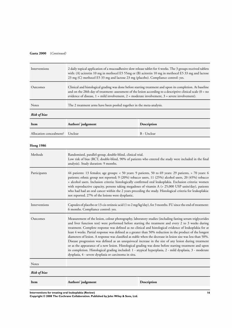
Gaeta 2000 (Continued)
Interventions 2 daily topical application of a mucoadhesive slow release tablet for 4 weeks. The 3 groups received tablets
with: (A) acitretin 10 mg in methocel E5 55mg or (B) acitretin 10 mg in methocel E5 33 mg and lactose
23 mg (C) methocel E5 33 mg and lactose 23 mg (placebo). Compliance control: yes.
Outcomes Clinical and histological grading was done before starting treatment and upon its completion. At baseline
and on the 28th day of treatment: assessment of the lesion according to a descriptive clinical scale (0 = no
evidence of disease, 1 = mild involvement, 2 = moderate involvement, 3 = severe involvement).
Notes The 2 treatment arms have been pooled together in the meta-analysis.
Risk of bias
Item Authors’ judgement Description
Allocation concealment? Unclear B - Unclear
Hong 1986
Methods Randomised, parallel-group, double-blind, clinical trial.
Low risk of bias (RCT, double-blind, 90% of patients who entered the study were included in the final
analysis). Study duration: 9 months.
Participants 44 patients: 13 females; age groups: < 50 years: 9 patients, 50 to 69 years: 29 patients, > 70 years: 6
patients; ethnic group not reported; 9 (20%) tobacco users, 11 (25%) alcohol users, 20 (45%) tobacco
+ alcohol users. Inclusion criteria: histologically confirmed oral leukoplakia. Exclusion criteria: women
with reproductive capacity, persons taking megadoses of vitamin A (> 25,000 USP units/day), patients
who had had an oral cancer within the 2 years preceding the study. Histological criteria for leukoplakia:
not reported. 27% of the lesions were dysplastic.
Interventions Capsules of placebo or 13-cis-retinoic acid (1 to 2 mg/kg/day), for 3 months. FU since the end of treatment:
6 months. Compliance control: yes.
Outcomes Measurement of the lesion, colour photography, laboratory studies (including fasting serum triglycerides
and liver function test) were performed before starting the treatment and every 2 to 3 weeks during
treatment. Complete response was defined as no clinical and histological evidence of leukoplakia for at
least 4 weeks. Partial response was defined as a greater than 50% reduction in the product of the longest
diameters of lesion. A response was classified as stable when the decrease in lesion size was less than 50%.
Disease progression was defined as an unequivocal increase in the size of any lesion during treatment
or as the appearance of a new lesion. Histological grading was done before starting treatment and upon
its completion. Histological grading included: 1 - atypical hyperplasia, 2 - mild dysplasia, 3 - moderate
dysplasia, 4 - severe dysplasia or carcinoma in situ.
Notes
Risk of bias
Item Authors’ judgement Description
16Interventions for treating oral leukoplakia (Review)
Copyright © 2008 The Cochrane Collaboration. Published by John Wiley & Sons, Ltd.

Hong 1986 (Continued)
Allocation concealment? Yes A - Adequate
Li 1999
Methods Randomised, parallel-group, double-blind, clinical trial.
Moderate risk of bias (unclear methods of allocation, double-blind, 92% of patients who entered the
study were included in the final analysis).
Participants 64 patients: 24 females, mean age 54.5 (range 23 to 78), ethnic group not reported, 46 (71.9%) tobacco
users. Histological criteria for leukoplakia: not reported. 20% of the lesions were dysplastic.
Interventions Systemic (capsules) and topic (paint) placebo or systemic (capsules) and topic (paint) mixed tea (3 g/day
and 3 paintings/day), for 6 months. Compliance control: no.
Outcomes Size and number of lesions of each subject were recorded at the baseline and at the end of the trial. Oral
biopsies were conducted at the beginning and at the end of the trial. Besides routine histopathological
examination lesional tissue investigations included also silver stained nucleolar organizer regions (Ag-
NOR), proliferation cell nuclear antigen (PCNA) and epidermal growth factor receptor (EGFR) analysis.
A complete regression was defined as the complete disappearance of the lesion. A partial regression was
defined as a 30% or more reduction in the size of a single lesion or in the sum of sizes of multiple lesions.
Lesions with no change in size were recorded as no change. Deterioration referred to the occurrence of
new lesions.
Notes
Risk of bias
Item Authors’ judgement Description
Allocation concealment? Unclear B - Unclear
Mulshine 2004
Methods Randomised, parallel-group, double-blind, clinical trial.
Moderate risk of bias (unclear methods of allocation, double-blind, 98% of patients who entered the
study were included in the final analysis).
Participants 57 patients: 19 females; age not reported; ethnic group: non white subjects: 6/57, white subjects: 51/57;
48/56 (86%) smokers, 40/56 (86%) alcohol users. Inclusion criteria: subjects with bidimensionally mea-
surable leukoplakia of the oral cavity or of the oral pharynx. In case of previous oral cancer diagnosis,
individuals had to be free from diseases for at least 3 months, excellent performance status, general good
health. Exclusion criteria: hypersensitivity to aspirin, lidocaine, NSAIDs, retinoids. Use of antibiotics,
steroids, NSAIDs, aspirin, probenecid, antihistamines for least > 10 consecutive days or any immuno-
suppressants, anticoagulants, dilantin, lithium, methotrexate, phenothiazines, or drugs that could com-
promise the test product safety during the 30 days immediately preceding the first treatment visit, debil-
itating oral conditions requiring extensive dental procedures or conditions interfering with compliance.
Respiratory or cardiovascular problems. Histological criteria for leukoplakia: not reported. Percentage of
17Interventions for treating oral leukoplakia (Review)
Copyright © 2008 The Cochrane Collaboration. Published by John Wiley & Sons, Ltd.
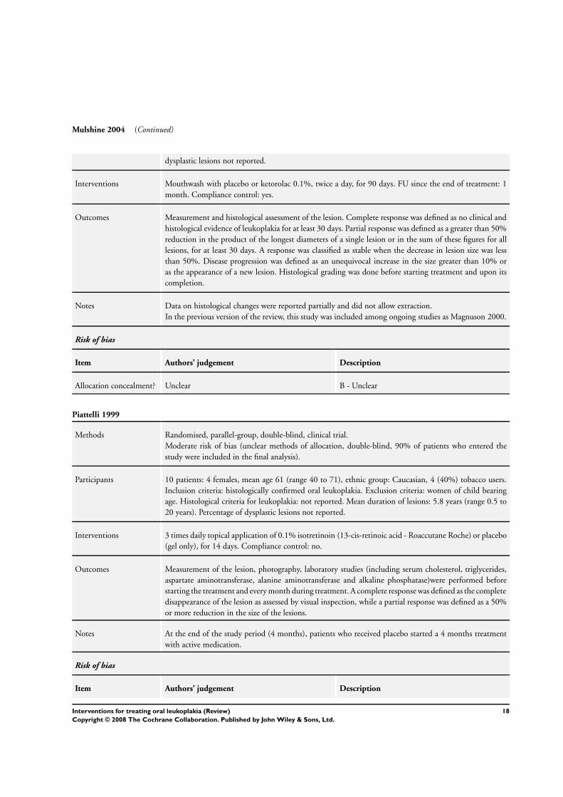
Mulshine 2004 (Continued)
dysplastic lesions not reported.
Interventions Mouthwash with placebo or ketorolac 0.1%, twice a day, for 90 days. FU since the end of treatment: 1
month. Compliance control: yes.
Outcomes Measurement and histological assessment of the lesion. Complete response was defined as no clinical and
histological evidence of leukoplakia for at least 30 days. Partial response was defined as a greater than 50%
reduction in the product of the longest diameters of a single lesion or in the sum of these figures for all
lesions, for at least 30 days. A response was classified as stable when the decrease in lesion size was less
than 50%. Disease progression was defined as an unequivocal increase in the size greater than 10% or
as the appearance of a new lesion. Histological grading was done before starting treatment and upon its
completion.
Notes Data on histological changes were reported partially and did not allow extraction.
In the previous version of the review, this study was included among ongoing studies as Magnuson 2000.
Risk of bias
Item Authors’ judgement Description
Allocation concealment? Unclear B - Unclear
Piattelli 1999
Methods Randomised, parallel-group, double-blind, clinical trial.
Moderate risk of bias (unclear methods of allocation, double-blind, 90% of patients who entered the
study were included in the final analysis).
Participants 10 patients: 4 females, mean age 61 (range 40 to 71), ethnic group: Caucasian, 4 (40%) tobacco users.
Inclusion criteria: histologically confirmed oral leukoplakia. Exclusion criteria: women of child bearing
age. Histological criteria for leukoplakia: not reported. Mean duration of lesions: 5.8 years (range 0.5 to
20 years). Percentage of dysplastic lesions not reported.
Interventions 3 times daily topical application of 0.1% isotretinoin (13-cis-retinoic acid - Roaccutane Roche) or placebo
(gel only), for 14 days. Compliance control: no.
Outcomes Measurement of the lesion, photography, laboratory studies (including serum cholesterol, triglycerides,
aspartate aminotransferase, alanine aminotransferase and alkaline phosphatase)were performed before
starting the treatment and every month during treatment. A complete response was defined as the complete
disappearance of the lesion as assessed by visual inspection, while a partial response was defined as a 50%
or more reduction in the size of the lesions.
Notes At the end of the study period (4 months), patients who received placebo started a 4 months treatment
with active medication.
Risk of bias
Item Authors’ judgement Description
18Interventions for treating oral leukoplakia (Review)
Copyright © 2008 The Cochrane Collaboration. Published by John Wiley & Sons, Ltd.
Piattelli 1999 (Continued)
Allocation concealment? Unclear B - Unclear
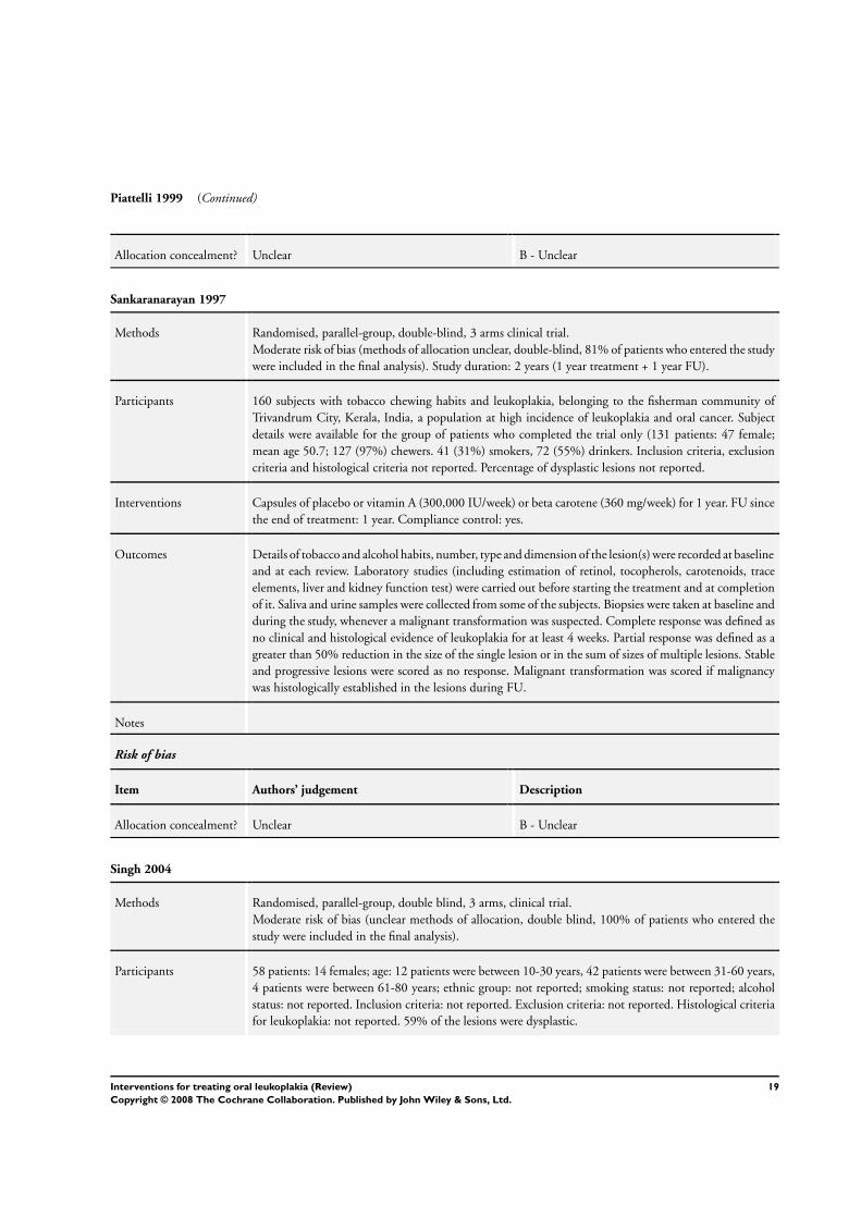
Sankaranarayan 1997
Methods Randomised, parallel-group, double-blind, 3 arms clinical trial.
Moderate risk of bias (methods of allocation unclear, double-blind, 81% of patients who entered the study
were included in the final analysis). Study duration: 2 years (1 year treatment + 1 year FU).
Participants 160 subjects with tobacco chewing habits and leukoplakia, belonging to the fisherman community of
Trivandrum City, Kerala, India, a population at high incidence of leukoplakia and oral cancer. Subject
details were available for the group of patients who completed the trial only (131 patients: 47 female;
mean age 50.7; 127 (97%) chewers. 41 (31%) smokers, 72 (55%) drinkers. Inclusion criteria, exclusion
criteria and histological criteria not reported. Percentage of dysplastic lesions not reported.
Interventions Capsules of placebo or vitamin A (300,000 IU/week) or beta carotene (360 mg/week) for 1 year. FU since
the end of treatment: 1 year. Compliance control: yes.
Outcomes Details of tobacco and alcohol habits, number, type and dimension of the lesion(s) were recorded at baseline
and at each review. Laboratory studies (including estimation of retinol, tocopherols, carotenoids, trace
elements, liver and kidney function test) were carried out before starting the treatment and at completion
of it. Saliva and urine samples were collected from some of the subjects. Biopsies were taken at baseline and
during the study, whenever a malignant transformation was suspected. Complete response was defined as
no clinical and histological evidence of leukoplakia for at least 4 weeks. Partial response was defined as a
greater than 50% reduction in the size of the single lesion or in the sum of sizes of multiple lesions. Stable
and progressive lesions were scored as no response. Malignant transformation was scored if malignancy
was histologically established in the lesions during FU.
Notes
Risk of bias
Item Authors’ judgement Description
Allocation concealment? Unclear B - Unclear
Singh 2004
Methods Randomised, parallel-group, double blind, 3 arms, clinical trial.
Moderate risk of bias (unclear methods of allocation, double blind, 100% of patients who entered the
study were included in the final analysis).
Participants 58 patients: 14 females; age: 12 patients were between 10-30 years, 42 patients were between 31-60 years,
4 patients were between 61-80 years; ethnic group: not reported; smoking status: not reported; alcohol
status: not reported. Inclusion criteria: not reported. Exclusion criteria: not reported. Histological criteria
for leukoplakia: not reported. 59% of the lesions were dysplastic.
19Interventions for treating oral leukoplakia (Review)
Copyright © 2008 The Cochrane Collaboration. Published by John Wiley & Sons, Ltd.
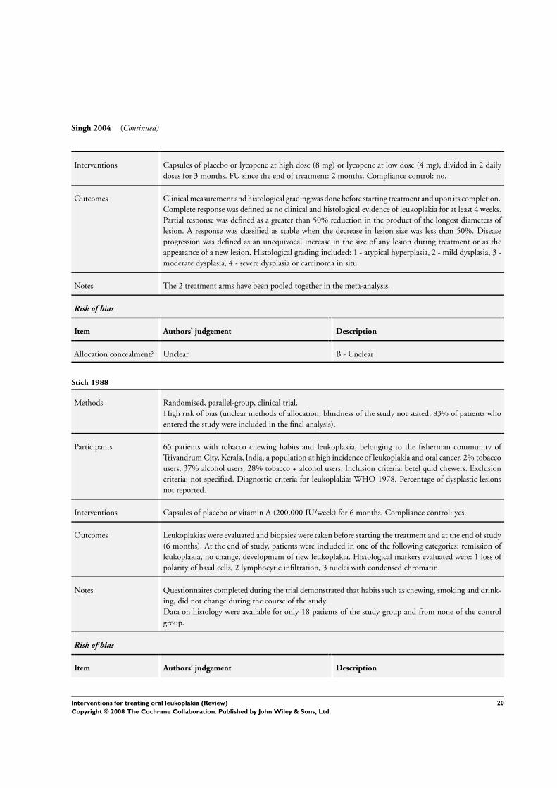
Singh 2004 (Continued)
Interventions Capsules of placebo or lycopene at high dose (8 mg) or lycopene at low dose (4 mg), divided in 2 daily
doses for 3 months. FU since the end of treatment: 2 months. Compliance control: no.
Outcomes Clinical measurement and histological grading was done before starting treatment and upon its completion.
Complete response was defined as no clinical and histological evidence of leukoplakia for at least 4 weeks.
Partial response was defined as a greater than 50% reduction in the product of the longest diameters of
lesion. A response was classified as stable when the decrease in lesion size was less than 50%. Disease
progression was defined as an unequivocal increase in the size of any lesion during treatment or as the
appearance of a new lesion. Histological grading included: 1 - atypical hyperplasia, 2 - mild dysplasia, 3 -
moderate dysplasia, 4 - severe dysplasia or carcinoma in situ.
Notes The 2 treatment arms have been pooled together in the meta-analysis.
Risk of bias
Item Authors’ judgement Description
Allocation concealment? Unclear B - Unclear
Stich 1988
Methods Randomised, parallel-group, clinical trial.
High risk of bias (unclear methods of allocation, blindness of the study not stated, 83% of patients who
entered the study were included in the final analysis).
Participants 65 patients with tobacco chewing habits and leukoplakia, belonging to the fisherman community of
Trivandrum City, Kerala, India, a population at high incidence of leukoplakia and oral cancer. 2% tobacco
users, 37% alcohol users, 28% tobacco + alcohol users. Inclusion criteria: betel quid chewers. Exclusion
criteria: not specified. Diagnostic criteria for leukoplakia: WHO 1978. Percentage of dysplastic lesions
not reported.
Interventions Capsules of placebo or vitamin A (200,000 IU/week) for 6 months. Compliance control: yes.
Outcomes Leukoplakias were evaluated and biopsies were taken before starting the treatment and at the end of study
(6 months). At the end of study, patients were included in one of the following categories: remission of
leukoplakia, no change, development of new leukoplakia. Histological markers evaluated were: 1 loss of
polarity of basal cells, 2 lymphocytic infiltration, 3 nuclei with condensed chromatin.
Notes Questionnaires completed during the trial demonstrated that habits such as chewing, smoking and drink-
ing, did not change during the course of the study.
Data on histology were available for only 18 patients of the study group and from none of the control
group.
Risk of bias
Item Authors’ judgement Description
20Interventions for treating oral leukoplakia (Review)
Copyright © 2008 The Cochrane Collaboration. Published by John Wiley & Sons, Ltd.
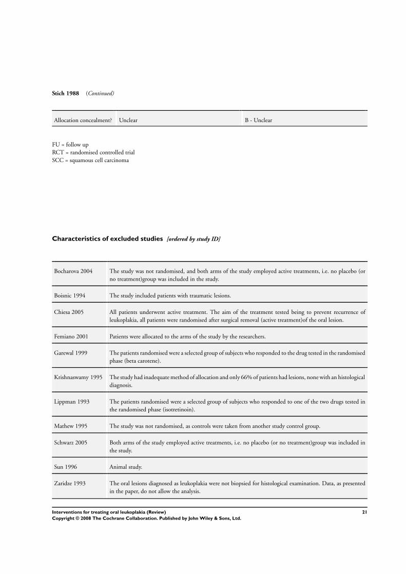
Stich 1988 (Continued)
Allocation concealment? Unclear B - Unclear
FU = follow up
RCT = randomised controlled trial
SCC = squamous cell carcinoma
Characteristics of excluded studies [ordered by study ID]
Bocharova 2004 The study was not randomised, and both arms of the study employed active treatments, i.e. no placebo (or
no treatment)group was included in the study.
Boisnic 1994 The study included patients with traumatic lesions.
Chiesa 2005 All patients underwent active treatment. The aim of the treatment tested being to prevent recurrence of
leukoplakia, all patients were randomised after surgical removal (active treatment)of the oral lesion.
Femiano 2001 Patients were allocated to the arms of the study by the researchers.
Garewal 1999 The patients randomised were a selected group of subjects who responded to the drug tested in the randomised
phase (beta carotene).
Krishnaswamy 1995 The study had inadequate method of allocation and only 66% of patients had lesions, none with an histological
diagnosis.
Lippman 1993 The patients randomised were a selected group of subjects who responded to one of the two drugs tested in
the randomised phase (isotretinoin).
Mathew 1995 The study was not randomised, as controls were taken from another study control group.
Schwarz 2005 Both arms of the study employed active treatments, i.e. no placebo (or no treatment)group was included in
the study.
Sun 1996 Animal study.
Zaridze 1993 The oral lesions diagnosed as leukoplakia were not biopsied for histological examination. Data, as presented
in the paper, do not allow the analysis.
21Interventions for treating oral leukoplakia (Review)
Copyright © 2008 The Cochrane Collaboration. Published by John Wiley & Sons, Ltd.
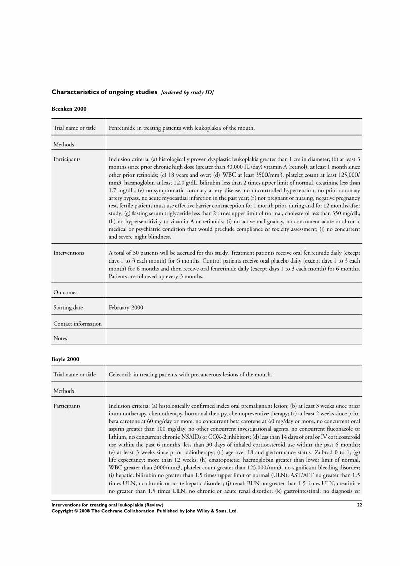
Characteristics of ongoing studies [ordered by study ID]
Beenken 2000
Trial name or title Fenretinide in treating patients with leukoplakia of the mouth.
Methods
Participants Inclusion criteria: (a) histologically proven dysplastic leukoplakia greater than 1 cm in diameter; (b) at least 3
months since prior chronic high dose (greater than 30,000 IU/day) vitamin A (retinol), at least 1 month since
other prior retinoids; (c) 18 years and over; (d) WBC at least 3500/mm3, platelet count at least 125,000/
mm3, haemoglobin at least 12.0 g/dL, bilirubin less than 2 times upper limit of normal, creatinine less than
1.7 mg/dL; (e) no symptomatic coronary artery disease, no uncontrolled hypertension, no prior coronary
artery bypass, no acute myocardial infarction in the past year; (f ) not pregnant or nursing, negative pregnancy
test, fertile patients must use effective barrier contraception for 1 month prior, during and for 12 months after
study; (g) fasting serum triglyceride less than 2 times upper limit of normal, cholesterol less than 350 mg/dL;
(h) no hypersensitivity to vitamin A or retinoids; (i) no active malignancy, no concurrent acute or chronic
medical or psychiatric condition that would preclude compliance or toxicity assessment; (j) no concurrent
and severe night blindness.
Interventions A total of 30 patients will be accrued for this study. Treatment patients receive oral fenretinide daily (except
days 1 to 3 each month) for 6 months. Control patients receive oral placebo daily (except days 1 to 3 each
month) for 6 months and then receive oral fenretinide daily (except days 1 to 3 each month) for 6 months.
Patients are followed up every 3 months.
Outcomes
Starting date February 2000.
Contact information
Notes
Boyle 2000
Trial name or title Celecoxib in treating patients with precancerous lesions of the mouth.
Methods
Participants Inclusion criteria: (a) histologically confirmed index oral premalignant lesion; (b) at least 3 weeks since prior
immunotherapy, chemotherapy, hormonal therapy, chemopreventive therapy; (c) at least 2 weeks since prior
beta carotene at 60 mg/day or more, no concurrent beta carotene at 60 mg/day or more, no concurrent oral
aspirin greater than 100 mg/day, no other concurrent investigational agents, no concurrent fluconazole or
lithium, no concurrent chronic NSAIDs or COX-2 inhibitors; (d) less than 14 days of oral or IV corticosteroid
use within the past 6 months, less than 30 days of inhaled corticosteroid use within the past 6 months;
(e) at least 3 weeks since prior radiotherapy; (f ) age over 18 and performance status: Zubrod 0 to 1; (g)
life expectancy: more than 12 weeks; (h) ematopoietic: haemoglobin greater than lower limit of normal,
WBC greater than 3000/mm3, platelet count greater than 125,000/mm3, no significant bleeding disorder;
(i) hepatic: bilirubin no greater than 1.5 times upper limit of normal (ULN), AST/ALT no greater than 1.5
times ULN, no chronic or acute hepatic disorder; (j) renal: BUN no greater than 1.5 times ULN, creatinine
no greater than 1.5 times ULN, no chronic or acute renal disorder; (k) gastrointestinal: no diagnosis or
22Interventions for treating oral leukoplakia (Review)
Copyright © 2008 The Cochrane Collaboration. Published by John Wiley & Sons, Ltd.
Boyle 2000 (Continued)
treatment of oesophageal, gastric, pyloric channel, or duodenal ulceration within past 30 days, no prior or
active pancreatic disease or inflammatory bowel disease; (l) other: completed a smoking cessation program, if
applicable, no prior hypersensitivity to COX-2 inhibitors, NSAIDs, salicylates, or sulphonamides, no prior
invasive cancer within the past 5 years except non-melanoma skin cancer or carcinoma in situ of the cervix,
no other concurrent condition that would preclude study, not pregnant or nursing, negative pregnancy test,
fertile patients must use effective contraception.
Interventions Arm I: patients receive lower-dose oral celecoxib twice daily. Arm II: patients receive higher-dose oral celecoxib
twice daily. Arm III: patients receive oral placebo twice daily. Treatment continues in all 3 arms for 12 weeks
in the absence of disease progression or unacceptable toxicity. Patients are followed at 18, 24, and 26 weeks.
Outcomes
Starting date October 2000.
Contact information Jay O Boyle, Study Chair, Memorial Sloan-Kettering Cancer Center.
Notes This study is no longer recruiting patients.
Chiang 2005
Trial name or title Photodynamic therapy for oral leukoplakia and erythroleukoplakia.
Methods
Participants Inclusion criteria: patients with leukoplakia or erythroleukoplakia. Exclusion criteria: oral cancers.
Interventions Photodynamic therapy.
Outcomes
Starting date August 2004.
Contact information Chun-Pin Chiang DMSc
tel: +886 2 23123456 ext: 6855
Notes Recruiting.
Goodin 2005
Trial name or title A phase II trial to assess the effects of green tea in oral leukoplakia.
Methods
23Interventions for treating oral leukoplakia (Review)
Copyright © 2008 The Cochrane Collaboration. Published by John Wiley & Sons, Ltd.
Goodin 2005 (Continued)
Participants Patients with oral leukoplakia without evidence of active infections in the mouth will be invited to participate
in this study. Patients must currently consume no more than 3 cups of tea a day, not be taking large doses of
vitamin A or its derivatives (more than 25,000 units), selenium, or beta carotene. Additionally, patients must
not participate in this study if they are allergic to caffeine, have GI ulcers, are pregnant, or have had previous
invasive mouth cancer.
Interventions Green tea lozenge versus placebo.
Outcomes
Starting date November 2003.
Contact information Susan Goodin PharmD, University of Medicine and Dentistry New Jersey.
Notes
Lippman 2004
Trial name or title Celecoxib or erlotinib or both in preventing oral cancer in patients with oral leukoplakia.
Methods
Participants Inclusion criteria: (a) diagnosis of oral aneuploid premalignant lesion within the past 3 months; (b) dysplastic
white or red patches in the oral cavity with DNA aneuploidy determined by high resolving image cytometry;
(c) must have sufficient lesion to biopsy; (d) no prior or active head and neck cancer or lung cancer; (e) no
known metastases to the head and neck area from other tumours.
Interventions Celecoxib or erlotinib or both may be effective in preventing the development of oral cancer in patients who
have oral leukoplakia.
Outcomes
Starting date
Contact information Scott M. Lippman MD, FACP, M.D. Anderson Cancer Center.
Notes Not yet recruiting.
24Interventions for treating oral leukoplakia (Review)
Copyright © 2008 The Cochrane Collaboration. Published by John Wiley & Sons, Ltd.
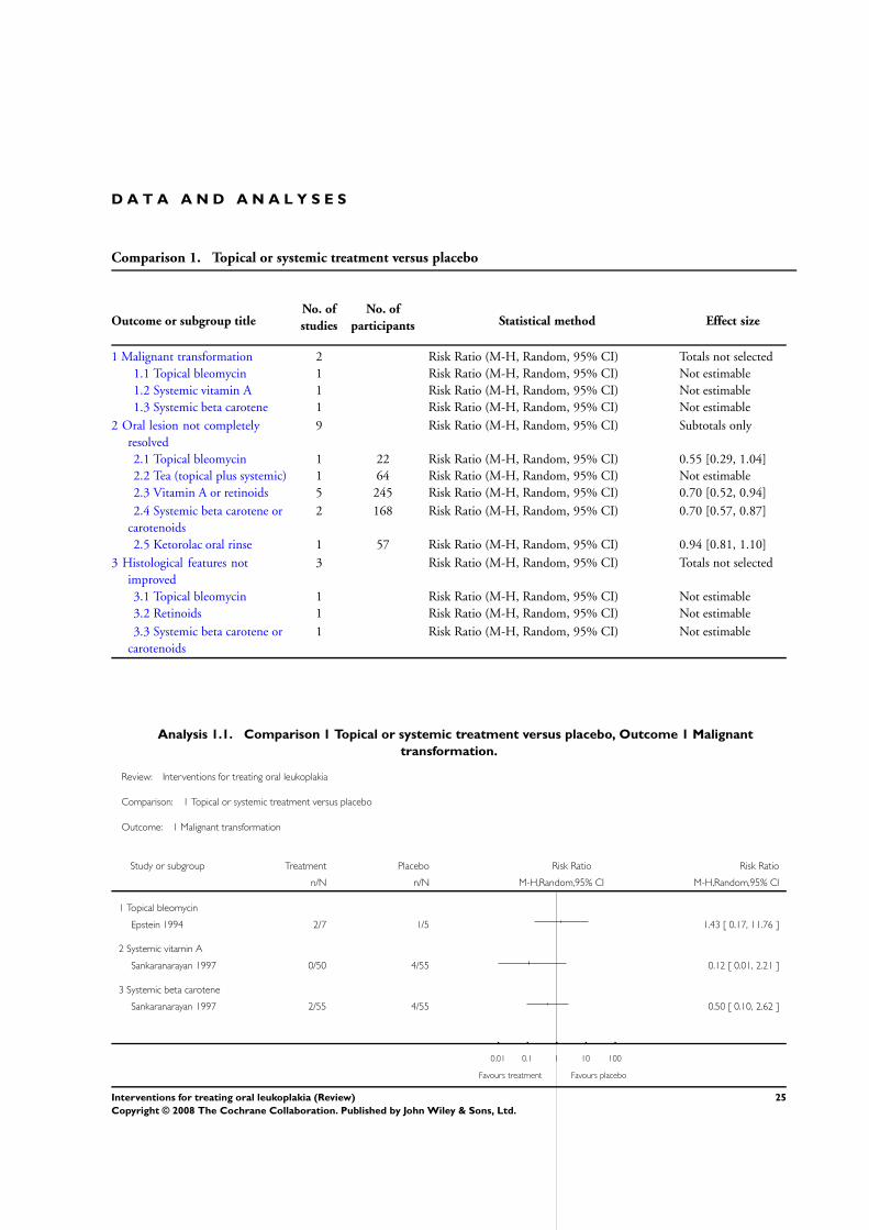
D A T A A N D A N A L Y S E S
Comparison 1. Topical or systemic treatment versus placebo
Outcome or subgroup titleNo. of
studies
No. of
participants Statistical method Effect size
1 Malignant transformation 2 Risk Ratio (M-H, Random, 95% CI) Totals not selected
1.1 Topical bleomycin 1 Risk Ratio (M-H, Random, 95% CI) Not estimable
1.2 Systemic vitamin A 1 Risk Ratio (M-H, Random, 95% CI) Not estimable
1.3 Systemic beta carotene 1 Risk Ratio (M-H, Random, 95% CI) Not estimable
2 Oral lesion not completely
resolved
9 Risk Ratio (M-H, Random, 95% CI) Subtotals only
2.1 Topical bleomycin 1 22 Risk Ratio (M-H, Random, 95% CI) 0.55 [0.29, 1.04]
2.2 Tea (topical plus systemic) 1 64 Risk Ratio (M-H, Random, 95% CI) Not estimable
2.3 Vitamin A or retinoids 5 245 Risk Ratio (M-H, Random, 95% CI) 0.70 [0.52, 0.94]
2.4 Systemic beta carotene or
carotenoids
2 168 Risk Ratio (M-H, Random, 95% CI) 0.70 [0.57, 0.87]
2.5 Ketorolac oral rinse 1 57 Risk Ratio (M-H, Random, 95% CI) 0.94 [0.81, 1.10]
3 Histological features not
improved
3 Risk Ratio (M-H, Random, 95% CI) Totals not selected
3.1 Topical bleomycin 1 Risk Ratio (M-H, Random, 95% CI) Not estimable
3.2 Retinoids 1 Risk Ratio (M-H, Random, 95% CI) Not estimable
3.3 Systemic beta carotene or
carotenoids
1 Risk Ratio (M-H, Random, 95% CI) Not estimable
Analysis 1.1. Comparison 1 Topical or systemic treatment versus placebo, Outcome 1 Malignant
transformation.
Review: Interventions for treating oral leukoplakia
Comparison: 1 Topical or systemic treatment versus placebo
Outcome: 1 Malignant transformation
Study or subgroup Treatment Placebo Risk Ratio Risk Ratio
n/N n/N M-H,Random,95% CI M-H,Random,95% CI
1 Topical bleomycin
Epstein 1994 2/7 1/5 1.43 [ 0.17, 11.76 ]
2 Systemic vitamin A
Sankaranarayan 1997 0/50 4/55 0.12 [ 0.01, 2.21 ]
3 Systemic beta carotene
Sankaranarayan 1997 2/55 4/55 0.50 [ 0.10, 2.62 ]
0.01 0.1 1 10 100
Favours treatment Favours placebo
25Interventions for treating oral leukoplakia (Review)
Copyright © 2008 The Cochrane Collaboration. Published by John Wiley & Sons, Ltd.
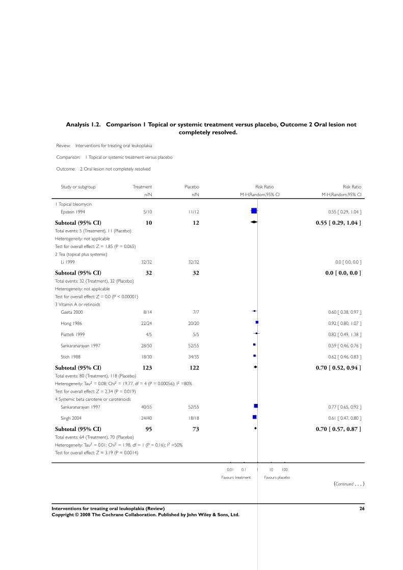
Analysis 1.2. Comparison 1 Topical or systemic treatment versus placebo, Outcome 2 Oral lesion not
completely resolved.
Review: Interventions for treating oral leukoplakia
Comparison: 1 Topical or systemic treatment versus placebo
Outcome: 2 Oral lesion not completely resolved
Study or subgroup Treatment Placebo Risk Ratio Risk Ratio
n/N n/N M-H,Random,95% CI M-H,Random,95% CI
1 Topical bleomycin
Epstein 1994 5/10 11/12 0.55 [ 0.29, 1.04 ]
Subtotal (95% CI) 10 12 0.55 [ 0.29, 1.04 ]
Total events: 5 (Treatment), 11 (Placebo)
Heterogeneity: not applicable
Test for overall effect: Z = 1.85 (P = 0.065)
2 Tea (topical plus systemic)
Li 1999 32/32 32/32 0.0 [ 0.0, 0.0 ]
Subtotal (95% CI) 32 32 0.0 [ 0.0, 0.0 ]
Total events: 32 (Treatment), 32 (Placebo)
Heterogeneity: not applicable
Test for overall effect: Z = 0.0 (P < 0.00001)
3 Vitamin A or retinoids
Gaeta 2000 8/14 7/7 0.60 [ 0.38, 0.97 ]
Hong 1986 22/24 20/20 0.92 [ 0.80, 1.07 ]
Piattelli 1999 4/5 5/5 0.82 [ 0.49, 1.38 ]
Sankaranarayan 1997 28/50 52/55 0.59 [ 0.46, 0.76 ]
Stich 1988 18/30 34/35 0.62 [ 0.46, 0.83 ]
Subtotal (95% CI) 123 122 0.70 [ 0.52, 0.94 ]
Total events: 80 (Treatment), 118 (Placebo)
Heterogeneity: Tau2 = 0.08; Chi2 = 19.77, df = 4 (P = 0.00056); I2 =80%
Test for overall effect: Z = 2.34 (P = 0.019)
4 Systemic beta carotene or carotenoids
Sankaranarayan 1997 40/55 52/55 0.77 [ 0.65, 0.92 ]
Singh 2004 24/40 18/18 0.61 [ 0.47, 0.80 ]
Subtotal (95% CI) 95 73 0.70 [ 0.57, 0.87 ]
Total events: 64 (Treatment), 70 (Placebo)
Heterogeneity: Tau2 = 0.01; Chi2 = 1.98, df = 1 (P = 0.16); I2 =50%
Test for overall effect: Z = 3.19 (P = 0.0014)
0.01 0.1 1 10 100
Favours treatment Favours placebo
(Continued . . . )
26Interventions for treating oral leukoplakia (Review)
Copyright © 2008 The Cochrane Collaboration. Published by John Wiley & Sons, Ltd.
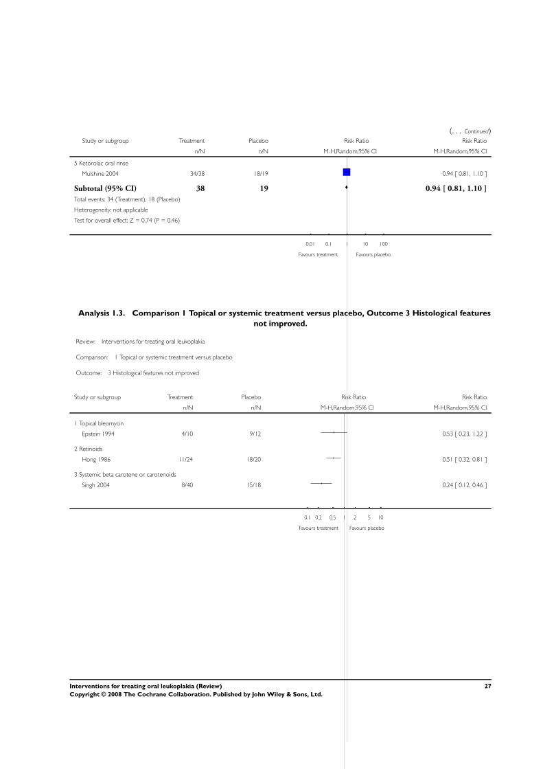
(. . . Continued)Study or subgroup Treatment Placebo Risk Ratio Risk Ratio
n/N n/N M-H,Random,95% CI M-H,Random,95% CI
5 Ketorolac oral rinse
Mulshine 2004 34/38 18/19 0.94 [ 0.81, 1.10 ]
Subtotal (95% CI) 38 19 0.94 [ 0.81, 1.10 ]
Total events: 34 (Treatment), 18 (Placebo)
Heterogeneity: not applicable
Test for overall effect: Z = 0.74 (P = 0.46)
0.01 0.1 1 10 100
Favours treatment Favours placebo
Analysis 1.3. Comparison 1 Topical or systemic treatment versus placebo, Outcome 3 Histological features
not improved.
Review: Interventions for treating oral leukoplakia
Comparison: 1 Topical or systemic treatment versus placebo
Outcome: 3 Histological features not improved
Study or subgroup Treatment Placebo Risk Ratio Risk Ratio
n/N n/N M-H,Random,95% CI M-H,Random,95% CI
1 Topical bleomycin
Epstein 1994 4/10 9/12 0.53 [ 0.23, 1.22 ]
2 Retinoids
Hong 1986 11/24 18/20 0.51 [ 0.32, 0.81 ]
3 Systemic beta carotene or carotenoids
Singh 2004 8/40 15/18 0.24 [ 0.12, 0.46 ]
0.1 0.2 0.5 1 2 5 10
Favours treatment Favours placebo
27Interventions for treating oral leukoplakia (Review)
Copyright © 2008 The Cochrane Collaboration. Published by John Wiley & Sons, Ltd.
A P P E N D I C E S
Appendix 1. CENTRAL search strategy
#1 LEUKOPLAKIA ORAL explode all trees (MeSH)
#2 leukoplaki*
#3 preneoplastic NEAR oral
#4 precancer* NEAR oral
#5 (#1 OR #2 OR #3 OR #4)
W H A T ’ S N E W
Last assessed as up-to-date: 3 July 2006.
5 August 2008 Amended Converted to new review format.
H I S T O R Y
Protocol first published: Issue 3, 1999
Review first published: Issue 4, 2001
4 July 2006 New citation required but conclusions have not changed Review updated. Two new included studies (Mulshine
2004; Singh 2004), three ongoing studies, three excluded
studies. Conclusions remained essentially the same.
25 May 2004 New citation required but conclusions have not changed Review updated. One new study (Gaeta 2000) has been
included, however the summary estimates did not change
significantly and conclusions remained essentially the
same.
C O N T R I B U T I O N S O F A U T H O R S
Giovanni Lodi: main review author, participation in all phases of the review’s preparation.
Cristina Bez: articles retrieval, data collection, interpretation of results.
Antonio Carrassi: group co-ordinator, selection of articles, interpretation of results.
Federica Demarosi: prospective handsearching, data collection, interpretation of results.
Andrea Sardella: selection of articles, critical appraisal of studies, interpretation of results.
28Interventions for treating oral leukoplakia (Review)
Copyright © 2008 The Cochrane Collaboration. Published by John Wiley & Sons, Ltd.
D E C L A R A T I O N S O F I N T E R E S T
None known.
S O U R C E S O F S U P P O R T
Internal sources
• University of Milan, Italy.
External sources
• No sources of support supplied
I N D E X T E R M S
Medical Subject Headings (MeSH)
Leukoplakia, Oral [∗therapy]; Randomized Controlled Trials as Topic
MeSH check words
Humans
29Interventions for treating oral leukoplakia (Review)
Copyright © 2008 The Cochrane Collaboration. Published by John Wiley & Sons, Ltd.

![FRP - StudiesToday.com Class 7... · History Picture Identification & discription- [10 Marks] Geography- Map ... Lodis Bahlul lodi Sikander Lodi Ibrahim Lodi](https://img.pdfslide.us/doc/110x75/5a9e2ce47f8b9a36788c4f5e/frp-class-7history-picture-identification-discription-10-marks-geography-.jpg)