Embed Size (px)
Citation preview
REVIEWFor reprint orders, please contact:[email protected]
Localized surface plasmon resonance biosensors
Jing Zhao1, Xiaoyu Zhang1, Chanda Ranjit Yonzon1, Amanda J Haes1 & Richard P Van Duyne1††Author for correspondence1Northwestern University, Department of Chemistry, 2145 Sheridan Road, Evanston, IL 60208-3113, USATel.: +1 847 491 3516;Fax: +1 847 491 7713;E-mail: [email protected]
Keywords: Alzheimer’s disease, biosensing, electrochemistry, localized surface plasmon resonance, multiplexed sensing, nanosphere lithography, silver nanoparticles, solution-phase nanoparticles
10.2217/17435889.1.1.219 © 2
In this review, the most recent progress in the development of noble metal nano-optical sensors based on localized surface plasmon resonance (LSPR) spectroscopy is summarized. The sensing principle relies on the LSPR spectral shifts caused by the surrounding dielectric environmental change in a binding event. Nanosphere lithography, an inexpensive and simple nanofabrication technique, has been used to fabricate the nanoparticles as the LSPR sensing platforms. As an example of the biosensing applications, the LSPR detection for a biomarker of Alzheimer’s disease, amyloid-derived diffusable ligands, in human brain extract and cerebrospinal fluid samples is highlighted. Furthermore, the LSPR sensing method can be modified easily and used in a variety of applications. More specifically, a LSPR chip capable of multiplex sensing, a combined electrochemical and LSPR protocol and a fabrication method of solution-phase nanotriangles are presented here.
The last 20 years have witnessed a rapid growthin the development of the highly selective andsensitive optical nanobiosensors for medicaldiagnosis and monitoring of diseases, drug dis-covery and the detection of environmental pol-lutants and biological agents [1]. Among themany advantages of optical biosensors, sensitiv-ity and real-time detection of biomolecularinteractions allow them to be applied widely.Optical sensing techniques are based on varioussensing transduction mechanisms, for example,chemiluminescence [2,3], fluorescence [4], lightabsorption and scattering [5,6], reflectance [7],surface plasmon resonance (SPR) [8–10] andRaman scattering [11–13].
This review will focus on optical sensors basedon localized surface plasmon resonance (LSPR) oftriangular nanoparticles. This nanoparticle-basedoptical sensing technique is effective for quantita-tive detection of chemical and biologicaltargets [5,10,14,15]. The sensing principle employedin these experiments relies on the high sensitivityof the LSPR spectrum of noble metal nanoparti-cles to adsorbate-induced changes in the dielectricconstant of the surrounding environment. LSPRis one of the signature optical properties of noblemetal nanoparticles, which arises when the inci-dent photon frequency is resonant with the collec-tive oscillation of the conduction electrons in themetal nanoparticles. The LSPR is dependent onthe size, shape, interparticle spacing and dielectricproperties of the material, as well as the dielectricproperties of the local environment that sur-rounds the nanoparticles. The LSPRextinction maximum (λmax) of the nanoparticle
arrays can be measured with UV-visible extinctionspectroscopy [16–20]. Since the LSPR of the nano-particles is highly dependent on the local environ-ment, λmax will shift when adsorbates bind to thenanoparticles. The LSPR shift induced by theadsorbates, ∆λmax, is given by Equation 1:
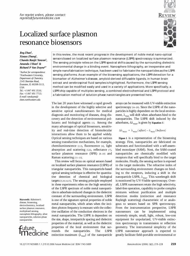
Figure 1 is a representation of the biosensingstrategy. First, nanoparticles are fabricated onsubstrates and functionalized with a self-assem-bled monolayer (SAM). Next, the SAM-coatednanoparticles are chemically modified withreceptors that will specifically bind to the targetmolecules. Finally, the sensing surface is exposedto the target molecules. The refractive index ofthe surrounding environment changes on bind-ing to the receptors, inducing a shift in thenanoparticle LSPR, λmax. This wavelength shiftis monitored by UV-Visible spectroscopy. Over-all, LSPR nanosensors retain the high selectivity,label-free operation, capability to probe complexmixtures without purification and multipledetection modes (extinction and resonanceRayleigh scattering) characteristic of or analo-gous to sensors based on SPR spectroscopy.From the instrumentation perspective, LSPRnanosensors can be implemented usingextremely simple, small, light, robust, low-costequipment for unpolarized, UV-visible extinc-tion spectroscopy in transmission or reflectiongeometry. The instrumental simplicity of theLSPR nanosensor approach is expected togreatly facilitate field-portable environmental or
∆λmax λmax after⟨ ⟩ λmax before⟨ ⟩–=
006 Future Medicine Ltd ISSN 1743-5889 Nanomedicine (2006) 1(2), 219–228 219
REVIEW – Zhao, Zhang, Ranjit Yonzon, Haes & Van Duyne
220
Figure 1. Representfor localized surface
Transmission UV-visible s(LSPR) of Ag nanoparticlewith unpolarized light. Thome-built flow cell wasnanoparticle substrates. figuredisplays the surfacesurface-confined Ag nanfabricated using nanosphmonolayer consisting of surface passivating moleccovalently attached. Fina
Light source
point-of-service medical diagnostic applications.A more comprehensive comparison of the twomethods is available in a recent review [21].
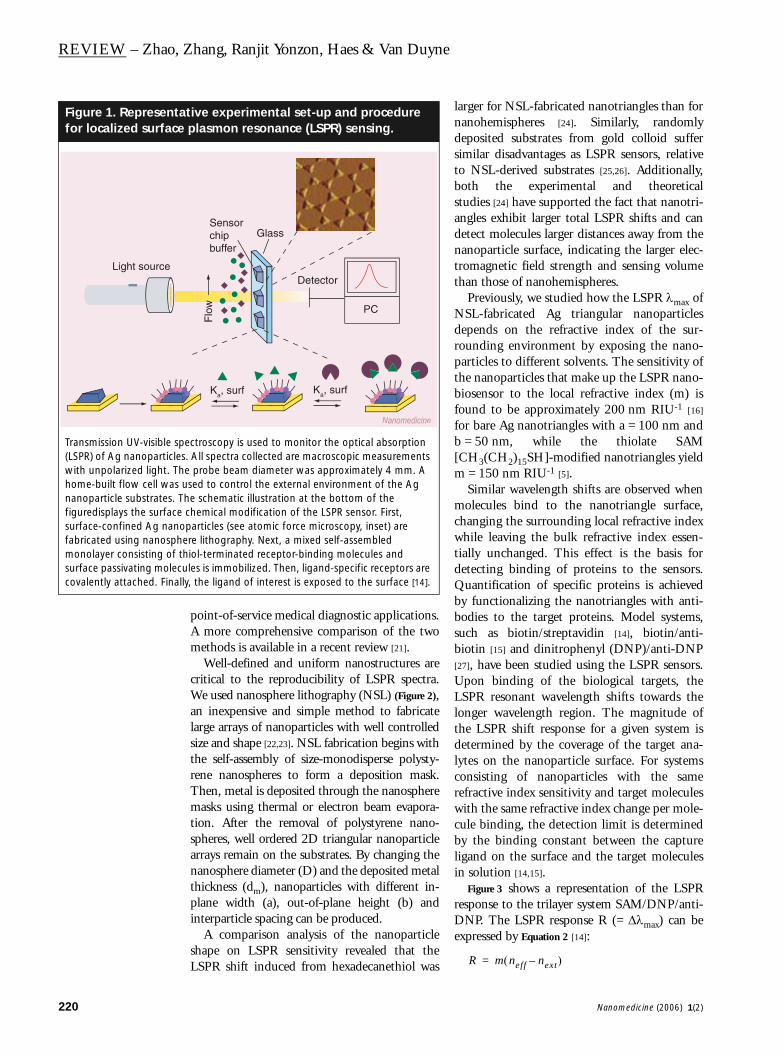
Well-defined and uniform nanostructures arecritical to the reproducibility of LSPR spectra.We used nanosphere lithography (NSL) (Figure 2),an inexpensive and simple method to fabricatelarge arrays of nanoparticles with well controlledsize and shape [22,23]. NSL fabrication begins withthe self-assembly of size-monodisperse polysty-rene nanospheres to form a deposition mask.Then, metal is deposited through the nanospheremasks using thermal or electron beam evapora-tion. After the removal of polystyrene nano-spheres, well ordered 2D triangular nanoparticlearrays remain on the substrates. By changing thenanosphere diameter (D) and the deposited metalthickness (dm), nanoparticles with different in-plane width (a), out-of-plane height (b) andinterparticle spacing can be produced.
A comparison analysis of the nanoparticleshape on LSPR sensitivity revealed that theLSPR shift induced from hexadecanethiol was
larger for NSL-fabricated nanotriangles than fornanohemispheres [24]. Similarly, randomlydeposited substrates from gold colloid suffersimilar disadvantages as LSPR sensors, relativeto NSL-derived substrates [25,26]. Additionally,both the experimental and theoreticalstudies [24] have supported the fact that nanotri-angles exhibit larger total LSPR shifts and candetect molecules larger distances away from thenanoparticle surface, indicating the larger elec-tromagnetic field strength and sensing volumethan those of nanohemispheres.
Previously, we studied how the LSPR λmax ofNSL-fabricated Ag triangular nanoparticlesdepends on the refractive index of the sur-rounding environment by exposing the nano-particles to different solvents. The sensitivity ofthe nanoparticles that make up the LSPR nano-biosensor to the local refractive index (m) isfound to be approximately 200 nm RIU-1 [16]
for bare Ag nanotriangles with a = 100 nm andb = 50 nm, while the thiolate SAM[CH3(CH2)15SH]-modified nanotriangles yieldm = 150 nm RIU-1 [5].
Similar wavelength shifts are observed whenmolecules bind to the nanotriangle surface,changing the surrounding local refractive indexwhile leaving the bulk refractive index essen-tially unchanged. This effect is the basis fordetecting binding of proteins to the sensors.Quantification of specific proteins is achievedby functionalizing the nanotriangles with anti-bodies to the target proteins. Model systems,such as biotin/streptavidin [14], biotin/anti-biotin [15] and dinitrophenyl (DNP)/anti-DNP[27], have been studied using the LSPR sensors.Upon binding of the biological targets, theLSPR resonant wavelength shifts towards thelonger wavelength region. The magnitude ofthe LSPR shift response for a given system isdetermined by the coverage of the target ana-lytes on the nanoparticle surface. For systemsconsisting of nanoparticles with the samerefractive index sensitivity and target moleculeswith the same refractive index change per mole-cule binding, the detection limit is determinedby the binding constant between the captureligand on the surface and the target moleculesin solution [14,15].
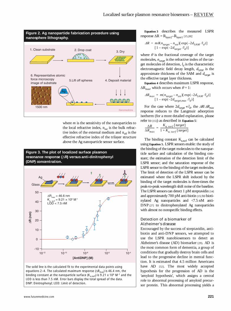
Figure 3 shows a representation of the LSPRresponse to the trilayer system SAM/DNP/anti-DNP. The LSPR response R (= ∆λmax) can beexpressed by Equation 2 [14]:
ative experimental set-up and procedure plasmon resonance (LSPR) sensing.
pectroscopy is used to monitor the optical absorption s. All spectra collected are macroscopic measurements he probe beam diameter was approximately 4 mm. A used to control the external environment of the Ag The schematic illustration at the bottom of the chemical modification of the LSPR sensor. First, oparticles (see atomic force microscopy, inset) are ere lithography. Next, a mixed self-assembled
thiol-terminated receptor-binding molecules and ules is immobilized. Then, ligand-specific receptors are lly, the ligand of interest is exposed to the surface [14].
Sensorchipbuffer
Flo
w PC
Ka, surf Ka, surf
Detector
Glass
Nanomedicine
R m neff next–( )=
Nanomedicine (2006) 1(2)
www.futuremedicine.com
Localized surface plasmon resonance biosensors – REVIEW
Figure 2. Ag nanopananosphere lithogr
Figure 3. The plot oresonance response(DNP) concentration
The solid line is the calcuequations 2–4. The calcubinding constant at the nLOD is less than 7.5 nM.DNP: Dinitrophenyl; LOD
1. Clean substrate
6. Representative atoforce microscopyimage of substrate
1500 nm
Glass
Ag
10-12
-10
0
10
20
30
40
50
R (
nm
)
Rmax = 4Ka, surf = 9LOD < 7.
where m is the sensitivity of the nanoparticles tothe local refractive index, next is the bulk refrac-tive index of the external medium and neff is theeffective refractive index of the trilayer structureabove the Ag nanoparticle sensor surface.
Equation 3 describes the measured LSPRresponse ∆R = Rlayer2–Rlayer1 [15,28]:
where θ is the fractional coverage of the targetmolecules, ntarget is the refractive index of the tar-get molecules of detection, ld is the characteristicelectromagnetic field decay length, dSAM is theapproximate thickness of the SAM and dtarget isthe effective target layer thickness.
Equation 4 describes maximum LSPR response,∆Rmax, which occurs when θ = 1:
For the case where 2dtarget « ld, the ∆R/∆Rmaxresponse reduces to the Langmuir adsorptionisotherm (for a more detailed explanation, pleaserefer to [15]) as described in Equation 5:
The binding constant Ka,surf can be calculatedusing Equation 5. LSPR sensors enable: the study ofthe binding of the target molecules to the nanopar-ticle surface and calculation of the binding con-stant; the estimation of the detection limit of theLSPR sensor; and the saturation response of theLSPR sensor to the binding of the target molecules.The limit of detection of the LSPR sensor can beestimated where the LSPR shift induced by thebinding of the target molecules is three-times thepeak-to-peak wavelength shift noise of the baseline.The LSPR sensors can detect 1 pM streptavidin [14]
and approximately 700 pM anti-biotin [15] to bioti-nylated Ag nanoparticles and <7.5 nM anti-DNP [27] to dinitrophenylated Ag nanoparticleswith almost no nonspecific binding effects.
Detection of a biomarker of Alzheimer’s diseaseEncouraged by the success of streptavidin, anti-biotin and anti-DNP sensors, we attempted touse the LSPR nanobiosensors to detect anAlzheimer’s disease (AD) biomarker [29]. AD isthe most common form of dementia, a group ofconditions that gradually destroy brain cells andlead to the progressive decline in mental func-tion. It is estimated that 4.5 million Americanshave AD [52]. The most widely acceptedhypothesis for the progression of AD is the‘amyloid hypothesis’, which assigns a centralrole to abnormal processing of amyloid precur-sor protein. This abnormal processing yields a
rticle fabrication procedure using aphy.
f localized surface plasmon (∆R) versus anti-dinitrophenyl .
lated fit to the experimental data points using lated maximum response (∆Rmax) is 46.4 nm, the anoparticle surface (Ka,surf) is 9.21 x 10⁶ M-1 and the
Error bars display the total spread of the data.: Limit of detection.
Ag
2. Drop coat 3. Dry
4. Deposit material5.Lift off spheres
mic
Nanomedicine
10-10 10-8 10-6 10-4
[AntiDNP] (M)
6.6 nm.21 × 106 M-1
5 nM
∆R mθ ntarget next–( ) 2dSAM ld⁄–( )exp[ ] 1 2dtarget ld⁄–( )exp–[ ]
=
∆Rmax m ntarget next–( ) 2dSAM ld⁄–( )exp[ ] 1 2dtarget,max ld⁄–( )exp–[ ]
=
∆R∆Rmax----------------
Ka surf, target[ ]1 Ka surf, target[ ]+------------------------------------------------=
221
REVIEW – Zhao, Zhang, Ranjit Yonzon, Haes & Van Duyne
222
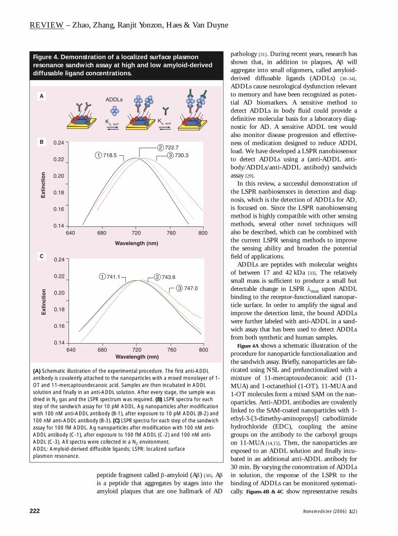
Figure 4. Demonstraresonance sandwichdiffusable ligand co
(A) Schematic illustrationantibody is covalently attaOT and 11-mercaptoundesolution and finally in an dried in N2 gas and the LSstep of the sandwich assawith 100 nM anti-ADDL a100 nM anti-ADDL antiboassay for 100 fM ADDL. AADDL antibody (C-1), afteADDL (C-3). All spectra wADDL: Amyloid-derived dplasmon resonance.
Ext
inct
ion
640
0.24
0.22
0.20
0.18
0.16
0.14
Ext
inct
ion
640
0.24
0.22
0.20
0.18
0.16
0.14
A
B
C
peptide fragment called β-amyloid (Aβ) [30]. Aβis a peptide that aggregates by stages into theamyloid plaques that are one hallmark of AD
pathology [31]. During recent years, research hasshown that, in addition to plaques, Aβ willaggregate into small oligomers, called amyloid-derived diffusable ligands (ADDLs) [30–34].ADDLs cause neurological dysfunction relevantto memory and have been recognized as poten-tial AD biomarkers. A sensitive method todetect ADDLs in body fluid could provide adefinitive molecular basis for a laboratory diag-nostic for AD. A sensitive ADDL test wouldalso monitor disease progression and effective-ness of medication designed to reduce ADDLload. We have developed a LSPR nanobiosensorto detect ADDLs using a (anti-ADDL anti-body/ADDLs/anti-ADDL antibody) sandwichassay [29].
In this review, a successful demonstration ofthe LSPR nanbiosensors in detection and diag-nosis, which is the detection of ADDLs for AD,is focused on. Since the LSPR nanobiosensingmethod is highly compatible with other sensingmethods, several other novel techniques willalso be described, which can be combined withthe current LSPR sensing methods to improvethe sensing ability and broaden the potentialfield of applications.
ADDLs are peptides with molecular weightsof between 17 and 42 kDa [33]. The relativelysmall mass is sufficient to produce a small butdetectable change in LSPR λmax upon ADDLbinding to the receptor-functionalized nanopar-ticle surface. In order to amplify the signal andimprove the detection limit, the bound ADDLswere further labeled with anti-ADDL in a sand-wich assay that has been used to detect ADDLsfrom both synthetic and human samples.
Figure 4A shows a schematic illustration of theprocedure for nanoparticle functionalization andthe sandwich assay. Briefly, nanoparticles are fab-ricated using NSL and prefunctionalized with amixture of 11-mercaptoundecanoic acid (11-MUA) and 1-octanethiol (1-OT). 11-MUA and1-OT molecules form a mixed SAM on the nan-oparticles. Anti-ADDL antibodies are covalentlylinked to the SAM-coated nanoparticles with 1-ethyl-3-[3-dimethy-aminopropyl] carbodiimidehydrochloride (EDC), coupling the aminegroups on the antibody to the carboxyl groupson 11-MUA [14,15]. Then, the nanoparticles areexposed to an ADDL solution and finally incu-bated in an additional anti-ADDL antibody for30 min. By varying the concentration of ADDLsin solution, the response of the LSPR to thebinding of ADDLs can be monitored systemati-cally. Figures 4B & 4C show representative results
tion of a localized surface plasmon assay at high and low amyloid-derived ncentrations.
of the experimental procedure. The first anti-ADDL ched to the nanoparticles with a mixed monolayer of 1-canoic acid. Samples are then incubated in ADDL
anti-ADDL solution. After every stage, the sample was PR spectrum was required. (B) LSPR spectra for each y for 10 pM ADDL. Ag nanoparticles after modification ntibody (B-1), after exposure to 10 pM ADDL (B-2) and dy (B-3). (C) LSPR spectra for each step of the sandwich g nanoparticles after modification with 100 nM anti-r exposure to 100 fM ADDL (C-2) and 100 nM anti-ere collected in a N2 environment.iffusible ligands; LSPR: localized surface
ADDLs
Ka, surfKa, surf
Wavelength (nm)
680 720 760 800
1 718.52 722.7
3 730.3
Wavelength (nm)680 720 760 800
1 741.1 2 743.6
3 747.0
Nanomedicine (2006) 1(2)
www.futuremedicine.com
Localized surface plasmon resonance biosensors – REVIEW
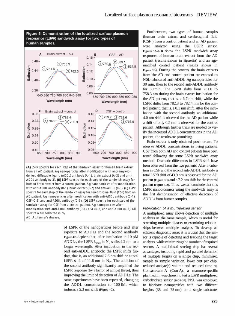
Figure 5. Demonstraresonance (LSPR) sahuman samples.
(A) LSPR spectra for eachfrom an AD patient. Ag nderived diffusable ligandADDL antibody (A-3). (B)human brain extract fromwith anti-ADDL antibodyspectra for each step of tAD patient. Ag nanopartCSF (C-2) and anti-ADDLsandwich assay for CSF fmodification with anti-ADspectra were collected inAD: Alzheimer’s disease.
Ext
inct
ion
Waveleng
0.48
0.44
0.40
0.36
640 680 720 760
1 751.6
2
Brain extr
Brain extra
Ext
inct
ion
Waveleng
0.36
0.32
0.28
0.24
0.20
0.16650 700 750
1 782.32
A
C
of LSPR of the nanoparticles before and afterexposure to ADDLs and the second antibody.Figure 4B depicts that, after incubation in 10 pMADDLs, the LSPR λmax in N2 shifts 4.2 nm to alonger wavelength. After incubation in the sec-ond anti-ADDL antibody, the LSPR shifts fur-ther, that is, an additional 7.6 nm shift or a totalLSPR shift of 11.8 nm in N2. The addition ofthe second antibody significantly amplified theLSPR response (by a factor of almost three), thusimproving the limit of detection of ADDLs. Thesame experiments have been repeated, changingthe ADDL concentration to 100 fM, whichinduces a 5.3 nm shift (Figure 4C).
Furthermore, two types of human samples(human brain extract and cerebrospinal fluid[CSF]) from a control patient and an AD patientwere analyzed using the LSPR sensor.Figures 5A & B show the LSPR sandwich assayresponses of human brain extract from the ADpatient (results shown in Figure 5A) and an age-matched control patient (results shown inFigure 5B). During the process, the brain extractsfrom the AD and control patient are exposed toNSL-fabricated anti-ADDL Ag nanoparticles for30 min, then to the second anti-ADDL antibodyfor 30 min. The LSPR shifts from 751.6 to758.3 nm during the brain extract incubation forthe AD patient, that is, a 6.7 nm shift; while theLSPR shifts from 782.3 to 782.4 nm for the con-trol patient, that is, a 0.1 nm shift. After the incu-bation with the second antibody, an additional4.0 nm shift is observed for the AD patient whilea shift of only 0.5 nm is observed for the controlpatient. Although further trials are needed to ver-ify the increased ADDL concentrations in the ADpatient, the results are promising.
Brain extract is only obtained postmortem. Toobserve ADDL concentrations in living patients,CSF from both AD and control patients have beentested following the same LSPR sandwich assaymethod. Dramatic differences in LSPR shift havebeen observed from the two patients. After incuba-tion in CSF and the second anti-ADDL antibody, atotal LSPR shift of 43.9 nm is observed for the ADpatient (Figure 5C) and a 7.2 nm shift for the controlpatient (Figure 5D). Thus, we can conclude that thisLSPR nanobiosensor using the sandwich assay isthe first demonstration of effective detection ofADDLs from human samples.
Fabrication of a multiplexed sensorA multiplexed assay allows detection of multipleanalytes in the same sample, which is useful forscreening multiple diseases or examining relation-ships between multiple analytes. To develop anefficient diagnostic assay, it is crucial that the sen-sor is capable of detecting and tracking the targetanalytes, while minimizing the number of requiredsensors. A multiplexed sensing chip has severaladvantages, including rapid and parallel detectionof multiple targets on a single chip, minimizedsample to sample variation, lower cost per chip,reduced analyte(s) volume and reduced time [9].Concanavalin A (Con A), a mannose-specificplant lectin, was chosen to test a LSPR multiplexedcarbohydrate sensor [10,35–37]. NSL was employedto fabricate nanoparticles with two differentheights (35 and 75 nm) on a single substrate.
tion of the localized surface plasmon ndwich assay for two types of
step of the sandwich assay for human brain extract anoparticles after modification with anti-amyloid-
(ADDL) antibody (A-1), brain extract (A-2) and anti- LSPR spectra for each step of the sandwich assay for a control patient. Ag nanoparticles after modification
(B-1), brain extract (B-2) and anti-ADDL (B-3). (C) LSPR he sandwich assay for cerebrospinal fluid (CSF) from an icles after modification with anti-ADDL antibody (C-1), antibody (C-3). (D) LSPR spectra for each step of the rom a control patient. Ag nanoparticles after DL antibody (D-1), CSF (D-2) and anti-ADDL (D-3). All
N2.
th (nm)
800 840 880
758.3
3 762.3
act – AD CSF – AD
ct – control CSF – control
th (nm)
800 850 900
782.43 782.9
Ext
inct
ion
Wavelength (nm)
0.16
0.14
0.12
0.10
0.08
0.06650 700 750 800 850 900 950
1 780.6 2 809.13 824.5
Ext
inct
ion
Wavelength (nm)
0.16
0.14
0.12
0.08
0.06
0.10
650 700 750 800 850 900
1 759.7 2 762.63 766.9
B
D
223
REVIEW – Zhao, Zhang, Ranjit Yonzon, Haes & Van Duyne
224
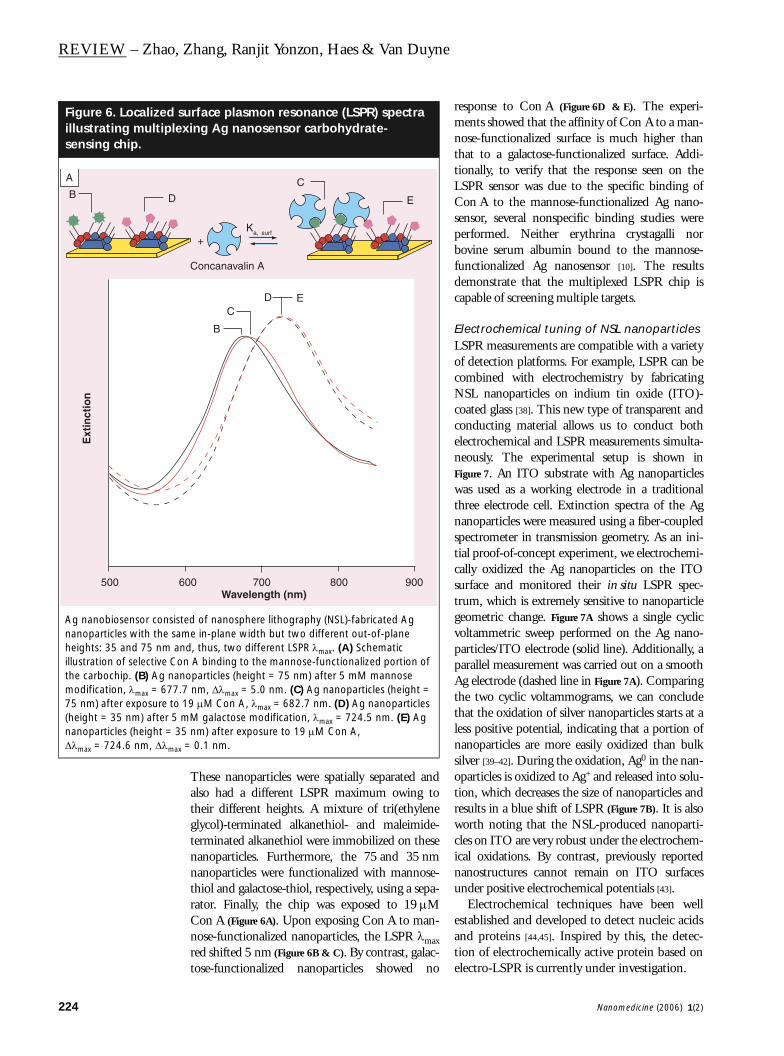
Figure 6. Localized sillustrating multiplesensing chip.
Ag nanobiosensor consisnanoparticles with the saheights: 35 and 75 nm aillustration of selective Cothe carbochip. (B) Ag namodification, λmax = 67775 nm) after exposure to(height = 35 nm) after 5nanoparticles (height = 3∆λmax = 724.6 nm, ∆λmax
Ext
inct
ion
500
B D
A
These nanoparticles were spatially separated andalso had a different LSPR maximum owing totheir different heights. A mixture of tri(ethyleneglycol)-terminated alkanethiol- and maleimide-terminated alkanethiol were immobilized on thesenanoparticles. Furthermore, the 75 and 35 nmnanoparticles were functionalized with mannose-thiol and galactose-thiol, respectively, using a sepa-rator. Finally, the chip was exposed to 19 µMCon A (Figure 6A). Upon exposing Con A to man-nose-functionalized nanoparticles, the LSPR λmaxred shifted 5 nm (Figure 6B & C). By contrast, galac-tose-functionalized nanoparticles showed no
response to Con A (Figure 6D & E). The experi-ments showed that the affinity of Con A to a man-nose-functionalized surface is much higher thanthat to a galactose-functionalized surface. Addi-tionally, to verify that the response seen on theLSPR sensor was due to the specific binding ofCon A to the mannose-functionalized Ag nano-sensor, several nonspecific binding studies wereperformed. Neither erythrina crystagalli norbovine serum albumin bound to the mannose-functionalized Ag nanosensor [10]. The resultsdemonstrate that the multiplexed LSPR chip iscapable of screening multiple targets.
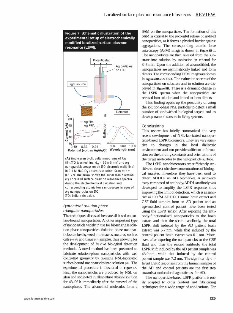
Electrochemical tuning of NSL nanoparticlesLSPR measurements are compatible with a varietyof detection platforms. For example, LSPR can becombined with electrochemistry by fabricatingNSL nanoparticles on indium tin oxide (ITO)-coated glass [38]. This new type of transparent andconducting material allows us to conduct bothelectrochemical and LSPR measurements simulta-neously. The experimental setup is shown inFigure 7. An ITO substrate with Ag nanoparticleswas used as a working electrode in a traditionalthree electrode cell. Extinction spectra of the Agnanoparticles were measured using a fiber-coupledspectrometer in transmission geometry. As an ini-tial proof-of-concept experiment, we electrochemi-cally oxidized the Ag nanoparticles on the ITOsurface and monitored their in situ LSPR spec-trum, which is extremely sensitive to nanoparticlegeometric change. Figure 7A shows a single cyclicvoltammetric sweep performed on the Ag nano-particles/ITO electrode (solid line). Additionally, aparallel measurement was carried out on a smoothAg electrode (dashed line in Figure 7A). Comparingthe two cyclic voltammograms, we can concludethat the oxidation of silver nanoparticles starts at aless positive potential, indicating that a portion ofnanoparticles are more easily oxidized than bulksilver [39–42]. During the oxidation, Ag0 in the nan-oparticles is oxidized to Ag+ and released into solu-tion, which decreases the size of nanoparticles andresults in a blue shift of LSPR (Figure 7B). It is alsoworth noting that the NSL-produced nanoparti-cles on ITO are very robust under the electrochem-ical oxidations. By contrast, previously reportednanostructures cannot remain on ITO surfacesunder positive electrochemical potentials [43].
Electrochemical techniques have been wellestablished and developed to detect nucleic acidsand proteins [44,45]. Inspired by this, the detec-tion of electrochemically active protein based onelectro-LSPR is currently under investigation.
urface plasmon resonance (LSPR) spectra xing Ag nanosensor carbohydrate-
ted of nanosphere lithography (NSL)-fabricated Ag me in-plane width but two different out-of-plane nd, thus, two different LSPR λmax. (A) Schematic n A binding to the mannose-functionalized portion of
noparticles (height = 75 nm) after 5 mM mannose .7 nm, ∆λmax = 5.0 nm. (C) Ag nanoparticles (height = 19 µM Con A, λmax = 682.7 nm. (D) Ag nanoparticles mM galactose modification, λmax = 724.5 nm. (E) Ag 5 nm) after exposure to 19 µM Con A, = 0.1 nm.
+Ka, surf
Concanavalin A
Wavelength (nm)600 700 800 900
C
E
B
CD E
Nanomedicine (2006) 1(2)
www.futuremedicine.com
Localized surface plasmon resonance biosensors – REVIEW
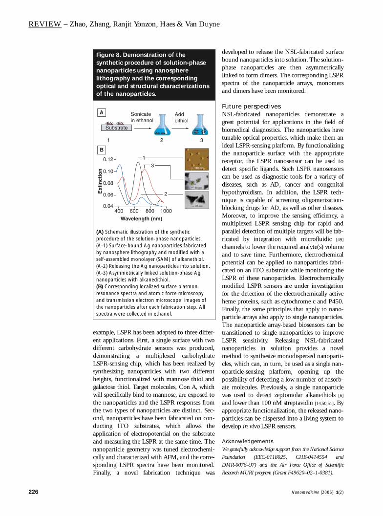
Synthesis of solution-phase triangular nanoparticlesThe techniques discussed here are all based on sur-face-bound nanoparticles. Another important typeof nanoparticle widely in use for biosensing is solu-tion-phase nanoparticles. Solution-phase nanopar-ticles can be dispersed into macrostructures, such ascells [46,47] and tissue [47] samples, thus allowing forthe development of in vivo biological detectionmethods. A novel method has been presented tofabricate solution-phase nanoparticles with wellcontrolled geometry by releasing NSL-fabricatedsurface-bound nanoparticles into solution [48]. Theexperimental procedure is illustrated in Figure 8A.First, the nanoparticles are produced by NSL onglass and incubated in alkanethiol ethanol solutionfor 48–96 h immediately after the removal of thenanospheres. The alkanethiol molecules form a
SAM on the nanoparticles. The formation of thisSAM is critical to the successful release of isolatednanoparticles, as it forms a physical barrier againstaggregations. The corresponding atomic forcemicroscopy (AFM) image is shown in Figure 8B-1.The nanoparticles are then released from the sub-strate into solution by sonication in ethanol for3–5 min. Upon the addition of alkanedithiol, thenanoparticles are asymmetrically linked and formdimers. The corresponding TEM images are shownin Figures 8B-2 & 8B-3. The extinction spectra of thenanoparticles on substrate and in solution are dis-played in Figure 8B. There is a dramatic change inthe LSPR spectra when the nanoparticles arereleased into solution and linked to form dimers.
This finding opens up the possibility of usingthe solution-phase NSL particles to detect a smallnumber of sandwiched biological targets and todevelop nanobiosensors in living systems.
ConclusionsThis review has briefly summarized the veryrecent development of NSL-fabricated nanopar-ticle-based LSPR biosensors. They are very sensi-tive to changes in the local dielectricenvironment and can provide sufficient informa-tion on the binding constants and orientations ofthe target molecules to the nanoparticle surface.
The LSPR nanobiosensors are sufficiently sen-sitive to detect ultralow concentrations of biologi-cal analytes. Therefore, they have been used todetect ADDLs: an AD biomarker. A sandwichassay composed of antibody/ADDL/antibody wasdeveloped to amplify the LSPR response, thusimproving the limit of detection, which is as sensi-tive as 100 fM ADDLs. Human brain extract andCSF fluid samples from an AD patient and anage-matched control patient have been testedusing the LSPR sensor. After exposing the anti-body-functionalized nanoparticles to the brainextract and then the second antibody, the totalLSPR shift induced by the AD patient brainextract was 6.7 nm, while that induced by thecontrol patient brain extract was 0.1 nm. More-over, after exposing the nanoparticles to the CSFfluid and then the second antibody, the totalLSPR shift induced by the AD patient sample was43.9 nm, while that induced by the controlpatient sample was 7.2 nm. The significantly dif-ferent LSPR responses from the human samples ofthe AD and control patients are the first steptowards a molecular diagnostic test for AD.
The nanoparticle-based LSPR platform is eas-ily adapted to other readout and fabricatingtechniques for a wide range of applications. For
Figure 7. Schematic illustration of the experimental setup of electrochemically modified localized surface plasmon resonance (LSPR).
(A) Single scan cyclic voltammograms of Ag film/ITO (dashed line, dm = 50 ± 5 nm) and Ag nanoparticle arrays on an ITO electrode (solid line) in 0.1 M NaClO4 aqueous solution. Scan rate = 0.1 V/s. The arrow shows the initial scan direction. (B) Localized surface plasmon resonance spectra during the electrochemical oxidation and corresponding atomic force microscopy images of Ag nanoparticles on ITO.ITO: Indium tin oxide.
0.40 0.32 0.24
80
40
0
-40
-80
Ag filmAg PPA
Cu
rren
t (µ
A)
Potential (volt vs Ag/AgCl)
Ext
inct
ion
Wavelength (nm)800 900 1000
Potentiostat
Light source
Detector
Ag particleson ITO
A B
225
REVIEW – Zhao, Zhang, Ranjit Yonzon, Haes & Van Duyne
226
example, LSPR has been adapted to three differ-ent applications. First, a single surface with twodifferent carbohydrate sensors was produced,demonstrating a multiplexed carbohydrateLSPR-sensing chip, which has been realized bysynthesizing nanoparticles with two differentheights, functionalized with mannose thiol andgalactose thiol. Target molecules, Con A, whichwill specifically bind to mannose, are exposed tothe nanoparticles and the LSPR responses fromthe two types of nanoparticles are distinct. Sec-ond, nanoparticles have been fabricated on con-ducting ITO substrates, which allows theapplication of electropotential on the substrateand measuring the LSPR at the same time. Thenanoparticle geometry was tuned electrochemi-cally and characterized with AFM, and the corre-sponding LSPR spectra have been monitored.Finally, a novel fabrication technique was
developed to release the NSL-fabricated surfacebound nanoparticles into solution. The solution-phase nanoparticles are then asymmetricallylinked to form dimers. The corresponding LSPRspectra of the nanoparticle arrays, monomersand dimers have been monitored.
Future perspectivesNSL-fabricated nanoparticles demonstrate agreat potential for applications in the field ofbiomedical diagnostics. The nanoparticles havetunable optical properties, which make them anideal LSPR-sensing platform. By functionalizingthe nanoparticle surface with the appropriatereceptor, the LSPR nanosensor can be used todetect specific ligands. Such LSPR nanosensorscan be used as diagnostic tools for a variety ofdiseases, such as AD, cancer and congenitalhypothyroidism. In addition, the LSPR tech-nique is capable of screening oligomerization-blocking drugs for AD, as well as other diseases.Moreover, to improve the sensing efficiency, amultiplexed LSPR sensing chip for rapid andparallel detection of multiple targets will be fab-ricated by integration with microfluidic [49]
channels to lower the required analyte(s) volumeand to save time. Furthermore, electrochemicalpotential can be applied to nanoparticles fabri-cated on an ITO substrate while monitoring theLSPR of these nanoparticles. Electrochemicallymodified LSPR sensors are under investigationfor the detection of the electrochemically activeheme proteins, such as cytochrome c and P450.Finally, the same principles that apply to nano-particle arrays also apply to single nanoparticles.The nanoparticle array-based biosensors can betransitioned to single nanoparticles to improveLSPR sensitivity. Releasing NSL-fabricatednanoparticles in solution provides a novelmethod to synthesize monodispersed nanoparti-cles, which can, in turn, be used as a single nan-oparticle-sensing platform, opening up thepossibility of detecting a low number of adsorb-ate molecules. Previously, a single nanoparticlewas used to detect zeptomolar alkanethiols [6]
and lower than 100 nM streptavidin [14,50,51]. Byappropriate functionalization, the released nano-particles can be dispersed into a living system todevelop in vivo LSPR sensors.
AcknowledgementsWe gratefully acknowledge support from the National ScienceFoundation (EEC-0118025, CHE-0414554 andDMR-0076–97) and the Air Force Office of ScientificResearch MURI program (Grant F49620–02–1-0381).
Figure 8. Demonstration of the synthetic procedure of solution-phase nanoparticles using nanosphere lithography and the corresponding optical and structural characterizations of the nanoparticles.
(A) Schematic illustration of the synthetic procedure of the solution-phase nanoparticles. (A-1) Surface-bound Ag nanoparticles fabricated by nanosphere lithography and modified with a self-assembled monolayer (SAM) of alkanethiol. (A-2) Releasing the Ag nanoparticles into solution. (A-3) Asymmetrically linked solution-phase Ag nanoparticles with alkanedithiol. (B) Corresponding localized surface plasmon resonance spectra and atomic force microscopy and transmission electron microscope images of the nanoparticles after each fabrication step. All spectra were collected in ethanol.
Wavelength (nm)
Ext
inct
ion
400 600 800 1000
0.12
0.10
0.08
0.06
0.04
1
3
2
Sonicatein ethanol
Adddithiol
1 2 3
Substrate
A
B
Nanomedicine (2006) 1(2)
www.futuremedicine.com
Localized surface plasmon resonance biosensors – REVIEW
Executiv
• Nanopasuitable
• LSPR ofsurroun
• The nannanostr
• The NSL(AD) – a
• The LSPresults a
• A multifsingle c
• Nanopananopacorrespo
• A novelinto solu
e summary
rticle-based localized surface plasmon resonance (LSPR) biosensing is a broad platform for many different sensing architectures.
noble metal nanoparticles is highly sensitive to the adsorbate-induced changes in the ding dielectric environment and, thus, can be used for biosensing.
osphere lithography (NSL) technique is an inexpensive method to produce well-ordered uctures that are good biosensing platforms.
-fabricated Ag nanoparticles were used to detect the biomarker of Alzheimer’s disease myloid-derived diffusable ligands.
R response has been tested using human samples from AD and control patients and the re significantly different.
unctionalized nanoparticle sensor has been fabricated to demonstrate multiplex sensing on a hip.
rticles have been fabricated using NSL on a transparent conducting substrate. The rticle structure can be finely tuned by applying a potential to the substrate and the nding optical properties have been monitored.
technique has been developed to release the NSL-fabricated nanoparticles from the substrate tion.
Bibliography1. Turner APF: Biosensors – sense and
sensitivity. Science 290, 1315–1317 (2000).2. Chan WCW, Nie SM: Quantum dot
bioconjugates for ultrasensitive nonisotopic detection. Science 281, 2016–2018 (1998).
3. Chan WCW, Maxwell DJ, Gao XH, Bailey RE, Han MY, Nie SM: Luminescent quantum dots for multiplexed biological detection and imaging. Curr. Opin. Biotechnol. 13, 40–46 (2002).
4. Mucic RC, Storhoff JJ, Mirkin CA, Letsinger RL: DNA-directed synthesis of binary nanoparticle network materials. J. Amer. Chem. Soc. 120, 12674–12675 (1998).
5. Malinsky MD, Kelly KL, Schatz GC, Van Duyne RP: Chain length dependence and sensing capabilities of the localized surface plasmon resonance of silver nanoparticles chemically modified with alkanethiol self-assembled monolayers. J. Amer. Chem. Soc. 123, 1471–1482 (2001).
6. McFarland AD, Van Duyne RP: Single silver nanoparticles as real-time optical sensors with zeptomole sensitivity. Nano Lett. 3, 1057–1062 (2003).
7. Hicks EM, Zhang XY, Zou SL et al.: Plasmonic properties of film over nanowell surfaces fabricated by nanosphere lithography. J. Phys. Chem. B 109, 22351–22358 (2005).
8. Mrksich M, Grunwell JR, Whitesides GM: Biospecific adsorption of carbonic-anhydrase to self-assembled monolayers of alkanethiolates that present
benzenesulfonamide groups on gold. J. Am. Chem. Soc. 117, 12009–12010 (1995).
9. Berger CEH, Beumer TAM, Kooyman RPH, Greve J: Surface plasmon resonance multisensing. Anal. Chem. 70, 703–706 (1998).
10. Yonzon CR, Jeoung E, Zou S, Schatz GC, Mrksich M, Van Duyne RP: A comparative analysis of localized and propagating surface plasmon resonance sensors: the binding of concanavalin A to a monosaccharide functionalized self-assembled monolayer. J. Am. Chem. Soc. 126, 12669–12676 (2004).
11. Nie S, Emory SR: Probing single molecules and single nanoparticles by surface-enhanced Raman scattering. Science 275, 1102–1106 (1997).
12. Zhang XY, Young MA, Lyandres O, Van Duyne RP: Rapid detection of an anthrax biomarker by surface-enhanced Raman spectroscopy. J. Amer. Chem. Soc. 127, 4484–4489 (2005).
13. Jiang J, Bosnick K, Maillard M, Brus L: Single molecule Raman spectroscopy at the junctions of large Ag nanocrystals. J. Phys. Chem. B 107, 9964–9972 (2003).
14. Haes AJ, Van Duyne RP: A nanoscale optical biosensor: sensitivity and selectivity of an approach based on the localized surface plasmon resonance spectroscopy of triangular silver nanoparticles. J. Am. Chem. Soc. 124, 10596–10604 (2002).
15. Riboh JC, Haes AJ, McFarland AD, Yonzon CR, Van Duyne RP: A nanoscale optical biosensor: real-time immunoassay in physiological buffer enabled by improved nanoparticle adhesion. J. Phys. Chem. B 107, 1772–1780 (2003).
16. Jensen TR, Duval ML, Kelly KL, Lazarides A, Schatz GC, Van Duyne RP: Nanosphere lithography: effect of the external dielectric medium on the surface plasmon resonance spectrum of a periodic array of silver nanoparticles. J. Phys. Chem. B 103, 9846–9853 (1999).
17. Jensen TR, Kelly KL, Lazarides A, Schatz GC: Electrodynamics of noble metal nanoparticles and nanoparticle clusters. J. Cluster Sci. 10, 295–317 (1999).
18. Jensen TR, Malinsky MD, Haynes CL, Van Duyne RP: Nanosphere lithography: tunable localized surface plasmon resonance spectra of silver nanoparticles. J. Phys. Chem. B 104, 10549–10556 (2000).
19. Malinsky MD, Kelly KL, Schatz GC, Van Duyne RP: Nanosphere lithography: effect of substrate on the localized surface plasmon resonance spectrum of silver nanoparticles. J. Phys. Chem. B 105, 2343–2350 (2001).
20. Hutter E, Fendler JH: Exploitation of localized surface plasmon resonance. Advanced Mater. 16, 1685–1706 (2004).
21. Stuart DA, Haes AJ, Yonzon CR, Hicks EM, Van Duyne RP: Biological applications of localised surface plasmonic phenomenae. IEE Proc. Nanobiotechnol. 152, 13–32 (2005).
227

REVIEW – Zhao, Zhang, Ranjit Yonzon, Haes & Van Duyne
22. Hulteen JC, Van Duyne RP: Nanosphere lithography: a materials general fabrication process for periodic particle array surfaces. J. Vacuum Sci. Technol. A 13, 1553–1558 (1995).
23. Haynes CL, Van Duyne RP: Nanosphere lithography: a versatile nanofabrication tool for studies of size-dependent nanoparticle optics. J. Phys. Chem. B 105, 5599–5611 (2001).
24. Haes AJ, Zou S, Schatz GC, Van Duyne RP: A nanoscale optical biosensor: the long range distance dependence of the localized surface plasmon resonance of noble metal nanoparticles. J. Phys. Chem. B 108, 109–116 (2004).
25. Nath N, Chilkoti A: A colorimetric gold nanoparticle sensor to interrogate biomolecular interactions in real time on a surface. Anal. Chem. 74, 504–509 (2002).
26. Okamoto T, Yamaguchi I, Kobayashi T: Local plasmon sensor with gold colloid monolayers deposited upon glass substrates. Optics Lett. 25, 372–374 (2000).
27. Yonzon CR, Zhang XY, Van Duyne RP: Localized surface plasmon resonance immunoassay and verification using surface-enhanced Raman spectroscopy. Proc. SPIE – Int. Soc. Optical Engin. (2003).
28. Campbell DJ, Herr BR, Hulteen JC, Van Duyne RP, Mirkin CA: Ion-gated electron transfer in self-assembled monolayer films. J. Am. Chem. Soc. 118, 10211–10219 (1996).
29. Haes AJ, Chang L, Klein WL, Van Duyne RP: Detection of a biomarker for Alzheimer’s disease from synthetic and clinical samples using a nanoscale optical biosensor. J. Amer. Chem. Soc. 127, 2264–2271 (2005).
30. Hardy J, Selkoe DJ: Medicine – the amyloid hypothesis of Alzheimer’s disease: progress and problems on the road to therapeutics. Science 297, 353–356 (2002).
31. Selkoe DJ, Hardy J: The search for an amyloid solution – response. Science 298, 963–964 (2002).
32. Gong YS, Chang L, Viola KL et al.: Alzheimer’s disease-affected brain: presence of oligomeric Aβ ligands (ADDLs) suggests
a molecular basis for reversible memory loss. Proc. Natl Acad. Sci. USA 100, 10417–10422 (2003).
33. Lambert MP, Barlow AK, Chromy BA et al.: Diffusible, nonfibrillar ligands derived from Aβ(1–42) are potent central nervous system neurotoxins. Proc. Natl Acad. Sci. USA 95, 6448–6453 (1998).
34. Walsh DM, Klyubin I, Fadeeva JV et al.: Naturally secreted oligomers of amyloid β protein potently inhibit hippocampal long-term potentiation in vivo. Nature 416, 535–539 (2002).
35. Hardman KD, Schiffer M, Wood MK, Ainswort Cf, Edmundso AB: X-ray crystallographic studies of concanavalin-A. Cold Spring Harbor Symp. Quant. Biol. 36, 271–276 (1971).
36. Hardman KD, Ainswort CF: Myoinositol binding-site of concanavalin A. Nat. New Biol. 237, 54–55 (1972).
37. Smith, EA, Thomas WD, Kiessling LL, Corn RM: Surface plasmon resonance imaging studies of protein–carbohydrate interactions. J. Am. Chem. Soc. 125, 6140–6148 (2003).
38. Zhang XY, Hicks EM, Zhao J, Schatz GC, Van Duyne RP: Electrochemical tuning of silver nanoparticles fabricated by nanosphere lithography. Nano Lett. 5, 1503–1507 (2005).
39. Plieth WJ: The work function of small metal particles and its relation to electrochemical properties. Surface Sci. 156, 530–535 (1985).
40. Ng KH, Liu H, Penner RM: Subnanometer silver clusters exhibiting unexpected electrochemical metastability on graphite. Langmuir 16, 4016–4023 (2000).
41. Chaki NK, Sharma J, Mandle AB, Mulla IS, Pasricha R, Vijayamohanan K: Size dependent redox behavior of monolayer protected silver nanoparticles (2–7 nm) in aqueous medium. Phys. Chem. Chem. Phys. 6, 1304–1309 (2004).
42. Redmond PL, Hallock AJ, Brus LE: Electrochemical Ostwald ripening of colloidal Ag particles on conductive substrates. Nano Lett. 5, 131–135 (2005).
43. Ung T, Giersig M, Dunstan D, Mulvaney P: Spectroelectrochemistry of colloidal silver. Langmuir 13, 1773–1782 (1997).
44. Kojima K, Hiratsuka A, Suzuki H, Yano K, Ikebukuro K, Karube I: Electrochemical protein chip with arrayed immunosensors with antibodies immobilized in a plasma-polymerized film. Anal. Chem. 75, 1116–1122 (2003).
45. Wang J: Towards genoelectronics: electrochemical biosensing of DNA hybridization. Chem. Eur. J. 5, 1681–1685 (1999).
46. Kneipp K, Haka AS, Kneipp H et al.: Surface-enhanced Raman spectroscopy in single living cells using gold nanoparticles. Appl. Spectroscopy 56, 150–154 (2002).
47. Thygesen LG, Jorgensen K, Moller BL, Engelsen SB: Raman spectroscopic analysis of cyanogenic glucosides in plants: development of a flow injection surface-enhanced Raman scatter (FI-SERS) method for determination of cyanide. Appl. Spectroscopy 58, 212–217 (2004).
48. Haes AJ, Zhao J, Zou SL et al.: Solution-phase, triangular Ag nanotriangles fabricated by nanosphere lithography. J. Phys. Chem. B 109, 11158–11162 (2005).
49. Wheeler AR, Throndset WR, Whelan RJ et al.: Microfluidic device for single-cell analysis. Anal. Chem. 75, 3581–3586 (2003).
50. Haes AJ, Stuart DA, Nie S, Van Duyne RP: Using solution-phase nanoparticles, surface-confined nanoparticle arrays and single nanoparticles as biological sensing platforms. J. Fluorescence 14, 355–367 (2004).
51. Van Duyne RP, Haes AJ, McFarland AD: Nanoparticle optics: sensing with nanoparticle arrays and single nanoparticles. Proc. SPIE – Int. Soc. Optical Engin. 5223, 197–207 (2003).
Website101. Alzheimer’s Association. About Alzheimer's
disease www.alz.org/
228 Nanomedicine (2006) 1(2)


![CoordinatedDispersionandAggregationofGoldNanorodin ...biosensors, which include surface plasmon resonance [12, 13], waveguide mode sensor [14], colorimetric [15], and RAMAN spectroscopy](https://img.pdfslide.us/doc/110x75/60df4ae239770b0b1f37c3cb/coordinateddispersionandaggregationofgoldnanorodin-biosensors-which-include.jpg)