Embed Size (px)
Citation preview

PEDIATRIC DENTISTRY/Copyright s 1981 byThe American Academy of PedodonticsVol. 3, Special Issue
Localized juvenile periodontitis(periodontosis)
Michael G. Newman, DOS
AbstractA discussion of the recent information regarding the
terminology, clinical manifestations and epidemiology of theperiodontal disease termed Localized Juvenile Periodontitis(LJP) is presented in the context of newer scientificadvances. Emphasis is given to the microbiological andimmunological parameters associated with the etiology ofthe disease previously referred to as Periodontosis. Newertherapeutic modalities including adjunctive antimicrobialtherapy appear to offer promise in the treatment of thishighly destructive disease.
Introduction
Interest in the rapidly destructive form of perio-dontal disease referred to as "periodontosis" hasgreatly increased since 1973 when the suggestion of aunique bacterial etiology was made. In preliminaryfindings Newman et al.1 reported that gram negativerods predominated at the forefront of molar incisor le-sions in patients diagnosed as having "periodontosis."In that study, a sampling device which permitted bac-terial sampling at the apical zone of the lesion wascombined with newly developed methods2 of increas-ing the viable recovery of plaque bacteria. Their re-port stimulated great interest among microbiologists,immunologists and clinicians since it suggested thatbacterial specificity may be an important etiologic fac-tor in this disease.
In the last seven years a major world-wide researcheffort has taken place in all areas of study. Particu-larly important has been the recognition that therapid periodontal breakdown which occurs in "perio-dontosis" may also occur during periods of exacerba-tion in more common adult forms of periodontitis.3'4
Understanding more about these periods of disease ac-tivity may provide the basis to unraveling key fea-tures associated with destructive periodontal diseasesin man. Since "periodontosis" can be sharply definedit may be considered as an ideal model in which exac-erbation and bone destruction can be more accuratelypredicted and studied.
Dr. Newman
The following discussion will highlight recent majorareas of research activity which have provided thebasis for our current understanding of this impor-tant periodontal disease. Many reviews have beenpublished.5*7
Terminology
Even though a more clear understanding of theetiology and pathogenesis of this disease currently ex-ists, the terminology used to describe this entity hasbecome more confusing. Confusion may result from afailure to clarify and correlate the clinical descriptorswith research findings. These findings have clearlydemonstrated that specific microbiologic, immuno-logic and histologic features can be associated withthis disease process (Table 1). It is suggested thatthese findings become incorporated into, and becomepart of, the diagnostic features and then become thebasis for classification of the disease process. Table 2lists some current terminology. The author prefers thedifferentiation of the molar-incisor type of bone lossfrom the generalized involvement since this conditioncan be distinctly separated on the basis of clinical cri-teria in moderate and advanced cases. Localized Juve-nile Periodontitis, (LJP), is the suggested term for themolar incisor involvement. Generalized terms can bereferred to as Juvenile Periodontitis, (JP). As withany classification system there are always exceptions.In a recent study Hormand and Frandsen8 describedthree types of bone loss in patients with JP. Type I in-cluded first molars and/or incisors. Type II includedfirst molars and/or incisors and some additional teeth(less than 14 involved teeth) and Type III patientswere generally involved. The authors referred to allthree types as JP.
In 1977 Sugarman and Sugarman9 argued that theterm Precocious Periodontitis be used to describe bothlocalized and generalized forms. The authors arguedthat this terminology more clearly describes the clini-cal manifestation of the disease process. In 1978 Baerand Kaslick pointed out that the term "Periodon-
PEDIATRIC DENTISTRY
Volume 3, Special Issue121

Table 1. Possible etiologic factors associated with LJP. Table 2. Current terminology.
Subgingival PlaqueGram Negative RodsPMN Dysfunction
Cell Mediated DeficiencyAltered Cementum
tosis’" was never intended to mean "degenerative."They suggested the etymological derivation was fromthe Greek and properly defined the term "Pe~iodon-tosis"as meaning "an abnormal or diseased conditionof the periodontium." It is clear that confusion overterminology and definitions exist. The following dis-cussion may help to clarify the confusion and providethe basis for agreement.
Definition m Clinical FeaturesAge of Onset: The circumpubertal period appears tobe the primary time when the disease process mani-fests. 11,1. In some cases however, bone destruction canbe documented to occur in the mid-teen years. LJP asdescribed in this article, does not occur in the pre-pubertal child, it is a disease of the immediate preteenand teenage years. If rapid or unusual periodontal de-struction occurs in young children it may be simplytermed, Periodontitis.
Clinical Manifestations: Table 3 summarizes the majorclinical manifestations associated with LJP. The keyfeatures of early and moderate forms of the disease areunlike the adult forms of periodontitis of similar in-volvement. Particularly important is the lack of thoseclinical signs which result from an acute inflammatoryresponse such as: gingival erythema, edema, andbleeding adjacent to involved teeth. When poor oralhygiene is present plaque and calculus may inducegingival inflammation. Later clinical findings (Table
Table 3. LJP.. Early clinical findings.
AgeSystemicBone Loss
Onset and CourseGingiva
Plaque
MicrobiologyOther
CircumpubertalNoneMolar -- Incisor -- Bilateraland Symmetrical
Insidious + RapidLittle Clinical Inflammation
Small Amounts
Characteristic Gram Neg. FloraRegional Lymph NodesFamilial PatternNo Primary Teeth InvolvementPMN Dysfunction
PeriodontosisJuvenile Periodontitis
*Localized Juvenile PeriodontitisIdiopathic Juvenile Periodontitis
Precocious PeriodontitisGottlieb Syndrome
Destructive Juvenile Periodontitis
* Preferred Term
4) are more typical of "inflammatory" chronic perio-dontitis. In these cases roentgenographic evidence ofbone loss and periodontal probing can be used to con-firm the diagnosis of LJP.
Many patterns of bone loss may occur among in-volved teeth. Maxillary first molars and incisors areaffected to a higher degree than their mandibularcounterparts. 8 There are numerous studies which havedocumented the bilateral (cross arch) symmetry bone loss. Bone loss occurs rapidly. One study18 hasdocumented an average attachment loss of 4-5microns per day.
General Health: LJP occurs in healthy patients. Nosystemic disease occurs with this condition. Whenrapid periodontal destruction occurs in patients withcertain systemic conditions such as Papillon Lefevresyndrome or cyclic neutropenia it is called "periodon-titis" not LJP. A recent study which examined the mi-crobiota associated with Papillon Lefevre syndrome’4
demonstrated that a flora similar to adult forms ofperiodontitis was present. The microbiota was re-markably different from that seen in patients withLJP (see below).
Recent studies (see below) have also suggested thatan underlying defect in polymorphonuclear leukocytes{PMN) may predispose affected patients to LJP.Clark et al. ’5 have suggested that the PMN dysfunc-tion is genetically transmitted and is re|atively mild.Affected patients have no other predisposition (as faras is known) to other infectious agents. This observa-tion does not alter the conceptual basis for definingLJP as occurring in healthy patients.
Table 4. LIP: Late clinica~ findings.
Mobility and MigrationDiasthemata
Pocket FormationRoot Sensitivity
Pain Upon MasticationAbscess Formation
Plaque and InflammationBurn Out
LOCALIZED JUVENILE PERIODONTITIS (PERIODONTOSIS)122 Newman

EpidemiologyIncidence: A recent unpublished study conducted byHew and Killoy TM examined 22,000 U.S. Air Forcerecruits for evidence of LJP. Their findings indicatedan overall incidence of 0.255%. When race was consid-ered, blacks had an overall incidence of 0.410% whilecaucasians had an overall incidence of 0.198%.
Sex Ratio: Hew and Killoy ~s and others reported anoverall female to male ratio of 1.05:1. When racial con-siderations were analyzed the female to male ratioamong blacks was 1.16:1, and among caucasians 1.19:1.Horman and Frandsen~ found that the overall femaleto male ratio was 2.5:1 in their caucasian population.When broken down according to age, younger females(12-18) had a female to male ratio of 5.3:1 while olderfemales had a ratio of 2.4:1.
Familial Pattern: The familial pattern of the occurrenceof LJP is well documented. Potential genetic pre-disposition is very likely based on recent immunologicstudies as well as from historical data.
Etiology: The Role of MicroorganismsWhile many details describing the microbiota of
the LJP lesions have not been clarified, a number ofobservations seem consistent (Table 5). The number organisms in LJP lesions is less than in most forms ofdestructive periodontal disease3 ranging from 1@-1@bacteria per pocket. This may be one to three ordersof magnitude lower than counts of organisms in adultperiodontitis pockets of similar depth and associatedbone loss. From microscopic studies of in situ plaque,it appears that the organisms at the apical portion ofthe pocket are loosely and sparsely attached to thetooth surfacemT.~ with little evidence of calcification.Clinically, the root surfaces feel hard and smooth.
The predominant cultivable microbiota of the LJPlesion is dominated by gram negative rods.’,’9.~.~’ Mostof the isolates from such lesions have been saccha-rolytic and capnophilic to anaerobic (Table 6). At thepresent time, three groups of organisms appear to bemore frequently encountered in lesions of LJP pa-tients, as well as in other patients with early onsethighly destructive periodontal disease.~ These organ-isms are not isolated in the same magnitude or fre-
Table 5. Microbiologic features associated with LJP.
Fewer bacteriaUnusual gram negative rodsMaj or proportion of plaque -- unattachedPathogenic potential in animal models~
Immunologic responseAntibacterial treatment
quency in diseases affecting adult patients.One group which has received considerable atten-
tion consists of fusiform-shaped, capnophilic rodswhich have the ability to glide on agar surfaces. Mem-bers of this group had been called Bacteroides ochra-ceus, but are now designated by the genus name Cap-nocytopl~aga.~,~.~ The creation of the new genus wasbased on the recognition of a typical "wet spreading"colony demonstrating the gliding ability of the organ-isms on the solid agar surface. The organisms are notanaerobic (as originally described by Newman et al.~),but require increased carbon dioxide tension for theirgrowth. The ability to glide on surfaces has long beenrecognized in organisms colonizing other sites in na-ture, particularly the soil. Since the oral isolates mostresembled the gliding aerobic genus Cy~opbaga, a newgenus Capnocytopl~aga was proposed to include thecarbon dioxide loving oral strains. ~ The genus Capno-cytopl~aga includes three species: Capnocytopl~aga
ochracea, Capnocytopt~aga sputigena and Capnocy-tophaga ging~valls. ~,~ In early publications, the Cap-nocytopl~aga strains had been designated Group II.’~,~
All three species have been detected in LJP lesionswith total Capnocytophaga counts averaging 25% ofthe organisms isolated.
A second group of capnophilic, gram negative rodsfrequently isolated from LJP lesions includes the spe-cies Actinobacillus ac~inomycetemcomitans.~,~ Strainsof this species were included in Groups III and IV inone of the original descriptions of the microbiota ofLJP lesions; ~,~ The colonies are small, convex andsmooth, while the cells are short, round-ended and oc-casionally slightly curved. Strains of this species pro-duce a protein exotoxin capable of destroying poly-morphonuclear leukocytes and gingival fibroblasts.~,~.~’
The exact role that this organism, its exotoxin and thehost response to the exotoxin play in the pathogenesisis under intensive investigation and is one of the mostexciting avenues of research concerning this diseaseentity.
Table 6. Changes in terminology for LJP associated bacteria.
Original CurrentClassific~ tion~°’~ Terminology *
¯ Group I ..........................Anaerobic Vibrio-STB¯ Group II ........................Capnocytopbaga¯ Group III .......................Group III STB & Actinobscillus¯ Group IV ....................... Actinobacillus (Y4).......................................... Bacteroides corrodens¯ Group V ......................... Eubacte~m
* Other bacteria within groups I - V are not listed under heading"Current Terminology" since they have not been characterizedfurther.
PEDIATRIC DENTISTRY123
Volume 3, Special Issue
A third group of organisms frequently encounteredin LJP lesions includes organisms previously placed in"periodontosis Group V.’’19,~ Strains of this groupoften demonstrate a type of spreading on the agar sur-face. Many isolates from Group V appear to fit de-scriptions of the species Eubacterium saburrheum2This species stains Gram positively in very youngcultures and Gram negatively in older preparations,cells are long blunt-ended rods which often producefilaments.
The three groups of organisms briefly discussedabove are by no means solely confined to LJP lesionsor lesions of other forms of early onset destructive dis-eases. However, their frequency of isolation and theirproportions when isolated are clearly much higher inthese lesions than in lesions of adult destructive dis-eases or healthy sites of adult patients or age-matchedcontrols.
In 1978, Allen and Brady18 found rod-shapedmicroorganisms which were continuously presentalong the entire cemental surface of extracted LJP in-volved teeth. The cultural and anatomical associationof microorganisms associated with LJP lesions pro-vides the basis for research which may clarify theexact role of these bacteria. These observations wereprerequisite to a more accurate assessment of the re-sponse (or failure to respond) by the host to poten-tially pathogenic bacteria and/or their products.
Immunologic StudiesAntibody: IgG antibody response to many of the sub-gingival bacteria associated with LIP lesions has beendocumented.~,u.~ In particular, antibody to A.actinomycetemcondtans and its leukotoxin hasreceived the greatest attention, u,~.~,~ This responsehas been interpreted by some as a blocking effect ofthe leukotoxinY The antibody response to A. ac-tinomycetemcomitans is also strain specificu,4! indicat-ing the possibility of virulent and avirulent strains.
Gingival fluid antibodies appear to parallel anti-body activity in serum~ but, antibody coating of sub-gingival bacteria from LIP lesions may not occur tohigh degree?2 These observations may provide insightinto the host-bacterial interactions which take placein the lesion. Also, the pathogenicity of the residentflora may be enhanced since the bacterial numberswould not be limited by antibody interactions occur-ring within the lesion.
Polymorphonuclear Leukocytes (PMN|: The current re-search investigating PMN functions in LIP may pro-vide a more clear understanding of the pathogenicmechanisms involved with the disease process.|8,43.~,~
Table 7 lists the major research findings associatedwith PMNs.
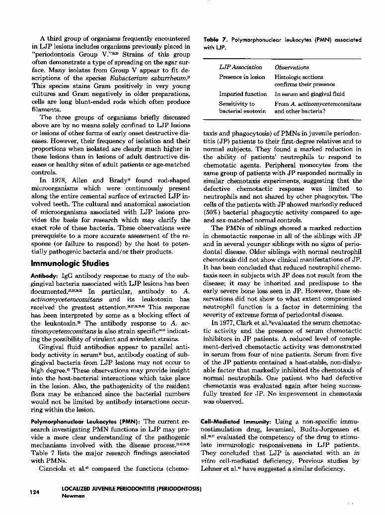
Cianciola et al?3 compared the functions (chemo-
Table 7. Polymorphonuclear leukocytes (PMN) associatedwith LJP.
IMP Association
Presence in lesion
Imparied function
Sensitivity tobacterial exotoxin
Observations
Histologic sectionsconirLrms their presence
In serum and gingival fluid
From A. actinomycetemcomitansand other bacteria?
taxis and phagocytosis) of PMNs in juvenile periodon-titis (JP) patients to their first-degree relatives and normal subjects. They found a marked reduction inthe ability of patients’ neutrophils to respond tochemotatic agents. Peripheral monocytes from thesame group of patients with JP responded normally insimilar chemotaxis experiments, suggesting that thedefective chemotactic response was limited toneutrophils and not shared by other phagocytes. Thecells of the patients with JP showed markedly reduced(5070) bacterial phagocytic activity compared to age-and sex-matched normal controls.
The PMNs of siblings showed a marked reductionin chemotactic response in all of the siblings with JPand in several younger siblings with no signs of perio-dontal disease. Older siblings with normal neutrophilchemotaxis did not show clinical manifestations of JP.It has been concluded that reduced neutrophil chemo-taxis seen in subjects with JP does not result from thedisease; it may be inherited and predispose to theearly severe bone loss seen in JP. However, these ob-servations did not show to what extent compromisedneutrophil function is a factor in determining theseverity of extreme forms of periodontal disease.
In 1977, Clark et al.%valuated the serum chemotac-tic activity and the presence of serum chemotacticinhibitors in JP patients. A reduced level of comple-ment-derived chemotactic activity was demonstratedin serum from four of nine patients. Serum from fiveof the JP patients contained a heat-stable, non-dialyz-able factor that markedly inhibited the chemotaxis ofnormal neutrophils. One patient who had defectivechemotaxis was evaluated again after being success-fully treated for JP. No improvement in chemotaxiswas observed.
Cell-Mediated Immunity: Using a non-specific immu-nostimulation drug, levamisol, Budtz-Jorgensen etal. ~,~ evaluated the competency of the drug to stimu-late immunologic responsiveness in LIP patients.They concluded that LIP is associated with an invitro cell-mediated deficiency. Previous studies byLehner et al?9 have suggested a similar deficiency.
124LOCALIZED JUVENILE PERIODONTITIS (PERIODONTOSIS)Newman

TreatmentSince the suggestion that subgingival plaque may
be etiologic and contribute to the destructive processin LJP, much of the treatment employed in these pa-tients has been directed towards the control of the as-sociated bacteria.1949 The results of treatment can beexcellent especially when initiated early in the clinicalcourse of the disease and/or when the destructiveprocess begins late in the teen years (Table 8). Table 4summarizes the current treatment modalities.
The clinical course of LJP is rapid and fairly pre-dictable. However, there is a phenomenon referred toas "burn out" which is seen in many cases. Burn outrefers to a sudden and unexplainable decrease in therate of bone destruction, and is almost always seen inthe mid- and late twenties. In these cases, the onset ofalveolar resorption apparently occurred in the mid- tolate teens rather than during the circumpubertal pe-riod. Although many theories have been postulated re-garding this phenomenon, none are based on scientificfact.
The use of systemic tetracycline in patients withLJP appears to enchance treatment success in some
Table 8. Diagnostic features associated with treatment ofLJP.
Diagnostic Feature
Age of onset
Depth of lesion
Furcation involvement
Oral hygiene
Occlusion
Effect on Treatment
Late onset favorssuccessful treatment
Incipient lesions favorstreatment
Decreases success
Improves prognosis
Excessive forcesdecreases success
Table 9. Treatment modalities currently used in LJP patients.
Nonsurgical
Plaque controlRoot planingAntibacterial agentsOrthodontic treatmentOcclusal adjustment
Surgical
Mucoperiosteal flapOsseous graftingAutologous tooth transplantationHemisection, root amputationExtraction
patients. When employed by the author, the combina-tion of surgical intervention, stablization, and sys-temic antibiotic appears to have success when thepatients were selected on the basis of antimicrobialsusceptibility testing of the organisms found in thelesions (Figure 1). The predictability of treatmentsuccess can be enchanced (Table 9) as in other formsof destructive periodontal disease in man. There isno better treatment than prevention through earlydiagnosis.
Figure 1. Roentgenographs of 18-year-old girl treated for LJP with
conventional therapy and systemic tetracycline. Patients bacteriawere tested for susceptibility to tetracycline prior to treatment. Top:
Bitewing roentgenographs at age 14 demonstrating moderate bone
loss around remaining first molars. Bottom: Bitewing roentgeno-
graphs four years later demonstrating cessation of visable bone
loss and improvement in roentgenographic picture. Clinically,
pocket depths were 3 mm or less in all areas. M. G. Newman,
E. Montierth and R. Williams.
Dr. Newman is adjunct associate professor, school of dentistry,University of California, The Center for the Health Sciences, LosAngeles, CA 90024. Requests for reprints should be sent to him atthat address.
References
1. Newman, M., Williams, R., Crawford, A., Manganiello, A. D.and Socransky, S. S.: Predominant cultivable microbiota of per-iodontitis and periodontosis. III. Periodontosis, Journal of DentRes, 52, Abst. 290,1973.
2. Manganiello, A. D., Socransky, S. S., Smith, C., Propas, D.,Orham, V., Digon, I.: Attempts to increase viable count recoveryof human supragingival dental plaque, J Period Res, 12:107,1977.
3. Socransky, S. S.: Microbiology of periodontal disease — presentstatus and future considerations, J Periodontol, 48:497,1977.
4. Newman, M. G.: The role of Bacteroides melaninogenicus andother anaerobes in periodontal infections, Reviews of InfectiousDisease, 2:313,1979.
5. Newman, M. G.: Periodontosis, Journal of the Western SocietyofPeriodontology, 24:5-16,1976.
PEDIATRIC DENTISTRY
Volume 3, Special Issue125

6. Hashim, J. R., Ruben, M. P., Kramer, G. M.: Cellular and im-mune mechanisms in juvenile periodontitis, J West Soc Perio,27:40, 1979.
7. Manoucheln-Pour, M. and Bissada, N. F.: Juvenile perlodontitis(periodontosis): A review of the literature, J West Soc Pe~o, 27:86, 1979.
8. Hormand, J., Frandsen, A.: Bone loss in juvenile periodontitis, JClin Pe~o, 6:417, 1979.
9. Sugerman, M. M. and Sugerman, E. F.: Precocious periodontitis:A clinical entity and a treatment responsibility, Journal of Per-iodontology, 48:397,409.
10.Baer, P. N. and Kaslick, R. S.: Periodontosis: A confusion ofterminology, JPe~odontol, 50:153, 1979.
11.Baer, P. N. and Benjamin, S.: Periodontal Disease in Cblldrenand Adoleecenta, J. B. Lipincott, 1974.
12.Baer, P. N.: The case for periodontosis as a clinical entity, JPer-iodontol, 42:516, 1971.
13.Waerhaug, J.: Subgingival plaque and loss of attachment in per-iodontosis as evaluated on extracted teeth, Journal of Pe~odon-tology, 48:125-130, 1977.
14.Newman, M. B., Angel, I., Karge, H., Weiner, M., Grinenko, V.,Schusterman, L.: Bacterial studies of the Papillon-Lefevre syn-drome, JDent Res, 56:545, 1977.
15.Clark, R. A., Page, R. C. and Wilde, G.: Defective neutrophilchemotaxis in juvenile periodontitis, Infect Immun, 18:694, 1977.
16.Hew, E. and Killoy, W.: The incidence of periodontosis in ayoung military population, Unpublished study, 1979.
17.Listgarten, M. A.: Structure of the microbial flora associatedwith periodontal health and disease in man, J Pe~odontol, 47:1,1976.
18.Allen, A. L. and Brady, J. M.: Periodontosis: A case report withscanning electron microscopic observations, J Periodont, 49:415-418, 1978.
19.Newman, M. G., Socransky, S. S., Savitt, E. D., Propas, D. A.and Crawford, A.: Studies of the microbiology of periodontosis,J Periodontol, 47:373-379, 1976.
20.Newman, M. G. and Socransky, S. S.: Predominant cultivablemicrobiota in periodontosis, JPeHodont Res, 12:120-128, 1977.
21.Slots, J.: The predominant cultivable organisms in juvenile per-iodontosis, Scand J Dent Res, 84:1, 1976.
22.Tanner, A. C. R., Haffer, C., Bratthall, G. T., Visconti, R. A. andSocransky, S. S.: A study of the bacteria associated with advanc-ing periodontal disease in man, J Cb’n Pe~odont, 5:278-307,1979.
23.Savitt, E. and Hammond, Fo B.: Capnocytophaga: new genus ofgram-negative gliding bacteria III. Physiological characteristics,Archives Microblol, 122:29, 1979.
24.Leadbetter, E. R., Holt, S. C. and Socransky, S. S.: Capnocy-tophaga: new genus of gram-negative gliding bacteria. I. Generalcharacteristics, taxonomic considerations and significance, ArchMlcrobiol, 122:9, 1979.
25.Newman, M. G., Sutter, V. L., Pickett, M. J., Blackman, M.,Greenwood, J. R., Grinenko, V. and Citron, D.: Capnocy-tophaga, B. ochraceus, and DF-I: seponomy, detection and iden-tification, J Clln Micro, In Press.
26.Williams, B. L., Hollis, D. G., Holdeman, L. V.: Synonomy ofCDC coup OF-1 with species of Capnocytophaga, J Clin Micro,In Press.
27.Tanner, A. C. R. and Socransky, S. S.: Unpublished data, 1979.28.Slots, J., Reynolds, H. S., Jobbins, P. M., Genco, R. J.: Actinoba-
cillus actinomytemcomitans: Selective culturing and oral ecol-ogy in patients with localized juvenile periodontitis, JDent Res,Abst. 244, 1980.
29.Tsai, C. C., Baehm, P., Hammond, B. F., Taichman, N. S. andMcArthur, W. P.: Characterization of Y4-derived leukotoxic fac-
tor(s), IADR Abstract, 446, 1979.30.Baehni, P., Tsai, C., McArthur, W., Hammond, B., Socransky,
S. S. Taicbanan, N.: Leukotoxicity of various Actinobacillusactinomycetemcomitans isolates, J Dent Res Abst, 223, 1980.
31.Taichman, W. S. and Wilton, J. M. A.: Cytotoxicity of Actinoba-cillas actinomycetemcomitans (Y4) Leukotoxin for gingival crev-ice PMNs, J Dent Res Abst., 224, 1980.
32.Nisengard, R. S., Myers, D. and Newman, M, G.: Human anti-body titers to periodontosis associated microbiota, J Dent Res,IADR Abstract 1977.
33.Kaslick, R. S., West, T. L., Singh, S. M., Chasens, A. I.: Serumimmunoglobulins in periodontosis patients, J Pe~odontol, 51:343, 1980.
34.Nisengard, R. J., Newman, M. N., Myers, D., Horkoashi, A.:Humeral responses in idiopathic juvenile peri.odontitis {perio-dontosis), J Periodontol, 51:30, 1980.
35.Murray, P. A., Genco, R. J.: Serum and gingival fluid antibodiesto A. actinomycetemcomitans in localized juvenile periodontitis,J Dent Res, Abst. 245, 1980.
36.McArthur, W., Tsai, C. C., Baehni, P., Hammond, F. G., Taich-man, N. S. and Genco, R.: Inhibition of Y4 leukotoxic activityby sera from juvenile periodontitis patients, IADR Abstract,116, 1979.
37.Genco, R. J., Taichman, N. A., Sadowski, L. A.: Precipitatingantibodies to A. actinomycetemcomitans in localized juvenileperiodontitis, JDent Res, Abst. 246, 1980.
38. Ebersole, J. L., Frey, D. E., Taubman, M. A., Smith, D. S. andGenco, R. J.: Serum antibody response to A. actinomycetem-comitans (Y4) in periodontal disease, J Dent Res, Abst. 249,1980.
39. Ebersole, J. L., Frey, D. E., Taubman, M. A., Smith, D. J.,Genco, B. F., Hammond, B. F.: Antibody response to antigensfrom A. actinomycetemcomitans (Y4): Relatiouship to localizedjuvenile periodontitis (LJP), JDent Res, Abst. 255, 1980.
40.Hammond, B. F., Darkes, M., Tsai, C. C.: Isolation and charac-terization of the group antigen of Actinobacillus actinomycetem-
comitans, J Dent Res, Abst. 973, 1980.41. Lai, G. H. and Listgarten, M. A.: Circulating antibody titers to
Actinobacillus actinomycetemcomitans in pati.ents with perio-dontal disease, J Dent Res, Abst. 975, 1980.
42. Nisengard, R. W. and Newman, M. G.: Unpublished results.43.Cianciola, L. J., Genco, R., Patters, M. and McKenna, J.: Defec-
tive polymorphonuclear leukocyte functions in a human perio-dontal disease, Nature, 165:445, 1977.
44.Altman, L. C., Page, R. C., Bowen, T., Ochs, H. and 0sterberg,S.: Neutrophil and monocyte chemotaxis in patients with var-ious forms of periodontal diseases, J Dent Res, Abst. 252, 1980.
45.Lavine, W. S., Maderazo, E. G., Stolman, J., Ward, P. A., Cogen,R. B., Greenblat, I. and Robertson, P. B.: Impaired neutrophilchemotaxis in patients with juvenile and rapidly progressingperiodontitis, JPeriodont Res, 14:10, 1979.
46.Budtz-J~’gensen, E., Ellegaard, J., Ellegaard, B., Jorgensen, F.and Kelstrup, J.: Cell-mediated immunity in juvenile periodon-titis and levamisole treatment, Scand Jour of Dent Res, 86, 124-129, 1978A.
47.Budtz-J~’gensen, E., Ellegaard, J., Ellegaard, B., Jorgensen, F.and Kelstrup, J.: The effect of levamisole on experimental gingi-vitis in juvenile periodontitis, J Pe~o Res, 11:460-473, 1978B.
48.Kehner, T., Wilton, J. M. A., Ivanyi, L. and Manson, J. D.:Immunological aspects of juvenile periodontitis (periodontosis),J Pe~o Res, 9:261-272, 1974.
49.Waerhaug, J.: Plaque control in the treatment of juvenile perio-dontitis, J Clln PeHo, 4:29, 1977.
LOCALIZED JUVENILE PERIODONTITIS (PERIODONTOSIS)126
Newman