Embed Size (px)
Citation preview
1
Localization of Unknown Primary Site with 68Ga-DOTATOC PET/CT in Patients with
Metastatic Neuroendocrine Tumor
Yusuf Menda, MD1*
Thomas M O’Dorisio, MD2
James R Howe, MD3
Michael Schultz, PhD1
Joseph S Dillon, MD2
David Dick, PhD1
G Leonard Watkins, PhD1
Timothy Ginader, MS4
David L Bushnell, MD1
John J Sunderland, PhD1
Gideon KD Zamba, PhD5
Michael Graham, MD, PhD1
M Sue O’Dorisio, MD, PhD6
1: Department of Radiology, University of Iowa Carver College of Medicine, Iowa City, Iowa, USA 2: Department of Medicine, University of Iowa Carver College of Medicine, Iowa City, Iowa, USA 3: Department of Surgery, University of Iowa Carver College of Medicine, Iowa City, Iowa, USA 4: Holden Comprehensive Cancer Center, University of Iowa Carver College of Medicine, Iowa City, Iowa, USA 5: Department of Biostatistics, University of Iowa College of Public Health, Iowa City, Iowa, USA 6: Department of Pediatrics, University of Iowa Carver College of Medicine, Iowa City, Iowa, USA Corresponding Author: Yusuf Menda, MD; [email protected] Department of Radiology, University of Iowa Hospitals and Clinics, 200 Hawkins Drive, Iowa City, IA 52246, USA
Running Title: 68Ga-DOTATOC in Unknown Primary NETs
Journal of Nuclear Medicine, published on February 2, 2017 as doi:10.2967/jnumed.116.180984by on April 7, 2018. For personal use only. jnm.snmjournals.org Downloaded from
2
ABSTRACT
Localization of the site of the unknown primary tumor is critical for surgical treatment of
patients presenting with neuroendocrine tumor (NET) with metastases. Methods: Forty
patients with metastatic NET and unknown primary site underwent 68Ga-DOTATOC
PET/CT in a single-site prospective study. The 68Ga-DOTATOC PET/CT was considered
true positive if the positive primary site was confirmed by histology or follow-up imaging.
The scan was considered false positive if no primary lesion was found corresponding to
68Ga-DOTATOC positive site. All negative scans for primary tumor were considered
false negative. A scan was classified unconfirmed if 68Ga-DOTATOC PET/CT suggested
a primary, however, no histology was obtained and imaging follow-up was not
confirmatory. Results: The true positive, false positive, false negative and unconfirmed
rates for unknown primary tumor were 38%, 7%, 50% and 5% respectively. Conclusions:
68Ga-DOTATOC PET/CT is an effective modality in localization of unknown primary in
patients with metastatic NET.
by on April 7, 2018. For personal use only. jnm.snmjournals.org Downloaded from
3
INTRODUCTION
Between 9-19% of patients with neuroendocrine tumors (NETs) present with
metastatic disease with an unknown primary tumor site (1). Localization of the primary
tumor is highly relevant in the management of this patient population as complete
resection of the primary tumor and metastases is the treatment goal for patients with
well-differentiated NET metastases (2). Even if the metastases are not completely
resectable, debulking surgery can improve symptom control in patients with endocrine
symptoms and may improve survival (2-4).
The standard imaging for staging of NETs includes computerized tomography
(CT) and magnetic resonance imaging (MRI) as well as somatostatin receptor
scintigraphy (SRS). CT and MRI are limited for evaluation of primary small bowel
neuroendocrine tumors; somatostatin receptor imaging with 111In-Octreotide
(Octreoscan®) also shows limited detectability with only 37% of small bowel primary
NETs detected preoperatively with 111In-Octreotide (5). More recently somatostatin
receptor imaging with positron emitters has been developed using Gallium-68 (68Ga; a
generator product with a half-life of 68 minutes) and DOTA as chelator. The most widely
studied 68Ga-DOTA-Octreotide analogs for positron emission tomography (PET) imaging
are 68Ga-DOTA-Tyr3-Thr8-octreotide (68Ga-DOTATATE), 68Ga-DOTA-D-Phe1-Try3–
Octreotide (68Ga-DOTATOC) and 68Ga-DOTA-1-Nal3-octreotide (68Ga-DOTANOC). All of
these radiopharmaceuticals have higher affinity than 111In-Octreotide for the
somatostatin receptor subtype 2, the primary target in NETs, and are more sensitive
than 111In-Octreotide in detection of NET lesions (6-8).
The objective of this study was to evaluate the accuracy of 68Ga-DOTATOC
PET/CT imaging in localization of the site of the unknown primary tumor in patients with
metastatic NET.
by on April 7, 2018. For personal use only. jnm.snmjournals.org Downloaded from
4
MATERIALS AND METHODS
Patients
A single center prospective study was performed to evaluate the safety and
efficacy of 68Ga-DOTATOC PET/CT in patients with previous diagnosis of or suspected
NET. A database of enrolled subjects was created and classified according to the scan
indications, which included initial staging and restaging of NETs, diagnosis of NET in
patients with suspected disease and localization of unknown primary in patients with
metastatic NET. Patients with histologically proven NET metastases and an unknown
primary were included in this analysis. This study was performed under a physician
sponsored Investigational New Drug application from the United States Food and Drug
Administration for 68Ga-DOTATOC (IND#: 114,398). The study protocol was approved
by the University of Iowa Institutional Review Board and all subjects signed a written
informed consent. The clinical trial was listed in clinicaltrials.gov (NCT01619865).
68Ga-DOTATOC PET/CT Imaging
68Ga-DOTATOC was synthesized using an automated 68Ge/68Ga generator
(IGG100, Eckert & Ziegler) coupled with a ModularLab PharmTracer synthesis module
(Eckert & Ziegler) as previously described(9-12). Briefly, 68Ga was eluted from the
generator with 0.1 M hydrochloric acid (6 ml) and passed through an in-line cation
exchange resin (STRATA-XC, Phenomenex). Purified 68Ga was then eluted with 98%
acetone/0.02 M HCl (0.8 ml) to a glass reaction vessel containing DOTATOC (39
nmoles, 55 μg) in sodium acetate buffer, pH 4 (2 ml). Radiolabeling was carried out at 95
°C for 6 min, and acetone removed by vent to waste during the radiolabeling step.
Following radiolabeling, 68Ga-DOTATOC was transferred to an in-line SPE tC-18
cartridge (Waters); free Ga-68 was removed by saline rinse. Pure 68Ga-DOTATOC was
first eluted with 47.5% ethanol: water (1.2 mL) to the product vial via a 0.22 μm sterilizing
by on April 7, 2018. For personal use only. jnm.snmjournals.org Downloaded from
5
filter followed by isotonic saline for injection (7 mL). QC parameters of radiochemical and
radionuclidic purity, half-life, pH, endotoxin content and sterility were measured by
standard techniques.
Whole body images from top of the head to proximal thighs were obtained on a
Siemens Biograph PET/CT scanner after a 60-min uptake period following the IV
administration of 148-185 MBq of 68Ga-DOTATOC. A low-dose, non-contrast whole body
CT scan (30-60mAs, 120 kV, 5-mm slice thickness and pitch of 0.8) was acquired for
attenuation correction. PET/CT images were iteratively reconstructed using 4 iterations
and 8 subsets with a 7-mm Gaussian post-processing filter.
Data Analysis
PET/CT images were interpreted qualitatively by an experienced nuclear
medicine specialist with focal uptake of 68Ga-DOTATOC above normal background
considered positive for neuroendocrine tumor. The clinical indication of the scan and
previous conventional imaging studies were available to the reading physicians. The
scan was considered true positive if the primary lesion suggested by 68Ga-DOTATOC
PET/CT was confirmed by histology or follow-up imaging. A scan was considered false
positive if no primary lesion was found corresponding to 68Ga-DOTATOC positive site on
surgery, endoscopy or imaging follow-up. All 68Ga-DOTATOC PET/CT scans negative
for primary tumor were considered false negative. None of the scans was considered
true negative for primary tumor as all patients had histologically proven NET metastases
(present or previously resected). A scan was classified as unconfirmed if a potential
primary tumor site was identified on 68Ga-DOTATOC PET/CT, but no histology was
obtained and imaging and clinical follow-up did not confirm the lesion. The effectiveness
of 68Ga-DOTATOC PET/CT scans was measured with regard to identification of primary
tumors.
by on April 7, 2018. For personal use only. jnm.snmjournals.org Downloaded from
6
RESULTS
Patients
Forty patients, 23 males and 17 females with an age range of 20-75 (55±12),
with metastatic NET and unknown primary site, were imaged with 68Ga-DOTATOC
PET/CT. Prior to 68Ga-DOTATOC PET/CT scan, all patients had a CT and/or MRI of the
abdomen-pelvis (conventional imaging) that failed to localize a primary tumor.
Conventional imaging was obtained within a median of 3 months prior to 68Ga-
DOTATOC PET/CT (range: 1-27 months; in 37 patients within 1 year). 17 patients also
had an 111In-Octreotide scan within 1 year of 68Ga-DOTATOC PET/CT scan (median: 4
months). The metastatic sites identified prior to 68Ga-DOTATOC PET/CT were liver
(n=29), lymph nodes (n=8), bones (n=6), lungs and pleura (n=5), peritoneum (n=2) and
one patient each with skin, brain, or kidney lesions; in 18 patients, liver was the only
known site of metastasis. The distribution of tumor grade for metastatic sites, available
in 38 patients, was low-grade in 13 patients, intermediate grade in 17 patients and high
grade in 8 patients. The initial diagnosis of metastatic NET was made between 1 and
178 months (median: 6.5 months) prior to 68Ga-DOTATOC PET/CT scans; 20 scans
were performed within 6 months of diagnosis, 6 scans within 6-12 months, 10 scans
within 1-5 years and 4 scans more than 5 years after initial diagnosis of neuroendocrine
tumor.
Localization of Unknown Primary
68Ga-DOTATOC PET/CT was true positive for primary tumor in 15 patients (38%,
confidence interval: 23-53%), unconfirmed positive in 2 patients (5%), false positive in 3
patients (7%); and was considered false negative in 20 patients (50%). The true positive
sites for primary NET on 68Ga-DOTATOC were the pancreas in 6 patients, ileum in 8
patients and retrorectal in 1 patient. The true positive tumor sites were confirmed with
histology in 11/15 patients and follow-up imaging in 4/15 patients. There was no
by on April 7, 2018. For personal use only. jnm.snmjournals.org Downloaded from
7
significant difference in detection rate of the primary tumor between patients imaged
within 1 year of diagnosis versus after 1 year (44% versus 27%). The true positive rate
for primary tumor detection was 5/13 for patients with low-grade metastases, 7/17 for
intermediate grade metastases and 2/8 for high-grade metastases, with no statistically
significant difference between the groups. Among 11 primary tumors with histopathologic
confirmation, 8 were low-grade and 3 were intermediate grade. There were 3 false
positive scans, 1 in the ileum in an area of lymphoid hyperplasia found on surgical
histopathology, 1 in the stomach with negative follow-up endoscopy and another false-
positive scan in the pancreatic head with negative conventional imaging and endoscopic
ultrasound on follow-up. In 17 patients who had both 111In-Octreoscan and 68Ga-
DOTATOC within 1 year, there were 1 true positive on 111In-Octreoscan (also positive on
68Ga-DOTATOC) and 7 true-positive 68Ga-DOTATOC (p=0.03). The performance
characteristics of 68Ga-DOTATOC PET/CT in detection of unknown primary NETs are
summarized in Table 2. Figures 1 and 2 show two true-positive scans of ileal and
pancreatic primary NETs and Figure 3 shows a false positive scan in ileal lymphoid
hyperplasia.
DISCUSSION
Neuroendocrine tumors of unknown primary are the third most common site of
origin for NETs after rectal and pulmonary carcinoids surpassing pancreatic and ileal
primary sites (13). Identification of primary tumor is critical if curative intent therapy is
planned with complete surgical resection of metastases and the primary tumor. Even in
patients with unresectable liver metastases, a systematic review of the literature has
found a potential benefit of resection of primary NET (3). In a multi-institutional
retrospective analysis of patients with metastatic liver disease from midgut NET,
resection of the primary tumor was an independent predictor of survival with a median
survival of 9.92 years after resection of the primary versus 4.68 years for patients with
by on April 7, 2018. For personal use only. jnm.snmjournals.org Downloaded from
8
no resection of the primary tumor (14). Improved survival was also reported in patients
after resection of primary pancreatic tumor in metastatic NET in 882 patients, with a
median survival of 5.42 years for patients who had resection versus 0.83 years for
patients who did not have surgery for primary tumor (4). It should be however noted
these studies reporting improved survival after resection were limited in their
retrospective nature and did not prospectively randomize patients to systemic therapy
versus surgery.
In our study, 68Ga-DOTATOC PET/CT identified the primary tumor in 38% of
patients with metastases after conventional imaging failed to detect the primary lesion.
68Ga-DOTATOC PET/CT was equally effective in localization of unknown primary tumor
in low-grade and intermediate-grade metastases. The 68Ga-DOTATOC PET/CT however
led to unnecessary invasive procedures in 3 patients (8%). In two patients, variants of
presumed normal uptake of 68Ga-DOTATOC were considered sufficiently suspicious in
the pancreatic head (uncinate process) and gastric fundus to recommend endoscopic
procedures, both of which failed to identify a primary tumor. In a third patient (Figure 3),
lymphoid hyperplasia in the ileum caused a false positive scan that led to unnecessary
small bowel resection. This likely reflected the high somatostatin receptor density in
inflammatory bowel disease reported previously by somatostatin receptor
autoradiography (15) and on 111In- Octreotide scintigraphy (16) .
There are few reports of case series in the literature on patients with unknown
primary tumors imaged with 68Ga-DOTA octreotide analogues (DOTATOC, DOTANOC
or DOTATATE). The largest study was reported by Prasad et al with 68Ga-DOTANOC in
59 patients with metastatic NETs (17). In this latter study the reported detectability of
primary tumor with 68Ga-DOTANOC was higher at 59%, although 49% of the positive
scans for primary tumor were unconfirmed and surgical histopathology was available in
17% of patients. There may be also differences in detectability of tumor between 68Ga-
DOTANOC and 68Ga-DOTATOC PET/CT although the receptor affinity of both agents is
by on April 7, 2018. For personal use only. jnm.snmjournals.org Downloaded from

9
similar for somatostatin receptor subtype 2 (sstr2), the predominant somatostatin
receptor on NETs, whereas 68Ga-DOTANOC has significantly higher affinity for sstr3 and
sstr5 (18). In another study by Schreiter et al, patients with metastatic NET and unknown
primary underwent either 68Ga-DOTATOC PET/CT (n=33) or 111In-Octreotide SPECT/CT
(n=50)(19). The detection rate of 46% for the unknown primary site with Ga-68
DOTATOC was similar to our findings and significantly better than 8% obtained with
111In-111 Octreotide. With 68Ga-DOTATATE PET/CT, Alonso et al reported a detection
rate of 59% of primary tumor in 29 patients with metastatic NETs, with a change in
management in 24% of patients (20). Findings of our study along with these studies
clearly demonstrate the value of PET/CT with 68Ga-DOTA Octreotide analogues in
patients with unknown primary NETs and suggest that for this indication 68Ga labeled
DOTATOC, DOTANOC and DOTATATE may be equivalent. At the time of writing of this
manuscript, only 68Ga-DOTATATE is approved by the United States Food and Drug
Administration for clinical use and 68Ga-DOTANOC and 68Ga-DOTATOC are
investigational.
Our study had some limitations. Not all primary tumor sites suggested by 68Ga-
DOTATOC PET/CT could be verified by histology because some patients were not
considered surgical candidates after work-up. Although we were able to confirm or
exclude primary tumor in 18/20 patients with positive 68Ga-DOTATOC PET/CT by
histology or imaging, 2 patients remained unconfirmed. The work-up prior to Ga-68
DOTATOC PET/CT scan was not homogenous as conventional imaging studies were
done at different institutions with potentially different protocols and interpreted by
different readers.
CONCLUSION
68Ga-DOTATOC PET/CT is an effective modality in localization of unknown
primary tumor site in patients with metastatic neuroendocrine tumors. .
by on April 7, 2018. For personal use only. jnm.snmjournals.org Downloaded from

10
ACKNOWLEDGMENTS
This study was funded in part by NIH grants 1R01CA167632 and
1P50CA174521-01A1. This study was partly presented at the 2015 Society of Nuclear
Medicine and Molecular Imaging Annual Meeting.
by on April 7, 2018. For personal use only. jnm.snmjournals.org Downloaded from
11
REFERENCES: 1. BellizziAM.Assigningsiteoforigininmetastaticneuroendocrineneoplasms:a clinically significant application of diagnostic immunohistochemistry. AdvAnatPathol.2013;20:285‐314.2. PavelM,BaudinE, CouvelardA, et al. ENETSConsensusGuidelines for themanagement of patients with liver and other distant metastases fromneuroendocrine neoplasms of foregut, midgut, hindgut, and unknown primary.Neuroendocrinology.2012;95:157‐176.3. Capurso G, Rinzivillo M, Bettini R, Boninsegna L, Delle Fave G, Falconi M.Systematicreviewofresectionofprimarymidgutcarcinoidtumourinpatientswithunresectablelivermetastases.BrJSurg.2012;99:1480‐1486.4. KeutgenXM,NilubolN,Glanville J, etal.Resectionofprimary tumor site isassociated with prolonged survival in metastatic nonfunctioning pancreaticneuroendocrinetumors.Surgery.2016;159:311‐319.5. Maxwell JE, Sherman SK, Menda Y, Wang D, O'Dorisio TM, Howe JR.Limitations of somatostatin scintigraphy in primary small bowel neuroendocrinetumors.JSurgRes.2014;190:548‐553.6. GabrielM,DecristoforoC,KendlerD,etal.68Ga‐DOTA‐Tyr3‐octreotidePETinneuroendocrinetumors:comparisonwithsomatostatinreceptorscintigraphyandCT.JNuclMed.2007;48:508‐518.7. Kowalski J,HenzeM,Schuhmacher J,MackeHR,HofmannM,HaberkornU.Evaluationofpositronemissiontomographyimagingusing[68Ga]‐DOTA‐DPhe(1)‐Tyr(3)‐Octreotideincomparisonto[111In]‐DTPAOCSPECT.Firstresultsinpatientswithneuroendocrinetumors.MolImagingBiol.2003;5:42‐48.8. Buchmann I, HenzeM, Engelbrecht S, et al. Comparison of 68Ga‐DOTATOCPET and 111In‐DTPAOC (Octreoscan) SPECT in patients with neuroendocrinetumours.EurJNuclMedMolImaging.2007;34:1617‐1626.9. MendaY,PontoLL,SchultzMK,etal.Repeatabilityofgallium‐68DOTATOCpositron emission tomographic imaging in neuroendocrine tumors. Pancreas.2013;42:937‐943.10. Zhernosekov KP, Filosofov DV, Baum RP, et al. Processing of generator‐produced68Gaformedicalapplication.JNuclMed.2007;48:1741‐1748.11. Mueller D, Klette I, Baum RP, Gottschaldt M, Schultz MK, Breeman WA.Simplified NaCl based (68)Ga concentration and labeling procedure for rapidsynthesis of (68)Ga radiopharmaceuticals in high radiochemical purity.BioconjugChem.2012;23:1712‐1717.
by on April 7, 2018. For personal use only. jnm.snmjournals.org Downloaded from
12
12. SchultzMK,MuellerD,BaumRP, LeonardWatkinsG,BreemanWA.AnewautomatedNaClbasedrobustmethodforroutineproductionofgallium‐68labeledpeptides.ApplRadiatIsot.2013;76:46‐54.13. Yao JC, Hassan M, Phan A, et al. One hundred years after "carcinoid":epidemiologyofandprognosticfactorsforneuroendocrinetumorsin35,825casesintheUnitedStates.JournalofClinicalOncology.2008;26:3063‐3072.14. AhmedA,TurnerG,KingB,etal.Midgutneuroendocrinetumourswithlivermetastases:resultsoftheUKINETSstudy.EndocrRelatCancer.2009;16:885‐894.15. ReubiJC,MazzucchelliL,LaissueJA.Intestinalvesselsexpressahighdensityofsomatostatinreceptorsinhumaninflammatoryboweldisease.Gastroenterology.1994;106:951‐959.16. Marko J, Lamba R, Miller F, Buchman A, Spies S, Nikolaidis P. OctreoScanpositive Crohn's disease mimicking an ileal carcinoid tumor. JClinGastroenterol.2008;42:66‐68.17. PrasadV,AmbrosiniV,HommannM,HoerschD,FantiS,BaumRP.Detectionofunknownprimaryneuroendocrinetumours(CUP‐NET)using(68)Ga‐DOTA‐NOCreceptorPET/CT.EurJNuclMedMolImaging.2010;37:67‐77.18. AntunesP,GinjM,ZhangH,etal.Areradiogallium‐labelledDOTA‐conjugatedsomatostatin analogues superior to those labelled with other radiometals? Eur JNuclMedMolImaging.2007;34:982‐993.19. SchreiterNF,BartelsAM,FroelingV,etal.Searchingforprimariesinpatientswith neuroendocrine tumors (NET) of unknown primary and clinically suspectedNET:EvaluationofGa‐68DOTATOCPET/CTandIn‐111DTPAoctreotideSPECT/CT.RadiolOncol.2014;48:339‐347.20. AlonsoO,Rodriguez‐TarocoM,SavioE,BentancourtC,GambiniJP,EnglerH.(68)Ga‐DOTATATE PET/CT in the evaluation of patients with neuroendocrinemetastaticcarcinomaofunknownorigin.AnnNuclMed.2014;28:638‐645.
by on April 7, 2018. For personal use only. jnm.snmjournals.org Downloaded from
13
Figure Legends:
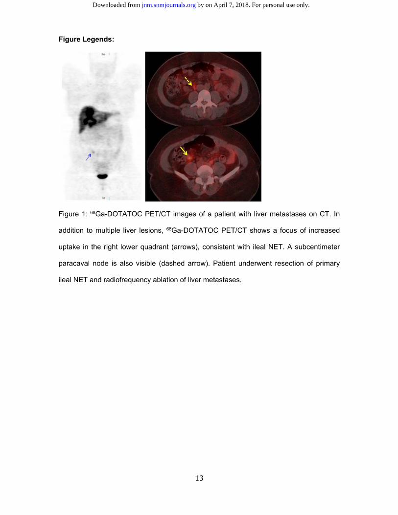
Figure 1: 68Ga-DOTATOC PET/CT images of a patient with liver metastases on CT. In
addition to multiple liver lesions, 68Ga-DOTATOC PET/CT shows a focus of increased
uptake in the right lower quadrant (arrows), consistent with ileal NET. A subcentimeter
paracaval node is also visible (dashed arrow). Patient underwent resection of primary
ileal NET and radiofrequency ablation of liver metastases.
by on April 7, 2018. For personal use only. jnm.snmjournals.org Downloaded from
14
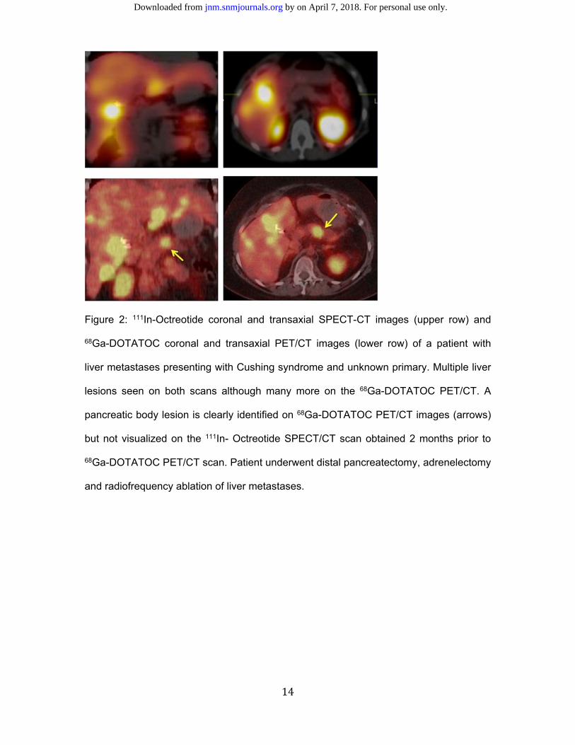
Figure 2: 111In-Octreotide coronal and transaxial SPECT-CT images (upper row) and
68Ga-DOTATOC coronal and transaxial PET/CT images (lower row) of a patient with
liver metastases presenting with Cushing syndrome and unknown primary. Multiple liver
lesions seen on both scans although many more on the 68Ga-DOTATOC PET/CT. A
pancreatic body lesion is clearly identified on 68Ga-DOTATOC PET/CT images (arrows)
but not visualized on the 111In- Octreotide SPECT/CT scan obtained 2 months prior to
68Ga-DOTATOC PET/CT scan. Patient underwent distal pancreatectomy, adrenelectomy
and radiofrequency ablation of liver metastases.
by on April 7, 2018. For personal use only. jnm.snmjournals.org Downloaded from
15
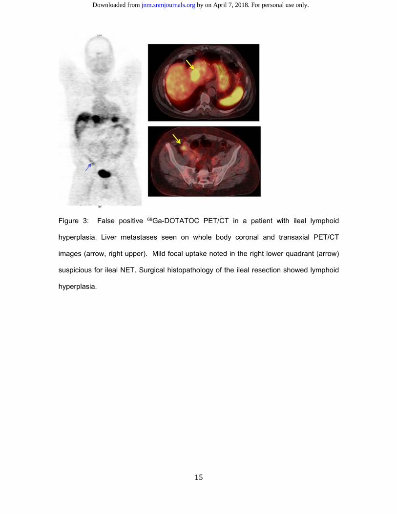
Figure 3: False positive 68Ga-DOTATOC PET/CT in a patient with ileal lymphoid
hyperplasia. Liver metastases seen on whole body coronal and transaxial PET/CT
images (arrow, right upper). Mild focal uptake noted in the right lower quadrant (arrow)
suspicious for ileal NET. Surgical histopathology of the ileal resection showed lymphoid
hyperplasia.
by on April 7, 2018. For personal use only. jnm.snmjournals.org Downloaded from
16
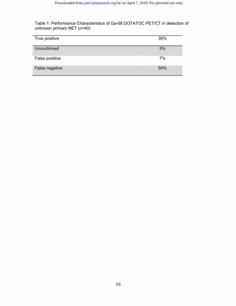
Table 1: Performance Characteristics of Ga-68 DOTATOC PET/CT in detection of unknown primary NET (n=40) True positive 38%
Unconfirmed 5%
False positive 7%
False negative 50%
by on April 7, 2018. For personal use only. jnm.snmjournals.org Downloaded from
Doi: 10.2967/jnumed.116.180984Published online: February 2, 2017.J Nucl Med. Timothy Ginader, David L. Bushnell, John Sunderland, Gideon KD Zamba, Michael M. Graham and M. Sue O'DorisioYusuf Menda, Thomas M. O'Dorisio, James Howe, Michael King Schultz, Joseph Dillon, David Dick, Leonard G. Watkins, Metastatic Neuroendocrine Tumor
Ga-DOTATOC PET/CT in Patients with68Localization of Unknown Primary Site with
http://jnm.snmjournals.org/content/early/2017/02/02/jnumed.116.180984This article and updated information are available at:
http://jnm.snmjournals.org/site/subscriptions/online.xhtml
Information about subscriptions to JNM can be found at:
http://jnm.snmjournals.org/site/misc/permission.xhtmlInformation about reproducing figures, tables, or other portions of this article can be found online at:
and the final, published version.proofreading, and author review. This process may lead to differences between the accepted version of the manuscript
ahead of print area, they will be prepared for print and online publication, which includes copyediting, typesetting,JNMcopyedited, nor have they appeared in a print or online issue of the journal. Once the accepted manuscripts appear in the
. They have not beenJNM ahead of print articles have been peer reviewed and accepted for publication in JNM
(Print ISSN: 0161-5505, Online ISSN: 2159-662X)1850 Samuel Morse Drive, Reston, VA 20190.SNMMI | Society of Nuclear Medicine and Molecular Imaging
is published monthly.The Journal of Nuclear Medicine
© Copyright 2017 SNMMI; all rights reserved.
by on April 7, 2018. For personal use only. jnm.snmjournals.org Downloaded from
![[68Ga]NOTA-Galactosyl Human Serum Albumin: a Tracer for ... · 68Ga]NOTA-GSA showed slower uptake in comparison with 68Ga-DTPA-GSA (123 ± 10 vs. 89 ± 3 s, p G 0.01). Conclusions:](https://img.pdfslide.us/doc/110x75/5f6d9cae33a64152a27bd022/68ganota-galactosyl-human-serum-albumin-a-tracer-for-68ganota-gsa-showed.jpg)


![68Ga] peptide high-output production on commercially · [68Ga] peptide high-output production on commercially available MiniAIO® synthesizer Objectives Optmization parameters Material](https://img.pdfslide.us/doc/110x75/5f95b4c0eecbac70717355d8/68ga-peptide-high-output-production-on-68ga-peptide-high-output-production-on.jpg)


![[68Ga]PSMA-HBED-CC Uptake in Osteolytic, Osteoblastic, and ... · Conclusions: [68Ga]PSMA-HBED-CC uptake is higher in osteolytic and bone marrow metastases compared to osteoblastic](https://img.pdfslide.us/doc/110x75/607572caf32e2d79681dbd86/68gapsma-hbed-cc-uptake-in-osteolytic-osteoblastic-and-conclusions-68gapsma-hbed-cc.jpg)
![Review The Search for an Alternative to [ Ga]Ga-DOTA-TATE ...thno.org/v09p1336.pdf · [68Ga]Ga-DOTA-TATE, [68Ga]Ga-DOTA-TOC, and [68Ga]Ga-DOTA-NOC allows for NET staging with high](https://img.pdfslide.us/doc/110x75/5e2a1b5b2104573c786ad22c/review-the-search-for-an-alternative-to-gaga-dota-tate-thnoorg-68gaga-dota-tate.jpg)

![Research Paper Lu]pentixather: Comprehensive Preclinical ...been paralleled by the use of [68Ga/177Lu]PSMA-I&T [34] 177and [68Ga/ Lu]PSMA-617 [35] for theranostics of metastatic castration](https://img.pdfslide.us/doc/110x75/6108d6669c3ce0590d229f48/research-paper-lupentixather-comprehensive-preclinical-been-paralleled-by.jpg)


![Labelling Efficiency DOTA PSMA Methods - Trasis 68Ga ISRS.pdf · Objectives [68Ga]Ga-HBED-11-PSMA (PSMA) and [68Ga]Ga-DOTA-tate (DOTAtate) are two well established PET tracers for](https://img.pdfslide.us/doc/110x75/5aae60737f8b9a6b308bf490/labelling-efficiency-dota-psma-methods-68ga-isrspdfobjectives-68gaga-hbed-11-psma.jpg)
![GMP-compliant production of [68Ga]Ga-NeoB for positron](https://img.pdfslide.us/doc/110x75/61b2306e3e680e78ed311d79/gmp-compliant-production-of-68gaga-neob-for-positron-.jpg)


![Extravasation of [177Lu]Lu-DOTATOC: case report and discussion](https://img.pdfslide.us/doc/110x75/6238d2331822df509e15b92c/extravasation-of-177lulu-dotatoc-case-report-and-discussion.jpg)