Embed Size (px)
Citation preview

Local Flaps: A Real-Time Finite ElementBased Solution to the Plastic Surgery
Defect Puzzle
Eftychios SIFAKIS a,1 , Jeffrey HELLRUNG a , Joseph TERAN a , Aaron OLIKER b
and Court CUTTING, M.D. c
a University of California, Los Angelesb BioDigital Systems
c NYU Medical Center
Abstract. One of the most fundamental challenges in plastic surgery is the alter-ation of the geometry and topology of the skin. The specific decisions made by thesurgeon concerning the size and shape of the tissue to be removed and the sub-sequent closure of the resulting wound may have a dramatic affect on the qualityof life for the patient after the procedure is completed. The plastic surgeon mustlook at the defect created as an organic puzzle, designing the optimal pattern toclose the hole aesthetically and efficiently. In the past, such skills were the distil-lation of years of hands-on practice on live patients, while relevant reference ma-terial was limited to two-dimensional illustrations. Practicing this procedure on apersonal computer [1] has been largely impractical to date, but recent technologi-cal advances may come to challenge this limitation. We present a comprehensivereal-time virtual surgical environment, built on finite element modeling and simula-tion of tissue cutting and manipulation. Our system demonstrates the fundamentalbuilding blocks of plastic surgery procedures on a localized tissue flap, and pro-vides a proof of concept for larger simulation systems usable in the authoring ofcomplex procedures on elaborate subject geometry.
Keywords. Reconstructive surgery, finite element models, simulation
The core principle of plastic surgery practice is the alteration of the geometry andtopology of the skin. For a patient diagnosed with malignant melanoma, the plastic sur-geon in many cases has to resect the tumor and the surrounding area. The extent of theskin that needs to be removed depends on the size and shape of the tumor. Furthermore,the removal of a large amount of tissue may have a dramatic affect on the patient’s re-covery and post-operative quality of life. The excision of tissue and subsequent defectclosure constitute a “puzzle” the surgeon has to solve. In the past it has been typical fora surgeon to require many years to master the craft of skin flap design. The only docu-mentation on how to solve this difficult three-dimensional problem would typically belimited to collections of two-dimensional illustrations. Currently a plastic surgeon canonly practice this skill on a live patient in an operating room. Doing this as a laptop sim-ulation [1] has long been a dream; yet only recently have computer hardware advancespromised sufficient computational capacity to accommodate the accuracy and real-timeperformance requirements of a tool usable in actual clinical practice.
1Corresponding Author: E-mail: [email protected]
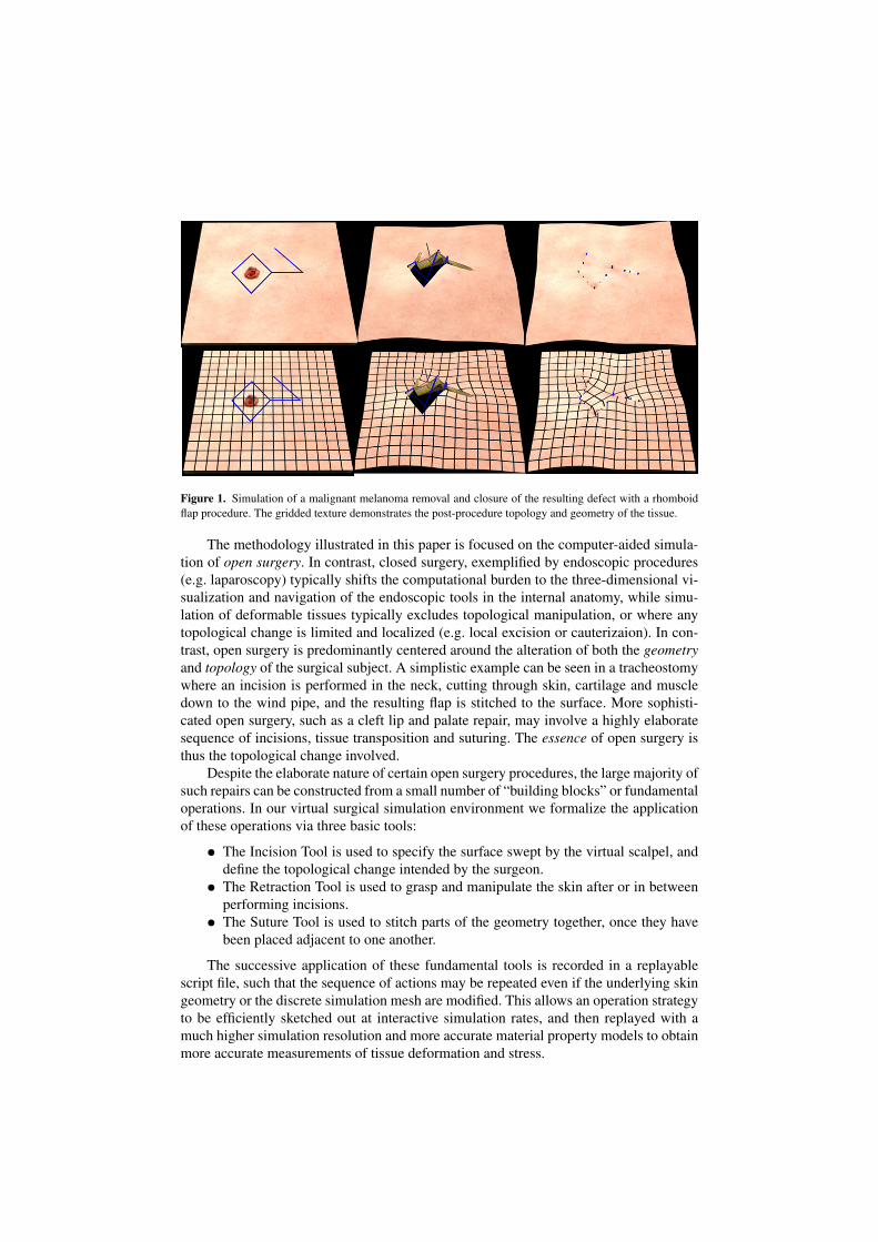
Figure 1. Simulation of a malignant melanoma removal and closure of the resulting defect with a rhomboidflap procedure. The gridded texture demonstrates the post-procedure topology and geometry of the tissue.
The methodology illustrated in this paper is focused on the computer-aided simula-tion of open surgery. In contrast, closed surgery, exemplified by endoscopic procedures(e.g. laparoscopy) typically shifts the computational burden to the three-dimensional vi-sualization and navigation of the endoscopic tools in the internal anatomy, while simu-lation of deformable tissues typically excludes topological manipulation, or where anytopological change is limited and localized (e.g. local excision or cauterizaion). In con-trast, open surgery is predominantly centered around the alteration of both the geometryand topology of the surgical subject. A simplistic example can be seen in a tracheostomywhere an incision is performed in the neck, cutting through skin, cartilage and muscledown to the wind pipe, and the resulting flap is stitched to the surface. More sophisti-cated open surgery, such as a cleft lip and palate repair, may involve a highly elaboratesequence of incisions, tissue transposition and suturing. The essence of open surgery isthus the topological change involved.
Despite the elaborate nature of certain open surgery procedures, the large majority ofsuch repairs can be constructed from a small number of “building blocks” or fundamentaloperations. In our virtual surgical simulation environment we formalize the applicationof these operations via three basic tools:
• The Incision Tool is used to specify the surface swept by the virtual scalpel, anddefine the topological change intended by the surgeon.
• The Retraction Tool is used to grasp and manipulate the skin after or in betweenperforming incisions.
• The Suture Tool is used to stitch parts of the geometry together, once they havebeen placed adjacent to one another.
The successive application of these fundamental tools is recorded in a replayablescript file, such that the sequence of actions may be repeated even if the underlying skingeometry or the discrete simulation mesh are modified. This allows an operation strategyto be efficiently sketched out at interactive simulation rates, and then replayed with amuch higher simulation resolution and more accurate material property models to obtainmore accurate measurements of tissue deformation and stress.

1. Technical background
A usable, practical and beneficial open surgery simulator needs to meet certain importantrequirements in order to address the needs of a clinical setting. First, the computationalperformance must enable real-time interaction when authoring a certain surgical strategy.The material models used must be accurate and representative of the (complex, typicallyhighly nonlinear) constitutive properties of the biological tissues involved. Furthermore,reconstructive surgery typically entails substantial tissue deformation, requiring the nu-merical and algorithmic robustness of any simulation techniques used. Finally, all theserequirements need to be reconciled with the need for a high level of visual detail, bothin terms of texture and geometrical detail, to reflect the geometric and visual complexityof the subject tissues and aid in the reproduction of the process in the operating room byproviding discernible surface landmarks for the various surgical operations.
1.1. Real-time simulation
A virtual simulation environment will have a vastly reduced potential for being used inactual practice if it does not offer the ability for a clinician to cut and manipulate the skinin real-time. In a finite-element discretization of a volumetric object representing a tissueflap, certain algorithmic and numerical factors may severely compromise the real-timeperformance of such a system. First, the resolution of the simulation mesh alone needs tobe limited enough to allow for real-time simulation; although discretizations with severalhundreds of thousands or millions of tetrahedral elements would be desired (and mayactually be feasible in the light of emerging massively parallel computing platforms),commodity computer hardware dictates stricter limits for simulations tractable with thecomputational resources of a mainstream laptop, for example. Instead of compromisingthe visual detail contained in our model for the sake of a coarser discretization, we em-ploy an embedded scheme (as in [3,4]) to allow a coarser simulation mesh to serve as a“framework” for a higher resolution geometry. The surface resolution may be substan-tially higher than that of the embedding grid, and certain aspects of simulation (suchas collision processing) may be handled on the high-resolution embedded geometry ifso desired. Additionally, this eliminates the risk of ill-conditioned simulation elementsnecessitated to resolve intricate geometrical features of the tissue surface. More impor-tant, this practice circumvents the need for remeshing of the tissue geometry in order toresolve the topological change incurred by incisions. At all times, the simulation gridis maintained at a regular, lower degree-of-freedom lattice (with a topology adhering asclosely as possible to the topology of the continuous tissue volume).
1.2. Accuracy and nonlinear deformation
Although linear material models and mass-spring networks have been widely used for in-teractive simulation of deformable solids, providing a virtual surgery simulation systemwith the ability to make reliable predictions about the behavior of real tissue necessitatesthe adoption of much more accurate, nonlinear, anisotropic constitutive material models.This requirement will be essential in making the virtual surgery framework presentedhere capable of reliable predictions of the surgical outcome, establishing a virtual sys-tem as a dependable platform for surgical planning. The constitutive models that accu-

Figure 2. Simulation of a z-plasty procedure for the elongation of a scar contracture.
rately convey the material properties of human flesh have to account for the inhomogene-ity of materials (e.g. anatomical parts consisting of passive fatty tissue, active muscula-ture, tendons, ligaments and connective tissue). Furthermore, the materials involved arenonlinear and near-incompressible, in sharp contrast with linear material approximationswhich may be employed in applications where deformation is limited to the small strainregime, or where physical accuracy is not essential. Our system supports arbitrary non-linear, inhomogeneous and anisotropic material properties, which may be defined on thebasis of every distinct simulation element in the underlying embedding mesh. This alsohighlights the ability of the system to facilitate a two-pass simulation process, where alower resolution embedding mesh (where the nonlinearity and inhomogeneity may man-ifest themselves in a limited capacity) can be used for crafting the surgical approach in-teractively, and a subsequent pass where the same sequence of actions is repeated off-line on a highly refined embedding mesh, which is able to resolve the intricacies of thenonlinear deformation.
1.3. Robustness under large deformation
The manipulation of flesh during plastic surgery operations is by no means limited tosmall geometric change; in fact, large strain deformation is quite typical of the config-urations involved in the closure of the tissue flaps created. Therefore, especially giventhe necessity for nonlinear constitutive models and the relatively under-resolved natureof the simulation (owing to the embedding approach), the simulation methods employedhave to address and survive extreme geometric configurations, such as element inversionor collapse. We employ the Invertible Finite Element method of [2] which allows suchsimulations to continue and gracefully recover when transitioning through such extremegeometric configurations, while still supporting the full gamut of nonlinear constitutivemodels.

2. Tools and Methods
We have created a "local flaps" simulator that will allow surgeons to practice their clos-ing designs in a three-dimensional environment with real-time interaction. This environ-ment uses the PhysBAM physics simulation library to allow the user to make incisions,move tissue flaps, and create virtual sutures to simulate closing of a skin defect, all in ascientifically accurate framework. The simulator consists of several simple surgical tools:
• The Incision Tool. This tool creates a triangulated surface which represents thearea swept by the scalpel during an incision. This incision surface is generatedby sketching a curve (either a sequence of straight line cuts, or a spline curve) onthe surface of the tissue, while controlling the angle which the scalpel forms withthe tissue being cut. The result of this operation is a discrete triangulated surfacerepresentation of the incision being made. The algorithm of [3] is subsequentlyused to determine the topology resulting from this manipulation of the tissue ge-ometry, generating the necessary degrees of freedom to enable the opening of thetissue at the location of the incision.
• The Retraction Tool. Once the topology of the incision has been resolved, the ac-tion of grasping and manipulating the tissue flap is performed using a simulatedhook-and-handle mechanism. By direct selection, a point on the surface of thetissue is defined as the anchor point of a retraction site (i.e. a hook point). At thesame time, a “handle” point is defined by offsetting the hook location a certainshort distance off the surface of the tissue. This handle can be arbitrarily posi-tioned in 3D space, giving rise to deformation of the simulated tissue. The targetposition of the handle is communicated to the deformable tissue via a spring forcethat aims to bring the hook at the specified 3D location.
• The Suture Tool. Once the retraction tool has been used to deform the tissue shapeinto its target location, this tool emulates the process of suturing two adjacentsurfaces together. The suture can be either a point-to-point connection, or a curvedpath connecting two sides of tissue. The geometry on either side of the suture arebrought together by the simulated action of a zero rest-length spring joining theparts of the tissue connected by the suture.
The successive application of these fundamental tools is recorded in a replayablescript file, such that the sequence of actions may be repeated even if the underlying skingeometry or discrete simulation mesh are modified; this allows an operation strategy tobe efficiently sketched out in interactive simulation rates, and then replayed with a muchhigher resolution simulation mesh to obtain more accurate measurements of tissue de-formation and stress. Using this simple combination of surgical tools, the surgeon is ableto practice existing procedures for closing the defect. A surgeon may also invent a newpattern altogether and assess its efficacy based on such physically quantifiable metrics aspost procedure tension in the tissue and suture. The elasticity of the tissue is simulatedusing the finite element method defined on a volumetric tetrahedral representation of thetissue. An implicit time stepping scheme is used to obtain a frame rate adequate for in-teractivity. Tissues involved typically undergo large deformation and the algorithms of[2] and [5] are used to guarantee robust performance in this challenging setting. Tissueincisions are represented using the novel tetrahedral cutting approach of [3] and suturesare modeled with the highly flexible embedding framework of [4].

Figure 3. Comparison of the simulated results of Z-plasty procedures with incision angles at 45, 60 and 90degrees respectively from left to write. Simulation confirms the conventional wisdom that 60 degrees is theoptimal incision angle. Also, the 90 degree incision reproduces the so called “dog ear” effect
3. Results
Figure 1 depicts the results of a local flaps simulation of a rhomboid flap procedure forremoving a malignant melanoma and repairing the skin near the excision region. This is avery common procedure and the simulated tissue configuration matches the conventionalwisdom very closely. Different stages in a z-plasty procedure (typically used for elon-gating scar contractures) shown in figures 2 and 3, show the effects of varying the anglesof the z-incision. In practice, the optimal angle of incision is 60 degrees. Our simulatedresults also suggest that 60 degrees is the optimal angle as lower angles fail to producesufficient elongation and large angles produce non-planar equilibrium configurations (orthe so-called “dog ear” effect).
4. Conclusions
The local flaps environment is an effective tool that the plastic surgeon can utilize toleverage physically accurate simulation in an interactive environment to improve manyaspects of his cognitive surgical practice. The uses of the environment fit into two basiccategories. The first is related to training in existing procedures. These scenarios demandrealtime interaction but not necessarily the highest degree of physical accuracy. The sec-ond is related to design of new procedures. Such applications require a higher degree ofphysical accuracy. Our finite element based approach allows for this and has the abilityfor future incorporation of subject specific constitutive models that can be used to studyrepair of unique injuries (e.g. those arising on the battlefield).
References
[1] S.D. Pieper, D.R. Laub Jr. and J.M. Rosen "A finite-element facial model for simulating plastic surgery",Plast. Reconstr. Surg., 96(5):1100-5, Oct 1995.
[2] G. Irving, J. Teran and R. Fedkiw, "Invertible finite elements for robust simulation of large deformation",ACM SIGGRAPH/Eurographics Symp. on Computer Animation (SCA), 131-140, 2004.
[3] E. Sifakis, K. Der, and R. Fedkiw, "Arbitrary cutting of deformable tetrahedralized objects", ACM SIG-GRAPH/Eurographics Symp. on Computer Animation (SCA), 73-80, 2007
[4] E. Sifakis, T. Shinar, G. Irving and R. Fedkiw, "Hybrid simulation of deformable solids", ACM SIG-GRAPH/Eurographics Symp. on Computer Animation (SCA), 81-90, 2007
[5] J. Teran, E. Sifakis, G. Irving and R. Fedkiw, "Robust quasistatic finite elements and flesh simulation.ACM SIGGRAPH/Eurographics Symp. on Computer Animation (SCA). 181-190, 2005.