Embed Size (px)
Citation preview

Rab8 controls AMPA receptor synaptic trafficking
1
Local control of AMPA receptor trafficking at the postsynaptic
terminal by a small GTPase of the Rab family
Nashaat Z. Gerges, Donald S. Backos and José A. Esteban*
Department of Pharmacology, University of Michigan Medical School, Ann Arbor, MI 48109-
0632
Running title: Rab8 controls AMPA receptor synaptic trafficking
*Correspondence should be addressed to J.A.E. ([email protected]):
1150 W Medical Center Drive
Ann Arbor, MI 48109-0632
Phone: 734-615-2686
Fax: 734-763-4450
Rab8 controls AMPA receptor synaptic trafficking
2
SUMMARY
The delivery of neurotransmitter receptors into the synaptic membrane is essential for
synaptic function and plasticity. However, the molecular mechanisms of these specialized
trafficking events and their integration with the intracellular membrane transport machinery are
virtually unknown. Here, we have investigated the role of the Rab family of membrane sorting
proteins in the late stages of receptor trafficking into the postsynaptic membrane. We have
identified Rab8, a vesicular transport protein associated to trans-Golgi network membranes, as a
critical component of the cellular machinery that delivers AMPA-type glutamate receptors into
synapses. Using electron microscopic techniques, we have found that Rab8 is localized in close
proximity to the synaptic membrane, including the postsynaptic density. Electrophysiological
studies indicated that Rab8 is necessary for the synaptic delivery of AMPA receptors during
plasticity (long-term potentiation) and during constitutive receptor cycling. In addition, Rab8 is
required for AMPA receptor delivery into the spine surface, but not for receptor transport from
the dendritic shaft into the spine compartment or for delivery into the dendritic surface.
Therefore, Rab8 drives specifically the local delivery of AMPA receptors into synapses. These
results demonstrate a new role for the cellular secretory machinery in the control of synaptic
function and plasticity directly at the postsynaptic membrane.
Rab8 controls AMPA receptor synaptic trafficking
3
INTRODUCTION
Synaptic plasticity is critical for the establishment and maturation of functional neuronal
circuits in the brain and is widely thought of as the cellular process responsible for learning and
memory. An important mechanism controlling synaptic maturation and remodeling is the
targeting and delivery of neurotransmitter receptors into synapses. However, very little is known
of the exocytic processes that sort neurotransmitter receptors into the postsynaptic membrane. In
addition, it remains to be determined how these specialized trafficking events are integrated with
the intracellular membrane transport machinery. In fact, most of our current knowledge on
membrane trafficking at the synapse derives from studies on neurotransmitter vesicle fusion at
the presynaptic terminal (reviewed in (1,2)).
The AMPA-type glutamatergic receptors (AMPARs) are highly dynamic components of
excitatory synapses. They can traffic in and out of the synaptic membrane constitutively or in an
activity-dependent manner, and this regulated trafficking is known to contribute to synaptic
plasticity during brain development and in adulthood (3-6). AMPARs are hetero-oligomeric
molecules composed of different combinations of GluR1 to GluR4 subunits (7). In hippocampus,
AMPARs containing GluR1 or GluR4 subunits are delivered to synapses in an activity-
dependent manner upon NMDA receptor activation, leading to long-lasting synaptic potentiation
(8-11). In contrast, AMPARs containing only GluR2 and GluR3 subunits cycle continuously in
and out of synapses in a manner largely independent from synaptic activity (10,12), but
dependent on NSF (13-15) and Hsp90 (16) function. These two distinct trafficking routes have
been coined as regulated and constitutive pathways, respectively (17). However, the cellular and
molecular mechanisms mediating membrane transport in these pathways are largely unknown.
Rab8 controls AMPA receptor synaptic trafficking
4
Members of the Rab family of small GTPases are important regulators of intracellular
membrane sorting in eukaryotes. In particular, they are proposed to mediate membrane transport
specificity (18-20). Rab proteins are likely to be important for neuronal function, since
alterations in Rab protein regulation can lead to mental retardation in humans (21). Rab3 is a
presynaptic member of this family (22). It controls the fusion of neurotransmitter vesicles with
the plasma membrane (23,24) and mediates some presynaptic forms of synaptic plasticity
(25,26). In contrast, the potential role of Rab proteins in neurotransmitter receptor targeting and
synaptic function at the postsynaptic terminal has never been tested.
Besides Rab3, three members of the Rab family are involved in exocytic delivery into the
plasma membrane: Rab4, Rab8 and Rab11. In neurons, Rab8 has an exclusive somatodendritic
distribution (27). In epithelial (28,29) and photoreceptor (30,31) cells, Rab8 mediates the
transport of trans-Golgi network-derived membranes into specific compartments of the plasma
membrane. Rab4 is involved in the recycling pathway from early endosomes back into the
plasma membrane, whereas Rab11 mediates membrane transport from late endosomes and trans-
Golgi network (32-39). These three Rab proteins act on late stages of the exocytic trafficking
into the plasma membrane, and therefore, are attractive candidates to mediate receptor targeting
into the postsynaptic membrane.
In this study, we have explored the role of these exocytic Rab proteins in synaptic
function and plasticity, and in particular, in the late trafficking events that deliver AMPARs into
synapses. Using a combination of molecular biology, electrophysiology and imaging techniques
on organotypic hippocampal slice cultures, we have found that Rab8, but not Rab4 or Rab11, are
required for the late stages of AMPAR synaptic delivery into the postsynaptic membrane.
Rab8 controls AMPA receptor synaptic trafficking
5
EXPERIMENTAL PROCEDURES
Constructs of recombinant proteins and expression. Rab8a, Rab4a and Rab11a coding
sequences were cloned by PCR from a commercial rat brain cDNA preparation (Clontech, cat. #
7150). The rat Rab8a sequence that we cloned has 96% and 97% identity to the mouse and
human sequences, respectively. The reported Rab8a rat sequence in the NCBI database
(accession numbers M83675 and NM_053998) has a significant N-terminal truncation (83 amino
acids) with respect to the mouse and human sequences and with respect to the rat sequence we
cloned. This is probably due to a sequencing error in the rat sequence submitted originally. Our
cloned Rab4a sequence was identical to the reported rat Rab4a sequence in the NCBI database
(accession number P05714) with the exception of two differences: Q47S and T75R. These two
amino acids in our sequence (47S) and (75R) are conserved in the mouse and human sequences
reported in the NCBI database (accession numbers NP_033029 and NP_004569, respectively).
Therefore, these two differences probably correspond to sequencing errors in the previously
reported rat sequence. Our cloned Rab11a sequence was identical to the reported one in the
NCBI database (accession number NM_031152). Rab8a, Rab4a and Rab11a coding sequences
were cloned as fusion proteins downstream from EGFP using the pEGFP-C1 plasmid (Clontech,
cat. # 6084). The dominant negative mutants (GDP-bound form) were generated by PCR
introducing a single-amino acid substitution previously described (T22N for Rab8dn, S22N for
Rab4dn and S25N for Rab11dn). The RFP fusion construct of Rab8dn was generated with a red
fluorescence protein variant (tdimer2(12); (40)) generously provided by Dr. Roger Tsien
(UCSD). All constructs were recloned into pSinRep5 for Sindbis virus preparation (41).
Hippocampal slices are prepared from young rats (postnatal day 5 to 7) and placed in culture on
Rab8 controls AMPA receptor synaptic trafficking
6
semiporous membranes (42). After 4-7 days in culture, the recombinant gene is delivered into the
slices. For the experiments shown in Fig. 3A and B, Fig. 4, Fig. 5B and C and Fig. 7, we used the
biolistic delivery method (43), which allowed us to co-express several proteins with plasmids
bearing the CMV promoter. For the rest of the experiments we use the Sindbis virus expression
system (44). This is a replication-deficient, low-toxicity, neurotropic virus, allowing the
expression of recombinant proteins exclusively in neurons by injecting the viral solution
extracellularly in the desired area of a hippocampal slice (41). Expression of the recombinant
proteins was for 36 hours when AMPA receptor subunits were expressed (Fig. 3A and B, Fig. 4
and Fig. 7), or for 15 hours in the rest of the cases. Neurons remain morphologically and
electrophysiologically intact during these expression times. All biosafety procedures and animal
care protocols were approved by the University of Michigan.
Electrophysiology. Simultaneous double whole-cell recordings were obtained from nearby pairs
of infected and uninfected CA1 pyramidal neurons, under visual guidance using fluorescence
and transmitted light illumination. The recording chamber was perfused with 119 mM NaCl, 2.5
mM KCl, 4 mM CaCl2, 4 mM MgCl2, 26 mM NaHCO3, 1 mM NaH2PO4, 11 mM glucose, 0.1
mM picrotoxin, 10 µM bicuculline and 2 µM 2-chloroadenosine, at pH 7.4, gassed with 5%
CO2/95% O2. Patch recording pipettes (3-6 MΩ) were filled with 115 mM cesium
methanesulfonate, 20 mM CsCl, 10 mM HEPES, 2.5 mM MgCl2, 4 mM Na2ATP, 0.4 mM
Na3GTP, 10 mM sodium phosphocreatine and 0.6 mM EGTA at pH 7.25. In the rectification
experiments, i.e. Fig 7, 0.1 mM spermine was added. Voltage-clamp whole-cell recordings were
carried out with multiclamp 700A amplifiers (Axon Instruments, Union City, California, USA).
Synaptic responses were evoked with two bipolar electrodes with single voltage pulses (200 µs,
Rab8 controls AMPA receptor synaptic trafficking
7
up to 20 V). The stimulating electrodes were placed over Schaffer collateral fibers between 300
µm and 500 µm from the recorded cells. Since only CA1, but not CA3 cells are infected, this
configuration ensures that recombinant proteins are always expressed exclusively in postsynaptic
cells. Synaptic AMPA receptor-mediated responses were measured at –60 mV and NMDA
receptor-mediated responses at +40 mV and were averaged over 50-100 trials. In the rectification
experiment (Fig 7), NMDA receptor-mediated responses were blocked pharmacologically using
0.1 mM DL-APV. Synaptic AMPA receptor-mediated responses were measured at –60 mV and
+40 mV and their ratio was used as an index of rectification. LTP experiments were carried out
as previously described (16), by pairing 0 mV postsynaptic depolarization with 3 Hz presynaptic
stimulation (300 pulses).
Biochemistry. Hippocampal extracts were prepared in homogenization buffer containing
protease inhibitors (10 mM HEPES, 500 mM NaCl, 10 mM EDTA, 4 mM EGTA, 0.1 mM
phenylmethylsulfonyl fluoride, 2 µg/ml chymostatin, 2 µg/ml leupeptin, 2 µg/ml antipain, 2
µg/ml pepstatin and 1% triton X-100) as previously described (8). Expression of Rab proteins
was analyzed by Western blot with anti-Rab4 (BD Biosciences), anti-Rab8 (Pharmingen) and
anti-Rab11 (Zymed Laboratories) antibodies. Phosphorylation of αCaMKII at Thr286 was
analyzed with phospho-specific and regular anti-αCaMKII antibodies (Upstate) using the
homogenization buffer described above supplemented with phosphatase inhibitors (10 mM NaF,
1 µM microcystin LR and 0.5 µM calyculin A).
Fluorescence immunohistochemistry. Immunohistochemical detection of recombinant AMPA
receptors was carried out with anti-GFP mouse antibody (Roche). Fluorescence labeling was
Rab8 controls AMPA receptor synaptic trafficking
8
achieved with anti-mouse biotinylated secondary antibody (Sigma) and streptavidin tagged with
AlexaFluor 594 (Molecular Probes; Fig. 3) or with Cy5 (Amersham Biosciences; Fig. 4).
Detergents were omitted in all incubations to evaluate surface expression. Images were taken
using Olympus FV 500 confocal microscopy. A 60 X lens with water immersion interface was
used. FluoView software was used for acquiring the images. ImageJ was used for 3D
reconstruction and quantification of fluorescence intensities. Analysis of surface immunostaining
at spines and dendrites was carried out as follows. Line plots of fluorescence intensity were
generated across spine heads and the adjacent dendritic shafts. Fluorescence intensity at each
compartment was quantified from the peaks corresponding to the spine and the dendrite after
background subtraction (Fig. 4B). Then, surface ratios are calculated as the ratio between the
GFP signal (total receptor) and the Cy5 signal (surface receptor).
Immunogold electron microscopy. Hippocampal slices were fixed and processed for osmium-
free post-embedding immunogold labeling, essentially as previously described (45). Rab8 was
labeled with anti-Rab8 antibody (Pharmingen) and an anti-mouse antibody coupled to 6 nm gold
particles (Electron Microscopy Sciences). Electron micrographs were obtained with a Philips
CM-100 transmission electron microscope and a Kodak 1.6 Megaplus digital camera.
Rab8 controls AMPA receptor synaptic trafficking
9
RESULTS
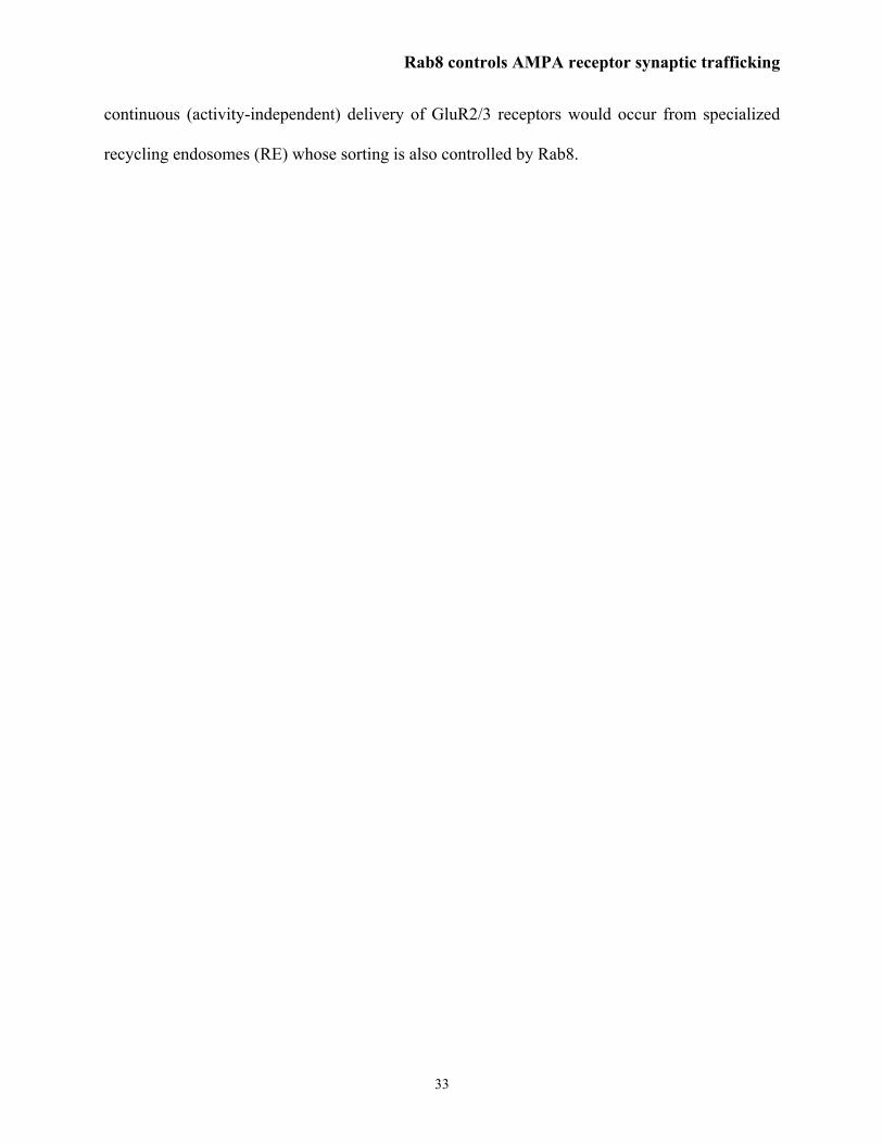
Role of Rab4, Rab8 and Rab11 in AMPA receptor-mediated synaptic transmission
As a first step to evaluate the role of these three exocytic Rab proteins in synaptic
transmission, we blocked their function individually by expressing the corresponding dominant
negative (Rab-dn) forms as GFP-fusion proteins in organotypic hippocampal slice cultures (see
Experimental Procedures). Point mutations known to confer dominant negative (GDP-bound)
phenotypes to these proteins have been employed in multiple occasions: Rab4(S22N) (46-48),
Rab8(T22N) (31,49-51), Rab11(S25N) (36-39,52-55). Also, GFP-Rab fusions have been used
successfully in numerous studies to study membrane trafficking in living cells (31,33,56-58)).
Rab4dn-, Rab8dn- and Rab11dn-GFP were efficiently expressed in hippocampal slice cultures,
as assayed by Western blot analysis (see left insets in Fig. 1).
The effect of these dominant negative proteins on AMPA and NMDA receptor function
was evaluated by recording simultaneously from nearby infected and uninfected CA1 neurons,
allowing direct comparison of synaptic responses evoked by stimulating the Schaffer collateral
pathway from the CA3 region. Importantly, only CA1 but not CA3 cells express the recombinant
dominant negative protein, and therefore, the site of action of these proteins when monitoring
CA3-to-CA1 synaptic transmission is necessarily postsynaptic. As shown in Fig. 1A, Rab8dn
significantly depressed AMPAR-mediated currents, without affecting NMDAR-mediated
currents. Other electrophysiological parameters were not significantly different between
uninfected and Rab8dn-infected neurons (input resistance: 168±23 MΩ -uninfected-, 192±25
MΩ -infected-; holding current: -68±13 pA –uninfected-, -57±9 pA –infected-). This result
suggests that Rab8 function is required for AMPAR delivery into synapses. In contrast, neither

Rab8 controls AMPA receptor synaptic trafficking
10
Rab4dn (Fig. 1B) nor Rab11dn (Fig. 1C) changed significantly AMPAR or NMDAR-mediated
synaptic responses. Furthermore, a dominant negative form of the endocytic Rab5 protein
(S34N) did not alter AMPA or NMDA transmission either (unpublished observations). These
results serve to verify that the depression of AMPAR responses by Rab8dn was not due to virus
infection or non-specific sequestration of regulatory proteins, like GDP/GTP exchange factors
(GEFs) or GDP dissociation inhibitors (GDIs).
To determine whether Rab8 is a rate-limiting factor for AMPAR delivery, we
overexpressed Rab8wt (Fig. 1D, left inset) and compared AMPA and NMDA R-mediated
responses from control and infected nearby neurons. As shown in Fig. 1D, overexpression of
Rab8wt did not produce any significant effect on AMPA or NMDA receptor-mediated synaptic
transmission. Since Rab8wt overexpression did not increase AMPA currents, this result suggests
that Rab8 is not rate-limiting for AMPAR delivery into synapses.
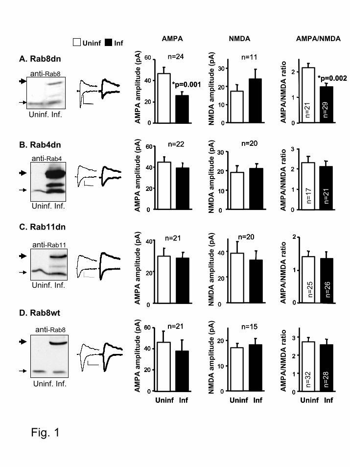
Expression and ultrastructural localization of Rab8 in hippocampal neurons.
The electrophysiological experiments shown above suggest a role for Rab8 in AMPA
receptor synaptic trafficking. To further investigate this possibility, we determined the
ultrastructural localization of Rab8 in hippocampal neurons from brain tissue. Although previous
studies have localized Rab8 at the somatodendritic compartment in dissociated neuronal cultures
(27), its localization relative to synaptic contacts has not been addressed before. To this end, we
used post-embedding anti-Rab8 immunogold labeling on the synaptic region of the CA1
hippocampus (stratum radiatum; see Experimental Procedures). Rab8 was abundant in
postsynaptic terminals (Fig. 2A, arrows; Fig. 2B, “Postsynaptic”), whereas the presynaptic
labeling was marginal (Fig. 2B, “Pre-”). Quantification of the immungold particles in the
Rab8 controls AMPA receptor synaptic trafficking
11
postsynaptic terminal indicated that Rab8 accumulates in intracellular compartments in close
proximity to the synapse (Fig. 2B, “Intra”), and in some cases directly at the postsynaptic density
(Fig. 2B, “PSD”). This result supports a potential role for Rab8 in the local transport of
membrane proteins into synapses.
We also examined Rab8wt-GFP distribution in CA1 neurons from hippocampal slices by
imaging GFP fluorescence with confocal microscopy. Rab8wt-GFP was broadly expressed in the
cell (Fig. 2C, left), including distal dendrites and spines (Fig. 2C, right). Although these results
do not show specific targeting of Rab8-GFP to spines, they do suggest that the recombinant
protein has access to membrane compartments in close proximity to synapses.
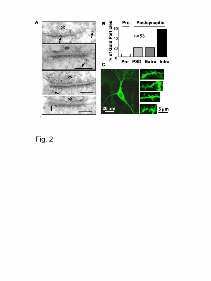
Rab8 is not required for the large-scale transport of AMPA receptors along dendrites.
Early studies using antisense oligonucleotides in dissociated neuronal cultures suggested
that Rab8 may be involved in the export of membrane proteins from the neuronal cell body into
dendrites (27). Therefore, it is conceivable that Rab8dn may depress AMPAR-mediated
transmission by interfering with their transport into dendrites. We tested this possibility by
coexpressing GFP-tagged GluR2 and Rab8dn using biolistic gene delivery (the same constructs
and transfection method used for the electrophysiological experiments shown in Fig. 7A; see
below). The surface distribution of the recombinant receptor was monitored by immunostaining
using an anti-GFP antibody in non-permeabilized conditions (the GFP tag is placed at the N-
terminus of GluR2, and it is therefore exposed to the extracellular side of the plasma membrane).
As shown in Fig. 3B, Rab8dn did not qualitatively alter the export of GluR2-GFP into the
dendritic surface, including secondary and tertiary distal dendrites (similar to the
immunostaining distribution obtained for GluR2-GFP expressed alone –Fig. 3A) (see Fig. 4,
Rab8 controls AMPA receptor synaptic trafficking
12
below, for a quantitative analysis of surface receptor delivery at spines and dendrites). As
control, neurons expressing Rab8dn-GFP alone did not show any surface immunostaining (Fig.
3C), consistent with the intracellular distribution of Rab8. These results suggest that Rab8dn
depresses AMPA transmission not by globally altering AMPAR transport into dendrites, but by
interfering with a local trafficking step, possibly the synaptic delivery of the receptor.
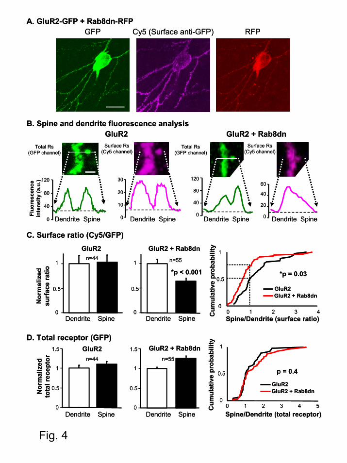
Rab8 is required for local AMPA receptor delivery into the spine surface.
To test whether Rab8 is involved in a local step of AMPAR trafficking, we quantitatively
evaluated the surface delivery of AMPARs at dendritic spines and their adjacent dendritic shafts.
For this experiment we substituted the GFP tag on Rab8dn with a red fluorescence protein
(Rab8dn-RFP, see Experimental Procedures), so its fluorescence can be separated from the
coexpressed GluR2-GFP (the functionality of Rab8dn-RFP was confirmed electrophysiologically
by coexpressing it with GluR2(R607Q)-GFP and monitoring receptor delivery into synapses
using a rectification assay; see below in description for Fig. 7). Surface immunostaining was then
carried out with an anti-GFP antibody under non-permeabilized conditions, as described above.
In this case the labeling was visualized with an infrared fluorophore (Cy5). Therefore, this
experimental design allows us to monitor the total amount of receptor (GFP channel), the
fraction exposed to the cell surface (Cy5 channel) and the presence of the coexpressed Rab8dn
(RFP channel) (see Fig. 4A as an example).
Line plots of fluorescence intensity were generated across the spine head and the adjacent
dendritic shaft. Surface ratios for spine and dendrites were then calculated as the ratio between
the corresponding peaks in the Cy5 channel (surface receptor) and the GFP channel (total
receptor) after background subtraction (see Fig. 4B). Spines are solely selected from their GFP
Rab8 controls AMPA receptor synaptic trafficking
13
image, precluding any bias with respect to their surface immunostaining. The results of this
analysis are shown in Fig. 4C. When expressing GluR2-GFP alone, surface ratios were similar in
spine and dendrites. In contrast, surface ratio was significantly lower in the spines as compared
to their adjacent dendrites when Rab8dn was coexpressed. Importantly, absolute surface ratio at
dendrites was not altered by Rab8dn (Cy5/GFP ratios were 0.31±0.05 for GluR2 alone versus
0.31±0.04 for GluR2 plus Rab8dn). These data indicate that Rab8dn specifically impairs the
delivery of AMPARs to the surface of the spine. Interestingly, Rab8dn did not decrease the total
amount of receptor (GFP channel) in the spine compared to the dendrite (Fig 4D) nor total
receptor abundance at dendrites (absolute GFP values were 50±4 for GluR2 alone versus 54±3
for GluR2 plus Rab8dn). These results support the interpretation that Rab8 is involved in a local
trafficking step from an intracellular membrane compartment inside the spine to the postsynaptic
plasma membrane.
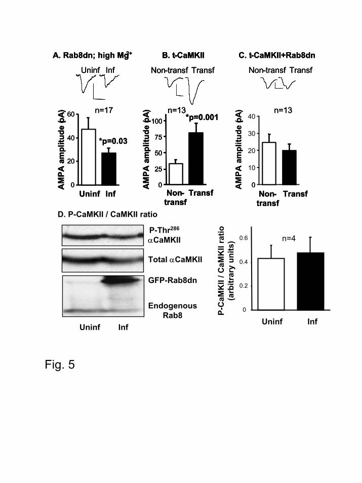
Rab8 is necessary for both the constitutive and regulated delivery of AMPA receptors into
synapses.
As described above, AMPARs may reach synapses through a constitutive pathway, in an
activity-independent manner, and through a regulated pathway, triggered by NMDA receptor
opening and CaMKII activation (17). In order to test the role of Rab8 in the constitutive
pathway, we expressed Rab8 dominant negative in slices kept in conditions of reduced neuronal
activity (12 mM Mg2+ was added to the culture medium immediately after infecting with the
virus expressing Rab8dn; recordings were carried out in the presence of the standard 4 mM
Mg2+). As shown in Fig. 5A, Rab8dn depresses AMPAR-mediated responses in conditions of

Rab8 controls AMPA receptor synaptic trafficking
14
reduced neuronal activity, suggesting that Rab8 is necessary for the constitutive (activity-
independent) delivery of AMPARs into synapses.
The role of Rab8 in the activity-dependent delivery of AMPARs was tested with a
truncated form of αCaMKII (t-CaMKII) that is constitutively active (8,59). Expression of t-
CaMKII potentiated AMPAR-mediated responses (Fig. 5B), as previously described (8,59).
However, this potentiation was fully blocked when Rab8dn was co-expressed with t-CaMKII
(Fig. 5C). This result is consistent with Rab8 being also necessary for the regulated delivery of
AMPARs triggered by CaMKII. To test whether the absence of potentiation is due to the
interference of Rab8dn with CaMKII activity, we quantified the autophosphorylation levels of
αCaMKII at Thr286 in control and Rab8dn-expressing neurons. As shown in Fig 5D, phospho-
CaMKII levels from CA1 dissected regions were similar between infected and uninfected slices.
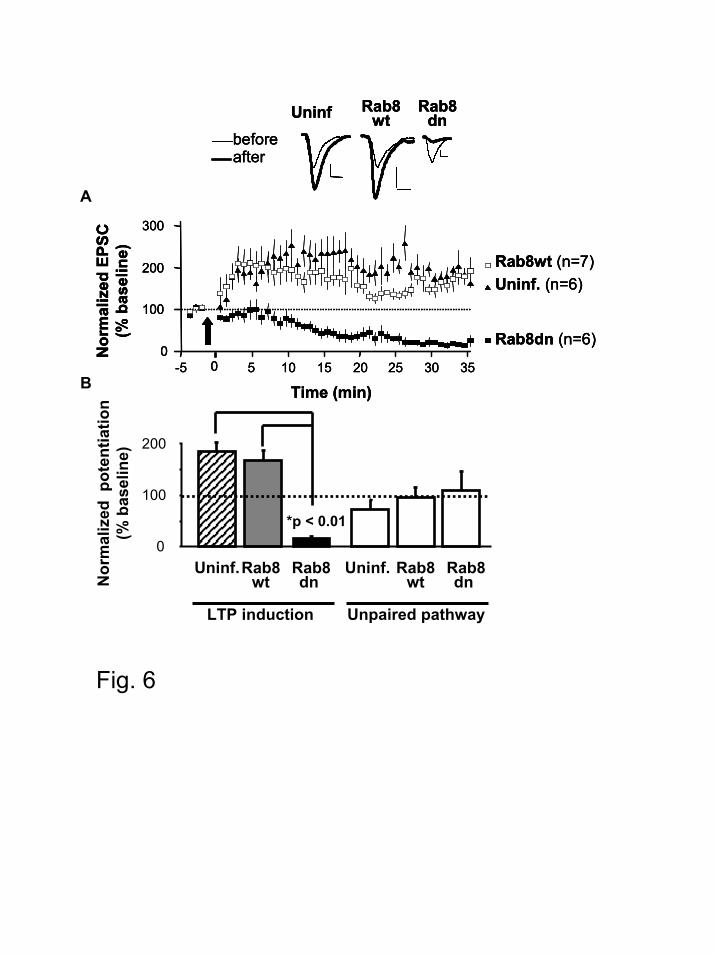
Rab8 is necessary for synaptic plasticity.
Long term potentiation (LTP) in the CA1 hippocampus is one of the most thoroughly
studied forms of synaptic plasticity, and it is accompanied by the synaptic delivery of GluR1-
containing AMPARs (8). To test whether Rab8 is necessary for the synaptic delivery of
AMPARs during synaptic plasticity, we examined pairing-induced LTP in CA1 neurons infected
with Rab8dn or Rab8wt. Consistent with its effect on CaMKII-induced AMPAR delivery,
Rab8dn blocked LTP expression (Fig. 6). Unexpectedly, Rab8dn led to synaptic depression. This
late depression upon LTP induction has been previously reported in situations when AMPAR
delivery was impaired (8). Importantly, LTP was normal in cells expressing Rab8wt, as
compared with uninfected cells (Fig. 6). These results indicate that Rab8 function is necessary
for LTP.
Rab8 controls AMPA receptor synaptic trafficking
15
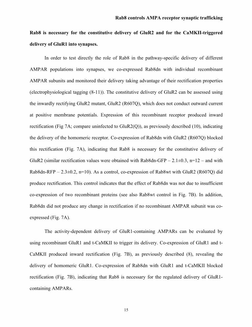
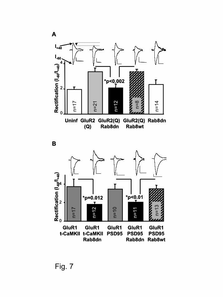
Rab8 is necessary for the constitutive delivery of GluR2 and for the CaMKII-triggered
delivery of GluR1 into synapses.
In order to test directly the role of Rab8 in the pathway-specific delivery of different
AMPAR populations into synapses, we co-expressed Rab8dn with individual recombinant
AMPAR subunits and monitored their delivery taking advantage of their rectification properties
(electrophysiological tagging (8-11)). The constitutive delivery of GluR2 can be assessed using
the inwardly rectifying GluR2 mutant, GluR2 (R607Q), which does not conduct outward current
at positive membrane potentials. Expression of this recombinant receptor produced inward
rectification (Fig 7A; compare uninfected to GluR2(Q)), as previously described (10), indicating
the delivery of the homomeric receptor. Co-expression of Rab8dn with GluR2 (R607Q) blocked
this rectification (Fig. 7A), indicating that Rab8 is necessary for the constitutive delivery of
GluR2 (similar rectification values were obtained with Rab8dn-GFP – 2.1±0.3, n=12 – and with
Rab8dn-RFP – 2.3±0.2, n=10). As a control, co-expression of Rab8wt with GluR2 (R607Q) did
produce rectification. This control indicates that the effect of Rab8dn was not due to insufficient
co-expression of two recombinant proteins (see also Rab8wt control in Fig. 7B). In addition,
Rab8dn did not produce any change in rectification if no recombinant AMPAR subunit was co-
expressed (Fig. 7A).
The activity-dependent delivery of GluR1-containing AMPARs can be evaluated by
using recombinant GluR1 and t-CaMKII to trigger its delivery. Co-expression of GluR1 and t-
CaMKII produced inward rectification (Fig. 7B), as previously described (8), revealing the
delivery of homomeric GluR1. Co-expression of Rab8dn with GluR1 and t-CaMKII blocked
rectification (Fig. 7B), indicating that Rab8 is necessary for the regulated delivery of GluR1-
containing AMPARs.
Rab8 controls AMPA receptor synaptic trafficking
16
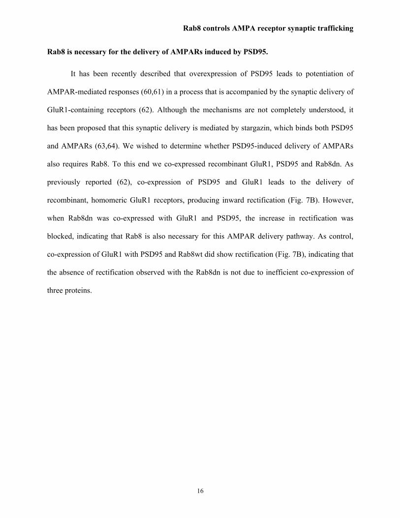
Rab8 is necessary for the delivery of AMPARs induced by PSD95.
It has been recently described that overexpression of PSD95 leads to potentiation of
AMPAR-mediated responses (60,61) in a process that is accompanied by the synaptic delivery of
GluR1-containing receptors (62). Although the mechanisms are not completely understood, it
has been proposed that this synaptic delivery is mediated by stargazin, which binds both PSD95
and AMPARs (63,64). We wished to determine whether PSD95-induced delivery of AMPARs
also requires Rab8. To this end we co-expressed recombinant GluR1, PSD95 and Rab8dn. As
previously reported (62), co-expression of PSD95 and GluR1 leads to the delivery of
recombinant, homomeric GluR1 receptors, producing inward rectification (Fig. 7B). However,
when Rab8dn was co-expressed with GluR1 and PSD95, the increase in rectification was
blocked, indicating that Rab8 is also necessary for this AMPAR delivery pathway. As control,
co-expression of GluR1 with PSD95 and Rab8wt did show rectification (Fig. 7B), indicating that
the absence of rectification observed with the Rab8dn is not due to inefficient co-expression of
three proteins.
Rab8 controls AMPA receptor synaptic trafficking
17
DISCUSSION
The Rab family of small GTPases is known to confer directionality to intracellular
membrane trafficking in several cell types (reviewed in (20)). However, it is surprising that very
few studies have addressed the role of Rab proteins in neuronal function, where polarized
membrane traffic is essential to maintain presynaptic and postsynaptic function. The notable
exception is Rab3, which is known to regulate neurotransmitter vesicle fusion and short-term
plasticity at the presynaptic terminal (26), although the precise mechanisms underlying this
regulation are still being elucidated (65,66).
We have now tested the role of the other three exocytic Rab proteins, namely Rab4, Rab8
and Rab11, at the postsynaptic terminal. These studies have identified Rab8 as a critical player in
postsynaptic membrane trafficking and plasticity. Rab8 was proposed to mediate dendritic
membrane trafficking and neurite outgrowth nearly a decade ago (27,67). These early studies,
using neuronal dissociated cultures, suggested the involvement of Rab8 in early stages of the
secretory pathway. Since then, no reports have tested whether Rab8 plays a more direct role in
synaptic function. Here we have shown that Rab8 is a necessary mediator of the local delivery of
AMPARs into excitatory synapses. Using quantitative surface immunostaining, we show that
Rab8 is required for the delivery of AMPARs into the spine surface, but not for their transport
from the dendritic shaft into the spine compartment or for their delivery into the dendritic plasma
membrane. In addition, by monitoring electrophysiologically the synaptic targeting of
endogenous and recombinant AMPARs, we demonstrate that Rab8 is necessary for both the
constitutive cycling of AMPARs and their regulated synaptic delivery, as triggered by CaMKII
activation, PSD95 overexpression or LTP induction. Therefore, we propose that Rab8 drives the
Rab8 controls AMPA receptor synaptic trafficking
18
local transport of AMPARs from an intracellular membrane compartment, possibly in the
dendritic spine, to the synaptic membrane.
Rab8 is a known mediator of exocytic transport from the trans-Golgi network into the
plasma membrane. As such, it is possibly involved in the surface delivery of a variety of
membrane proteins in neurons. However, an unexpected result of the present study is that Rab8
drives the local targeting of AMPARs into the highly specialized compartment that constitutes
the postsynaptic membrane. Several lines of evidence support this role of Rab8 in the short-range
synaptic delivery of AMPARs. Firstly, Rab8, besides being present in the dendritic shaft, is
localized in very close proximity to the synapse, including the postsynaptic density. Secondly,
Rab8 is necessary for the continuous synaptic cycling of GluR2/3-containing AMPARs.
Although the molecular details of this activity-independent pathway are not well understood, its
fast kinetics (13,15) suggests that it occurs in close proximity to the synapse. And thirdly,
quantitative immunohistochemical analysis showed that blockade of endogenous Rab8 function
by expression of a Rab8 dominant negative does not affect the transport of AMPARs in
dendrites, but it does impair receptor delivery into the spine surface. This result is in contrast
with a previous publication showing that Rab8 was necessary for the dendritic transport of a
recombinant viral glycoprotein (27). Although we do not know the reason for this discrepancy, it
is possible that Rab8 plays a more general role in membrane trafficking in developing neurons,
such as those in dissociated primary cultures, as compared with the more differentiated neurons
present in our organotypic slice cultures. This interpretation is consistent with the proposed role
of Rab8 in neurite outgrowth before axons and dendrites are differentiated (67).
As mentioned above, Rab8 mediates exocytic transport from the trans-Golgi network.
Interestingly, it has been recently described that distal dendrites contain intracellular
Rab8 controls AMPA receptor synaptic trafficking
19
compartments belonging to the trans-Golgi network (68,69). Our functional results fit very well
with these morphological observations, and suggest that AMPARs are driven locally from distal
trans-Golgi network outposts into synapses via a Rab8-mediated process. Exocytic delivery of
membrane proteins from trans-Golgi network can proceed directly into the plasma membrane or
through intermediate recycling endosomes (29,70). Rab8 is present in both trans-Golgi network
membranes and recycling endosomes (29), and to date, a possible role of Rab8 in controlling
membrane sorting from both compartments has not been ruled out. Interestingly, we have found
that Rab8 mediates both the continuous cycling of GluR2/3 receptors, and the transient, activity-
dependent delivery of GluR1/2 receptors. At this point, we cannot resolve whether these two
populations of receptors are delivered from different intracellular compartments, or whether a
single but sub-compartmentalized storage place is the source for both types of exocytic events.
However, biochemical membrane fractionations (71) and imaging experiments (10,72) suggest
that GluR1/2 and GluR2/3 receptors have different subcellular distributions. Therefore, we
propose that the synaptic delivery of GluR1/2 receptors during synaptic plasticity is regulated
from the trans-Golgi network, whereas the activity-independent cycling of GluR2/3 receptors
occurs from a specialized recycling compartment in close proximity to the synapse (see Fig. 8).
In this sense, it is important to note that neither Rab4 nor Rab11 seem to be involved in
the continuous cycling of AMPARs (the corresponding dominant negative mutants did not affect
basal AMPAR-mediated synaptic transmission). This observation is somehow surprising, since
these Rab proteins are known to mediate the trafficking of various populations of recycling
endosomes (32-36,52). Our results, then, support the interpretation that GluR2/3 synaptic
delivery occurs from a specialized membrane compartment, whose cycling is controlled by Rab8
but not by Rab4 or Rab11. This interpretation does not rule out a role for Rab4 and/or Rab11 in

Rab8 controls AMPA receptor synaptic trafficking
20
other steps of AMPAR trafficking. For instance, it has been reported that AMPARs that return to
the plasma membrane after activity-dependent internalization colocalize with Rab4-positive
compartments (73,74). However, our results indicate that Rab8 is the Rab family member that
directly mediates the late stages of membrane transport that result in the synaptic delivery of
AMPARs both during constitutive receptor cycling and during activity-dependent delivery
(LTP).
In conclusion, these results expand our view of the cellular functions accomplished by the
Rab protein-driven exocytic machinery in neurons, and shed light into the membrane trafficking
events that control the dynamic organization of the postsynaptic terminal.
Rab8 controls AMPA receptor synaptic trafficking
21
ACKNOWLEDGEMENTS
We thank Roberto Malinow, Ronald Holz, Lori Isom, Edward Stuenkel and María S. Soengas for
critical reading of the manuscript. We also thank Kristen Verhey for helpful discussions. This
work was supported by grants from the National Institutes of Health (MH070417), National
Alliance for Research on Schizophrenia and Depression (NARSAD) and the Alzheimer’s
Association to J.A.E.
ABBREVIATIONS
AMPA R, alpha-amino-3-hydroxy-5-methyl-4-isoxazolepropionate receptors; NMDA R, N-
methyl-D-aspartate receptor; GluR1-4, glutamate receptor subunit 1-4; CaMKII, Ca+2/
calmodulin dependent protein kinase II; LTP, long term potentiation.

Rab8 controls AMPA receptor synaptic trafficking
22
REFERENCES
1. Sudhof, T. C. (2000) Neuron 28, 317-320
2. Murthy, V. N., and De Camilli, P. (2003) Annu Rev Neurosci 26, 701-728
3. Sheng, M., and Lee, S. H. (2001) Cell 105, 825-828
4. Barry, M. F., and Ziff, E. B. (2002) Curr Opin Neurobiol 12, 279-286
5. Malinow, R., and Malenka, R. C. (2002) Annu Rev Neurosci 25, 103-126
6. Song, I., and Huganir, R. L. (2002) Trends Neurosci 25, 578-588
7. Hollmann, M., and Heinemann, S. (1994) Annu Rev Neurosci 17, 31-108
8. Hayashi, Y., Shi, S. H., Esteban, J. A., Piccini, A., Poncer, J. C., and Malinow, R. (2000)
Science 287, 2262-2267
9. Zhu, J. J., Esteban, J. A., Hayashi, Y., and Malinow, R. (2000) Nat Neurosci 3, 1098-1106
10. Shi, S., Hayashi, Y., Esteban, J. A., and Malinow, R. (2001) Cell 105, 331-343
11. Esteban, J. A., Shi, S. H., Wilson, C., Nuriya, M., Huganir, R. L., and Malinow, R. (2003)
Nat Neurosci 6, 136-143
12. Passafaro, M., Piech, V., and Sheng, M. (2001) Nat Neurosci 4, 917-926
13. Nishimune, A., Isaac, J. T., Molnar, E., Noel, J., Nash, S. R., Tagaya, M., Collingridge, G.
L., Nakanishi, S., and Henley, J. M. (1998) Neuron 21, 87-97
14. Song, I., Kamboj, S., Xia, J., Dong, H., Liao, D., and Huganir, R. L. (1998) Neuron 21, 393-
400
15. Luscher, C., Xia, H., Beattie, E. C., Carroll, R. C., von Zastrow, M., Malenka, R. C., and
Nicoll, R. A. (1999) Neuron 24, 649-658
Rab8 controls AMPA receptor synaptic trafficking
23
16. Gerges, N. Z., Tran, I. C., Backos, D. S., Harrell, J. M., Chinkers, M., Pratt, W. B., and
Esteban, J. A. (2004) J Neurosci 24, 4758-4766
17. Malinow, R., Mainen, Z. F., and Hayashi, Y. (2000) Curr Opin Neurobiol 10, 352-357
18. Pfeffer, S. R. (2001) Trends Cell Biol 11, 487-491
19. Stenmark, H., and Olkkonen, V. M. (2001) Genome Biol 2, REVIEWS3007
20. Zerial, M., and McBride, H. (2001) Nat Rev Mol Cell Biol 2, 107-117.
21. D'Adamo, P., Menegon, A., Lo Nigro, C., Grasso, M., Gulisano, M., Tamanini, F., Bienvenu,
T., Gedeon, A. K., Oostra, B., Wu, S. K., Tandon, A., Valtorta, F., Balch, W. E., Chelly, J.,
and Toniolo, D. (1998) Nat Genet 19, 134-139
22. Fischer von Mollard, G., Mignery, G. A., Baumert, M., Perin, M. S., Hanson, T. J., Burger,
P. M., Jahn, R., and Sudhof, T. C. (1990) Proc Natl Acad Sci U S A 87, 1988-1992
23. Geppert, M., Bolshakov, V. Y., Siegelbaum, S. A., Takei, K., De Camilli, P., Hammer, R. E.,
and Sudhof, T. C. (1994) Nature 369, 493-497
24. Geppert, M., Goda, Y., Stevens, C. F., and Sudhof, T. C. (1997) Nature 387, 810-814
25. Castillo, P. E., Janz, R., Sudhof, T. C., Tzounopoulos, T., Malenka, R. C., and Nicoll, R. A.
(1997) Nature 388, 590-593
26. Lonart, G., Janz, R., Johnson, K. M., and Sudhof, T. C. (1998) Neuron 21, 1141-1150
27. Huber, L. A., de Hoop, M. J., Dupree, P., Zerial, M., Simons, K., and Dotti, C. (1993) J Cell
Biol 123, 47-55.
28. Huber, L. A., Pimplikar, S., Parton, R. G., Virta, H., Zerial, M., and Simons, K. (1993) J Cell
Biol 123, 35-45
29. Ang, A. L., Folsch, H., Koivisto, U. M., Pypaert, M., and Mellman, I. (2003) J Cell Biol 163,
339-350
Rab8 controls AMPA receptor synaptic trafficking
24
30. Deretic, D., Huber, L. A., Ransom, N., Mancini, M., Simons, K., and Papermaster, D. S.
(1995) J Cell Sci 108 ( Pt 1), 215-224
31. Moritz, O. L., Tam, B. M., Hurd, L. L., Peranen, J., Deretic, D., and Papermaster, D. S.
(2001) Mol Biol Cell 12, 2341-2351.
32. van der Sluijs, P., Hull, M., Webster, P., Male, P., Goud, B., and Mellman, I. (1992) Cell 70,
729-740.
33. Sonnichsen, B., De Renzis, S., Nielsen, E., Rietdorf, J., and Zerial, M. (2000) J Cell Biol
149, 901-914.
34. Sheff, D. R., Daro, E. A., Hull, M., and Mellman, I. (1999) J Cell Biol 145, 123-139
35. De Renzis, S., Sonnichsen, B., and Zerial, M. (2002) Nat Cell Biol 4, 124-133.
36. Ullrich, O., Reinsch, S., Urbe, S., Zerial, M., and Parton, R. G. (1996) J Cell Biol 135, 913-
924
37. Chen, W., Feng, Y., Chen, D., and Wandinger-Ness, A. (1998) Mol Biol Cell 9, 3241-3257
38. Prekeris, R., Klumperman, J., and Scheller, R. H. (2000) Mol Cell 6, 1437-1448
39. Wang, X., Kumar, R., Navarre, J., Casanova, J. E., and Goldenring, J. R. (2000) J Biol Chem
275, 29138-29146
40. Campbell, R. E., Tour, O., Palmer, A. E., Steinbach, P. A., Baird, G. S., Zacharias, D. A., and
Tsien, R. Y. (2002) Proc Natl Acad Sci U S A 99, 7877-7882
41. Malinow, R., Hayashi, Y., Maletic-Savatic, M., Zaman, S., Poncer, J.-C., Shi, S.-H. and
Esteban, J.A. (1999) (Yuste, R., Lanni, F. and Konnerth A., ed), Cold Spring Harbor Press,
Cold Spring Harbor, NY
42. Gahwiler, B. H., Capogna, M., Debanne, D., McKinney, R. A., and Thompson, S. M. (1997)
Trends Neurosci 20, 471-477
Rab8 controls AMPA receptor synaptic trafficking
25
43. Lo, D. C., McAllister, A. K., and Katz, L. C. (1994) Neuron 13, 1263-1268
44. Schlesinger, S. (1993) Trends Biotechnol 11, 18-22
45. Phend, K. D., Rustioni, A., and Weinberg, R. J. (1995) J Histochem Cytochem 43, 283-292
46. Shirakawa, R., Yoshioka, A., Horiuchi, H., Nishioka, H., Tabuchi, A., and Kita, T. (2000) J
Biol Chem 275, 33844-33849
47. McCaffrey, M. W., Bielli, A., Cantalupo, G., Mora, S., Roberti, V., Santillo, M., Drummond,
F., and Bucci, C. (2001) FEBS Lett 495, 21-30
48. Mohrmann, K., Leijendekker, R., Gerez, L., and van Der Sluijs, P. (2002) J Biol Chem 277,
10474-10481
49. Peranen, J., Auvinen, P., Virta, H., Wepf, R., and Simons, K. (1996) J Cell Biol 135, 153-167
50. Hattula, K., Furuhjelm, J., Arffman, A., and Peranen, J. (2002) Mol Biol Cell 13, 3268-3280
51. Ren, M., Zeng, J., De Lemos-Chiarandini, C., Rosenfeld, M., Adesnik, M., and Sabatini, D.
D. (1996) Proc Natl Acad Sci U S A 93, 5151-5155
52. Wilcke, M., Johannes, L., Galli, T., Mayau, V., Goud, B., and Salamero, J. (2000) J Cell Biol
151, 1207-1220
53. Fan, G. H., Lapierre, L. A., Goldenring, J. R., and Richmond, A. (2002) Blood
54. Volpicelli, L. A., Lah, J. J., Fang, G., Goldenring, J. R., and Levey, A. I. (2002) J Neurosci
22, 9776-9784
55. Roosterman, D., Schmidlin, F., and Bunnett, N. W. (2003) Am J Physiol Cell Physiol 284,
C1319-1329
56. Roberts, R. L., Barbieri, M. A., Pryse, K. M., Chua, M., Morisaki, J. H., and Stahl, P. D.
(1999) J Cell Sci 112 ( Pt 21), 3667-3675
Rab8 controls AMPA receptor synaptic trafficking
26
57. Barbieri, M. A., Hoffenberg, S., Roberts, R., Mukhopadhyay, A., Pomrehn, A., Dickey, B.
F., and Stahl, P. D. (1998) J Biol Chem 273, 25850-25855
58. Barbero, P., Bittova, L., and Pfeffer, S. R. (2002) J Cell Biol 156, 511-518
59. Poncer, J. C., Esteban, J. A., and Malinow, R. (2002) J Neurosci 22, 4406-4411
60. El-Husseini, A. E., Schnell, E., Chetkovich, D. M., Nicoll, R. A., and Bredt, D. S. (2000)
Science 290, 1364-1368
61. Stein, V., House, D. R., Bredt, D. S., and Nicoll, R. A. (2003) J Neurosci 23, 5503-5506
62. Ehrlich, I., and Malinow, R. (2004) J Neurosci 24, 916-927
63. Chen, L., Chetkovich, D. M., Petralia, R. S., Sweeney, N. T., Kawasaki, Y., Wenthold, R. J.,
Bredt, D. S., and Nicoll, R. A. (2000) Nature 408, 936-943
64. Schnell, E., Sizemore, M., Karimzadegan, S., Chen, L., Bredt, D. S., and Nicoll, R. A. (2002)
Proc Natl Acad Sci U S A 99, 13902-13907
65. Schluter, O. M., Khvotchev, M., Jahn, R., and Sudhof, T. C. (2002) J Biol Chem 277, 40919-
40929
66. Schluter, O. M., Schmitz, F., Jahn, R., Rosenmund, C., and Sudhof, T. C. (2004) J Neurosci
24, 6629-6637
67. Huber, L. A., Dupree, P., and Dotti, C. G. (1995) Mol Cell Biol 15, 918-924
68. Horton, A. C., and Ehlers, M. D. (2003) J Neurosci 23, 6188-6199
69. Pierce, J. P., Mayer, T., and McCarthy, J. B. (2001) Curr Biol 11, 351-355
70. Folsch, H., Pypaert, M., Maday, S., Pelletier, L., and Mellman, I. (2003) J Cell Biol 163,
351-362
71. Lee, S. H., Valtschanoff, J. G., Kharazia, V. N., Weinberg, R., and Sheng, M. (2001)
Neuropharmacology 41, 680-692

Rab8 controls AMPA receptor synaptic trafficking
27
72. Piccini, A., and Malinow, R. (2002) J Neurosci 22, 5387-5392
73. Ehlers, M. D. (2000) Neuron 28, 511-525
74. Steiner, P., Sarria, J. C., Glauser, L., Magnin, S., Catsicas, S., and Hirling, H. (2002) J Cell
Biol 157, 1197-1209.

Rab8 controls AMPA receptor synaptic trafficking
28
FIGURE LEGENDS:
Figure 1. Rab8 is necessary for AMPA receptor delivery into synapses. (A-D) Insets,
Western blot analysis of the expression of recombinant Rab-GFP in hippocampal slices using the
Sindbis virus method. Big arrowheads indicate recombinant Rab-GFP proteins (about 52 KDa);
small arrows indicate endogenous Rab proteins (about 25 KDa). Sample trace of evoked AMPA-
and NMDA-receptor mediated synaptic responses recorded at –60 mV and +40 mV,
respectively, from uninfected and infected cells. Scale bars, 20 pA and 40 ms. Left, average
AMPAR-mediated current amplitude (i.e. the peak of the response recorded at –60 mV) from
infected (inf) neurons expressing Rab8dn (A), Rab4dn (B), Rab11dn (C) or Rab8wt (D) and
control neighboring cells not expressing the recombinant protein (uninf). n represents the number
of pathways from cell pairs; p is the probability value according to the Wilcoxon test. Middle,
average NMDA R-mediated current amplitude (recorded at +40 mV at a latency when AMPA
responses are fully decayed, 60 ms) from uninfected and infected cells (n also represents the
number of pathways from cell pairs). Right, average AMPA/NMDA ratios for uninfected and
infected cells (n represents the number of pathways).
Figure 2. Localization of Rab8 at postsynaptic terminals. (A) Ultrastructural localization of
Rab8 at CA1 hippocampal synapses by electron microscopy. Rab8 immunogold particles
(arrows) were found at the postsynaptic terminal, including the postsynaptic density. Labeling
was also detected in dendritic shafts (not shown). Presynaptic terminals are labeled with
asterisks. Scale bar, 100 nm. (B) Quantification of immunogold labeling. Most of the gold
particles were found postsynaptically, with marginal presence presynaptically (Pre), indicating

Rab8 controls AMPA receptor synaptic trafficking
29
specificity for the immunogold labeling. The majority of the gold particles were found
intracelullarly (Intra). Gold particles were also found at the postsynaptic density (PSD) as well as
extrasynaptic membranes (Extra). For this quantification, only gold particles within 600 nm of
the synapse were included. (C) Rab8-GFP is present at dendritic spines. Representative confocal
images showing the distribution of the recombinant Rab8-GFP in hippocampal neurons from
slices. Rab8 is present in cell body and dendrites (left), as well as in dendritic spines (right).
Figure 3. Immunohistochemical localization of surface AMPA receptors in slices expressing
Rab8dn. (A) Confocal imaging of GluR2-GFP transfected neuron. GFP fluorescence signal
(top) and surface anti-GFP immunoreactivity of the same neuron in non-permeabilized
conditions (bottom). (B) Confocal imaging for a neuron cotransfected with GluR2-GFP and
Rab8dn-GFP. GFP signal and surface anti-GFP immunoreactivity, as in A. (C) Control neurons
expressing Rab8dn alone. Negative surface immunostaining (bottom) is expected given the
intracellular distribution of Rab8-GFP.
Fig. 4. Rab8dn decreases AMPAR surface delivery locally at spines. (A) Representative
confocal image of a neuron cotransfected with GluR2-GFP and Rab8dn-RFP. Left: GFP
fluorescence signal showing total GluR2 receptor distribution. Middle: surface GluR2-GFP
receptors assayed by anti-GFP immunoreactivity of the same neuron in non-permeabilized
conditions. Right: RFP fluorescence signal from the same neuron indicating expression of the
cotransfected Rab8dn-RFP (neurons transfected only with GluR2-GFP do not show any RFP
signal above background; not shown). Scale bar: 30 µm. (B) Representative line plot analysis of
total (left; GFP signal) and surface (right; Cy5 signal) receptor across a spine and the adjacent
Rab8 controls AMPA receptor synaptic trafficking
30
dendritic shaft of GluR2 transfected or GluR2 plus Rab8dn cotransfected neurons, as indicated.
Values for surface and total receptors were taken from the fluorescence intensity peaks after
background subtraction (dashed line). Scale bar: 2 µm. (C) Surface ratio from pairs of spines and
dendrites in GluR2 transfected neurons (left) or in GluR2 and Rab8dn cotransfected neurons
(middle). Surface ratios for spines and dendrites were calculated by dividing the corresponding
background-subtracted Cy5 and GFP values. Surface ratios are normalized by the mean dendrite
value (0.31±0.05 for GluR2 alone and 0.31±0.04 for GluR2 plus Rab8dn). Cumulative
probabilities (right) of spine/dendrite ratios show a significant difference between the surface
ratio distribution of GluR2 and GluR2 plus Rab8dn transfected neurons. For comparison, dashed
lines indicate that 50% of the spine-dendrite pairs have less surface ratio in the spine than in the
dendrite for cells transfected with GluR2 alone (black line: cumulative probability=0.5 for
spine/dendrite=1). In contrast, 75% of the spine-dendrite pairs have less surface ratio in the spine
than in the dendrite for cells cotransfected with Rab8dn (red line: cumulative probability=0.75
for spine/dendrite=1). (D) Total receptor amount (GFP signal) in spines and dendrites are
calculated in GluR2 transfected neurons (left) and in GluR2 plus Rab8dn cotransfected neurons
(middle). Values are normalized by the mean obtained at dendrites (50±4 for GluR2 alone and
54±3 for GluR2 plus Rab8dn). The cumulative probability (right) indicates that Rab8dn does not
significantly change the total receptor distribution in spines versus dendrites.
Figure 5. Rab8 is necessary for both the constitutive and regulated delivery of AMPA
receptors into synapses. (A-C) Inset, sample trace of evoked AMPA receptor mediated synaptic
responses recorded at –60 mV from uninfected/non-transfected and infected/transfected cells.
Scale bars, 20 pA and 10 ms. (A) Rab8dn significantly reduced AMPA transmission in the

Rab8 controls AMPA receptor synaptic trafficking
31
presence of high Mg2+ (12 mM MgCl2). Average current amplitude at –60 mV from uninfected
and infected cells was 46.9±9.9 pA and 26.7±4.5 pA, respectively. n represents the number of
pathways from cell pairs. p = 0.03, according to the Wilcoxon test. (B) Expression of t-CaMKII
significantly increases AMPAR-mediated transmission. Non-transfected: 32.5±6.1 pA;
transfected with t-CaMKII: 81.5±14.4 pA. p = 0.001, according to the Wilcoxon test; n
represents the number of the pathways from cell pairs. (C) Rab8dn blocked t-CaMKII-induced
potentiation of AMPA responses. Non-transfected: 25.0±4.2 pA; transfected with t-CaMKII and
Rab8dn: 19.7±3.8 pA. (D) Rab8dn does not change the phosphorylation levels of CaMKII. Left,
Western blot analysis showing the P-Thr286 αCaMKII / total αCaMKII ratios in Rab8dn-infected
slices and uninfected slices. Each lane in the western blot is the result of pooling together
extracts from three dissected CA1 regions. Right, Quantification by densitometric scanning of
four independent experiments as the one shown in the left.
Figure 6. Rab8 is necessary for LTP. Organotypic slice cultures were infected with virus
expressing either Rab8wt or Rab8dn. Whole-cell recordings were established from neurons
expressing the recombinant proteins or uninfected cells, and LTP was induced by pairing 3 Hz
presynaptic stimulation with 0 mV postsynaptic depolarization, as previously described (8). (A)
Pairing significantly increased AMPA receptor-mediated responses in uninfected (p = 0.002,
paired t-test) and Rab8wt-infected (p = 0.001, paired t-test) neurons. However, pairing
significantly decreased synaptic responses in Rab8dn expressing neurons (p = 0.001, paired t-
test). Inset, sample trace of evoked AMPA receptor-mediated synaptic responses recorded at –60
mV before pairing (thin line) and 30 min after pairing (thick line) from control (uninfected)
neurons or infected cells, as indicated. Scale bars, 20 pA and 10 ms. (B) Normalized steady-state
Rab8 controls AMPA receptor synaptic trafficking
32
AMPAR response amplitudes in paired (LTP induction) and control (Unpaired) pathways in cells
expressing Rab8wt-GFP, Rab8dn-GFP and uninfected control cells. AMPAR responses from
control pathways were not statistically different from baseline responses.
Figure 7. Rab8 is necessary for the constitutive delivery of GluR2 and for the delivery of
GluR1 triggered by t-CaMKII or by PSD95 into synapses. Rectification values were
calculated as the ratio between the amplitude of the synaptic response at -60 mV over the
amplitude at +40 mV. Endogenous receptors conduct current at -60 mV and +40 mV, whereas
recombinant receptors conduct only at negative membrane potentials. Therefore, delivery of the
recombinant homomeric receptors is accompanied by an increase in the rectification value (a
decrease in the outward current, depicted by the arrow head in A, with respect to the inward
current). (A) Average rectification values were: control (uninfected), 1.9±0.2; GluR2(R586Q),
3.3±0.2; GluR2(R586Q) plus Rab8dn, 2.1±0.3; GluR2(R586Q) plus Rab8wt, 3.3±0.2; Rab8dn,
2.3±0.3. (B) Average rectification values were: GluR1 plus t-CaMKII, 3.9±0.7; GluR1 plus t-
CaMKII and Rab8dn, 1.8±0.2; GluR1 plus PSD95, 3.5±0.5; GluR1 plus PSD95 and Rab8dn,
2.0±0.2; GluR1 plus PSD95 and Rab8wt, 3.5±0.4. n represents the number of pathways, p is the
probability value according to the Student’s t-test. Inset, sample traces of evoked AMPA
receptor mediated synaptic responses recorded at –60 mV and +40 mV from control or
transfected cells as indicated. Scale bars, 20 pA and 20 ms.
Figure 8. Membrane sorting model for the control of AMPAR synaptic delivery by Rab8.
The regulated (activity-dependent) delivery of GluR1/2 receptors would be mediated by Rab8
from a membrane compartment belonging to the trans-Golgi network (TGN). In contrast, the
Rab8 controls AMPA receptor synaptic trafficking
33
continuous (activity-independent) delivery of GluR2/3 receptors would occur from specialized
recycling endosomes (RE) whose sorting is also controlled by Rab8.
AMPA NMDA AMPA/NMDAUninf Inf
AM
PA a
mpl
itude
(pA
)
NM
DA
am
plitu
de (p
A)
AM
PA/N
MD
A ra
tio
AM
PA a
mpl
itude
(pA
)
NM
DA
am
plitu
de (p
A)
AM
PA/N
MD
A ra
tio
AM
PA a
mpl
itude
(pA
)
NM
DA
am
plitu
de (p
A)
AM
PA/N
MD
A ra
tio
0
20
40
60 n=22
0
10
20
30n=20
0
1
2
3
n=17
n=21
0
1
2
n=25
n=26
n=21
0
20
40n=20
0
20
40
0
20
40
60 n=21
0
10
20
n=15
0
1
2
3
n=32
n=28
Uninf Inf Uninf InfUninf Inf
0
20
40
60 n=22
0
20
40
60
0
20
40
60 n=22
0
10
20
30n=20
0
10
20
30
0
10
20
30n=20
0
1
2
3
0
1
2
3
n=17
n=21
0
1
2
n=25
n=26
0
1
2
0
1
2
0
1
2
n=25
n=26
n=21
0
20
40 n=21
0
20
40n=20
0
20
40
n=20
0
20
40
0
20
40
60 n=21
0
20
40
60
0
20
40
60 n=21
0
10
20
n=15
0
10
20
n=15
0
1
2
3
n=32
n=28
0
1
2
3
n=32
n=28
Uninf Inf Uninf InfUninf Inf
0
20
40
60
AM
PA a
mpl
itude
(pA
)
n=24
0
10
20
30
NM
DA
am
plitu
de (p
A)
n=11
*p=0.001
AM
PA/N
MD
A ra
tio
0
1
2
n=21
n=29
*p=0.002
0
20
40
0
20
40
60 n=24
0
10
20
30
0
10
20
30
n=11
*p=0.001
0
1
2
n=21
n=29
0
1
2
n=21
n=29
*p=0.002
A. Rab8dnanti-Rab8
Uninf. Inf.
B. Rab4dnanti-Rab4
Uninf. Inf.
C. Rab11dn
anti-Rab11
Uninf. Inf.
D. Rab8wt
Uninf. Inf.
anti-Rab8
Fig. 1
20 µm20 µm∗
∗
∗
∗
0
20
40
60
% o
f Gol
d Pa
rtic
lesB
Pre PSD Extra Intra
PostsynapticPre-
0
20
40
60
0
20
40
60
% o
f Gol
d Pa
rtic
lesB
Pre PSD Extra Intra
PostsynapticPre-A
∗
∗A
∗
∗
C
5 µm5 µm
....
n=53
Fig. 2
mmmm
20 m
20 µm
20 m
20 µm20 µm20 µm 20 µm
GluR2-GFP GluR2-GFPRab8dn-GFP
Rab8dn-GFP
( )( )A B C
GFPsignal
Surfaceanti-GFP
Fig. 3
0
0.5
1
0 1 2 3 4Spine/Dendrite (surface ratio)C
umul
ativ
e pr
obab
ility
0
0.5
1
0 1 2 3 4 5Spine/Dendrite (total receptor)
Cum
ulat
ive
prob
abili
ty
0
0.5
1
1.5
Nor
mal
ized
su
rfac
e ra
tio
A. GluR2-GFP + Rab8dn-RFPGFP Cy5 (Surface anti-GFP) RFP
B. Spine and dendrite fluorescence analysis
C. Surface ratio (Cy5/GFP)
D. Total receptor (GFP)
GluR2
GluR2
GluR2 + Rab8dn
GluR2 + Rab8dn
GluR2GluR2 + Rab8dn
GluR2GluR2 + Rab8dn
p = 0.4
*p = 0.03
Dendrite0
0.5
1
*p < 0.001
0
0.5
1
Nor
mal
ized
to
tal r
ecep
tor
0
0.5
1
1.5
Surface Rs(Cy5 channel)
Total Rs(GFP channel)
0
40
80
120
Fluo
resc
ence
inte
nsity
(a.u
.)
SpineDendrite 0
10
20
30
SpineDendrite
Spine Dendrite Spine
Dendrite SpineDendrite Spine
n=44
n=44
n=55
n=55
0
40
80
120
0
20
40
Total Rs(GFP channel)
Surface Rs(Cy5 channel)
GluR2 GluR2 + Rab8dn
SpineDendrite SpineDendrite
60
0
0.5
1
0 1 2 3 4Spine/Dendrite (surface ratio)C
umul
ativ
e pr
obab
ility
0
0.5
1
0 1 2 3 4 5Spine/Dendrite (total receptor)
Cum
ulat
ive
prob
abili
ty
0
0.5
1
1.5
Nor
mal
ized
su
rfac
e ra
tio
A. GluR2-GFP + Rab8dn-RFPGFP Cy5 (Surface anti-GFP) RFP
B. Spine and dendrite fluorescence analysis
C. Surface ratio (Cy5/GFP)
D. Total receptor (GFP)
GluR2
GluR2
GluR2 + Rab8dn
GluR2 + Rab8dn
GluR2GluR2 + Rab8dn
GluR2GluR2 + Rab8dn
p = 0.4
*p = 0.03
Dendrite0
0.5
1
*p < 0.001
0
0.5
1
Nor
mal
ized
to
tal r
ecep
tor
0
0.5
1
1.5
Surface Rs(Cy5 channel)
Total Rs(GFP channel)
0
40
80
120
Fluo
resc
ence
inte
nsity
(a.u
.)
SpineDendrite 0
10
20
30
SpineDendrite
Spine Dendrite Spine
Dendrite SpineDendrite Spine
n=44
n=44
n=55
n=55
0
40
80
120
0
20
40
Total Rs(GFP channel)
Surface Rs(Cy5 channel)
GluR2 GluR2 + Rab8dn
SpineDendrite SpineDendrite
60
Fig. 4
D. P-CaMKII / CaMKII ratio
0
0.2
0.4
0.6
Uninf Inf
P-C
aMK
II/ C
aMK
IIra
tio(a
rbitr
ary
units
)
0
20
40
60 n=17
*p=0.03
Uninf Inf
Uninf InfA
MPA
am
plitu
de ( p
A)A. Rab8dn; high Mg2+
0
25
50
75
100n=13
*p=0.001
Non- Transftransf
B. t-CaMKII
Non-transf Transf
AM
PA a
mpl
itude
(pA)
0
20
40
60 n=17
*p=0.03
Uninf Inf
Uninf InfA
MPA
am
plitu
de ( p
A)A. Rab8dn; high Mg2+
0
20
40
60 n=17
*p=0.03
Uninf Inf
Uninf InfA
MPA
am
plitu
de ( p
A)A. Rab8dn; high Mg2+
n=13
C. t-CaMKII+Rab8dn
Non- Transftransf
Non-transf Transf
0AM
PA a
mpl
itude
(pA) n=13
C. t-CaMKII+Rab8dn
Non- Transftransf
Non-transf Transf
0
n=13
C. t-CaMKII+Rab8dn
Non- Transftransf
Non-transf Transf
0
10
AM
PA a
mpl
itude
(pA)
0
25
50
75
100n=13
*p=0.001
Non- Transftransf
B. t-CaMKII
Non-transf Transf
AM
PA a
mpl
itude
(pA)
0
25
50
75
100n=13
*p=0.001
Non- Transftransf
B. t-CaMKII
Non-transf Transf
0
25
50
75
100n=13
*p=0.001
Non- Transftransf
B. t-CaMKII
Non-transf Transf
AM
PA a
mpl
itude
(pA)
n=4
20
30
40
P-Thr286
αCaMKII
Total αCaMKII
GFP-Rab8dn
EndogenousRab8
Uninf Inf
Fig. 5
Rab8wt (n=7)Uninf. (n=6)
Rab8dn (n=6)
Nor
mal
ized
EPS
C(%
bas
elin
e)
00
100
200
300
-5 5 10 15 20 25 30 35
Time (min)
Rab8wt (n=7)Uninf. (n=6)
Rab8dn (n=6)
Nor
mal
ized
EPS
C(%
bas
elin
e)
00
100
200
300
-5 5 10 15 20 25 30 35
Time (min)
Rab8wt (n=7)Uninf. (n=6)
Rab8dn (n=6)
Nor
mal
ized
EPS
C(%
bas
elin
e)
00
100
200
300
-5 5 10 15 20 25 30 35
Time (min)
beforeafter
Uninf Rab8wt
Rab8dn
beforeafter
Uninf Rab8wt
Rab8dn
beforeafter
Uninf Rab8wt
Rab8dn
Nor
mal
ized
pot
entia
tion
(% b
asel
ine)
0
100
200
*p < 0.01
Uninf. Rab8wt
Rab8dn
Uninf. Rab8wt
Rab8dn
LTP induction Unpaired pathway
A
B
Fig. 6
Rec
tific
atio
n (I -
60/I +
40)
I-60
I+40
GluR1t-CaMKII
GluR1t-CaMKIIRab8dn
A
B
GluR1PSD95
GluR1PSD95Rab8dn
GluR1PSD95Rab8wt
Rec
tific
atio
n (I -
60/I +
40)
0
2
4
*p=0.012
n=17
n=12
n=10
n=11
GluR2(Q)
Uninf Rab8dnGluR2(Q)Rab8dn
n=21
0
2
4
n=17
*p<0.002
n=12
n=14
GluR2(Q)Rab8wt
*p<0.01
n=8
n=13
Rec
tific
atio
n (I -
60/I +
40)
I-60
I+40
GluR1t-CaMKII
GluR1t-CaMKIIRab8dn
A
B
GluR1PSD95
GluR1PSD95Rab8dn
GluR1PSD95Rab8wt
Rec
tific
atio
n (I -
60/I +
40)
0
2
4
*p=0.012
n=17
n=12
n=10
n=11
GluR2(Q)
Uninf Rab8dnGluR2(Q)Rab8dn
n=21
0
2
4
n=17
*p<0.002
n=12
n=14
GluR2(Q)Rab8wt
*p<0.01
n=8
n=13
Fig. 7
GluR2/3 GluR1/2
rab8
activity-dependent:
LTPCaMKIIPSD95
TGN
RE
constitutivecycling
rab8
GluR2/3 GluR1/2
rab8
activity-dependent:
LTPCaMKIIPSD95
TGN
RE
constitutivecycling
rab8rab8
Fig. 8


![Reduced aggression in AMPA-type glutamate receptor GluR …0].pdf · Reduced aggression in AMPA-type glutamate receptor GluR-A subunit-deficient mice ... conditioning responses](https://img.pdfslide.us/doc/110x75/5aa910cb7f8b9a72188c6780/reduced-aggression-in-ampa-type-glutamate-receptor-glur-0pdfreduced-aggression.jpg)