Embed Size (px)
DESCRIPTION
endo paper
Citation preview
Endodontic Topics 2002, 1, 26–39 Copyright C Blackwell MunksgaardPrinted in Denmark. All rights reserved ENDODONTIC TOPICS 2002
1601-1538
Local anesthetic failure inendodontics:Mechanisms and Management
KENNETH M. HARGREAVES & KARL KEISER
Many patients fear endodontic procedures due to a concern about pain. Although pain treatment is wellmanaged in many endodontic patients, there exists a group of patients who do not receive adequate localanesthesia. This article reviews the mechanisms of local anesthetic failure and focuses on available evidence fordeveloping effective and efficient approaches in local anesthesia.
IntroductionSuccessful management of pain emergencies is astrong component of clinical excellence. Althoughclinical studies indicate that comparatively few pa-tients experience pain after endodontic procedures, ithas been estimated that about 20% of patients experi-ence moderate-to-severe pain after treatment (1, 2,3). An additional small fraction of patients (about 1–2%) will experience a sudden ‘flare-up’ of severe painor swelling after endodontic treatment. To com-pound the problem, the clinical management of en-dodontic patients is often problematic due to inad-equate local anesthesia.
Many of these issues will be reviewed in articles insubsequent issues of Endodontic Topics, which willsummarize the latest clinical research and relevantbiological foundations from the perspective of provid-ing guidance for the efficient and effective manage-ment of endodontic pain conditions. In this inauguralissue, we will review the problem of local anestheticfailures in endodontic patients. Why are local anes-thetics less effective in endodontic pain patients? Howcan we better identify these patients before treatment?And, more importantly, what is the best current evi-dence available for recommending strategies in pro-ducing clinically acceptable anesthesia in these pa-tients?
26
Prevalence of local anestheticfailuresWhen planning dental procedures on teeth with clin-ically normal (i.e. uninflamed) pulps, effective localanesthesia is the bedrock of dental pain control. Localanesthetics administered by the infiltration route ofinjection are highly effective in producing clinicalanesthesia in normal tissue. Although nerve-block in-jections are considered more technically difficult, andtherefore somewhat less predictable than infiltrationinjections, clinical studies suggest success rates ofabout 75–90% or more in patients with clinically nor-mal teeth (4, 5, 6, 7, 8, 9, 10).
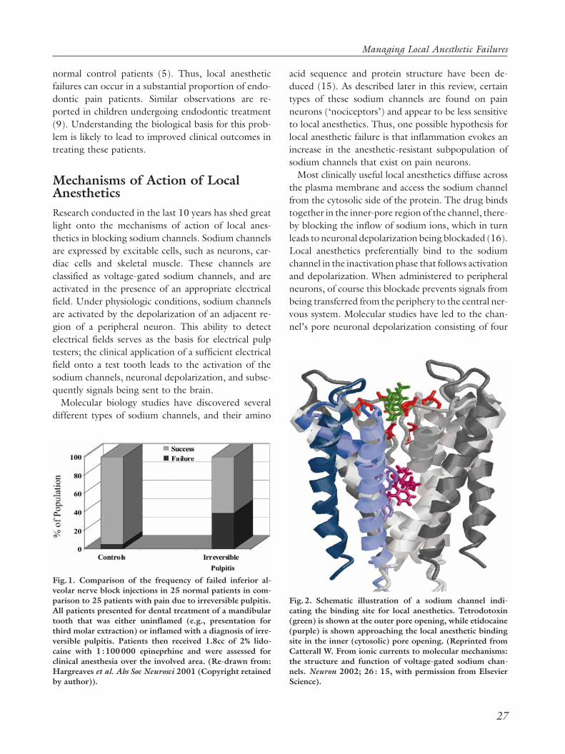
However, local anesthetics are generally much lesseffective when administered to patients with inflamedtissue (11). For the purposes of this review, we willfocus on studies that evaluate patients with odonto-genic pain due to inflamed pulpal or periradiculartissue. Clinical studies have reported that a single in-ferior alveolar nerve (IAN) block injection of localanesthetic(1.8 cc) is ineffective in 30–80% of patientswith a diagnosis of irreversible pulpitis (5, 12, 13,14). Figure 1 compares the frequency of anestheticfailures after a single IAN nerve block injection of1.8cc of 2% lidocaine with 1 :100000 epinephrine.Patients with irreversible pulpitis had an 8-fold higherfailure of local anesthetic injections in comparison to
Managing Local Anesthetic Failures
normal control patients (5). Thus, local anestheticfailures can occur in a substantial proportion of endo-dontic pain patients. Similar observations are re-ported in children undergoing endodontic treatment(9). Understanding the biological basis for this prob-lem is likely to lead to improved clinical outcomes intreating these patients.
Mechanisms of Action of LocalAnestheticsResearch conducted in the last 10 years has shed greatlight onto the mechanisms of action of local anes-thetics in blocking sodium channels. Sodium channelsare expressed by excitable cells, such as neurons, car-diac cells and skeletal muscle. These channels areclassified as voltage-gated sodium channels, and areactivated in the presence of an appropriate electricalfield. Under physiologic conditions, sodium channelsare activated by the depolarization of an adjacent re-gion of a peripheral neuron. This ability to detectelectrical fields serves as the basis for electrical pulptesters; the clinical application of a sufficient electricalfield onto a test tooth leads to the activation of thesodium channels, neuronal depolarization, and subse-quently signals being sent to the brain.
Molecular biology studies have discovered severaldifferent types of sodium channels, and their amino
Fig.1. Comparison of the frequency of failed inferior al-veolar nerve block injections in 25 normal patients in com-parison to 25 patients with pain due to irreversible pulpitis.All patients presented for dental treatment of a mandibulartooth that was either uninflamed (e.g., presentation forthird molar extraction) or inflamed with a diagnosis of irre-versible pulpitis. Patients then received 1.8cc of 2% lido-caine with 1:100000 epineprhine and were assessed forclinical anesthesia over the involved area. (Re-drawn from:Hargreaves et al. Abs Soc Neurosci 2001 (Copyright retainedby author)).
27
acid sequence and protein structure have been de-duced (15). As described later in this review, certaintypes of these sodium channels are found on painneurons (‘nociceptors’) and appear to be less sensitiveto local anesthetics. Thus, one possible hypothesis forlocal anesthetic failure is that inflammation evokes anincrease in the anesthetic-resistant subpopulation ofsodium channels that exist on pain neurons.
Most clinically useful local anesthetics diffuse acrossthe plasma membrane and access the sodium channelfrom the cytosolic side of the protein. The drug bindstogether in the inner-pore region of the channel, there-by blocking the inflow of sodium ions, which in turnleads to neuronal depolarization being blockaded (16).Local anesthetics preferentially bind to the sodiumchannel in the inactivation phase that follows activationand depolarization. When administered to peripheralneurons, of course this blockade prevents signals frombeing transferred from the periphery to the central ner-vous system. Molecular studies have led to the chan-nel’s pore neuronal depolarization consisting of four
Fig.2. Schematic illustration of a sodium channel indi-cating the binding site for local anesthetics. Tetrodotoxin(green) is shown at the outer pore opening, while etidocaine(purple) is shown approaching the local anesthetic bindingsite in the inner (cytosolic) pore opening. (Reprinted fromCatterall W. From ionic currents to molecular mechanisms:the structure and function of voltage-gated sodium chan-nels. Neuron 2002; 26: 15, with permission from ElsevierScience).

Hargreaves & Keiser
transmembrane domains, much like staves forming abarrel. Local anesthetics bind to sites located on all fourof these domains and thereby block sodium inflow(Fig.2) (11, 17). Knowledge of the properties of thesebinding sites has provided important clues in the devel-opment of new local anesthetic drugs (18).
Local anesthetics display a higher binding affinityto sodium channels that are in an inactivated form.This property predisposes local anesthetics to producea use-dependent blockade and indicates that thesedrugs are especially effective in blocking rapidly firingnerves (19, 20) (Fig.3). Since faster firing rates meansthat the sodium channel goes through the inactiveform more often as it goes through the cycle, there isincreased opportunity for local anesthetic binding.This is believed to be the basis for the effectivenessof systemic local anesthetics in treating chronic painpatients, since the local anesthetic would block themost rapidly firing neurons which are, presumably,nociceptors. However, this property does not explainlocal anesthetic failures in odontogenic pain patients,as such theories predict that these drugs would actu-ally work even more effectively under conditionswhere peripheral nerves are rapidly firing. Thus, othermechanisms must be considered in order to explainthe clinical problem of local anesthetic failure.
Although dental textbooks report that local anes-thetics show differential blockade of nerves, ourunderstanding of this process continues to evolve.Many of us may remember learning that the unmy-
Fig.3. Demonstration of use-dependent block by lidocaine (80mM) to sodium channels. Lidocaine or vehicle were continu-ously exposed to separate groups of channels and the channels were stimulated by electrical pulses (200ms pulses at 60mV).These data indicate that the binding site for local anesthetics is available only after activation of the sodium channel. Thus,local anesthetics exhibit greater blocking activity in rapidly firing neurons. This property is termed use-dependent inhibition.(Data redrawn from: Grant et al. Circulation Res 1989; 65:1247–62).
28
elinated C fibers are the most sensitive to local anes-thetic blockade, followed by the lightly myelinatedneurons (A-delta fibers), with the heavily myelinatedneurons (A-beta fibers) being the least sensitive tothese drugs (7). Under normal conditions, pain per-ception is mediated by the C and A-delta fibers,whereas touch and proprioception are mediated bythe A-beta fibers. As textbooks have suggested that apositive lip sign (i.e. lack of touch sensation due tothe blockade of A-beta fibers) predicts that pulpalpain fibers are anesthetized and the patient is readyfor treatment, this has classic dental implications (7).
However, this classic lesson is based on researchconducted in the 1930s using an older method ofwhole nerve recording of shorter fiber lengths (21,22). More recent studies have re-tested this hypoth-esis using a single fiber recording technique (23, 24).Under these experimental conditions, the results indi-cate that local anesthetics block the heavily myelinat-ed A-beta fibers and lightly myelinated A-delta fibersat much lower concentrations than unmyelinated C-fibers (24) (Fig.4). Moreover, this finding is sup-ported by careful behavioral studies conducted in ani-mals. Injection of lidocaine into the sciatic nerve pro-duces a complete blockade of innocuous touch atdoses that only partially block nociception (25).Therefore, these data indicate that local anestheticscan block myelinated fibers preferentially over the un-myelinated fibers.
Clinical research also does not support the classic

Managing Local Anesthetic Failures
interpretation. In dental patients receiving an IANblock, lip numbness indicates the lack of A-beta me-diated touch. In a clinical trial in 40 normal subjectsafter IAN block with 3% mepivacaine, 100% of thesubjects reported lip numbness, but only 80% of thesesubjects had pulpal anesthesia (defined as no responseto electrical testing). In another study on 30 normalsubjects, an inferior alveolar nerve block with 2% lido-caine (and epinephrine at either 1 :100000, 1 :80000or 1 :50000) produced 100% lip numbness, but onlyabout 50–75% incidence of pulpal anesthesia in mo-lars (26). Similar discrepancies between lip numbnessand pulpal anesthesia in normal subjects have beenreported in other clinical trials (4, 27, 28).
However, a positive lip sign is even more misleadingin endodontic pain patients. In a clinical trial of 61patients with irreversible pulpitis of a mandibular mo-lar, 100% of the patients reported lip numbness afterIAN anesthetic block, but only 62% of these patientshad pulpal anesthesia (as defined by no response tothermal testing) (12). In a study on 26 patients withmandibular pulpitis, IAN block (2% lidocaine with 1:100000 epinephrine) resulted in a 100% incidenceof lip numbness, but only a 38% incidence of pulpalanesthesia (13). These studies indicate that IAN anes-thetic blocks given to patients with irreversible pul-pitis in a mandibular tooth have, on average, a 55%incidence of pulpal anesthesia, even in the presenceof 100% lip numbness. Thus, a positive lip sign (i.e.
Fig.4. Demonstration of the relative susceptibility of pe-ripheral neurons to lidocaine using single fiber recordingtechnique. The myelinated A-beta fibers (squares) and A-delta fibers (triangles) are the most sensitive to lidocaine.The unmyelinated C fibers (circles) are the most resistantto lidocaine. N Ω 45. (Data re-drawn from: Huang et al. JPharm Expt Therap 1997).
29
lack of touch sensation) does not necessarily indicatepulpal anesthesia in the endodontic pain patient.
The results of other studies have also brought intoquestion another classical belief of local anesthetics.In 1942, Takeuchi and Tasaki (29) reported thatcomplete anesthesia occurs when three consecutivenodes of Ranvier are blocked, and this finding con-tinues to be reported in dental textbooks today (7).According to this view, conduction blockade occursregardless of the length of the nerve anesthetized –just as long as three nodes of Ranvier are included inthe area of local anesthetic administration. However,recent studies have demonstrated that anestheticblockade can be cumulative along the axon length,resulting in a gradual reduction in conduction velo-city that eventually leads to a complete blockade (30,31). This is shown in Fig.5, where the lidocaine-me-diated reduction in conduction velocity increases withthe length of the nerve exposed to the drug. Thiswork has called into question the traditional view thatthe three nodes of Ranvier should be blocked (30).
The clinical implication of this finding is that thesuccess of clinical anesthesia may be increased by in-creasing the length of the nerve exposed to the localanesthetic. This finding might suggest that, if an IANblock fails, then the clinician may wish to perform asecond injection via the Gow-Gates technique, as thiswould lead to an increase in the length of IAN bathedin local anesthetic (the classic technique fills the in-ferior pterygomandibular space). Alternatively, otherstudies have shown that an increased speed of injec-tion leads to a greater distribution of the drug in thetissues (due to increased pressure), and this may leadto longer sections of nerve being exposed to local an-esthetic (32). Given the potential for discomfort, thistechnique might be administered as a second IAN in-jection into anesthetized soft tissue, using appropriateaspiration methods. These hypotheses should first betested in clinical trials on pulpitis patients.
Hypotheses for Local AnestheticFailureDespite the large prevalence of local anesthetic fail-ures in endodontic pain patients, there have beencomparatively few studies that have attempted to de-termine the mechanism(s) for this effect. This is animportant problem, as identification of the mechan-ism(s) mediating local anesthetic failure are likely to

Hargreaves & Keiser
have immediate and long-term benefits in revealingtechniques that will provide more effective pain con-trol to these patients. As well as our own hypothesesthat we have developed based on a review of contem-porary pain physiology to explain local anesthetic fail-ures, listed below are several leading hypotheses ad-vanced in the dental literature. Clearly, this is an areathat needs continued research. For each hypothesis,we review the proposed mechanism and the clinicalimplications for improved anesthetic success in endo-dontic pain patients.
1. Anatomical Causes for Anesthetic Failures
While one could argue that the inability of the oper-ator to deposit anesthetic solution in close proximityto the targeted nerve would lead to inadequate block-ade in both normal and uninflamed states, it may bepossible that a partial blockade would be adequatein neurons that were not sensitized by inflammatorymediators (see below). Thus, it is critical to know thenerve supply to the tissue to be anesthetized, as wellas the anatomy of the injected site and its variations.Because anatomic variation would have a lesser im-pact on infiltration anesthesia (commonly used in the
Fig.5. Demonstration that lidocaine blockade of nerve conduction depends, in part, upon the length of the nerve exposedto the anesthetic solution. Lidocaine (0.8mM) was exposed to varying lengths of frog sciatic nerve with the compoundaction potential being measured. (Figure redrawn from: Raymond S, Steffensen S, Gugliano L, Strichartz G. Anesth Analg1989; 68:563–70).
30
maxilla), this discussion will be limited to mandibularanesthesia.
Traditionally, the pulps of mandibular teeth havebeen anesthetized by a blockade of the inferior al-veolar nerve via an intraoral approach to deliver thelocal anesthetic to the pterygomandibular space. Inthe classic technique, the needle is advanced to apoint where a pool of anesthetic is deposited near themandibular foramen, which lies below the lingula,and in the sulcus colli mandibulae (33). Because thebony prominence of the lingula projects medially, itis often difficult to place the tip of the needle in thesulcus colli, and it has been suggested that the bevelshould be orientated towards the midline in order totake advantage of the lateral deflection that would beprovide via tissue resistance (34). However, evenwhen needle placement is optimized with ultrasoundguidance, failure of the inferior alveolar nerve blockoccurs (35). This may be due to the erratic post-injec-tion distribution of anesthetic solution in the pteryg-omandibular space over which the operator has nocontrol (36).
Accessory innervation to the mandibular teeth fromseveral sources has also been suggested as the causefor inadequate anesthesia. In particular, the nerve to

Managing Local Anesthetic Failures
the mylohyoid muscle has been implicated in carryingafferent fibers from the mandibular teeth (37, 38,39). In a study of 37 cadavers, Wilson et al. (39)found the point at which the mylohyoid nervebranched from the inferior alveolar nerve to be anaverage of 14.7mm above the mandibular foramen;a distance which may be great enough to prevent ablockade of the mylohyoid nerve when the classictechnique is used. To overcome accessory innervationfrom the mylohyoid nerve, the clinician has severaloptions, including the use of a block technique thatdeposits anesthetic solution higher in the pterygom-andibular space (i.e. Gow-Gates or Akinosi), infil-tration on the lingual surface of the mandible ad-jacent to the tooth operated, or techniques that de-posit anesthetic solution in the medullary spacesurrounding the operated tooth, such as the intraliga-mentary or intraosseous routes of injection.
Other nerves which have been suggested as supply-ing afferent impulses from mandibular teeth includethe lingual, buccal, and transverse cervical (for reviewsee [40]); however, convincing evidence for these in-nervation sources does not exist. Regardless of theorigin, the technique that would predictably block allsources of accessory innervation to the mandibularteeth would be one in which the anesthetic solutionis deposited at the apices of the teeth in question (i.e.intraligamentary or intraosseous routes). While bothof these techniques appear to increase the efficacy ofinferior alveolar block anesthesia, randomized clinicaltrials show a greater duration of lower molar pulpalanesthesia (as determined by a reading of 80 on anelectric pulp tester) to be provided by the intraosse-ous technique (41, 42).
2. Acute Tachyphylaxis of Local Anesthetics
It is well known in pharmacology that administrationof receptor agonist drugs often leads to reduced re-sponsiveness to a subsequent administration of thedrug; an effect called tachyphylaxis. Because local an-esthetics are often administered together with vaso-constrictors, there is the possibility that the drug per-sists in the tissue for a sufficient amount of time toproduce tachyphylaxis at the sodium channel. It hasbeen proposed that this contributes to reduced anes-thetic effectiveness, especially after repeated injections(43).
However, it is not clear that local anesthetics pro-
31
duce substantial, or in fact any, tachyphylaxis underclinical conditions. Several clinical trials have evalu-ated repeated or continuous local anesthetic adminis-tration to treat chronic pain patients. Despite con-tinuous infusion or daily administration for periods ofup to several years, these studies have not reportedtachyphylaxis to local anesthetics (44, 45). Thus, thishypothesis may have comparatively little merit for ex-plaining local anesthetic failures.
3. Effect of Inflammation on Local TissuepH
As described above, most clinically useful local anes-thetics diffuse across the cell membrane and thenblock the sodium channel by accessing the proteinfrom the cell’s cytoplasm. This action requires thedrug to shift between its acid form (an ionized orcharged molecule) and its base form (an unchargedmolecule). The pH of most local anesthetics in car-tridge form is purposefully low (pHΩ3–4), becausethe charged, acid form of the molecule is more stable(as is the vasoconstrictor) at a low pH, and thus givesa longer shelf life (7, 46). Once injected, the localtissue pH and the drug’s strength as an acid (meas-ured as the pKa value) regulate the distribution ofthe local anesthetic between the acid and base formsaccording to the well-known Henderson-Hasselbalchequation (pH – Pka)Ω log (Base/Acid). The pro-portion of the drug that exists in the uncharged baseform is available to diffuse across the cell membrane.Once inside the cell, the drug repartitions into theacid and base forms, and it is the acid form of thedrug that blocks the sodium channel.
This is a potentially important issue because inflam-mation-induced tissue acidosis may cause ‘ion trap-ping’ of local anesthetics. According to this hypoth-esis, the low tissue pH will result in a greater pro-portion of the local anesthetic being trapped in thecharged acid form of the molecule and, therefore, un-able to cross cell membranes. This hypothesis hasbeen advanced as a major mechanism for local anes-thetic failures in conditions such as endodontic pain(6, 7). This calculated relationship for lidocaine,mepivacaine and bupivacaine is presented in Figure6.As the figure shows, the reduction in tissue pH resultsin a substantial proportion of the drug being trappedin the charged acid form. A second interpretation ofthis data is that tissue pH does not equally ion trap all

Hargreaves & Keiser
local anesthetics as they differ in their pKa properties.Thus, over the pH range of 7.4–6.6, in comparisonto lidocaine or bupivacaine, mepivacaine is relativelyresistant to ion trapping. To the extent that this hy-pothesis explains local anesthetic failure, mepivacainerepresents a logical local anesthetic for use in patientswith irreversible pulpitis.
However, there are considerations that may limitthe local pH hypothesis. First, the acidosis may beminor in magnitude. Although severe forms ofliquefaction necrosis (e.g. an abscess) may have pHlevels as low as 4–5, the affected area is restricted tothe actual abscess. Studies on cutaneous inflammationindicate that tissue pH may be only marginally re-duced to pH values of about 5.8–7.2 (46). In ad-dition, inflamed tissue possesses greater buffering ca-pacity than normal tissue (possibly due to extrava-sation of protein or erythrocytes into the inflamedtissue) (46). Thus, the actual pH change may not belarge enough to produce substantial ion trapping oflocal anesthetics. In addition, a reduction in tissue pHis likely to be a localized event and, with the excep-tion of mandibular second and third molars, mostprobably does not involve distinct fascial space com-partments that isolate the site for an IAN nerve blockfrom the mandibular teeth. Thus, even in severeforms of inflammation, local tissue pH may explainproblems with infiltration anesthesia in maxillaryteeth, but is unlikely to explain local anesthetic fail-ures in nerve block anesthesia.
To the extent of its validity, the local pH hypothesis
Fig.6. The relationship between the proportion of local an-esthetic in the cationic acid form of the drug as a functionof tissue pH. Note that the cationic acid form cannot dif-fuse across cell membranes and is referred to as the ‘iontrapped’ proportion of the molecule. This proportion is de-rived from the Henderson-Hasselbach equation and thepKa value for each drug.
32
has at least two clinical implications. Firstly, it sug-gests that local anesthetics with lower pKa values arelikely to be more effective in endodontic pain pa-tients. As seen in Fig.6, the data suggests that 3%mepivacaine might be able to produce greater anes-thesia in patients with irreversible pulpitis. This rec-ommendation is based on the physical properties andavailable formulations of these drugs, and it shouldbe evaluated in a prospective clinical trial. Secondly,the temporary adjustment of tissue pH may be usedto augment clinical anesthesia. This strategy has beenemployed by anesthesiologists with sodium bicarbon-ate to alkalinize the local anesthetic and tissue pH andthereby enhance local anesthesia (47, 48). Additionof sodium bicarbonate also raises the pCO2 of theanesthetic solution bathing the nerve. When CO2
crosses the nerve membrane and decreases the intra-cellular pH, the ionized form of the drug is favored,and as mentioned previously, it is this form that bindsto the sodium channel to effect blockade.
Although alkalinization may have theoretical utility,there is a paucity of clinical trials in dental pain patientsto support its use. In one study, compared with a stan-dard lidocaine formulation, a buffered lidocaine for-mulation demonstrated no significant difference whengiven by infiltration injection into inflamed maxillaryincisors (49). Although other formulations may war-rant testing in additional studies, there does not appearto be a preponderance of clinical evidence to supportthe use of alkalinization of local anesthetic solutions.
4. Effect of Inflammation on Blood Flow
Inflammation has several other effects on local tissuephysiology. For example, it has been proposed thatperipheral vasodilation induced by inflammatory me-diators would reduce the concentration of local anes-thetics by increasing the rate of systemic absorption(43). This is a potentially important mechanism, be-cause local anesthetics are well-recognized vaso-dilators that, in most cases, require formulation withvasoconstrictor agents. Although inflamed dentalpulp experiences regional changes in blood flow (50),less is known about inflammation-induced vascularchanges in periradicular tissue. Moreover, it is likelythat this vasodilation may be localized and notevident at distant sites of injection (i.e. nerve blockinjection sites). Thus, this hypothesis may havegreater utility in explaining difficulties with infil-

Managing Local Anesthetic Failures
tration anesthesia when compared with nerve blockanesthesia.
To the extent that this hypothesis predicts local an-esthetic failure, there are clinical implications thatmay improve the success of local anesthesia. If vaso-dilation leads to increased drug absorption, then theuse of higher concentrations of vasoconstrictors mayproduce more profound or longer duration anes-thesia. Thus, in patients who can tolerate it, the useof 1 :50000 epinephrine may improve clinical successin anesthetizing patients with endodontic pain. How-ever, to date, the results from clinical trials have beenequivocal. The use of 1 :50000 epinephrine producesa greater degree of vasoconstriction in patients than1 :100000 epinephrine (51), and yet, there is no dif-ference in the magnitude or duration of clinical anes-thesia in normal subjects (26). In this latter study,the clinical anesthesia for 2% lidocaine was the same,regardless of whether the epinephrine was present at1 :50000, 1 :80000 or 1 :100000 (26). Knoll-Kohl-er and Fortsch, however, showed a dose-dependentrelationship between the onset and duration ofanesthesia and the concentration of epinephrine(1 :200000, 1 :100000, 1 :50000) when used with2% lidocaine for infiltration anesthesia (52). It shouldbe noted that these studies were conducted in normalsubjects and, to date, no clinical trial has testedwhether these higher concentrations of epinephrinealter anesthesia in endodontic pain patients in whomtissue vasodilation may be increased.
5. Effect of Inflammation on Nociceptors
Substances released from inflamed tissue have twomajor effects on nociceptive (‘pain detecting’) neu-rons (53). Firstly, they change the functional activityof these neurons. As might be expected, nociceptorsare thought to be quiescent throughout much of ourlives and only discharge in the presence of stimulistrong enough to damage the tissue or chemicals thatstimulate receptors on these neurons. Inflammatorymediators activate or sensitize these neurons by inter-acting with specific receptors. An example of a me-diator that activates nociceptors is bradykinin: its ad-ministration causes a brisk firing of unmyelinated Cnociceptors via activation of cell surface bradykininreceptors (BK1 or BK2). Prostaglandin E2 is an ex-ample of a mediator that sensitizes nociceptors: ad-ministration of PGE2 reduces the threshold for firing
33
to the point where gentle stimuli can now activatethese neurons. For example, the throbbing nature ofpulpal pain is thought to be due to pulpal nociceptorssensitized to the point where they discharge in re-sponse to the patient’s heartbeat. Thus, activationand sensitization are two major mechanisms by whichinflammatory mediators alter the activity of these nor-mally quiescent neurons. Although local anestheticsdisplay use-dependent blockading properties, periph-eral sensitization and activation have been reportedto cause an increase in the resistance of nerves to an-esthetics (54).
In addition, inflammatory mediators, including cer-tain growth factors, have a profound effects on theseneurons by altering their structural properties. In par-ticular, the elegant studies by Byers and her col-leagues have led to the realization that the terminalsof peripheral nerves literally grow (‘sprout’) into areasof inflammation in dental pulp and periradiculartissue (55). Clinical studies have confirmed that asimilar sprouting occurs in inflamed human dentalpulp. This increase in nerve terminals in inflamedtissue increases the size of their receptive field, indi-cating that pain neurons may now be more easily acti-vated by a spatial summation of stimuli (56).
Inflammation also changes the synthesis of severalproteins in nociceptors, leading to an increase in neu-ropeptides, such as substance P and calcitonin gene-related peptide. These neuropeptides play importantroles in regulating pulpal inflammation (57). In ad-dition, tissue injury may alter the composition, distri-bution or activity of sodium channels expressed onnociceptors (58, 59, 60). The effect of inflammationon these sodium channels may have profound impli-cations in local anesthetic failures.
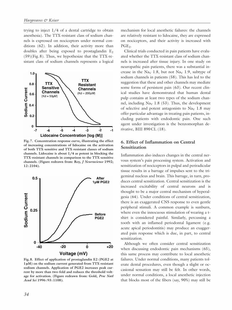
As mentioned earlier in the review, several types ofsodium channels have been discovered over the lastdecade. One particular group of channels is character-ized as being resistant to the puffer fish toxin, tetro-dotoxin (TTX). At least two channels are members ofthe TTX-resistant class, including the PN3 (alsoknown as SNS, or NaV 1.8) and NaN (also known asthe SNS2 or NaV 1.9) sodium channels. The TTX-resistant class of sodium channels is of interest sincethey are less sensitive to lidocaine (61) (Fig. 7). AsFig. 7 demonstrates, increasing concentrations oflidocaine provides increasing blockade of the sodiumchannels. However, the TTX-resistant channels areabout four times less sensitive to lidocaine (imagine
Hargreaves & Keiser
trying to inject 1/4 of a dental cartridge to obtainanesthesia). The TTX-resistant class of sodium chan-nels is expressed on nociceptors under normal con-ditions (62). In addition, their activity more thandoubles after being exposed to prostaglandin E2
(59)(Fig.8). Thus, we hypothesize that the TTX-re-sistant class of sodium channels represents a logical
Fig.7. Concentration response curve, illustrating the effectof increasing concentrations of lidocaine on the activationof both TTX-sensitive and TTX-resistant classes of sodiumchannels. Lidocaine is about 1/4 as potent in blocking theTTX-resistant channels in comparison to the TTX-sensitivechannels. (Figure redrawn from: Roy, J Neuroscience 1992;12:2104).
Fig.8. Effect of application of prostaglandin E2 (PGE2 at1mM) on the sodium current generated from TTX-resistantsodium channels. Application of PGE2 increases peak cur-rent by more than two-fold and reduces the threshold volt-age for activation. (Figure redrawn from: Gold, Proc NatlAcad Sci 1996:93:1108).
34
mechanism for local anesthetic failures: the channelsare relatively resistant to lidocaine, they are expressedon nociceptors, and their activity is increased withPGE2.
Clinical trials conducted in pain patients have evalu-ated whether the TTX-resistant class of sodium chan-nels is increased after tissue injury. In one study onneuropathic pain patients, there was a substantial in-crease in the NaV 1.8, but not NaV 1.9, subtype ofsodium channels in patients (58). This has led to thesuggestion that these and other channels may mediatesome forms of persistent pain (63). Our recent clin-ical studies have demonstrated that human dentalpulp contains at least two types of the sodium chan-nel, including NaV 1.8 (53). Thus, the developmentof selective and potent antagonists to NaV 1.8 mayoffer particular advantage in treating pain patients, in-cluding patients with endodontic pain. One suchagent under investigation is the benzomorphan de-rivative, BIII 890CL (18).
6. Effect of Inflammation on CentralSensitization
Inflammation also induces changes in the central ner-vous system’s pain processing system. Activation andsensitization of nociceptors in pulpal and periradiculartissue results in a barrage of impulses sent to the tri-geminal nucleus and brain. This barrage, in turn, pro-duces central sensitization. Central sensitization is theincreased excitability of central neurons and isthought to be a major central mechanism of hyperal-gesia (64). Under conditions of central sensitization,there is an exaggerated CNS response to even gentleperipheral stimuli. A common example is sunburn,where even the innocuous stimulation of wearing a t-shirt is considered painful. Similarly, percussing atooth with an inflamed periodontal ligament (e.g.acute apical periodontitis) may produce an exagger-ated pain response which is due, in part, to centralsensitization.
Although we often consider central sensitizationwhen discussing endodontic pain mechanisms (65),this same process may contribute to local anestheticfailures. Under normal conditions, many patients tol-erate dental procedures, even though a slight or oc-casional sensation may still be felt. In other words,under normal conditions, a local anesthetic injectionthat blocks most of the fibers (say, 90%) may still be

Managing Local Anesthetic Failures
clinically successful. This has been reported in otherclinical models (for example, IV cannulation of thearm), where patients treated with a topical anestheticreported that they did not experience pain, eventhough their visual analog pain scores were greaterthan zero (66). However, under conditions of centralsensitization, there is an exaggerated response to pe-ripheral stimuli and, under these conditions, the same90% block may permit sufficient signaling to occur tolead to the perception of pain. Thus, central sensitiza-tion may contribute to local anesthetic failures.
Unfortunately, there are no selective drugs forblocking central sensitization. The only clinical impli-cation would be to reduce the afferent barrage andthereby reduce central sensitization. This is done rou-tinely by clinicians via cleaning and shaping tech-niques, but this is a conundrum, as the endodontictreatment is performed after local anesthesia. One in-teresting study has demonstrated that intraosseous in-jection of steroid (methylprednisolone acetate 40mg)reduces endodontic pain in 24 h (67). If confirmed,then this approach may reduce peripheral and centralmechanisms sufficiently to obtain predictable localanesthesia.
7. Psychological Factors
Patient anxiety may also contribute to local anestheticfailure. Experienced clinicians understand that appre-hensive patients have a reduced pain threshold andare more likely to report an unpleasant dental experi-ence (68, 69, 11, 70). Fear of seeing and/or feelingthe needle and the sound of the dental handpiece areroutinely cited as causative agents in the creation ofanxiety in the dental patient (71). Moreover, patientsmay be particularly anxious about impending root ca-nal therapy (72). Investigators have also demon-strated that patient anxiety predicts a poor outcomefor clinical procedures involving local anesthetics ap-plied to the arm before IV cannulation (66). Thus,patient anxiety should be considered when managingthe endodontic pain patient.
Several methods have been advocated for managinganxious emergency pain patients (69, 11). First, theclinician should establish a positive and confident re-lationship and avoid exposing the patient to obviousfear-producing stimuli. Many clinicians report that asense of humor often helps to relax apprehensive pa-tients. For extremely fearful patients, cognitive behav-
35
ior-based programs have shown significant long-termreduction in predental treatment anxiety (73). Otherstudies have demonstrated that instructing patients tofocus on sensory stimuli significantly reduces intra-operative endodontic pain (74, 75). This effect wasmost evident in patients who were characterized ashaving a high desire for control and a low perceivedcontrol over their clinical care.
Second, pharmacologic agents can be administeredto control patient anxiety. While these agents can bedelivered via oral, inhalation (N2O) or intravenousroutes, a decreased likelihood of serious morbidity,reduced monitoring and demonstrated efficacy havemade oral or a combination of oral and inhalationroutes attractive (76, 77). Kaufman et al. (46)showed that oral triazolam 0.25mg was equally effec-tive in comparison to intravenous diazepam in reduc-ing anxiety in patients undergoing oral surgery.
One could certainly consider an integrated ap-proach that involves both non-pharmacologic andpharmacologic techniques. Regardless of the tech-nique utilized, providing some means of anxiety con-trol should enhance the clinician’s ability to provideadequate local anesthesia for endodontic pain pa-tients.
Therapeutic Approaches forManaging Local AnestheticFailuresBefore using the best current evidence to deal withthe problem of clinically inadequate local anesthesiain endodontic patients, the clinician would do well tofirst identify those patients who are likely pose such aproblem. This includes individuals who present withsigns and symptoms of irreversible pulpitis and/oracute periradicular periodontitis, secondary to eitheran apical extension of pulpal inflammation or pulpalnecrosis and bacterial invasion. It also includes pa-tients with a history of experiencing inadequate localanesthesia for dental procedures and those with anobviously high level of anxiety over the pending treat-ment.
In the above-mentioned patients, the clinicianshould consider the modifications discussed below be-fore beginning treatment, as repeated painful stimulicaused by endodontic therapy initiated in the pres-ence of inadequate anesthesia will, for reasons alreadydiscussed, tend to make the problem worse.

Hargreaves & Keiser
1. Supplemental Local Anesthesia
In the endodontic pain patient without an inordinatelevel of anxiety, supplemental local anesthesia has beenthe obvious first choice for dealing with potential anes-thetic failures. Supplementing a single-cartridge in-ferior alveolar block has the potential to deal with fail-ures created by several of the hypotheses previously putforth. By increasing the dose of local anesthetic, onecould expose a greater length of the inferior alveolarnerve (IAN) and increase the likelihood of conductionblockade (23). Increased dosage would also help toblock the population of TTX-resistant sodium chan-nels that may be elevated in the inflamed state (61). Byusing an anesthetic with a lower pKa, such as 3% mepi-vacaine, one could decrease the potential for ion trap-ping. This would increase the concentration of localanesthetic molecules in the base form necessary for dif-fusion across the nerve membrane, enhancing block-ade and increasing the onset of anesthesia (7). Deliver-ing the second cartridge of anesthetic higher in thepterygomandibular space would have the effect of in-creasing the length of nerve exposed, as well as possiblyblocking the nerve to the mylohyoid muscle before itbranches from the IAN (39).
As mentioned previously, supplemental anesthesiacan also be provided via other routes. Both intraliga-mentary and intraosseous techniques deliver the anes-thetic to the cancellous bone surrounding the apicesof injected teeth (6, 79). While the intraligamentaryinjection can be provided with no additional arma-mentaria, there are limitations to the volume of anes-thetic deposited, and a significant incidence of post-operative pain may ensue (80). The intraosseousroute does afford the possibility of delivering higherdoses but requires cortical perforation and delivery ofthe anesthetic via specialized instruments. It also hasthe potential to cause perforation of nearby toothroots if used incorrectly. Clinical trials indicate thatthe intraosseous route of injection significantly en-hances pulpal anesthesia after IAN block injection inendodontic pain patients (42)
The intrapulpal injection is generally used as a finaloption for the patient who is comfortable until thepulp is exposed (or nearly exposed) (6). This tech-nique delivers the solution directly into inflamed pul-pal tissues and requires some means of preventingback flow (81, 82). With this in mind, a limited accessopening to the pulp to provide a narrow channel for
36
the intrapulpal injection might be considered (e.g.using a .2 round bur to bore an opening into thepulp chamber); the final access preparation can be ac-complished after the pulp has been anesthetized.
As previously discussed, in order to evaluate thedepth of pulpal anesthesia provided by any technique,prior to initiating endodontic treatment (i.e. prior toplacement of the rubber dam), the clinician shouldtest the tooth in question rather than rely on a posi-tive lip sign. Probably the simplest way to do thiswould be to repeat the cold test. A lack of responsegives both the operator and the patient a certain de-gree of confidence and, thus, reduces anxiety duringthe operation.
2. Adjunctive Drugs or Techniques
The first part of this review has highlighted evidencethat has allowed a greater appreciation of the effects ofinflammation on peripheral nociceptors, as well as cen-tral nervous system processing of pain signals. It is verylikely that inflammation contributes to local anesthesiafailure in inflamed dental pulps via both mechanisms.With this in mind, the astute clinician may consider theuse of fast-acting anti-inflammatory drugs as an ad-junct to the provision of local anesthesia to teeth withinflamed pulps and/or periradicular tissues.
Reducing pulpal levels of the inflammatory me-diator PGE2 would be beneficial in two ways. Firstly,decreasing pulpal nociceptor sensitization would miti-gate an increase in resistance to local anesthetics (54).Secondly, it may diminish a prostanoid-inducedstimulation of TTX-resistant sodium channel activity(Fig.8); these channels also display relative resistanceto lidocaine (59, 61). Reduction of PGE2 could beaccomplished with either NSAIDS or steroids.Double-blind clinical trials have shown that the in-jectable non-steroidal anti-inflammatory drug ketoro-loac tromethamine, when injected intraorally or intra-muscularly, produces significant analgesia in patientswith severe odontogenic pain prior to definitive treat-ment (83, 84). Although it has yet to be evaluated inendodontic pain patients, ibuprofen in a liquid gelformulation (e.g. Advil Liquid GelA (White-HallRobbins, Madison, NJ)) may have similar effects. Inpatients with a diagnosis of irreversible pulpitis, adouble-blinded, randomized clinical trial showedthat, for the 7 day period preceding endodontic ther-apy, subjects that received an intraosseous injection of

Managing Local Anesthetic Failures
40mg methylprednisolone experienced significantlyless pain and required significantly less pain medi-cation than those receiving the placebo (67). Finally,for the overtly fearful patient, reducing anxiety bymethods previously discussed, such as sublingual tria-zolam (78) or nitrous oxide (76), and a caring chair-side manner, should actually increase the likelihood ofeffective local anesthesia in endodontic pain patients.
ConclusionsHopefully the reader has found this review to be in-formative and practical. Our objectives were to reviewthe pharmacological mechanisms of local anesthesiaand pain from the perspective of identifying potentialmechanisms for local anesthetic failures. These mech-anisms provide the basis for evidence-based treatmentstrategies and, importantly, point out areas where fu-ture research is needed. Given the greater understand-ing of acute pain mechanisms that we currently enjoyand the ongoing research efforts in pain laboratoriesthroughout the world, it is not too difficult to imaginea time when local anesthesia will be as predictable in in-flamed teeth as it is in the normal, uninflamed tooth.
References1. Georgopoulou M, Anastassiadis P, Sykaras S. Pain after
chemomechanical preparation. Int Endodont J 1996: 19:309.
2. Harrison JW, Baumgartner JC, Zielke DR. Analysis of in-terappointment pain associated with the combined use ofendodontic irrigants and medicaments. J Endodont 1981:7: 272–276..
3. Seltzer S, Bender I, Ehrenreich J. Incidence and durationof pain following endodontic therapy. Oral Surg OralMedical Oral Path 1961: 14: 74.
4. Guglielmo A, Reader A, Nist R, Beck M, Weaver J. Anes-thetic efficacy and heart rate effects of the supplementalintraosseous injection of 2% mepivacaine with 1: 20,000levonordefrin. Oral Surg Oral Med Oral Path Oral RadiolEndod 1999: 87: 284–293.
5. Hargreaves KM. Neurochemical Factors in Injury and In-flammation in Orofacial Tissues. In: Lavigne G, Lund J, Ses-sle B, Dubner R. eds. Orofacial Pain: Basic Sciences to Clin-ical Management. Chicago: Quintessence Publications2001
6. Jastak J, Yagiela J, Donaldson D. Local Anesthesia of the OralCavity. Philadelphia: Saunders 1995: 1–339.
7. Malamed S. Handbook of Local Anesthesia 3rd edn. StLouis: Mosby 1990: 1–332.
8. Milles M. The missed inferior alveolar block: a new look atan old problem. Anesth Prog 1984: 31: 87–90.
9. Nakai Y, Milgrom P, Mancl L, Coldwell SE, Domoto PK,
37
Ramsay DS. Effectiveness of local anesthesia in pediatricdental practice. J Am Dent Assoc 2000: 131: 1699–1705.
10. Vinckier F. ‘What is the cause of failure of local anesthesia?’[French]. Revue Belge Medecine Dentaire 2000: 55: 41–50.
11. Walton R, Torabinejad M. Managing local anesthesia prob-lems in the endodontic patient. J Am Dent Assoc 1992: 123:97–102.
12. Cohen HP, Cha BY, Spangberg LS. Endodontic anesthesiain mandibular molars: a clinical study. J Endod 1993: 19:370–373.
13. Nusstein J, Reader A, Nist R, Beck M, Meyers WJ. Anes-thetic efficacy of the supplemental intraosseous injection of2% lidocaine with 1: 100,000 epinephrine in irreversiblepulpitis. J Endod 1998: 24: 487–491.
14. Reisman D, Reader A, Nist R, Beck M, Weaver J. Anestheticefficacy of the supplemental intraosseous injection of 3%mepivacaine in irreversible pulpitis. Oral Surg Oral MedOral Path Oral Rad Endod 1997: 84: 676–682.
15. Catterall W. From ionic currents to molecular mechanisms:the structure and function of voltage-gated sodium chan-nels. Neuron 2000: 26: 13–25.
16. Hille B. Ionic Channels of Excitable Membranes 1st edn Sun-derland MA: Sinauer Assoc 1984.
17. Ragsdale D, McPhee J, Scheuer T, Catterall W. Moleculardeterminants of state-dependent block of Naπ channels bylocal anesthetics. Science 1994: 265: 1724–1728.
18. Carter A, Grauert M, Pschorn U, Bechtel W, Bartmann-Lindholm C, Qu Y, Scheuer T, Catterall W, Weiser T. Potentblockade of sodium channels and protection of brain tissuefrom ischemic by BIII 890 CL. Proc Natl Acad Sci 2000:97: 4944–4949.
19. Grant A, Dietz M, Gilliam F, Starmer C. Blockade of cardiacsodium channels by lidocaine. Single-channel analysis. CircRes 1989: 65: 1247–1262.
20. Ragsdale D, Scheuer T, Catterall W. Frequency and voltage-dependent inhibition of type IIA Naπ channels, expressedin a mammalian cell line, by local anesthetic, antiarrhythmic,and anticonvulsant drugs. Mol Pharm 1991: 40: 756–765.
21. Gasser H, Erlanger J. The role of fiber size in the establish-ment of a nerve block by pressure or cocaine. Am J Physiol1929: 88: 581–591.
22. Heinbecker B, Bishop G, O’Leary J. Analysis of sensation interms of nerve impulse. Arch Neurol Psych 1934: 31: 34–53.
23. Franz D, Perry R. Mechanisms for differential block amongsingle myelinated and nonmyelinated axons by procaine. JPhysiol 1974: 236: 193–210.
24. Huang J, Thalhammer J, Raymond S, Strichartz G. Suscep-tibility to lidocaine of impulses in different somatosensoryafferent fibers of rat sciatic nerve. J Pharm Exp Therap 1997:282: 802–811.
25. Thalhammer J, Vladimrova M, Bershadsky B, Strichartz G.Neurologic evaluation of the rat during sciatic nerve blockwith lidocaine. Anesthesiology 1995: 82: 1013–1025.
26. Bou Dagher F, Yared G, Machtou P. An evaluation of 2%lidocaine with different concentrations of epinephrine forinferior alveolar nerve block. J Endod 1997: 23: 178–180.
27. McLean C, Reader A, Beck M, Meryers W. An evaluation of4% prilocaine and 3% mepivacaine compared with 2% lido-caine (1: 100,000 epinephrine) for inferior alveolar nerveblock. J Endod 1993: 19: 146–150.
28. Vreeland D, Reader A, Beck M, Meyers W, Weaver J. An

Hargreaves & Keiser
evaluation of volumes and concentrations of lidocaine in hu-man inferior alveolar nerve block. J Endod 1989: 15: 6–12.
29. Takeuchi T, Tasaki I. Ubertrangung de nervenimpulses inder polarisierten nervenfaser. Pfluegers Arch Gesamte PhysiolMenshchen Tiere 1942: 246: 32–43.
30. Fink B, Cairns A. Differential showing and block of conduc-tion by lidocaine in individual afferent myelinated and un-myelinated axons.. Anesthesiology 1984: 60: 111–112.
31. Raymond S, Steffensen S, Gugliano L, Strichartz G. Therole of length of nerve exposed to local anesthetics in im-pulse blocking action. Anesth Analg 1989: 68: 563–570.
32. Rucci F, Pippa P, Boccaccini A, Barbagli R. Effect of injec-tion speed on anaesthetic spread during auxillary blockusing the orthogonal two-needle technique. Eur J Anaesth1995: 12: 505–511.
33. Bremer G. Measurements of special significance in connec-tion with anesthesia of the inferior alveolar nerve. Oral SurgOral Med Oral Path 1952: 5: 966–988.
34. Davidson MJ. Bevel-oriented mandibular injections: needledelflection can be beneficial. General Dent 1989: 37: 410–412.
35. Hannan L, Reader A, Nist R, Beck M, Meyers WJ. The useof ultrasound for guiding needle placement for inferior al-veolar nerve blocks. Oral Surg Oral Med Oral Pathol OralRadiol Endod 1999: 87: 658–665.
36. Berns JM, Sadove MS. Mandibular block injection: amethod of study using an injected radiopaque material. JAm Dent Assoc 1962: 65: 735–745.
37. Frommer J, Mele FA, Monroe CW. The possible role of themylohyoid nerve in mandibular posterior tooth sensation. JAm Dent Assoc 1972: 85: 113–117.
38. Jablonski NG, Cheng CM, Cheng LC, Cheung HM. Un-usual origins of the buccal and mylohyoid nerves. Oral SurgOral Med Oral Pathol 1985: 60: 487–488.
39. Wilson S, Johns P, Fuller PM. The inferior alveolar and my-lohyoid nerves. an anatomic study and relationship to localanesthesia of the anterior mandibular teeth. J Am Dent Assoc1984: 108: 350–352.
40. Roda R, Blanton P. The anatomy of local anesthesia. QuintInt 1994: 25: 27–38.
41. Childers M, Reader A, Nist R, Beck M, Meyers WJ. Anes-thetic efficacy of the periodontal ligament injection after aninferior alveolar nerve block. J Endod 1996: 22: 317–320.
42. Dunbar D, Reader A, Nist R, Beck M, Meyers WJ. Anes-thetic efficacy of the intraosseous injection after an inferioralveolar nerve block. J Endod 1996: 22: 481–486.
43. Vandermeulen E. Pain perception, mechanisms of action oflocal anesthetics and possible causes of failure. Revue BelgeMedecine Dentaire 2000: 55: 29–40.
44. Ang E, Lassale B, Goldfarb G. Continuous axillary brachialplexus block – a clinical and anatomical study. Anesth Analg1984: 63: 680–684.
45. Dahm P, Nitescu P, Appelgren L, Curelaru I. Efficacy andtechnical complications of long-term continuous intraspinalinfusions of opioid and/or bupivacaine in refractory non-malignant pain: a comparison between the epidural and theintrathecal approach with externalized or implanted cath-eters and infusion pumps. Clin J Pain 1998: 14: 4–16.
46. Punnia-Moorthy A. Buffering capacity of normal and in-flamed tissues following the injection of local anaestheticsolutions. Br J Anaesth 1988: 6: 154–159.
38
47. DiFazio C, Carron H, Grosslight K et al. Comparison ofpH-adjusted lidocaine solutions for epidural anesthesia.Anesth Analg 1986: 64: 760–764.
48. Gerancher JC. Upper extremity nerveblocks. Anesth ClinicsNorth Am 2000: 18: 297–317.
49. Rood J. Some anatomic and physiologic causes of failure toachieve local mandibular analgesia. Br J Oral Surg 1977: 15:75–82.
50. Suda H, Ikeda H. The circulation of the pulp. In: Har-greaves K, Goodis, H, eds. Seltzer and Bender’s Dental Pulp.Chicago: Quintessence, 2002.
51. Buckley J, Ciancio S, McMullen J. Efficacy of epinephrineconcentration in local anesthesia during periodontalsurgery. J Periodontol 1984: 55: 653–657.
52. Knoll-Kohler E, Fortsch G. Pulpal anesthesia dependent onepinephrine dose in 2% lidocaine. Oral Surg Oral Med OralPathol 1992: 73: 537–540.
53. Hargreaves KM, Dryden J, Schwarze M, Gracia N, MartinW, Flores CM. Development of a model to evaluate pheno-typic plasticity in human nociceptors. Abstract Soc Neurosci2000.
54. Rood JP, Pateromichelakis S. Inflammation and peripheralnerve sensitisation. Br J Oral Surg 1981: 19: 67–72.
55. Byers MR, Taylor PE, Khayat BG, Kimberly CL. Effects ofinjury and inflammation on pulpal and periapical nerves. JEndod 1990: 16: 78–84.
56. Byers MR, Narhi MVO. Dental injury models: Experimen-tal tools for understanding neuro-inflammatory interactionsand polymodal nociceptor functions. Crit Rev Oral BiolMedical 1999: 104–139.
57. Byers M, Narhi MV. Nerve supply of the pulpodentin com-plex and responses to injury. In: Hargreaves KM, Goodis H,eds. Seltzer and Bender’s Dental Pulp. Chicago: Quintess-ence 2002.
58. Coward K, Plumpton C, Facer P, Birch R, Carlstedt T, TateS, Bountra C, Anand P. Immunolocalization of SNS/PN3and NaN/SNS2 sodium channels in human pain states.Pain 2000: 85: 41–50.
59. Gold M, Reichling D, Shuster M, Levine JD. Hyperalgesicagents increase a tetrodotoxin-resistant Naπ current in no-ciceptors, Proc Nat Acad Sci 1996: 93: 1108.
60. Novakovic S, Tzoumaka E, McGiven J, Haragauchi M, San-gameswaran L, Gogas K, Eglen R, Hunter J. Distribution ofthe tetrodotoxin-resistant sodium channel PN3 in rat sen-sory nerves in normal and neuropathic conditions. J Neuros-ci 1998: 18: 2174–2187.
61. Roy ML, Narahashi T. Differential properties of tetrodo-toxin-sensitive and tetrodotoxin-resistant sodium channelsin rat dorsal root ganglion neurons. J Neuroscience 1992:12: 2104–2111.
62. Arbuckle JB, Docherty RJ. Expression of tetrodotoxin-re-sistant sodium channels in capsaicin-sensitive dorsal rootganglion neurons of adult rats. Neurosci Lett 1995: 85: 70.
63. Waxman SG. The molecular pathophysiology of pain: ab-normal expression of sodium channel genes and its contri-butions to hyperexcitability of primary sensory neurons.Pain Supplement 1999: 6: s133–s140.
64. Woolf C. Windup and central sensitization are not equiva-lent. Pain 1996: 66: 105.
65. Hargreaves KM. Pain mechanisms of the pulpodentincomplex. In: Hargreaves, KM, Goodis H, eds. Seltzer and

Managing Local Anesthetic Failures
Bender’s Dental Pulp. Chicago: Quintessence Publications2002.
66. Lander J, Hodgins M, Nazarali S, McTavish J, OuelletteJ, Friesen E. Determinants of success and failure of EMLA.Pain 1996: 64: 89–97.
67. Gallatin E, Reader A, Nist R, Beck M. Pain reduction inuntreated irreversible pulpitis using an intraosseous injec-tion of Depo-Medrol. J Endod 2000: 26: 633–638.
68. Dworkin S. Anxiety and performance in the dental en-vironment: an experimental investigation. J Am Soc PsychDent Medical 1967: 14: 88–103.
69. Fiset L, Getz T, Milgrom P, Weinstein P. Local anestheticfailure: diagnosis and management strategies. GeneralDent 1989: 37: 414–417.
70. Wong M, Jacobsen P. Reasons for local anesthesia failures.J Am Dent Assoc 1992: 12: 69–73.
71. Gale EN. Fears of the dental situation. J Dent Res 1972:51: 964–966.
72. Wong M, Lytle WR. A comparison of anxiety levels associ-ated with root canal therapy and oral surgery treatment. JEndod 1991: 17: 461–465.
73. Thom A, Sartory G, Johren P. Comparison between one-session psychological treatment and benzodiazepine indental phobia. J Consult Clin Psychol 2000: 68: 378–387.
74. Morse D. Use of meditative state for hypnotic inductionin the practice of endodontics. Oral Surg Oral Med OralPath Oral Radiol Endodon 1976: 41: 664–672.
75. Morse D, Wilcko J. Nonsurgical endodontic therapy for a
39
vital tooth with meditation-hypnosis as the sole anesthetic:case report. Am J Clin Hyp 1979: 21: 258–262.
76. Dionne R. Oral sedation. Compend Contin Educ Dent.1998: 868– 870, 872, 874 passim.
77. Haas DA. Oral and inhalation conscious sedation. DentClin North Am 1999: 43: 341–359.
78. Kaufman E, Hargreaves KM, Dionne RA. Comparison oforal triazolam and nitrous oxide with lacebo and intra-venous diazepam for outpatient sedation. Oral Surg OralMed Oral Oathol 1993: 75: 156–164.
79. Smith GN, Walton RE. Periodontal ligament injection:Distribution of injected solutions. Oral Surg Oral MedOral Pathol 1983: 55: 232–238.
80. D’Souza J, Walton RE, Peterson L. Periodontal ligamentinjection: an evaluation of extent of anesthesia and postin-jection discomfort. J Am Dent Assoc 1987: 114: 341–344.
81. Smith GN, Smith SA. Intrapulpal injection: Distributionof an injected solution. J Endod 1983: 9: 167–170.
82. VanGheluwe J, Walton R. Intrapulpal injection: factors re-lated to effectiveness. Oral surg Oral Med Oral Pathol OralRad Endod 1997: 83: 38–40.
83. Curtis P, Gartman LA, Green DB. Utilization of ketorolactromethamine for control of severe odontogenic pain. JEndod 1994: 20: 457–459.
84. Penniston S, Hargreaves KM. Evaluation of periapical in-jection of Ketorolac for management of endodontic pain.J Endod 1996: 22: 55–59.