Embed Size (px)
Citation preview
RESEARCH ARTICLE
Local and global effects of neck muscle vibrationduring stabilization of upright standing
Julius Verrel • Remy Cuisinier • Ulman Lindenberger •
Nicolas Vuillerme
Received: 19 December 2010 / Accepted: 9 March 2011 / Published online: 26 March 2011
� Springer-Verlag 2011
Abstract Neck muscle vibration (NMV) during upright
standing is known to induce forward leaning, which has
been explained as a global response to the (illusory) per-
ception of a lengthening of the dorsal neck muscles.
However, the effects of NMV both at the level of indi-
vidual joints and on whole-body postural coordination, and
its potential modulation by vision, have not yet been ana-
lyzed in detail. Eight healthy young adult participants
completed a total of ten trials each, with a 10-s period of
unperturbed standing followed by a 10-s period of NMV.
Participants were instructed to stand ‘‘as still as possible’’.
This postural task was executed under two visual condi-
tions: eyes open (EO) and eyes closed (EC). Postural
responses were measured in terms of center of pressure
(CoP) and center of mass (CoM) profiles, and whole-body
kinematics. Responses to NMV at the level of individual
body segments and joints were assessed by decomposing
the time series into linear trends and residual fluctuations.
Inter-segmental coordination was analyzed using a decor-
relation technique, assessing motor-equivalent stabilization
of four task-related variables: CoM position, trunk orien-
tation, as well as head position and orientation. NMV
induced a general forward leaning response under both
visual conditions, visible in CoP, CoM, segment positions
and orientations. Locally, NMV induced a pronounced
extension of the atlanto-occipital joint. Residual fluctua-
tions were higher with EC and unaffected by NMV.
Coordination analysis showed that stabilization of different
body parts was differentially affected by NMV. Head ori-
entation was consistently stabilized across all conditions,
with weaker coordination in the EC condition. In contrast,
motor-equivalent stabilization of CoM and head position,
and trunk orientation was only observed during no-vibration
periods. Taken together, our results demonstrate specific
effects of vision and proprioception on different aspects of
local and global postural control. While perturbed neck
proprioception seemed to affect the postural ‘‘set point’’
(inducing forward leaning), vision appeared to mainly serve
in noise reduction (residual fluctuations) and control of head
orientation.
Keywords Posture �Neck muscle vibration �Kinesthesia �Orientation �Multisensory integration � Coordination
Introduction
Human erect posture is known to depend on the coordi-
nation of numerous biomechanical degrees of freedom
(Creath et al. 2005; Hsu et al. 2007; Pinter et al. 2008),
based on the integration of vestibular, visual and somato-
sensory information (Peterka 2002), to stabilize equilib-
rium and orientation (Horak and MacPherson 1996;
Massion 1994). Here, equilibrium refers to the biome-
chanical requirements of upright stance (stabilizing the
center of mass, CoM, within the base of support), while
orientation refers to the relative or absolute positions of
different body parts such as trunk and head. Proprioception
of the neck may be particularly important to integrate
J. Verrel (&) � U. Lindenberger
Max Planck Institute for Human Development,
Center for Lifespan Psychology, Lentzeallee 94,
14195 Berlin, Germany
e-mail: [email protected]
R. Cuisinier � N. Vuillerme (&)
FRE3405, AGIM (AGeing Imaging Modeling),
CNRS-UJF-EPHE, Faculty of Medicine, Batiment Jean Roget,
38706 La Tronche Cedex, France
e-mail: [email protected]
123
Exp Brain Res (2011) 210:313–324
DOI 10.1007/s00221-011-2636-9
vestibular and visual information with somatosensory
information from the rest of the body to estimate the
position and motion of different body parts in space (e.g.,
Cohen 1961; Mergner et al. 1997).
Perturbation of neck proprioception, by means of neck
muscle vibration (NMV), is known to affect postural control
during upright quiet standing (Eklund and Hagbarth 1966;
Eklund 1971, 1972; Gregoric et al. 1978; Lund 1980).
Symmetric NMV induces a forward leaning of the body,
which can be detected both based on center of pressure (CoP)
profiles (Kavounoudias et al. 1999) and kinematic mea-
surements (Ivanenko et al. 1999; Gomez et al. 2009).
This effect has been explained as a corrective reaction to
perceived backward leaning of the body (e.g., Kavounou-
dias et al. 1999; Lekhel et al. 1997), in the sense that neck
muscle vibration induces the illusory perception of a
lengthening of the neck muscles (Goodwin et al. 1972a,
1972b; Lackner and Levine 1979; Gilhodes et al. 1986),
which may signify a distal movement (forward tilt of the
head) or a proximal movement (forward movement of the
body below the space-fixed head). Vestibular or visual
information indicating constant head orientation provides
sensory evidence against the former (distal) interpretation.
Therefore, the NMV-induced perceived neck lengthening is
hypothesized to be interpreted by the postural control sys-
tem as a backward leaning that would lead to a corrective
response, i.e., the observed forward leaning of the body.
To our knowledge, postural responses to neck muscle
vibration at the level of individual joints along the central
body axis and their coordination have not yet been inves-
tigated. We consider this issue relevant for two main rea-
sons. On the one hand, analysis at the joint level allows
assessing local effects of stimulation. In particular, the
local effect of neck muscle vibration on neck and head
orientation has not been explicitly addressed in previous
research. On the other hand, global analysis of inter-
segmental coordination may reveal which specific aspects
of posture (equilibrium vs. orientation) are affected by the
perturbation of neck proprioception.
For most motor activities, the number of degrees of
freedom (DOF) available to the motor system exceeds
those specified by the performance requirements. For
instance, in a simple (sagittal plane) postural model, the
human body can be modeled with six DOF (ankle, knee,
hip, lower back, upper back, neck), while important task-
related variables, such as the anterior–posterior CoM
position, are one dimensional. Analyzing the organization
of variability across the DOF allows formally testing
whether the motor system stabilizes specific task-related
variables (Schoner 1995; Scholz and Schoner 1999; Latash
et al. 2007). The rationale of this approach is that the
structure of intrinsic fluctuations is informative about how
the motor system deals with naturally occurring
perturbations. Stabilization of a task-related variable does
not necessarily mean that variability is minimized in each
DOF that affects this variable, but that variability is orga-
nized across DOF in a way that minimizes fluctuations in
the task-related variable. Importantly, this notion of sta-
bilization differs fundamentally from using raw variability
scores as indicators of stability.
Several methods have been proposed to analyze task-
specific organization of variability in multi-DOF effector
systems (Cusumano and Cesari 2006; Muller and Sternad
2003; Scholz and Schoner 1999). The general idea in such
an analysis is to relate variability at the level of the effector
system (e.g., joint angles) to variability of specific task-
related variables. Due to motor equivalence, the extent to
which joint angle variability results in variability in task-
related variables depends on the coordination among the
joint angles. In the case of upright postural control, CoM
and head position, as well as trunk and head orientation,
have been proposed as relevant (Horak and MacPherson
1996; Hsu et al. 2007). Here, we follow the approach of
Muller and Sternad (2003), in which joint angles time
series are randomly permuted to produce ‘‘decorrelated’’
surrogate data. The difference between actual and decor-
related performance, measured at the level of task-related
variables (e.g., variability of CoM position), is used to
quantify coordination (see ‘‘Methods’’ for details). Using
this method, it can be assessed whether and to what extent
joint angles are organized in a way that they stabilize a
specific task-related variable (compensatory organization
among joint angles), are independent (no compensation) or
‘‘destabilize’’ (inducing a change in) the task-related vari-
able. It has to be noted that this latter notion of ‘‘destabi-
lization’’ is independent of a potential new stable point
reached after the change.
Availability of visual feedback generally has a stabilizing
influence on postural control, providing additional infor-
mation on position and orientation (e.g., Paulus et al. 1984;
Redfern et al. 2001; Vuillerme et al. 2006). Indeed, with-
drawal of visual information was found to increase the for-
ward leaning induced by NMV, both in response to short
(Bove et al. 2009) and prolonged (Gomez et al. 2009) stim-
ulation intervals. Vision has also been found to influence
postural coordination during unperturbed upright standing
(Zhang et al. 2007). However, the combined effects of NMV
and availability of visual information on segmental vari-
ability and intersegmental coordination stabilizing different
balance-related variables (e.g., head orientation or CoM
position) have not been investigated to our knowledge.
In the present study, we studied the immediate effects of
short (10 s) neck muscle vibration on upright posture,
under conditions with and without visual feedback (eyes
open, eyes closed). Participants were instructed to stand as
still as possible (Zok et al. 2008). Postural responses to the
314 Exp Brain Res (2011) 210:313–324
123
proprioceptive perturbation were assessed in the sagittal
plane, both using a force plate (measuring CoP profiles)
and whole-body kinematics. Kinematic data were analyzed
both ‘‘locally’’ (markers, segments, joints), and ‘‘globally’’,
looking at whole-body coordination. Local variability was
decomposed into (linear) trends and fluctuations (residual
variability) to distinguish between vibration-induced pos-
tural shifts of set point (forward leaning) and fluctuations
around this set point. The former are assumed to reflect
changes in the internal reference for upright posture
(Gurfinkel et al. 1995), while the latter are taken to be
indicators of postural instability. Whole-body coordination
was assessed using a decorrelation technique (Muller and
Sternad 2003), with respect to motor-equivalent stabiliza-
tion of anterior–posterior CoM and head position, as well
as sagittal trunk and head orientation.
Methods
Subjects
Eight young male university students (mean age =
21.4 ± 2.4 years; body weight = 73.0 ± 5.2 kg; height =
179.6 ± 5.5 cm) voluntarily participated in the experi-
ment. None of the subjects presented any history of motor
problems, neurological diseases or other impairments that
could have influenced their balance, and none of them had
consumed alcohol or other drugs on the day prior to the
experiment, according to self-report. Subjects gave written
consent to the experimental procedure. The study was
conducted in accordance with the Helsinki Declaration and
was approved by the local ethics committee.
Muscle vibrator
A DC motor with an eccentric mass on the shaft, embedded
in a plastic tube of 7-cm length and 2.5-cm wide (Techno-
concept VB 115, France), produced vibration of an
amplitude of 0.85 mm and a maximal force of 5 N at
100 Hz (Bove et al. 2009). Vibrators were secured by
straps over the paravertebral muscles on either side of the
neck by a custom-made collar (Gomez et al. 2009). The
activation of the vibrators was computer controlled
(ADwin-pro Keithley Instruments Inc., Cleveland, OH,
US).
Data acquisition
A force platform (AMTI, Model 0R6-5-1) was used to
measure the displacement of the center of foot pressure
(CoP) by computation of the three orthogonal components of
the ground reaction forces and their associated torque.
Signals from the force platform were sampled at 100 Hz
(12 bit A/D conversion). An active opto-electronic motion
capture system (Optotrak 3020, Northern Digital, Waterloo,
ON, Canada) was used to record body movements. Eight
infrared-emitting markers were placed at the head (os
zygomaticum, ZYG, and orbit, ORB), shoulder (glenohu-
meral axis, SHLD), pelvis (anterior superior iliac spine,
ASIS), femur (great trochanter, GTR), knee (lateral epi-
condyle of femur, KNEE), and feet (lateral malleolus, ANK,
and fifth metatarsal head, TOE). The marker positions
(reported 3D accuracy at 2.25-m distance: 0.1 mm) were
sampled at a frequency of 100 Hz (12-bit A/D conversion).
Procedure
Subjects were asked to stand ‘‘as still as possible’’ (Zok et al.
2008), with the head facing forward, the hands at the back
and their feet 10 cm apart. Each trial lasted 20 s and was
divided into two intervals of 10 s: (1) no vibration (NOVIB)
for the first 10 s, and (2) vibration (VIB) of the neck muscles
for the second 10 s. This postural task was executed under
two visual conditions: eyes open (EO) and eyes closed (EC).
In the EC condition, subjects were asked to close their eyes,
whereas in the EO condition they were asked to look at the
intersection point of a black cross (20 9 25 cm) attached at
eye level to the white wall at a distance of about 1 m in front
of them. Between trials, 30 s of rest were provided. Before
each trial started, subjects were given 3 s to stabilize their
upright posture on the force platform, under the vision con-
dition (EO or EC) of the subsequent trial. One practice trial
and four experimental trials were performed in each vision
condition, intermixed in pseudorandom order.
Data analysis: univariate variability
All the analyses were performed using custom-written
Matlab routines. Kinematic and force place (CoP) data
were low pass filtered using a forward and reverse second-
order Butterworth filter with a cutoff frequency of 10 Hz.
Data from the NOVIB and VIB intervals of each trial (first
vs. last 10 s) were analyzed separately. Kinematic and
force plate data were only analyzed in the sagittal plane,
since symmetric neck tendon vibration had previously been
found to affect posture mainly in this plane (e.g., Kavou-
noudias et al. 1999). Preliminary analyses showed that
ankle and toe marker positions varied only minimally
during trials (within-trial SD less than 1 mm for all par-
ticipants, trials and spatial dimensions). Therefore, data of
these markers were replaced by their within-trial means,
allowing the use of the foot as a stable basis for the bio-
mechanical model (see below).
Position data of consecutive markers along the kine-
matic chain from foot to head were used to compute
Exp Brain Res (2011) 210:313–324 315
123
(absolute) segment positions and orientations. Whole-body
CoM position was estimated from segment data by taking
the relative mass-weighted sum of individual segments’
CoM positions (Winter 2004, p. 63/64, Table 3.1). Seg-
ment elevation angles were computed for the following
segments: foot, shank, thigh, pelvis, chest, neck and head
(taking the markers SHLD and ZYG as approximations of
the cervico-thoracic and atlanto-occipital joints in the
sagittal plane). From these, relative orientations of neigh-
boring segments were computed, yielding joint angles for
the following joints: ankle, knee, hip, lumbosacral (LS),
cervico-thoracic (CT) and atlanto-occipital (AO). Angle
orientation followed the mathematical convention, i.e.,
positive values correspond to counterclockwise rotation.
Therefore, for the knee joint, positive values correspond to
flexion, for all other joints positive values correspond to
extension (or plantar flexion for the ankle).
Positional (markers, CoM) and angular (segments,
joints) time series frequently showed linear drifts, in par-
ticular during VIB (see raw data, Fig. 1). Therefore, their
variability (within the two 10-s VIB and NOVIB intervals)
was split into a linear component (trend) and residual
variability (fluctuations). The trend was quantified as the
difference between the start and end value in the interval,
and fluctuations were quantified by means of the standard
deviation of the residual variability.
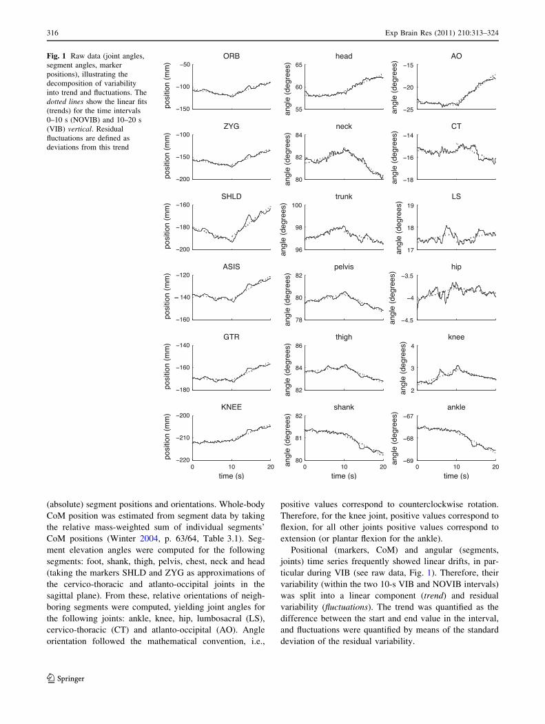
Fig. 1 Raw data (joint angles,
segment angles, marker
positions), illustrating the
decomposition of variability
into trend and fluctuations. The
dotted lines show the linear fits
(trends) for the time intervals
0–10 s (NOVIB) and 10–20 s
(VIB) vertical. Residual
fluctuations are defined as
deviations from this trend
316 Exp Brain Res (2011) 210:313–324
123
Data analysis: motor-equivalent coordination
Whole-body postural coordination was analyzed using a
decorrelation technique (Muller and Sternad 2003). Joint
angles were randomly permuted (‘‘decorrelated’’) across
time (within VIB/NOVIB intervals of trials). Using a bio-
mechanical forward model (see below), the effect of dec-
orrelation on a specific task-related variable (e.g., CoM
position) can be determined. The rationale of this approach
is that coordination among the joints is destroyed by the
decorrelation procedure. Therefore, coordination (with
respect to the task-related variable) can be quantified as the
normalized difference between empirically observed vari-
ance Vemp and decorrelated variance V0 in the task-related
variable. Specifically, a generalized correlation coefficient
can be defined as q = Vemp/V0 - 1. Coordination is indi-
cated when, statistically, Vemp \ V0, or equivalently q\ 0,
with perfect coordination corresponding to q = -1. On the
other hand, positive q indicates an organization of joint
angles that destabilizes the task-related variable. It can be
shown (Muller and Sternad 2003) that for the special case
of the forward model f(x,y) = x?y, q equals the conven-
tional correlation coefficient.
The decorrelation analysis requires a forward model,
mapping elemental variables (joint angles) to the task-
related variable at hand. This model was defined individ-
ually for each participant, estimating segment lengths by
taking the mean across all trials of each participant. These
were combined with joint angle data to compute segment
orientations and positions, which were then used to
compute the considered task-related variables: anterior–
posterior CoM and head position, as well as sagittal trunk
and head orientation (pitch). Thus, for each of these vari-
ables, Vemp,V0 and q were determined, allowing to test the
presence and amount of coordination with respect to these
variables. To increase reliability of the generalized corre-
lation estimate, V0 was computed by taking the average
over 100 repetitions of the decorrelation procedure. These
measures were computed for each trial separately.
Statistical analysis
Statistical analysis was performed using R statistical soft-
ware package (R Development Core Team 2008).
To correct for non-normal distribution, the fluctuations
(but not trends) of CoP and marker positions, segment
elevation and joint angles were log-transformed prior to
statistical analysis. Since preliminary analyses did not
show any systematic order effects, data from the four
experimental trials of one Vision/Vibration condition were
averaged for each participant.
Subsequently, dependent variables were submitted to
separate repeated measures ANOVAs with Vision (EO,
EC) and Vibration (NOVIB, VIB) as within-subject factors.
Effect sizes are reported as Cohen’s d (Cohen 1988), cor-
responding to the contrasts EC–EO (Vision), VIB–NOVIB
(Vibration), and their interaction. In addition, generalized
correlation with respect to each of the four task-related
variables under consideration (CoM position, head position,
trunk orientation, head orientation) were tested for being
different from zero, separately for each Vision and Vibra-
tion condition, using two-tailed one-sample t tests.
The alpha level for statistical significance testing was set
to 0.05.
Results
Sample data
Exemplary raw data from a single trial in the EO condition
are shown in Fig. 1, illustrating the decomposition of
univariate time series into trends and fluctuations. Neck
muscle vibration started after 10 s. Note that the subject
responded to the vibratory stimulation ‘‘locally’’ by
extending the AO joint (right column) by about 7�. In
contrast, head orientation is only shifted by about 4�,
indicating compensatory coordination of the other joints
stabilizing head orientation. Indeed, the CT, knee and ankle
joints show clear trends in the opposite direction, partially
compensating for the effect of neck extension on head
orientation. Note also that this kind of whole-body strategy
does affect body equilibrium (i.e., CoM position), as can be
seen from the considerable forward shift of all markers
plotted here (left column).
Center of pressure and center of mass
Figure 2 shows trends and fluctuations for CoP and CoM.
Due to the high correlation between these variables, the
patterns are very similar.
Statistical analysis of CoM trend showed a main effect
of Vibration [F(1,7) = 108.8, P \ 0.0001, d = 3.41] and a
Vision-by-Vibration interaction [F(1,7) = 9.56, P \ 0.05,
d = -1.86]. CoM fluctuations showed a main effect of
Vision [F(1,7) = 40.0, P \ 0.0005, d = 1.90]. The effects
for the CoP were similar: CoP trend showed a main effect
of Vibration [F(1,7) = 76.3, P \ 0.0001, d = 3.48] and a
Vision-by-Vibration interaction [F(1,7) = 10.24, P \ 0.05,
d = -1.69]; CoP fluctuations showed a main effect of
Vision [F(1,7) = 55.0, P \ 0.0005, d = 1.61].
In sum, neck muscle vibration affected postural equi-
librium, inducing a significant forward shift of CoM and
CoP position trends. The Vibration-by-Vision interaction
indicates that the difference in leaning trends between
NOVIB and VIB periods tended to be larger in EO than in
Exp Brain Res (2011) 210:313–324 317
123
EC conditions. In contrast, CoM and CoP fluctuations were
only affected by Vision, with higher fluctuations in the EC
condition.
The Vision-by-Vibration interaction observed for CoM
and CoP trends was unexpected and has not been reported
in previous research, to our knowledge. Since it may be
partly due to differences in leaning behavior during EO/
NOVIB and EC/NOVIB periods, we conducted an addi-
tional statistical analysis of Vision within the EC condition.
Directly comparing CoM and CoP trends in EO/VIB and
EC/VIB did not reveal a significant effect for either mea-
sure [CoM: F(1,7) = 2.06, P = 0.19; CoP: F(1,7) = 2.13,
P = 0.19]. This issue will be discussed in more detail
below.
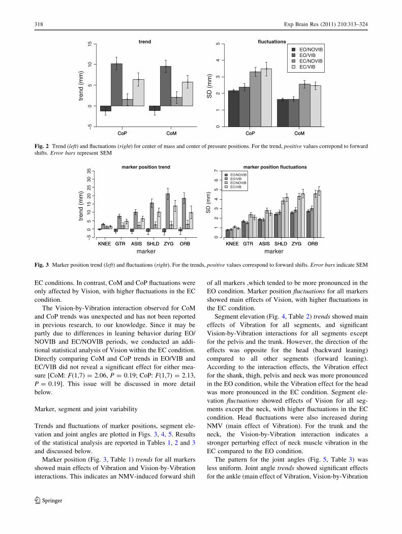
Marker, segment and joint variability
Trends and fluctuations of marker positions, segment ele-
vation and joint angles are plotted in Figs. 3, 4, 5. Results
of the statistical analysis are reported in Tables 1, 2 and 3
and discussed below.
Marker position (Fig. 3, Table 1) trends for all markers
showed main effects of Vibration and Vision-by-Vibration
interactions. This indicates an NMV-induced forward shift
of all markers ,which tended to be more pronounced in the
EO condition. Marker position fluctuations for all markers
showed main effects of Vision, with higher fluctuations in
the EC condition.
Segment elevation (Fig. 4, Table 2) trends showed main
effects of Vibration for all segments, and significant
Vision-by-Vibration interactions for all segments except
for the pelvis and the trunk. However, the direction of the
effects was opposite for the head (backward leaning)
compared to all other segments (forward leaning).
According to the interaction effects, the Vibration effect
for the shank, thigh, pelvis and neck was more pronounced
in the EO condition, while the Vibration effect for the head
was more pronounced in the EC condition. Segment ele-
vation fluctuations showed effects of Vision for all seg-
ments except the neck, with higher fluctuations in the EC
condition. Head fluctuations were also increased during
NMV (main effect of Vibration). For the trunk and the
neck, the Vision-by-Vibration interaction indicates a
stronger perturbing effect of neck muscle vibration in the
EC compared to the EO condition.
The pattern for the joint angles (Fig. 5, Table 3) was
less uniform. Joint angle trends showed significant effects
for the ankle (main effect of Vibration, Vision-by-Vibration
CoP CoMCoP CoM
−5
05
1015
trend
tren
d (m
m)
CoP CoM
EO/NOVIBEO/VIBEC/NOVIBEC/VIB
CoP CoM
01
23
45 fluctuations
SD
(m
m)
Fig. 2 Trend (left) and fluctuations (right) for center of mass and center of pressure positions. For the trend, positive values correpond to forward
shifts. Error bars represent SEM
KNEE GTR ASIS SHLD ZYG ORBKNEE GTR ASIS SHLD ZYG ORB
−5
05
1015
2025
3035
marker position trend
marker
tren
d (m
m)
KNEE GTR ASIS SHLD ZYG ORB
EO/NOVIBEO/VIBEC/NOVIBEC/VIB
KNEE GTR ASIS SHLD ZYG ORB
01
23
45
67
marker position fluctuations
marker
SD
(m
m)
Fig. 3 Marker position trend (left) and fluctuations (right). For the trends, positive values correspond to forward shifts. Error bars indicate SEM
318 Exp Brain Res (2011) 210:313–324
123
interaction, dorsiflexion in EO/NOVIB compared to the
other conditions), knee (Vibration, extension in VIB con-
dition) and AO joint (Vibration, extension in VIB condi-
tion). Joint angle fluctuations showed significant effects for
the ankle (Vision, higher fluctuations in EC), pelvis (lower
fluctuations in VIB), CT joint (Vision, lower fluctuations in
EC; Vibration by Vision, stronger perturbing effect of
vibration in EC condition) and AO joint (Vibration-by-
Vision interaction: stronger perturbing effect of vibration
in EC condition).
As for CoM and CoP, a Vision-by-Vibration interaction
was found in the trends of several variables (in particular,
marker positions, but also some shank elevation and ankle
joint angles), indicating that, unexpectedly, the forward
shank thigh pelvis chest neck headshank thigh pelvis chest neck head
−3
−2
−1
01
23
45 segment angle trend
segment
tren
d (d
eg)
shank thigh pelvis chest neck head
EO/NOVIBEO/VIBEC/NOVIBEC/VIB
shank thigh pelvis chest neck head
0.0
0.1
0.2
0.3
0.4
0.5
0.6
0.7 segment angle fluctuations
segment
SD
(de
g)
Fig. 4 Segment elevation angle trends (left) and fluctuations (right). For the trends, positive values correpond to backward leaning. Error barsindicate SEM
ankle knee hip LS CT AOankle knee hip LS CT AO
−2
02
46
8 joint angle trend
joint
tren
d (d
eg)
ankle knee hip LS CT AO
EO/NOVIBEO/VIBEC/NOVIBEC/VIB
ankle knee hip LS CT AO
0.0
0.1
0.2
0.3
0.4
0.5
0.6
0.7 joint angle fluctuations
joint
SD
(de
g)
Fig. 5 Joint angle trends (left) and fluctuations (right). For the trends, positive values correspond to extension (except for the knee joint, where
positive values correspond to flexion). Error bars indicate SEM
Table 1 T values (t(7)) and
effect sizes (Cohen’s d) for
marker position trends and
fluctuations
Significant effects are indicated
(* P \ 0.05, ** P \ 0.01,
*** P \ 0.001, uncorrected)
Measure Vision Vibration Vision 9 vibration
KNEE trend t = 0.10, d = 0.06 t = 4.26, d = 1.79** t = -3.91, d = -1.85**
GTR trend t = 0.08, d = 0.05 t = 8.17, d = 2.59*** t = -3.19, d = -1.86*
ASI trend t = -0.02, d = -0.01 t = 10.57, d = 3.09*** t = -3.08, d = -1.87*
SHLD trend t = -0.24, d = -0.15 t = 7.95, d = 3.68*** t = -2.57, d = -1.40*
ZYG trend t = -0.41, d = -0.24 t = 7.14, d = 3.55*** t = -2.90, d = -1.30*
ORB trend t = -0.52, d = -0.32 t = 5.47, d = 2.73*** t = -3.07, d = -1.39*
KNEE fluctuations t = 4.18, d = 0.98** t = -0.98, d = -0.20 t = -1.36, d = -0.54
GTR fluctuations t = 4.54, d = 1.29** t = -1.79, d = -0.46 t = -0.20, d = -0.08
ASI fluctuations t = 5.43, d = 1.53*** t = -1.20, d = -0.38 t = -0.36, d = -0.12
SHLD fluctuations t = 6.72, d = 2.19*** t = 0.53, d = 0.21 t = 0.74, d = 0.17
ZYG fluctuations t = 6.49, d = 2.63*** t = 0.56, d = 0.24 t = -0.08, d = -0.02
ORB fluctuations t = 6.85, d = 2.82*** t = 0.52, d = 0.23 t = -0.12, d = -0.04
Exp Brain Res (2011) 210:313–324 319
123
leaning was more pronounced in the EC condition. A direct
comparison of the forward trend in EO/VIB and EC/VIB
did not reach significance (P [ 0.1) for any of these
variables.
In summary, the effects on marker position trends and
fluctuations parallel the effects observed for CoM and CoP,
with a general forward leaning of the body and generally
increased fluctuations in the EC condition. The analysis of
segment elevation angles reveals differences along the
central body axis: in contrast to all other segments (that
showed a forward leaning trend upon vibration), the head
tended to lean backward. This is explained by a pro-
nounced extension of the AO joint, the joint most directly
affected by neck tendon vibration, presumably due to the
tonic vibration response (e.g., Eklund and Hagbarth 1966).
Note that the vibration-induced extension of the AO
joint was larger [mean ± SD, EO: 4.20� ± 2.06�, EC:
4.39� ± 2.38�] than the resulting backward leaning of the
head [EO: 1.91� ± 1.55�, EC: 2.84� ± 2.03�], suggesting
that the rest of the body compensated for the local effect of
the neck vibration to stabilize head orientation.
Motor-equivalent coordination
The results of the decorrelation analysis are shown in
Fig. 6. Generalized correlations were first tested for devi-
ating from zero (at the group level) with a one-sample
t test, separately for each of the four Vision and Vibration
conditions and for each the four task-related variables.
Coordination stabilizing the task-related variable under
consideration is indicated by negative generalized corre-
lation, while a positive correlation indicates a postural
organization that destabilizes the task-related variable.
Generalized correlations deviating from zero were found
for CoM position control in the EO/NOVIB condition
[t(7) = -4.05, P \ 0.005; negative], for head position in
both NOVIB conditions [EO: t(7) = -10.44, P \ 0.0005;
EC: t(7) = -4.12, P \ 0.01; both negative], and for trunk
orientation control in both NOVIB conditions [EO:
t(7) = -3.39, P \ 0.05, EC: t(7) = -4.11, P \ 0.005;
both negative] and in the EC/VIB condition [t(7) = 2.59,
P \ 0.05, positive]. In contrast, regarding head orientation
control, generalized correlations deviated from zero in all
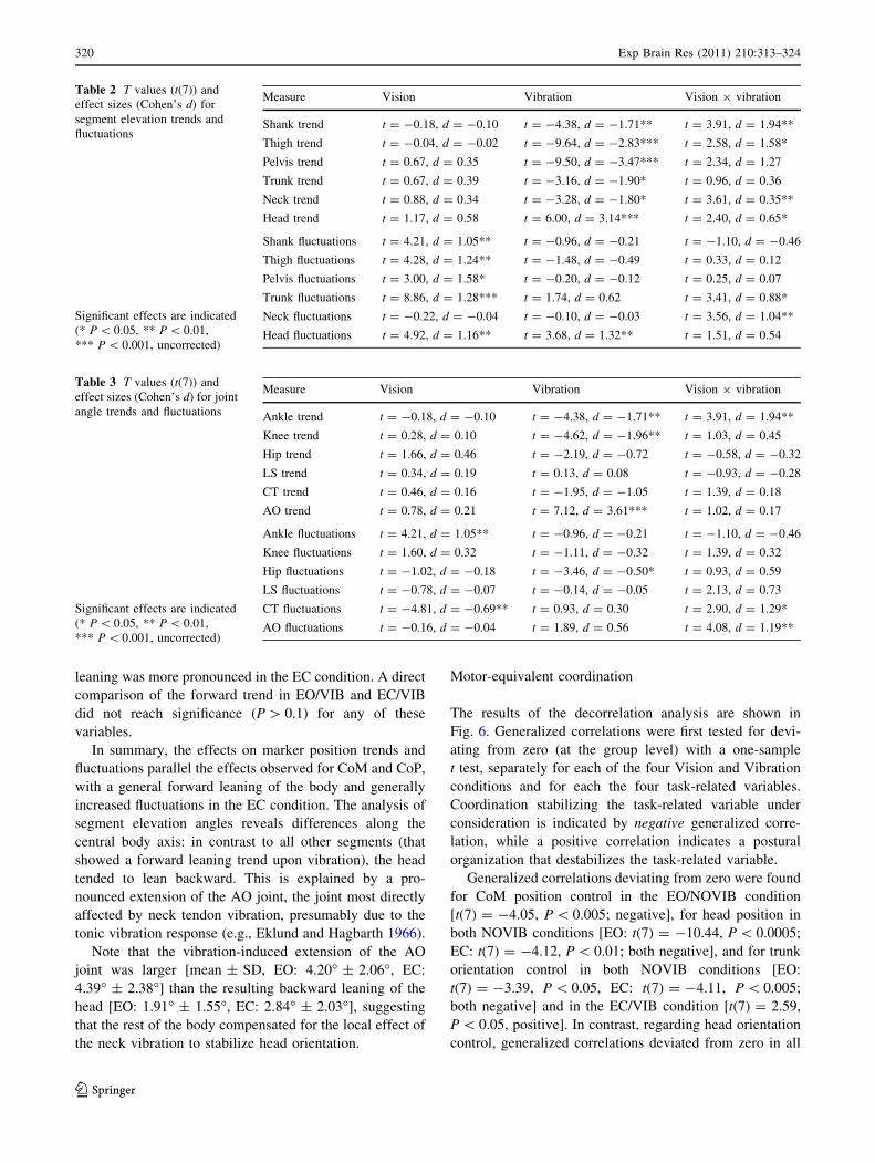
Table 2 T values (t(7)) and
effect sizes (Cohen’s d) for
segment elevation trends and
fluctuations
Significant effects are indicated
(* P \ 0.05, ** P \ 0.01,
*** P \ 0.001, uncorrected)
Measure Vision Vibration Vision 9 vibration
Shank trend t = -0.18, d = -0.10 t = -4.38, d = -1.71** t = 3.91, d = 1.94**
Thigh trend t = -0.04, d = -0.02 t = -9.64, d = -2.83*** t = 2.58, d = 1.58*
Pelvis trend t = 0.67, d = 0.35 t = -9.50, d = -3.47*** t = 2.34, d = 1.27
Trunk trend t = 0.67, d = 0.39 t = -3.16, d = -1.90* t = 0.96, d = 0.36
Neck trend t = 0.88, d = 0.34 t = -3.28, d = -1.80* t = 3.61, d = 0.35**
Head trend t = 1.17, d = 0.58 t = 6.00, d = 3.14*** t = 2.40, d = 0.65*
Shank fluctuations t = 4.21, d = 1.05** t = -0.96, d = -0.21 t = -1.10, d = -0.46
Thigh fluctuations t = 4.28, d = 1.24** t = -1.48, d = -0.49 t = 0.33, d = 0.12
Pelvis fluctuations t = 3.00, d = 1.58* t = -0.20, d = -0.12 t = 0.25, d = 0.07
Trunk fluctuations t = 8.86, d = 1.28*** t = 1.74, d = 0.62 t = 3.41, d = 0.88*
Neck fluctuations t = -0.22, d = -0.04 t = -0.10, d = -0.03 t = 3.56, d = 1.04**
Head fluctuations t = 4.92, d = 1.16** t = 3.68, d = 1.32** t = 1.51, d = 0.54
Table 3 T values (t(7)) and
effect sizes (Cohen’s d) for joint
angle trends and fluctuations
Significant effects are indicated
(* P \ 0.05, ** P \ 0.01,
*** P \ 0.001, uncorrected)
Measure Vision Vibration Vision 9 vibration
Ankle trend t = -0.18, d = -0.10 t = -4.38, d = -1.71** t = 3.91, d = 1.94**
Knee trend t = 0.28, d = 0.10 t = -4.62, d = -1.96** t = 1.03, d = 0.45
Hip trend t = 1.66, d = 0.46 t = -2.19, d = -0.72 t = -0.58, d = -0.32
LS trend t = 0.34, d = 0.19 t = 0.13, d = 0.08 t = -0.93, d = -0.28
CT trend t = 0.46, d = 0.16 t = -1.95, d = -1.05 t = 1.39, d = 0.18
AO trend t = 0.78, d = 0.21 t = 7.12, d = 3.61*** t = 1.02, d = 0.17
Ankle fluctuations t = 4.21, d = 1.05** t = -0.96, d = -0.21 t = -1.10, d = -0.46
Knee fluctuations t = 1.60, d = 0.32 t = -1.11, d = -0.32 t = 1.39, d = 0.32
Hip fluctuations t = -1.02, d = -0.18 t = -3.46, d = -0.50* t = 0.93, d = 0.59
LS fluctuations t = -0.78, d = -0.07 t = -0.14, d = -0.05 t = 2.13, d = 0.73
CT fluctuations t = -4.81, d = -0.69** t = 0.93, d = 0.30 t = 2.90, d = 1.29*
AO fluctuations t = -0.16, d = -0.04 t = 1.89, d = 0.56 t = 4.08, d = 1.19**
320 Exp Brain Res (2011) 210:313–324
123
conditions [EO/NOVIB: t(7) = -23.87, P \ 0.0001; EO/
VIB: t(7) = -10.43, P \ 0.0001; EC/NOVIB: t(7) =
-6.92, P \ 0.0005; EC/VIB: t(7) = -3.58, P \ 0.01; all
negative].
Analysis of variance regarding the influence of the
Vision and Vibration manipulations yielded the following
results. For CoM control, there was a main effect of
Vibration [F(1,7) = 31.44, P \ 0.001, d = 1.69], reflect-
ing a weaker (less negative) generalized correlation in the
VIB condition compared to the NOVIB condition. For
head position control, a main effect of Vibration
[F(1,7) = 13.4, P \ 0.01, d = 1.68] and a Vision-
by-Vibration interaction [F(1,7) = 8.11, P \ 0.05, d =
-1.13] were found, showing weaker coordination in the
VIB condition and a weaker effect of vibration in the EC
condition. For trunk orientation control, a main effect of
Vibration [F(1,7) = 24.2, P \ 0.005, d = 1.92] was
found, indicating weaker coordination in the VIB condi-
tion. Regarding head orientation control, a main effect
of Vision was present [F(1.7) = 29.35, P \ 0.001,
d = 1.48], with less negative generalized correlation in
the EC condition.
In summary, sagittal head orientation was the only task-
related variable for which generalized correlations were
consistently negative. This shows that, across all condi-
tions, all joints considered here were coordinated in a way
stabilizing head pitch. This coordination was weaker (less
negative generalized correlation) in the EC condition,
consistent with the role of vision for head stabilization, but
was not affected by NMV. In contrast, CoM and head
position only showed negative generalized correlations in
the EO/NOVIB (CoM position) or in both NOVIB (head
position, trunk orientation) conditions. As shown by the
ANOVA, whole-body coordination stabilizing these vari-
ables was compromised by NMV.
Discussion
We studied the immediate effects of symmetric vibratory
stimulation of the dorsal neck muscles on postural control,
as well as their potential interaction with vision. Postural
effects were analyzed locally (position and orientation of
individual body parts) and globally (whole-body coordi-
nation). Besides replicating previously observed effects of
NMV, i.e., a general forward learning of the body, the
present analysis allows refining previous accounts of this
phenomenon, as discussed below.
Postural trends and fluctuations are differentially
affected by NMV and vision
Postural variability was decomposed into (linear) trends
and (residual) fluctuations, based on the assumption that
the former capture NMV-induced changes in the internal
reference for upright posture (Gurfinkel et al. 1995), while
the latter are taken to be indicators of postural instability.
This decomposition showed differential effects of vibration
and vision. Consistent with previous research (Eklund and
Hagbarth 1966; Eklund 1971, 1972; Gregoric et al. 1978;
Lund 1980), NMV induced a forward leaning of the body.
This forward leaning was evident in trends of center of
pressure and center of mass positions, marker positions,
body segment orientations, and ankle and knee joint angles.
In contrast, NMV did not affect residual fluctuations of the
variables considered here, suggesting that the induced
forward leaning did not lead to a breakdown of posture-
stabilizing processes. On the other hand, in agreement with
previous findings on the posture-stabilizing role of vision
(e.g., Paulus et al. 1984; Redfern et al. 2001; Vuillerme
et al. 2006), postural fluctuations were higher in the EC
compared to the EO condition.
In addition, postural trends showed a Vision-by-Vibra-
tion interaction, indicating that the effect of NMV was
stronger with EO compared to EC. This was unexpected
and opposite to the typically observed stabilizing effect of
vision on posture (Paulus et al. 1984; Redfern et al. 2001),
as well as a recent analysis of modulation of NMV-induced
postural effects by vision (Bove et al. 2009). However, a
direct comparison of EO/VIB and EC/VIB in the present
study did not show a significant difference for any of the
forward leaning measures. This suggests that the observed
interaction effect may be at least partly explained by
vision-related differences in postural trend during the pre-
vibration interval.
CoM pos. head pos. trunk orient. head orient.
EO/NOVIBEO/VIBEC/NOVIBEC/VIB
CoM pos. head pos. trunk orient. head orient.−0.
8−
0.6
−0.
4−
0.2
0.0
0.2
0.4
0.6
intersegmental postural coordination
task variable
gene
raliz
ed c
orre
latio
n
Fig. 6 Generalized correlation, with respect to center of mass
position, head position, trunk orientation and head orientation.
Coordination is indicated when the generalized correlation is
negative, with perfect coordination corresponding to a value of -1.
Error bars represent SEM
Exp Brain Res (2011) 210:313–324 321
123
Differences in instruction may play a role as well. We
instructed participants to stand as still as possible. In
contrast, Bove et al. (2009) gave no specific instruction
about postural stabilization (Marco Schieppati, January
2010, personal communication). The considerably larger
forward leaning in this latter study (more than 40 mm in
some conditions, for a 5-s vibration) compared to the
present experiment (7–10 mm on average, for a 10-s
vibration) may be related to this difference in instructions.
Experimental instructions may also lead to a top–down
modulated reweighting of sensory information and task
priorities. In vestibular-loss patients, NMV-induced for-
ward leaning was found to be reduced or absent (Lekhel
et al. 1998). Thus, degraded information about head ori-
entation may actually (and somewhat counter-intuitively)
reduce the whole-body postural effects of NMV. Theoret-
ically, the availability of vision, providing additional
information about head orientation, may modulate NMV
effects in a similar way as the availability of vestibular
input. It remains to be investigated, in detail, how stabi-
lizing and (potential) destabilizing mechanisms of visual
feedback interact with experimental instruction.
Neck muscle vibration-induced extension of the upper
neck
Locally, NMV induced a pronounced extension of the
atlanto-occipital (AO) joint, leading to a backward leaning
of the head. Such local responses to muscle or tendon
vibration have previously been found for a number of
stimulation sites across the body and are typically inter-
preted as a corrective response to the perceived (illusory)
lengthening muscle induced by the vibration (Goodwin
et al. 1972a, b; Lackner and Levine 1979; Gilhodes et al.
1986). Our findings show that the effect of NMV on
upright postural control has a local component, besides the
previously hypothesized global effect on the ‘propriocep-
tive chain’ (Roll and Roll 1988).
To our knowledge, this local effect has not previously
been reported in unrestrained standing in healthy subjects.
Clinical studies found such a local response in vestibular
loss patients (Lekhel et al. 1998) and patients with torti-
collis (Lekhel et al. 1997), who in return showed no or
reduced forward leaning of the body. Lekhel et al. (1998)
report (without reference) that head backward tilt had been
observed in healthy individuals only when the trunk was
artificially restrained.
A simple and suitable reason why this effect was not
previously reported may be that postural data were not
acquired or analyzed accordingly. Numerous studies have
analyzed NMV effects on posture only using force plates
(Kavounoudias et al. 1999; Lekhel et al. 1998), while
others have measured body kinematics but not analyzed
movement in individual joints (Gomez et al. 2009). In the
study by (Lekhel et al. 1997), head orientation was mea-
sured in patients with torticollis, but only visually assessed
in control subjects.
More interestingly, another possible reason why previ-
ous studies did not find the local effect of neck extension
might be that its influence on sagittal head orientation
(pitch) was partially compensated by the forward leaning
of the body. For instance, in the EO condition, average AO
extension was about 4�, while the backward leaning of the
head was less than half as large. Therefore, neck extension
may go unnoticed when it is assessed by visual inspection
or even on measurement of absolute (but not relative) head
orientation.
Motor-equivalent coordination: differential effects
of NMV and vision
Whole-body coordination was analyzed using a decorre-
lation technique (Muller and Sternad 2003). In this method,
coordination with respect to variables in which stabiliza-
tion is assumed to be task relevant (here: CoM and head
position, trunk and head orientation) is quantified as a
generalized correlation coefficient. This measure is defined
by comparing variability in actual task performance to
surrogate data, obtained by decorrelating (reshuffling) joint
angle time series. The analysis is possible because the
human motor system exhibits motor equivalence with
respect to these task-related variables, since it has more
DOF available (six joint angles, in the present model) than
are specified at the task level (here, one DOF for each of
the four variables).
Negative generalized correlations indicate coordinated
stabilization of the studied variable, with -1 corresponding
to perfect coordination, i.e., full compensation among the
joint angles stabilizing the task-related variable. It has to be
noted that, with this operationalization, a postural change
that leads to a shift in the task-related variable (as observed
in particular for the CoM) may be considered destabilizing
(positive generalized correlation), even if the body reaches
a new stable equilibrium after the shift.
Coordination analysis revealed that, of the variables
under consideration, only head orientation was consistently
stabilized across all vision and vibration conditions.
Notably, coordination with respect to head orientation was
affected by vision (poorer stabilization in EC condition)
but not by NMV, indicating that participants coordinated
all joints along the central body axis to stabilize head
orientation even under conditions of proprioceptive per-
turbation. Note that this does not mean that head pitch was
as stable during VIB and NOVIB conditions (this was
actually not the case, as shown by the analysis of segment
elevation angles), but that under all conditions, the joints
322 Exp Brain Res (2011) 210:313–324
123
included in the present analysis were coordinated to a
similar extent in a way that stabilized head orientation. In
contrast, coordination underlying stabilization of CoM and
head position, as well as trunk orientation, was compro-
mised by the NMV.
Thus, NMV did not disrupt whole-body coordination
stabilizing head orientation, despite the fact that the neck is
most directly affected by NMV. In contrast, it did lead to a
destabilization of whole-body equilibrium. This indicates
that, probably within certain stability margins, orientation
may be prioritized over equilibrium by the postural control
system. This finding also suggests an alternative (or com-
plementary) explanation of NMV-induced forward leaning,
namely to compensate for the effect of neck extension (i.e.,
the local response to NMV) on head orientation, in contrast
(or in addition) to a response to illusory backward leaning
as suggested in previous accounts (e.g., Kavounoudias
et al. 1999; Lekhel et al. 1997). The present study does not
allow discrimination between these two hypotheses. A way
to address this question may be to experimentally vary task
requirements, either by instruction or by introducing dif-
ferent postural constraints concerning head stabilization
(e.g., balancing an object on the top of the head).
Conclusions and outlook
In conclusion, our study amends and extends previous
accounts of NMV-induced forward leaning in several
ways. First, the decomposition of univariate variability (in
CoM, CoP, markers, segments and joints) into linear trends
and residual fluctuations revealed differential effects of
Vision and NMV. Second, NMV also induced local pos-
tural responses, resulting in an extension of the upper neck
(backward leaning of the head). Finally, analysis of whole-
body coordination showed that, out of four analyzed task-
related variables (CoM and head position, trunk and head
orientation), only head orientation was consistently stabi-
lized by motor-equivalent coordination across Vision and
Vibration conditions. Future research should investigate
how explicit instructions or additional task constraints may
affect postural stabilization under conditions of perturbed
proprioception introduced by NMV.
Acknowledgments During his dissertation, the first author was a
pre-doctoral fellow of the International Max Planck Research School
‘‘The Life Course: Evolutionary and Ontogenetic Dynamic’’. The
study has been supported by a grant from the Centre de La Recherche
Scientifique (CNRS, France) to the last author.
References
Bove M, Fenoggio C, Tacchino A, Pelosin E, Schieppati M (2009)
Interaction between vision and neck proprioception in the
control of stance. Neuroscience 164(4):1601–1608. doi:
10.1016/j.neuroscience.2009.09.053
Cohen LA (1961) Role of eye and neck proprioceptive mechanisms in
body orientation and motor coordination. J Neurophysiol
24:1–11
Cohen J (1988) Statistical power analysis for the behavioral sciences,
2nd edn. Lawrence Earlbaum Associates, Hillsdale
Creath R, Kiemel T, Horak F, Peterka R, Jeka J (2005) A unified view
of quiet and perturbed stance: simultaneous co-existing excitable
modes. Neurosci Lett 377(2):75–80. doi:10.1016/j.neulet.2004.
11.071
Cusumano J, Cesari P (2006) Body-goal variability mapping in an
aiming task. Biol Cybern 94:367–379
Eklund G (1971) On muscle vibration in man; an amplitude-
dependent inhibition, inversely related to muscle length. Acta
Physiol Scand 83(3):425–426
Eklund G (1972) General features of vibration-induced effects on
balance. Ups J Med Sci 77(2):112–124
Eklund G, Hagbarth KE (1966) Normal variability of tonic vibration
reflexes in man. Exp Neurol 16(1):80–92
Gilhodes JC, Roll JP, Tardy-Gervet MF (1986) Perceptual and motor
effects of agonist–antagonist muscle vibration in man. Exp Brain
Res 61(2):395–402
Gomez S, Patel M, Magnusson M, Johansson L, Einarsson EJ,
Fransson PA (2009) Differences between body movement
adaptation to calf and neck muscle vibratory proprioceptive
stimulation. Gait Posture 30(1):93–99. doi:10.1016/j.gaitpost.
2009.03.009
Goodwin GM, McCloskey DI, Matthews PB (1972a) The contribution
of muscle afferents to kinaesthesia shown by vibration induced
illusions of movement and by the effects of paralysing joint
afferents. Brain 95(4):705–748
Goodwin GM, McCloskey DI, Matthews PB (1972b) Proprioceptive
illusions induced by muscle vibration: contribution by muscle
spindles to perception? Science 175(28):1382–1384
Gregoric M, Takeya T, Baron JB, Bessineton JC (1978) Influence of
vibration of neck muscles on balance control in man. Agressol-
ogie 19(A):37–38
Gurfinkel VS, YuP I, YuS L, Babakova IA (1995) Kinesthetic
reference for human orthograde posture. Neuroscience 68(1):
229–243
Horak FB, MacPherson JM (1996) Postural orientation and equilib-
rium. In: Rowell L, Shepherd J (eds) Handbook of physiology.
American Physiological Society, New York, pp 255–292
Hsu WL, Scholz JP, Schoner G, Jeka JJ, Kiemel T (2007) Control and
estimation of posture during quiet stance depends on multijoint
coordination. J Neurophysiol 97:3024–3035
Ivanenko YP, Talis VL, Kazennikov OV (1999) Support stability
influences postural responses to muscle vibration in humans. Eur
J Neurosci 11(2):647–654
Kavounoudias A, Gilhodes JC, Roll R, Roll JP (1999) From balance
regulation to body orientation: two goals for muscle proprio-
ceptive information processing? Exp Brain Res 124(1):80–88
Lackner JR, Levine MS (1979) Changes in apparent body orientation
and sensory localization induced by vibration of postural
muscles: vibratory myesthetic illusions. Aviat Space Environ
Med 50(4):346–354
Latash ML, Scholz JP, Schoner G (2007) Toward a new theory of
motor synergies. Mot Control 11:276–308
Lekhel H, Popov K, Anastasopoulos D, Bronstein A, Bhatia K,
Marsden CD, Gresty M (1997) Postural responses to vibration
of neck muscles in patients with idiopathic torticollis. Brain
120(4):583–591
Lekhel H, Popov K, Bronstein A, Gresty M (1998) Postural responses
to vibration of neck muscles in patients with uni- and bilateral
vestibular loss. Gait Posture 7(3):228–236
Exp Brain Res (2011) 210:313–324 323
123
Lund S (1980) Postural effects of neck muscle vibration in man.
Experientia 36(12):1398
Massion J (1994) Postural control system. Curr Opin Neurobiol
4(6):877–887
Mergner T, Huber W, Becker W (1997) Vestibular–neck interaction
and transformation of sensory coordinates. J Vestib Res
7(4):347–367
Muller H, Sternad D (2003) A randomization method for the
calculation of covariation in multiple nonlinear relations:
illustrated with the example of goal-directed movements. Biol
Cybern 89(1):22–33
Paulus WM, Straube A, Brandt T (1984) Visual stabilization of
posture. physiological stimulus characteristics and clinical
aspects. Brain 107(4):1143–1163
Peterka RJ (2002) Sensorimotor integration in human postural
control. J Neurophysiol 88(3):1097–1118
Pinter IJ, van Swigchem R, van Soest AJK, Rozendaal LA (2008) The
dynamics of postural sway cannot be captured using a one-
segment inverted pendulum model: a PCA on segment rotations
during unperturbed stance. J Neurophysiol 100(6):3197–3208.
doi:10.1152/jn.01312.2007
R Development Core Team (2008) R: a language and environment for
statistical computing. R Foundation for Statistical Computing,
Vienna, Austria, http://www.R-project.org, ISBN 3-900051-07-0
Redfern MS, Yardley L, Bronstein AM (2001) Visual influences on
balance. J Anxiety Disord 15(1–2):81–94
Roll J, Roll R (1988) From eye to foot: a proprioceptive chain
involved in postural control. In: Amblard B, Berthoz A, Clarac F
(eds) Posture and gait. Elsevier, Amsterdam, pp 155–164
Scholz JP, Schoner G (1999) The uncontrolled manifold concept:
identifying control variables for a functional task. Exp Brain Res
126(3):289–306
Schoner G (1995) Recent developments and problems in human
movement science and their conceptual implications. Ecol Psych
7(4):291–314
Vuillerme N, Burdet C, Isableu B, Demetz S (2006) The magnitude of
the effect of calf muscles fatigue on postural control during
bipedal quiet standing with vision depends on the eye–visual
target distance. Gait Posture 24(2):169–172. doi:10.1016/
j.gaitpost.2005.07.011
Winter DA (2004) Biomechanics and control of human movement.
3rd edition. John Wiley & Sons, Hoboken
Zhang Y, Kiemel T, Jeka J (2007) The influence of sensory
information on two-component coordination during quiet stance.
Gait Posture 26(2):263–271. doi:10.1016/j.gaitpost.2006.09.007
Zok M, Mazza C, Cappozzo A (2008) Should the instructions issued to
the subject in traditional static posturography be standardised? Med
Eng Phys 30(7):913–916. doi:10.1016/j.medengphy.2007.12.002
324 Exp Brain Res (2011) 210:313–324
123