Embed Size (px)
Citation preview

Rician Noise Removal in Diffusion Tensor MRI
Saurav Basu, Thomas Fletcher, and Ross Whitaker
University of Utah,School of Computing,
Salt Lake City, UT 84112, USA
Abstract. Rician noise introduces a bias into MRI measurements thatcan have a significant impact on the shapes and orientations of ten-sors in diffusion tensor magnetic resonance images. This is less of aproblem in structural MRI, because this bias is signal dependent andit does not seriously impair tissue identification or clinical diagnoses.However, diffusion imaging is used extensively for quantitative evalua-tions, and the tensors used in those evaluations are biased in ways thatdepend on orientation and signal levels. This paper presents a strat-egy for filtering diffusion tensor magnetic resonance images that ad-dresses these issues. The method is a maximum a posteriori estima-tion technique that operates directly on the diffusion weighted imagesand accounts for the biases introduced by Rician noise. We account forRician noise through a data likelihood term that is combined with aspatial smoothing prior. The method compares favorably with severalother approaches from the literature, including methods that filter dif-fusion weighted imagery and those that operate directly on the diffusiontensors.
1 Introduction
The quality of DT-MRI images is limited by the relatively long acquisition timesnecessary to obtain data at high spatial resolutions. Because acquisition time isrestricted by issues of patient comfort and system demand, the signal-to-noiseratio(SNR) in DT-MRI is often low. Thus, post processing techniques to removenoise in the acquired data are important. The diffusion weighted images (DWIs),from which the tensors are derived, are corrupted by Rician noise, which intro-duces a positive bias in those measurements. These signal-dependent biases arenot so detrimental to structural imaging, because they typically do not inter-fere with diagnostic decisions or tissue classification. However, DT-MRI mea-surements are being used extensively for quantitative comparisons, and severalstudies [1,2,3] have shown that bias can affect tensor properties such as traceand fractional anisotropy (FA).
Previous DT-MRI filtering methods fall into two categories: filters that op-erate on the DWIs and those that operate directly on the tensors. For instanceParker et al. [4] use Perona & Malik (P&M) [5] anisotropic diffusion to filterDWIs, and show that it produces less distortion in FA than filtering images ofFA. Wang et al. [6] formulate a variational approach to regularize DWIs while
R. Larsen, M. Nielsen, and J. Sporring (Eds.): MICCAI 2006, LNCS 4190, pp. 117–125, 2006.c© Springer-Verlag Berlin Heidelberg 2006
118 S. Basu, T. Fletcher, and R. Whitaker
constraining the estimated tensors to remain positive definite. Martin et al. [7]develop a Gaussian Markov Random Field model to regularize the estimateddiffusion tensor images. Pennec et al. [8] describe a framework for performinganisotropic diffusion on tensors which preserves the property of tensors beingsymmetric and positive definite. Their filter is based on the idea that the spaceof all positive definite symmetric matrices forms a Riemannian manifold witheach point representing a diffusion tensor. None of these techniques explicitlyaccount for the effects of bias in the original DWI measurements. After sub-mission of this work, we became aware of the work by Fillard et al. [9] thatadds a Rician noise model to smoothing of tensor images in a Log-Euclideanframework.
In this paper we show Monte Carlo simulations that add new insights intothe effects of Rician bias on tensor measurements. These results demonstratethe need for realistic noise models in DT-MRI filtering. We describe a filter-ing strategy that explicitly models the Rician noise as a data likelihood termin a maximum a posteriori framework. To assess the performance of our tech-nique, we propose a new method for producing low noise DWIs using a max-imum likelihood estimate (MLE) from repeated scans of a healthy volunteer.We present a comparison of filtering performance for tensor based methodsand methods that smooth the DWIs. Our results show that filtering on theoriginal DW images gives better results than filtering on tensor images, andthat our method using an explicit model of Rician noise gives the best overallresults.
2 Rician Noise and Its Effects on Diffusion Tensors
It is well known that MR magnitude images are corrupted by Rician noise, whicharises from complex Gaussian noise in the original frequency domain (k-space)measurements. The Rician probability density function for the corrupted imageintensity x is given by
p(x) =x
σ2 exp(
−x2 + A2
2σ2
)I0
(xA
σ2
), (1)
where A is the underlying true intensity, σ is the standard deviation of the noise,and I0 is the modified zeroth-order Bessel function of the first kind.
Previous studies on the effect of noise on diffusion tensor measurements haveshown that as noise increases, the tensor trace decreases [1] and FA increases[1,2,3]. Here we show that these effects can actually be quite different dependingon the orientation of the diffusion tensor with respect to the measurement gradi-ents. Using power series analysis, Anderson [2] shows that the major eigenvalueincreases with higher noise, causing FA to increase. This analysis assumes themajor eigenvalue is a combination of several diffusion weighted measurements,which happens when the major eigenvector lies in between several gradient di-rections. However, consider the special case of six gradient directions where the
Rician Noise Removal in Diffusion Tensor MRI 119
0 0.05 0.1 0.15 0.21.5
1.6
1.7
1.8
1.9
2
2.1
2.2x 10
−3
1 / SNR
Tra
ce
FA=0.25, unalignedFA=0.55, unalignedFA=0.70, unalignedFA=0.87, unalignedFA=0.25, alignedFA=0.55, alignedFA=0.70, alignedFA=0.87, aligned
0 0.05 0.1 0.15 0.20.2
0.3
0.4
0.5
0.6
0.7
0.8
0.9
1 / SNR
FA
FA=0.25, unalignedFA=0.55, unalignedFA=0.70, unalignedFA=0.87, unalignedFA=0.25, alignedFA=0.55, alignedFA=0.70, alignedFA=0.87, aligned
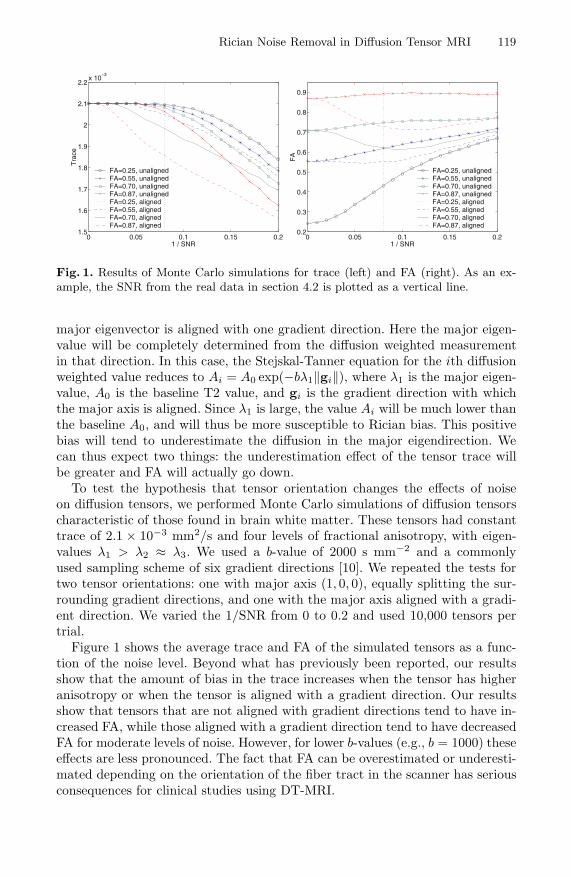
Fig. 1. Results of Monte Carlo simulations for trace (left) and FA (right). As an ex-ample, the SNR from the real data in section 4.2 is plotted as a vertical line.
major eigenvector is aligned with one gradient direction. Here the major eigen-value will be completely determined from the diffusion weighted measurementin that direction. In this case, the Stejskal-Tanner equation for the ith diffusionweighted value reduces to Ai = A0 exp(−bλ1‖gi‖), where λ1 is the major eigen-value, A0 is the baseline T2 value, and gi is the gradient direction with whichthe major axis is aligned. Since λ1 is large, the value Ai will be much lower thanthe baseline A0, and will thus be more susceptible to Rician bias. This positivebias will tend to underestimate the diffusion in the major eigendirection. Wecan thus expect two things: the underestimation effect of the tensor trace willbe greater and FA will actually go down.
To test the hypothesis that tensor orientation changes the effects of noiseon diffusion tensors, we performed Monte Carlo simulations of diffusion tensorscharacteristic of those found in brain white matter. These tensors had constanttrace of 2.1 × 10⁻³ mm2/s and four levels of fractional anisotropy, with eigen-values λ1 > λ2 ≈ λ3. We used a b-value of 2000 s mm−2 and a commonlyused sampling scheme of six gradient directions [10]. We repeated the tests fortwo tensor orientations: one with major axis (1, 0, 0), equally splitting the sur-rounding gradient directions, and one with the major axis aligned with a gradi-ent direction. We varied the 1/SNR from 0 to 0.2 and used 10,000 tensors pertrial.
Figure 1 shows the average trace and FA of the simulated tensors as a func-tion of the noise level. Beyond what has previously been reported, our resultsshow that the amount of bias in the trace increases when the tensor has higheranisotropy or when the tensor is aligned with a gradient direction. Our resultsshow that tensors that are not aligned with gradient directions tend to have in-creased FA, while those aligned with a gradient direction tend to have decreasedFA for moderate levels of noise. However, for lower b-values (e.g., b = 1000) theseeffects are less pronounced. The fact that FA can be overestimated or underesti-mated depending on the orientation of the fiber tract in the scanner has seriousconsequences for clinical studies using DT-MRI.
120 S. Basu, T. Fletcher, and R. Whitaker
3 Rician Bias Correction Filter
Our Rician bias correction filter is based on a maximum a posteriori (MAP)approach to the image reconstruction problem. Given an initial noisy image u0,we construct the filtered image u that maximizes the log-posterior probability
log p(u|u0) ∝ log p(u0|u) + log p(u), (2)
where p(u0|u) is the likelihood term, or noise model, and p(u) is the prior.For DWIs we consider u to be a vector-valued image, each gradient direction(including b = 0) representing a vector component. The formulation in thissection would also be valid for structural MRI.
3.1 The Rician Likelihood Term
The formulation of the filtering problem as maximization of a posterior p(u|u0)is useful as it allows us to incorporate the Rician bias correction as a data attach-ment term which can be added to the prior model. Using the Rician distribution(1) as the likelihood term and assuming independent noise, the pointwise log-likelihood becomes
log p(u0|u) = logu0
σ2 − u20 + u2
2σ2 + log I0
(u0u
σ2
). (3)
The derivative of (3) with respect to u, gives Rician data attachment term
B = − u
σ2 +[I1
(u0u
σ2
)/I0
(u0u
σ2
)] u0
σ2 . (4)
3.2 Combining the Rician Model with a Prior
The data likelihood term can be combined with any image prior model. In thispaper we use a Gibb’s prior model based on a P&M energy functional, given by
p(u) =1z
exp(−E(u)), E(u) = λ
∫U
c(‖∇u‖2) dx dy, (5)
where z is a suitable normalization, U is the image domain and c is the conduc-tance given by c(‖∇u‖2) = exp(−‖∇u‖2/2k2), k is the conductance parameterand λ is a constant weighting term.
By adding the Rician likelihood term (4) with the variational of the P&Menergy functional we form the update equation for the filtered image,
∂u
∂t= B + λdiv
(c(‖∇u‖2) ∇u
). (6)
Rician Noise Removal in Diffusion Tensor MRI 121
4 Results
The performance comparisons were made on four different filtering methods:Euclidean tensor filtering, Riemannian tensor filtering [8] and vector anisotropicdiffusion on DWIs with and without the Rician likelihood term as describedin section 3.1. Here Euclidean filtering refers to vector anisotropic diffusion onthe tensor components. To compare these methods, we used both synthetic andreal datasets. We used three different error metrics - root mean squared (RMS)error in the tensor, trace and fractional anisotropy. The error between tensorsis computed using the Frobenius norm. The parameters for each method wereoptimized for the RMS error on the tensor components. Both synthetic and realdatasets use seven images for each slice, one without diffusion gradient (b=0) andthe remaining six with b=1000s/mm2 and diffusion gradients along the standardsix orientations [10].
4.1 Synthetic Data
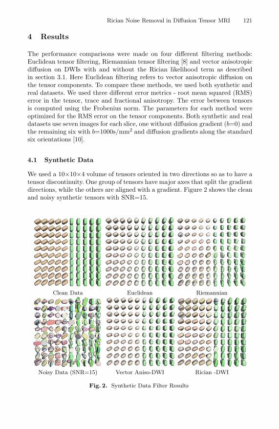
We used a 10×10×4 volume of tensors oriented in two directions so as to have atensor discontinuity. One group of tensors have major axes that split the gradientdirections, while the others are aligned with a gradient. Figure 2 shows the cleanand noisy synthetic tensors with SNR=15.
Clean Data Euclidean Riemannian
Noisy Data (SNR=15) Vector Aniso-DWI Rician -DWI
Fig. 2. Synthetic Data Filter Results

122 S. Basu, T. Fletcher, and R. Whitaker
Fig. 3. Plots of error metrics for the various filters on synthetic data
Fig. 4. Plots of error metrics for the various filters on real data
4.2 Real Data
DTI Ground Truth Generation: A key challenge in quantitatively evaluat-ing filtering methods on real diffusion tensor data is the lack of a ground truth.While realistic simulated brain datasets exists for structural MRI, no such nor-malized data is available for DT-MRI. For this paper we develop a new approachfor generating high SNR diffusion weighted image data. This technique buildsa ground truth image as a maximum likelihood (ML) estimate from a set ofrepeated scans of the same subject. If {xi} is the set of intensities from the samevoxel in N repeated scans, then the ML estimate of the true intensity A is foundby maximizing the log-likelihood, log L =
∑Ni=1 log p(xi|A), where p(xi|A) is the
Rician pdf as given in (1). The properties of the ML estimate are investigatedby Sijbers et al. [11]. This ML estimate is superior to a simple averaging of theintensities as it incorporates a priori knowledge of the noise statistics. Also, itis well known that in the limit the ML estimate is most precise.
About the data: We generated our ground truth ML images from a set of fivescans of a healthy volunteer on a Siemens head-only 3T scanner (Allegra). Foreach sequence, a single shot echo planar (EPI) diffusion tensor sequence withtotal scan time of approximately 12 minutes was used. The imaging parameterswere: TR=5400ms, TR=73ms, isotropic voxels with 2mm slice distance and in-plane resolution = 2 × 2mm, 20 averages. We added known Rician noise to the

Rician Noise Removal in Diffusion Tensor MRI 123
Clean Data Noisy Data (SNR=15)
Euclidean Riemannian
Vector Aniso-DWI Rician -DWI
Fig. 5. Real Data Filtering Results
ML estimated DWIs at SNR levels of 10, 15 and 20 with respect to the averagewhite matter signal level in the b = 0 image. A slice from the ground truth andSNR=15 tensor images is shown in Figure 5.

124 S. Basu, T. Fletcher, and R. Whitaker
4.3 Performance
The resulting error metrics for the various filtering methods on the syntheticand real data are shown in Figures 3 and 4. The original, noisy and filteredimages for SNR=15 are shown with superquadric glyphs [12] in Figures 2 and5. The results demonstrate that the Rician filter with the bias correction termgives better RMS error on tensor components. On both the real and syntheticdata the Rician filter is superior to all the other filtering techniques.The dataalso shows that for most of the error metrics the filtering methods on the DWI’syields better results than smoothing on the tensor space. The Riemannian filterrequires all tensors to be positive definite and is thus disadvantaged by theprocess of adjusting for negative eigenvalues.
5 Conclusions
We presented a new method for denoising diffusion tensor images that includesa Rician noise model as part of MAP estimation framework. To the best of ourknowledge, this is the first work to explicitly model and remove the bias effectsof Rician noise in DT-MRI. We presented Monte Carlo simulations that showthat noise can distort tensors in a manner that is dependent on the orientationand anisotropy of the underlying tensor. Our filtering results demonstrated thatfiltering on the original DWIs yields superior results to filtering methods thatoperate on the estimated tensors. Filtering on the DWIs with our Rician noisemodel gave the best overall results.
Acknowldgments
This work is part of the National Alliance for Medical Image Computing(NAMIC), funded by the National Institutes of Health through the NIH Roadmapfor Medical Research, Grant U54 EB005149. Information on the National Centersfor Biomedical Computing can be obtained from http://nihroadmap.nih.gov/bioinformatics. Funding for this work has also been provided by Center forIntegrative Biomedical Computing, NIH NCRR Project 2-P41-RR12553-07. Wethank Weili Lin and Guido Gerig from the University of North Carolina forproviding us with the DW-MRI data. Glyph visualizations created with Teem(http://teem.sf.net).
References
1. Pierpaoli, C., Basser, P.: Toward a quantitative assessment of diffusion anisotropy.Magnetic Resonance in Medicine 36(6) (1996) 893–906
2. Anderson, A.W.: Theoretical analysis of the effects of noise on diffusion tensorimaging. Magnetic Resonance in Medicine 46(6) (2001) 1174–1188
3. Skare, S., Li, T., Nordell, B., Ingvar, M.: Noise considerations in the determinationof diffusion tensor anisotropy. Magnetic Resonance Imaging 18(6) (2000) 659–669
Rician Noise Removal in Diffusion Tensor MRI 125
4. J., P.G.: Nonlinear smoothing for reduction of systematic and random errors indiffusion tensor imaging. J Magn. Reson Imaging 11(6) (2000) 702–710
5. Perona, P., Malik, J.: Scale-space and edge detection using anisotropic diffusion.IEEE Transactions on Pattern Analysis Machine Intelligence 17(4) (1990) 629–639
6. Wang, Z., Vemuri, B., Chen, Y., Mareci, T.: A constrained variational principle fordirect estimation and smoothing of the diffusion tensor field from complex DWI.IEEE Transactions on Medical Imaging 23(8) (2004) 930–939
7. Martin Fernandez, C.F. Westin, C.A.L.: 3d bayesian regularization of diffusiontensor mri using multivariate gaussian markov random fields. In: Medical ImageComputing and Computer-Assisted Intervention (MICCAI’04). (2004)
8. Pennec, X., Fillard, P., Ayache, N.: A riemannian framework for tensor computing.International Journal of Computer Vision 66(1) (2006) 41–66
9. Fillard, P., Arsigny, V., Pennec, X., Ayache, N.: Clinical DT-MRI estimation,smoothing and fiber tracking with log-Euclidean metrics. In: Proceedings of theThird IEEE International Symposium on Biomedical Imaging (ISBI 2006), CrystalGateway Marriott, Arlington, Virginia, USA (2006) 786–789
10. Basser, P., Pierpaoli, C.: Microstructural and physiological features of tissues elu-cidated by quantitative-diffusion-tensor MRI. J. Mag Res. 111(3) (1996) 209–219
11. Sijbers, J., den Dekker, A., Scheunders, P., Dyck, D.V.: Maximum-LikelihoodEstimation of Rician Distribution Parameters. IEEE Transactions on MedicalImaging 17(3) (1998) 357–361
12. Kindlmann, G.: Superquadric tensor glyphs. In: Proceedings of IEEE TVCG/EGSymposium on Visualization 2004. (2004) 147–154