Embed Size (px)
Citation preview
8561
Abstract. – OBJECTIVE: To investigate the role of HOXD-AS1 in preeclampsia and its under-lying mechanism.
PATIENTS AND METHODS: A total of 50 pre-eclampsia primiparas and 34 normal pregnan-cies admitted in our hospital from July 2015 to July 2017 were selected as the study group and control group, respectively. Age, body weight, blood pressure, 24-h urinary protein and neo-natal weight were compared between the two groups. HOXD-AS1 expression in the placen-ta tissues was detected by quantitative Re-al-time PCR (qRT-PCR). Preeclampsia patients were further assigned into high and low expres-sion group according to their HOXD-AS1 ex-pressions. The relationship between HOXD-AS1 expression and blood pressure, 24-h urinary protein and neonatal weight in preeclampsia pa-tients were analyzed. For in vitro experiments, transfection efficacy of pcDNA-HOXD-AS1 and si-HOXD-AS1 were detected by qRT-PCR. Pro-liferative and colony formation abilities in Be-Wo and Wish cells were detected by CCK-8 and colony formation assay, respectively. Moreover, protein expressions of p-p38, p-JNK, and p-ERK were detected by Western blot.
RESULTS: The systolic blood pressure, dia-stolic blood pressure and urinary protein in pre-eclampsia patients were higher than those of normal pregnancies. However, neonatal weight in preeclampsia patients was lower than that of normal pregnancies. HOXD-AS1 expressions were gradually increased in normal pregnan-cies, patients with late onset preeclampsia and patients with early onset preeclampsia sequen-tially. Additionally, higher levels of systolic pres-sure, diastolic pressure and 24-h urinary protein, as well as lower neonatal weight, were observed in preeclampsia patients with high expression of HOXD-AS1 than those with low expression. In vi-tro results demonstrated that proliferative and colony formation abilities in trophoblasts were elevated after HOXD-AS1 knockdown. Western blot data illustrated that protein expressions of p-p38 and p-JNK were decreased, while p-ERK expression was increased after overexpression of HOXD-AS1 in trophoblasts.
CONCLUSIONS: HOXD-AS1 participates in the development and progression of preeclampsia
by regulating trophoblast proliferation via the MAPK pathway.
Key Words:Preeclampsia, HOXD-AS1, Proliferation, MAPK path-
way.
Introduction
Preeclampsia is a multifactorial disorder uni-que to the third trimester of pregnancy, which is characterized by hypertension (≥ 140/90 mm Hg), proteinuria (≥ 0.3 g/24 h) and systemic dysfun-ction1. The incidence of preeclampsia in primipa-ras is 2% to 7%, which is one of the major causes of death in mother and child2. Perinatal mortality in preeclampsia patients is almost five times as those normal pregnancies in developed countries. More seriously, mortality in preeclampsia patien-ts accounts for 20-80% in developing countries. The study of preeclampsia pathogenesis has been well recognized3. Researches have shown that the imbalance of immune tolerance, genetic factors, and placental ischemia and hypoxia are involved in the development of preeclampsia4. The specific mechanism, however, has not been fully elucida-ted. Long non-coding RNA (lncRNA) is a kind of non-coding RNA with 200 nucleotides in length, which is responsible for regulating cellular activi-ties5. Researches6 have shown that lncRNA exer-ts a crucial role in biological processes, such as chromatin modification, transcription, translation, dose-compensation effect, X chromosome inacti-vation, gene imprinting, variable cleavage of RNA, and regulation of protein activities. Abnor-mally expressed lncRNAs are related to the deve-lopment of various human diseases7. It is reported that lncRNA is involved in cell proliferation, mi-gration and invasion8. For example, lncRNA p219, DQ78624310 and MALAT111 may participate in the abnormal activities of trophoblasts. Zou et al12 first demonstrated that lncRNA SPRY4-IT1 regu-
European Review for Medical and Pharmacological Sciences 2018; 22: 8561-8568
J. JIANG, Z.-M. ZHAO
Department of Obstetric, The Second People’s Hospital of Liaocheng, Linqing, China
Corresponding Author: Zhiming Zhao, MM; e-mail: [email protected]
LncRNA HOXD-AS1 promotes preeclampsia progression via MAPK pathway
J. Jiang, Z.-M. Zhao
8562
lates the proliferation, migration and apoptosis of trophoblast cell line HTR-8/SVneo. HOXD-AS1 is an antisense lncRNA encoded by the homeobox D family (HOXD) on chromosome 2, which be-longs to the HOX family. The HOX gene family, as a class of evolutionarily conserved genes, was originally found in the Drosophila homomorphi-sm. HOX gene is greatly involved in embryonic development13. Some investigations14-19 have con-firmed that HOX mutations can cause dysplasia, organ dysfunction, and even tumors. However, little study has been conducted on the role of HOXD-AS1 in preeclampsia. Our study aims to investigate the effect of HOXD-AS1 on pree-clampsia and its underlying mechanism.
Patients and Methods
Sample Collection50 preeclampsia primiparas admitted in the
Second People’s Hospital of Liaocheng from July 2015 to July 2017 were selected as the stu-dy group, and 34 cases of normal pregnancies in the same period were randomly selected as the control group. Blood pressure and 24-h urinary protein levels of pregnancies, as well as their neonatal weight were recorded. After the pla-centa was delivered, two placental tissues (1.0 cm×1.0 cm×0.6 cm) at the center of the placen-ta attached to the root of the umbilical cord were harvested. Placental tissues were required to be non-calcified without hemorrhage and necrosis. After being washed in DEPC water containing 0.1% normal saline, samples were then preserved at -80°C for further experiments. This study was approved by the Ethics Committee of the Second People’s Hospital of Liaocheng and all subjects were informed consent. Pregnancies with other diseases, abnormal pregnancy hormone, medical history of antibiotic medication and adosculation were excluded. General characteristics of enrolled subjects were listed in Table I.
Cell Culture and TransfectionHuman preeclampsia cell line was cultured
in 1640 medium supplemented with 10% fetal bovine serum, 100 U/mL penicillin and 100 μg/mL streptomycin (Gibco, Rockville, MD, USA), and maintained in a 5% CO2 incubator at 37°C. Cells in logarithmic growth period were seeded in 6-well plates. When cell confluence was up to 60-80%, pcDNA-HOXD-AS1 and si-HOXD-AS1 (GenePharma, Shanghai, China) were transfected
into BeWo and Wish cells, respectively according to the instructions of Lipofatamine 2000 (Ther-mo Fisher Scientific, Inc. Waltham, MA, USA). Transfection efficacy was verified by quantitative reverse transcriptase-polymerase chain reaction (qRT-PCR).
RNA Extraction and qRT-PCRThe mRNAs of cells were extracted by TRI-
zol reagent (Invitrogen, Carlsbad, CA, USA) and then reversely transcribed to cDNAs. The reaction conditions were as follows: denaturation at 95°C for 30 s, followed by annealing at 95°C for 5 s and extension at 60°C for 31 s, for a total of 40 cycles. Each sample was repeatedly performed for 3 times. The mRNA level was calculated based on the 2-ΔΔCt method. Primers used in this study were as follows: HOXD-AS1, F: GGCTCTTCCCTA-ATGTGTGG; R: CAGGTCCAGCATGAAACA-GA; GAPDH, F: CGCTCTCTGCTCCTCCTGT-TC, R: ATCCGTTGACTCCGACCTTCAC.
Cell Counting Kit-8 (CCK-8) AssayTransfected cells were collected and seeded
into a 96-well plate at a dose of 2×10³/mL. After 24 h-inoculation, 10 μL of CCK-8 solution were added into each well at 6 h, 24 h, 48 h, 72 h, 96 h, respectively. The absorbance (OD) values at the wavelength of 450 nm were accessed with a mi-croplate reader.
Colony Formation Assay BeWo and Wish cells were harvested and ino-
culated into a 6-well plate at a dose of 500/mL for 2-week culture in complete medium. 2 mL of 4% paraformaldehyde were applied to fix the co-lonies for 30 min and 0.1% crystal violet solution was added for 20-min staining. After washed with phosphate-buffered saline (PBS) for 3 times, 3 fields in each well were randomly selected for ob-serving and capturing colonies using an inverted microscope.
Western BlotThe total protein of the transfected cells
was extracted. The concentration of each pro-tein sample was determined by a bicinchoninic acid (BCA) kit (Pierce, Rockford, IL, USA). Briefly, 50 μg of total protein were separated by sodium dodecyl sulphate-polyacrylamide gel electrophoresis (SDS-PAGE) under denaturing conditions and transferred to polyvinylidene di-fluoride (PVDF) membranes (Millipore, Bille-rica, MA, USA). Membranes were blocked with
LncRNA HOXD-AS1 promotes preeclampsia progression via MAPK pathway
8563
5% skimmed milk, followed by the incubation of specific primary antibodies (p-p38, p-JNK and p-ERK) overnight. Membranes were then incubated with the secondary antibody at room temperature for 1 h. Immunoreactive bands were exposed by enhanced chemiluminescence (ECL) method.
Statistical AnalysisStatistical Product and Service Solutions
(SPSS) 13.0 statistical software (SPSS Inc., Chi-cago, IL, USA) was used for data analysis. Mea-surement data were expressed as mean ± standard deviation (`x±s). Comparison of measurement data was conducted using t-test. p<0.05 was con-sidered statistically significant.
Results
Clinical Data of Preeclampsia Patients and Normal Pregnancies
Clinical data of enrolled subjects were recor-ded, including the age, blood pressure and 24-h urinary protein levels, etc. No significant differen-ces in age and body weight were found between the two groups (p>0.05). However, higher levels of systolic pressure (148.31±13.55 mmHg), dia-stolic pressure (107.89±17.35 mmHg) and 24-h urinary protein (0.42±0.16 g/day) were observed in preeclampsia patients than those of normal pregnancies (113.04±18.32 mmHg, 76.31±19.13 mmHg and 0.15±0.18 g/day, respectively). All the above differences were statistically significant (all p<0.05). We also recorded the neonatal weight of each subject. The data suggested that lower ne-onatal weight was found in preeclampsia patien-ts (1785.25±530.17 g) compared with those of normal pregnancies (3317.13±389.16 g, p<0.05, Table I).
HOXD-AS1 Was Upregulated in Preeclampsia Patients
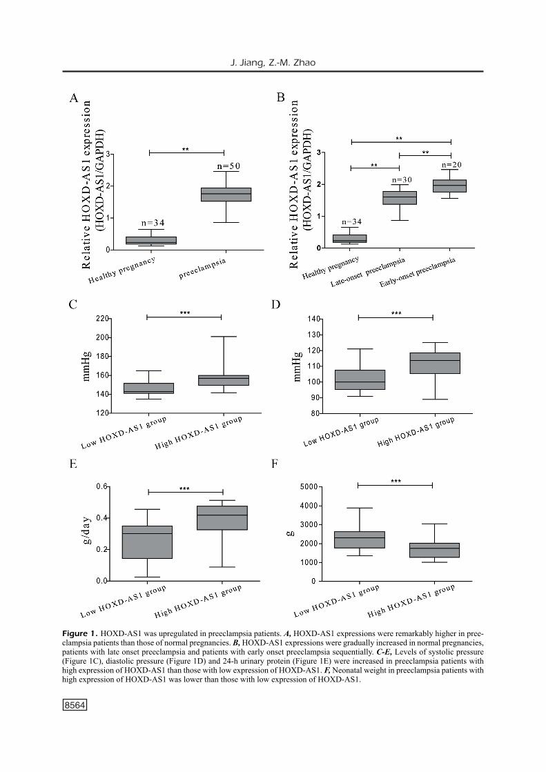
The expression of lncRNA HOXD-AS1 in the placenta tissues was detected by qRT-PCR. Our results demonstrated that HOXD-AS1 expres-sions were remarkably higher in placental tissues of preeclampsia patients than those of normal pregnancies (Figure 1A). Besides, HOXD-AS1 expressions were gradually increased in normal pregnancies, patients with late onset preeclampsia and patients with early onset preeclampsia sequen-tially (Figure 1B). Preeclampsia patients were fur-ther assigned into high and low expression group based on their HOXD-AS1 expressions. The data suggested that higher levels of systolic pressure (Figure 1C), diastolic pressure (Figure 1D) and 24-h urinary protein (Figure 1E) were observed in preeclampsia patients with high expression of HOXD-AS1 than those with low expression. Ad-ditionally, the neonatal weight in preeclampsia patients with high expression of HOXD-AS1 was remarkably lower than those with low expression (Figure 1F).
HOXD-AS1 Suppressed Cell Proliferation of Trophoblasts
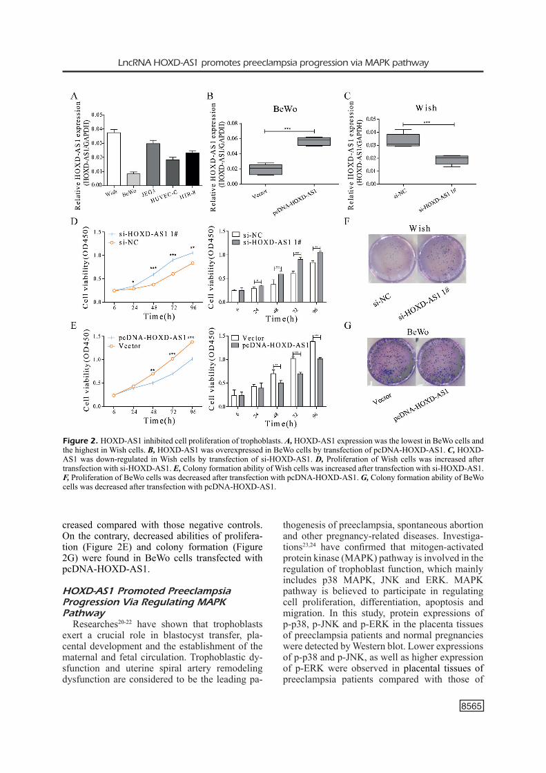
We detected HOXD-AS1 expression in 5 trophoblast cell lines by qRT-PCR. The results found that HOXD-AS1 expression was the lowest in BeWo cells and the highest in Wish cells (Fi-gure 2A). Therefore, BeWo cells and Wish cel-ls were selected for the following experiments. HOXD-AS1 was overexpressed in BeWo cells by transfection of pcDNA-HOXD-AS1 (Figure 2B), whereas HOXD-AS1 was down-regulated in Wish cells by transfection of si-HOXD-AS1 (Figure 2C). CCK-8 and colony formation assay showed that proliferative (Figure 2D) and colony formation abilities (Figure 2F) in Wish cells tran-sfected with si-HOXD-AS1 were remarkably in-
Table I. Clinical characteristics of normal and pre-eclamptic pregnacies
Healthy pregnancy PreeclampsiaVariable (n=34) (n=50) p-value
Maternal age (year) 31.12±4.03 31.54±3.59 >0.05Maternal weight (kg) 69.56±7.84 68.71±8.05 >0.05Proteinuria (g/day) 0.15±0.18 0.42±0.16 <0.05Systolic blood pressure (mm Hg) 113.04±18.32 148.31±13.55 <0.05Diastolic blood pressure (mm Hg) 76.31±19.13 107.89±17.35 <0.05Body weight of infant (g) 3317.13±389.16 1785.25±530.17 <0.05
J. Jiang, Z.-M. Zhao
8564
Figure 1. HOXD-AS1 was upregulated in preeclampsia patients. A, HOXD-AS1 expressions were remarkably higher in pree-clampsia patients than those of normal pregnancies. B, HOXD-AS1 expressions were gradually increased in normal pregnancies, patients with late onset preeclampsia and patients with early onset preeclampsia sequentially. C-E, Levels of systolic pressure (Figure 1C), diastolic pressure (Figure 1D) and 24-h urinary protein (Figure 1E) were increased in preeclampsia patients with high expression of HOXD-AS1 than those with low expression of HOXD-AS1. F, Neonatal weight in preeclampsia patients with high expression of HOXD-AS1 was lower than those with low expression of HOXD-AS1.
LncRNA HOXD-AS1 promotes preeclampsia progression via MAPK pathway
8565
creased compared with those negative controls. On the contrary, decreased abilities of prolifera-tion (Figure 2E) and colony formation (Figure 2G) were found in BeWo cells transfected with pcDNA-HOXD-AS1.
HOXD-AS1 Promoted Preeclampsia Progression Via Regulating MAPK Pathway
Researches20-22 have shown that trophoblasts exert a crucial role in blastocyst transfer, pla-cental development and the establishment of the maternal and fetal circulation. Trophoblastic dy-sfunction and uterine spiral artery remodeling dysfunction are considered to be the leading pa-
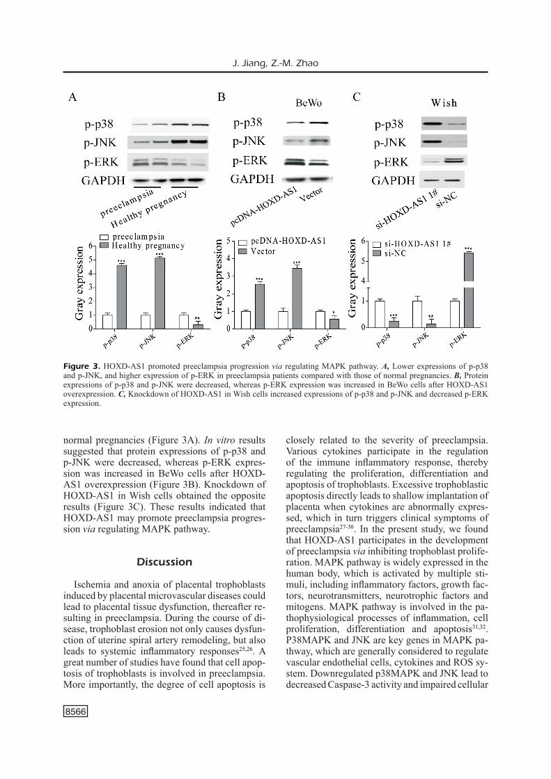
thogenesis of preeclampsia, spontaneous abortion and other pregnancy-related diseases. Investiga-tions23,24 have confirmed that mitogen-activated protein kinase (MAPK) pathway is involved in the regulation of trophoblast function, which mainly includes p38 MAPK, JNK and ERK. MAPK pathway is believed to participate in regulating cell proliferation, differentiation, apoptosis and migration. In this study, protein expressions of p-p38, p-JNK and p-ERK in the placenta tissues of preeclampsia patients and normal pregnancies were detected by Western blot. Lower expressions of p-p38 and p-JNK, as well as higher expression of p-ERK were observed in placental tissues of preeclampsia patients compared with those of
Figure 2. HOXD-AS1 inhibited cell proliferation of trophoblasts. A, HOXD-AS1 expression was the lowest in BeWo cells and the highest in Wish cells. B, HOXD-AS1 was overexpressed in BeWo cells by transfection of pcDNA-HOXD-AS1. C, HOXD-AS1 was down-regulated in Wish cells by transfection of si-HOXD-AS1. D, Proliferation of Wish cells was increased after transfection with si-HOXD-AS1. E, Colony formation ability of Wish cells was increased after transfection with si-HOXD-AS1. F, Proliferation of BeWo cells was decreased after transfection with pcDNA-HOXD-AS1. G, Colony formation ability of BeWo cells was decreased after transfection with pcDNA-HOXD-AS1.
J. Jiang, Z.-M. Zhao
8566
closely related to the severity of preeclampsia. Various cytokines participate in the regulation of the immune inflammatory response, thereby regulating the proliferation, differentiation and apoptosis of trophoblasts. Excessive trophoblastic apoptosis directly leads to shallow implantation of placenta when cytokines are abnormally expres-sed, which in turn triggers clinical symptoms of preeclampsia27-30. In the present study, we found that HOXD-AS1 participates in the development of preeclampsia via inhibiting trophoblast prolife-ration. MAPK pathway is widely expressed in the human body, which is activated by multiple sti-muli, including inflammatory factors, growth fac-tors, neurotransmitters, neurotrophic factors and mitogens. MAPK pathway is involved in the pa-thophysiological processes of inflammation, cell proliferation, differentiation and apoptosis31,32. P38MAPK and JNK are key genes in MAPK pa-thway, which are generally considered to regulate vascular endothelial cells, cytokines and ROS sy-stem. Downregulated p38MAPK and JNK lead to decreased Caspase-3 activity and impaired cellular
normal pregnancies (Figure 3A). In vitro results suggested that protein expressions of p-p38 and p-JNK were decreased, whereas p-ERK expres-sion was increased in BeWo cells after HOXD-AS1 overexpression (Figure 3B). Knockdown of HOXD-AS1 in Wish cells obtained the opposite results (Figure 3C). These results indicated that HOXD-AS1 may promote preeclampsia progres-sion via regulating MAPK pathway.
Discussion
Ischemia and anoxia of placental trophoblasts induced by placental microvascular diseases could lead to placental tissue dysfunction, thereafter re-sulting in preeclampsia. During the course of di-sease, trophoblast erosion not only causes dysfun-ction of uterine spiral artery remodeling, but also leads to systemic inflammatory responses25,26. A great number of studies have found that cell apop-tosis of trophoblasts is involved in preeclampsia. More importantly, the degree of cell apoptosis is
Figure 3. HOXD-AS1 promoted preeclampsia progression via regulating MAPK pathway. A, Lower expressions of p-p38 and p-JNK, and higher expression of p-ERK in preeclampsia patients compared with those of normal pregnancies. B, Protein expressions of p-p38 and p-JNK were decreased, whereas p-ERK expression was increased in BeWo cells after HOXD-AS1 overexpression. C, Knockdown of HOXD-AS1 in Wish cells increased expressions of p-p38 and p-JNK and decreased p-ERK expression.
LncRNA HOXD-AS1 promotes preeclampsia progression via MAPK pathway
8567
DNA33. In addition, ERK is activated by different external stimuli, thereby protecting cell apoptosis via the promotion of cell proliferation34-36. In the present study, higher levels of systolic pressure, diastolic pressure and 24-h urinary protein were observed in preeclampsia patients than those of normal pregnancies. Moreover, neonatal weight of preeclampsia patients was lower than that of normal pregnancies. By detecting key factors in MAPK pathway, we found increased expressions of p-p38 and p-JNK, as well as decreased ERK expression in placental tissues of preeclampsia pa-tients compared with those of normal pregnancies, which were regulated by HOXD-AS1. For in vitro experiments, transfection of pcDNA-HOXD-AS1 or si-HOXD-AS1 remarkably changed the proli-ferative and colony formation abilities via MAPK pathway, indicating that HOXD-AS1 is capable of regulating trophoblasts in preeclampsia patients.
Conclusions
We observed that HOXD-AS1 participates in the development and progression of preeclampsia by regulating trophoblast proliferation via MAPK pathway.
Conflict of InterestThe Authors declare that they have no conflict of interest.
References
1) CraiCi iM, Wagner SJ, WeiSSgerber TL, grande JP, garoviC vd. Advances in the pathophysiology of pre-eclampsia and related podocyte injury. Kid-ney Int 2014; 86: 275-285.
2) biLodeau JF. Review: maternal and placental an-tioxidant response to preeclampsia - impact on vasoactive eicosanoids. Placenta 2014; 35 Suppl: S32-S38.
3) Sibai b, dekker g, kuPFerMinC M. Pre-eclampsia. Lancet 2005; 365: 785-799.
4) ChaiWoraPongSa T, ChaeMSaiThong P, Yeo L, roMero r. Pre-eclampsia part 1: current understanding of its pa-thophysiology. Nat Rev Nephrol 2014; 10: 466-480.
5) Zhang Cg, Yin dd, Sun SY, han L. The use of ln-cRNA analysis for stratification management of prognostic risk in patients with NSCLC. Eur Rev Med Pharmacol Sci 2017; 21: 115-119.
6) PonTing CP, oLiver PL, reik W. Evolution and functions of long noncoding RNAs. Cell 2009; 136: 629-641.
7) Li JL, Li r, gao Y, guo WC, Shi PX, Li M. LncRNA CCAT1 promotes the progression of preeclampsia
by regulating CDK4. Eur Rev Med Pharmacol Sci 2018; 22: 1216-1223.
8) dong L, hui L. HOTAIR promotes proliferation, migration, and invasion of ovarian cancer SKOV3 cells through regulating PIK3R3. Med Sci Monit 2016; 22: 325-331.
9) Wu g, Cai J, han Y, Chen J, huang ZP, Chen C, Cai Y, huang h, Yang Y, Liu Y, Xu Z, he d, Zhang X, hu X, PineLLo L, Zhong d, he F, Yuan gC, Wang dZ, Zeng C. LincRNA-p21 regulates neointima for-mation, vascular smooth muscle cell proliferation, apoptosis, and atherosclerosis by enhancing p53 activity. Circulation 2014; 130: 1452-1465.
10) Sun L, Jiang C, Xu C, Xue h, Zhou h, gu L, Liu Y, Xu Q. Down-regulation of long non-coding RNA RP11-708H21.4 is associated with poor prognosis for colorectal cancer and promotes tumorigenesis through regulating AKT/mTOR pathway. Oncotar-get 2017; 8: 27929-27942.
11) Yang Mh, hu ZY, Xu C, Xie LY, Wang XY, Chen SY, Li Zg. MALAT1 promotes colorectal cancer cell proli-feration/migration/invasion via PRKA kinase anchor protein 9. Biochim Biophys Acta 2015; 1852: 166-174.
12) Zou Y, Jiang Z, Yu X, Sun M, Zhang Y, Zuo Q, Zhou J, Yang n, han P, ge Z, de W, Sun L. Upregulation of long noncoding RNA SPRY4-IT1 modulates proli-feration, migration, apoptosis, and network forma-tion in trophoblast cells HTR-8SV/neo. PLoS One 2013; 8: e79598.
13) grahaM a, PaPaLoPuLu n, kruMLauF r. The murine and drosophila homeobox gene complexes have common features of organization and expression. Cell 1989; 57: 367-378.
14) ahn d, ho rk. Tri-phasic expression of posterior Hox genes during development of pectoral fins in zebrafish: implications for the evolution of ver-tebrate paired appendages. Dev Biol 2008; 322: 220-233.
15) kouSSouLakoS S. Vertebrate limb development: from Harrison’s limb disk transplantations to targeted disruption of hox genes. Anat Embryol (Berl) 2004; 209: 93-105.
16) kruMLauF r. Hox genes in vertebrate development. Cell 1994; 78: 191-201.
17) neWMan Sa. Sticky fingers: hox genes and cell adhesion in vertebrate limb development. Bioes-says 1996; 18: 171-174.
18) SCoTT MP. Vertebrate homeobox gene nomencla-ture. Cell 1992; 71: 551-553.
19) ZakanY J, dubouLe d. The role of hox genes during vertebrate limb development. Curr Opin Genet Dev 2007; 17: 359-366.
20) kauFMann P, bLaCk S, huPPerTZ b. Endovascular trophoblast invasion: implications for the patho-genesis of intrauterine growth retardation and preeclampsia. Biol Reprod 2003; 69: 1-7.
21) boSe P, kadYrov M, goLdin r, hahn S, baCkoS M, re-gan L, huPPerTZ b. Aberrations of early trophoblast differentiation predispose to pregnancy failure: lessons from the anti-phospholipid syndrome. Placenta 2006; 27: 869-875.
J. Jiang, Z.-M. Zhao
8568
22) TorrY dS, Labarrere Ca, MCinTYre Ja. Uteropla-cental vascular involvement in recurrent spon-taneous abortion. Curr Opin Obstet Gynecol 1998; 10: 379-382.
23) ChakraborTY C, gLeeSon LM, MCkinnon T, LaLa Pk. Re-gulation of human trophoblast migration and invasi-veness. Can J Physiol Pharmacol 2002; 80: 116-124.
24) Wong Ch, Cheng CY. Mitogen-activated protein ki-nases, adherens junction dynamics, and sperma-togenesis: a review of recent data. Dev Biol 2005; 286: 1-15.
25) hung Th, SkePPer Jn, CharnoCk-JoneS dS, burTon gJ. Hypoxia-reoxygenation: a potent inducer of apoptotic changes in the human placenta and possible etiological factor in preeclampsia. Circ Res 2002; 90: 1274-1281.
26) braMhaM k, briLeY aL, Seed P, PoSTon L, Shennan ah, ChaPPeLL LC. Adverse maternal and perinatal outcomes in women with previous preeclampsia: a prospective study. Am J Obstet Gynecol 2011; 204: 511-512.
27) Shi Z, hou W, hua X, Zhang X, Liu X, Wang X, Wang X. Overexpression of calreticulin in pre-eclamptic placentas: effect on apoptosis, cell invasion and severity of pre-eclampsia. Cell Biochem Biophys 2012; 63: 183-189.
28) Jiang r, Teng Y, huang Y, gu J, Li M. Protein kina-se C-alpha activation induces NF-kB-dependent VCAM-1 expression in cultured human umbilical vein endothelial cells treated with sera from pre-eclamptic patients. Gynecol Obstet Invest 2010; 69: 101-108.
29) he g, Xu W, Chen Y, Liu X, Xi M. Abnormal apop-tosis of trophoblastic cells is related to the up-re-gulation of CYP11A gene in placenta of pree-clampsia patients. PLoS One 2013; 8: e59609.
30) Wang S, he g, Yang Y, Liu Y, diao r, Sheng k, Liu X, Xu W. Reduced expression of Enac in placenta tissues of patients with severe preeclampsia is related to compromised trophoblastic cell migration and inva-sion during pregnancy. PLoS One 2013; 8: e72153.
31) PeTrova i, SedMikova M, PeTr J, vodkova Z, PYTLoun P, ChMeLikova e, rehak d, CTrnaCTa a, raJMon r, Ji-Lek F. The roles of c-Jun N-terminal kinase (JNK) and p38 mitogen-activated protein kinase (p38 MAPK) in aged pig oocytes. J Reprod Dev 2009; 55: 75-82.
32) Liang r, niCkkhoLgh a, hoFFMann k, kern M, SChnei-der h, SobireY M, Zorn M, buChLer MW, SCheMMer P. Melatonin protects from hepatic reperfusion injury through inhibition of IKK and JNK pathways and modification of cell proliferation. J Pineal Res 2009; 46: 8-14.
33) ravindran J, guPTa n, agraWaL M, baLa ba, Lak-ShMana rP. Modulation of ROS/MAPK signaling pathways by okadaic acid leads to cell death via, mitochondrial mediated caspase-dependent me-chanism. Apoptosis 2011; 16: 145-161.
34) daviS rJ. Signal transduction by the JNK group of MAP kinases. Cell 2000; 103: 239-252.
35) dhanaSekaran dn, reddY eP. JNK signaling in apoptosis. Oncogene 2008; 27: 6245-6251.
36) WeSTon Cr, daviS rJ. The JNK signal transduction pathway. Curr Opin Cell Biol 2007; 19: 142-149.