Embed Size (px)
Citation preview

FATTY MATERIAL IN BACTERIA AND FUNGI REVEALEDBY STAINING DRIED, FIXED SLIDE PREPARATIONS'
KENNETH L. BURDONDepartment of Bacteriology and Immunology, Baylor University College of Medicine, Houston,
Texas
Received for publication September 3,1946
Following Hartman, who first suggested the use of Sudan black B, in place ofred Sudans, as a bacterial fat stain (Hartman, 1940), Burdon, Stokes, and Kim-brough (1942a) confirmed the greater value of this dye and modified the pro-cedure for demonstrating intracellular fatty material in bacteria by preparing,from suspensions of the organisms in alcoholic Sudan black B solution, driedfilms counterstained with safranine. Previously it had been thought that dried,fixed films were entirely unsuitable for fat stains (Lewis, 1941).These permanent films were regarded as an obvious improvement over the wet
preparations used by earlier workers, and they were shown to be of practical aidin the classification of aerobic sporeforming bacilli (Burdon, Stokes, and Kim-brough, 1942b), but the staining method still had a number of undesirablefeatures.
Further experimentation has resulted in the development of the much superiorprocedure to be described here. The new method is not only simpler, requiringno more effort than a gram stain, but it is also far more rewarding, for the filmsnow reveal clearly intracellular lipid matter that previously has not been seen oreven suspected. The improved stain has increased differential value. More.over, its application, to various bacteria has resulted in certain general findings ofunusual interest.
SUDAN BLACK B FAT STAIN FOR FIXED PREPARATIONS
Technique. 1. Prepare the lilm, let it dry thoroughly in the air, and fix it byheat in the usual way. (Chemical fixation has no special advantages and mayresult in some loss of demonstrable lipid.) 2. Flood the entire slide with Sudanblack solution (0.3 g of the powdered stain2 in 100 ml of 70 per cent ethyl aclohol),and allow the slide to remain undisturbed at room temperature for 5 to 15 min-utes. (A staining period of less than 5 minutes will often suffice, but the intra-
" Presented, in part, before the 45th General Meeting of the Society of American Bac-teriologists at New York, N. Y., May 3, 1944.
2The original, imported Sudan black B dye is not available commercially at the time ofwriting. But recently the National Aniline Division, Allied Chemical and Dye Corpora-tion, 40 Rector Street, New York 6, N. Y., has developed a duplicate of this stain, and thecompany promises to have a supply of the American-made Sudan black B on the marketshortly. In my hands this new product has given results equal to those obtained with theimported dye. The writer is indebted to Dr. H. J. Conn, president of the Biological StainCommission, for his kindness in making various dye samples available for comparativetests.
665
on July 9, 2018 by guesthttp://jb.asm
.org/D
ownloaded from

KENNETH L. BURDON
cellular lipid is colored somewhat more intensely when the staining is continuedfor 5 minutes or longer. No further staining apparently occurs after thesolution precipitates and turns a greenish or brownish color, but no harm is doneif the stain is allowed to dry completely over the- film. 3. Drain off excessstain and blot the slide thoroughly dry. 4. Clear the slide with cp xylQl bydipping it in and out of the solvent in a Coplin jar or by adding xylol from adropping bottle. Blot the cleared slide dry. 5. Counterstain with safranine(0.5 per cent aqueous solution) for 5 to 10 seconds (for ordinary bacteria or fungi),or with dilute carbol fuchsin (Ziehl's carbol fuchsin diluted 1:10 with distilledwater) for 1 to 3 minutes (for acid-fast organisms). (Overstaining with thecounterstain must be avoided). 6. Wash in water, blot, and dry the slide.
Comment. After the bulk of the dye has been dissolved, the Sudan black Bsolution should be thoroughly shaken at intervals, then allowed to stand over-night before use. It remains good for several months at room temperature,provided it is kept in a well-stoppered, chemically clean container.The entire slide is flooded with the staining solution to prevent the too rapid
evaporation that otherwise occurs. (For reasons not entirely understood, thestaining is generally unsatisfactory when slides are immersed in the Sudan blackB solution in a Coplin jar.) Since the cellular lipid in most organisms takes upthe characteristic blue-black color almost at once, it is possible to complete thewhole fat-staining procedure within a minute or two if desired. On the otherhand, if the technician is occupied with other tasks, the stain may simply beallowed to dry on the slide; then the clearing with xylol and counterstaining maybe carried out later at a more convenient time. If the Sudan black B solutionis allowed to stand on the slide for about 15 minutes and is then set afireby apply-ing the Bunsen flame to the fluid, followed by blotting and xylol-clearing as usual,the intracellular fat in some organisms (e.g., gonococci) is brought out moreclearly. Ordinarily, this step is unnecessary.
Examination of the cleared preparation without counterstaining is sometimesinteresting and revealing. Care should be taken'to avoid obscuring very tinyfat droplets by too strong a counterstain. Films must be examined with theoil immersion lens under critical illumination. To discern the smallest lipoidparticles the observer must have a good sense for the color distinction betweenthe blue-black or blue-gray of the fat droplets and the pink of the counterstain.
RESULTS OF APPLYING THE STAIN TO VARIOUS SPECIES OF BACTERIAAND FUNGI
Methods. The stain has now been applied to films from cultures of virtuallyall the chief species of bacteria, and of many fungi. In order to obtain a generalpicture of the occurrence of demonstrable intracellular lipid in these organismsand to permit a comparison between species in this respect, films were made fromall cultures at approximately the same stages of growth, i.e., in early maturity(12 to 24 hours old). For the purpose of this preliminary survey, the organismswere grown on the media customarily employed for routine cultures of the speciesconcerned, such as plain extract or infusion agar, potato slants, blood or ascitic
666 [VOL. 52
on July 9, 2018 by guesthttp://jb.asm
.org/D
ownloaded from

FATTY MATERIAL IN BACTERIA AND FUNGI
fluid agar (for pneumococci, streptococci, gonococci, etc.), Loeffler's serum slants(for the Corynebacterium group), blood agar slants or thioglycolate media (foranaerobes), coagulated egg media and glycerine agar (for acid-fast bacilli), andSabouraud's medium (for fungi). The majority of the films were made fromstock cultures, under conditions permitting accurate comparisons. A smallernumber of observations were made on preparations from primary (mixed)cultures, derived from the dust or from the throat, skin, etc., of human beings,and on direct films from the human mouth or from sputum, pus, and the like.
General findings. The films showed that fatty material staining with Sudanblack B is present, often in conspicuous amounts, in the great majority of micro-organisms, whether aerobic or anaerobic, saprophytic or parasitic, pathogenicor nonpathogenic, and that it is to be seen in a considerable number of bac-teria-such as the diphtheria bacilli, anaerobic sporeforming bacilli, and thecommon species of cocci-which have been described in the earlier literature aslacking any microscopically demonstrable fat (Lewis, 1941). The preparationsrevealed in a striking way the abundant lipoid matter in fungi of all kinds.
Intracellular lipid was observed in organisms in primary cultures, as well as inpure stock cultures, and some bacteria and fungi in films made directly from bodysurfaces or excretions were found to contain typical fatty inclusions..A high proportion of gram-positive bacteria were discovered to be fat storers.
Many familiar varieties of gram-negative bacteria, on the other hand, were shownto be free of stainable lipid when in aetive growth on common media. 'A ten-dency for saprophytic varieties to contain more fat than the parasitic species ofthe same genera was noted in certain cases, notably in Mycobacterium andCorynebacterium.
It was found that the appearance of distinct intracellular fat droplets, or otherSudan black B staining matter, in bacteria is not influenced directly by the pres-ence or absence of glycerol, or other fermentable carbohydrate, in the medium.Accumulation of the intracellular lipid is affected, however, in any medium by therapidity of growth, and if cell division is retarded, the relative amount of demon-strable fat is usually increased. In the case of both the aerobic and anaerobicsporeforming bacilli (genera BaciUus and Clostridium), the fatty material wasobserved to be reduced somewhat just before active spore formation began.A considerable amount remained, however, and often sizable fat droplets wereseen in the tags of protoplasm around incompletely free spores. If sporulationwas for any reason delayed, fatty substances continued to accumulate within thebacilli, and this material persisted indefinitely in situ, even after the stainablecytoplasm had disintegrated.The Sudan black B not only stained all cytoplasmic inclusions of lipoid na-
ture, but also colored parts of the cell structure (apparently the cytoplasmicmembrane) in the case of certain bacteria and fungi.The regularity with which fatty inclusions appeared, and the general pattern
exbibited by all the fat-staining material within the cells, were found to be re-markably constant for any one kind of organism. The fat-storing habits of aparticular species were not appreciably different in the numerous variant strains
1946] 667-
on July 9, 2018 by guesthttp://jb.asm
.org/D
ownloaded from
KENNETH L. BURDON
observed in this study, whether the variants were naturally encountered ordeliberately produced.Most impressive was the finding that, with only occasional exceptions, the
closer the relationship between varieties of bacteria in other respects, the morenearly alike was their content of stainable lipid.
Occurrence ofSudan blackB staining material in particular species. The variousorganisms observed in pure culture may be divided into three groups accordingto the results of this preliminry survey of their fat-staining propensities (table 1).
In one group (I) stainable intracellular lipid was present regularly in consider-able amounts in nearly all the mature cells whenever microscopic examinationwas made of the growth on the usual culture media. Included here are thelarger, common species of Bacillus; all representatives of the genera Clostridium,Corynebacterium, and Mycobacterium; Actinomyces species and the fungi; andsome of the more saprophytic gram-positive cocci. Also among the organismsregularly containing conspicuous fatty inclusions are a relatively few species ofgram-negative bacteria, including the nitrogen-fixing organisms (Azotobacterand Rhizobium), and, unexpectedly, such saprophytic species as Acetobacteraceti, Alkaligenes fecalis, and Spirillum rubrum.
In the case of other bacteria (group II) intracellular fat-staining material wasusually present, but the organisms in certain cultures on common media failedto show any fat. These organisms contained relatively small amounts of stain-able lipid at most. A clear distinction between these bacteria of group II andthose of group I may not be borne out by further investigations. It is convenient,however, to place in this second group, for the present purpose, such organisms asthe common, smaller-celled species of the genus Bacillus, the human and bovinetubercle bacilli, the familiar varieties of staphylococci, streptococci, and pneumo-cocci, and the gram-negative diplococci, which sometimes grow on the customarymedia without development of stainable lipid.No systematic attempt was made to discover the precise circumstances required
for the regular formation of intracellular fat by these group II organisms. Inci-dental observations indicated, however, that at least the majority of them willform characteristic fatty inclusions regularly when a suitable special medium isprovided. For example, Bacillus subtilis (Marburg) and its close relativesamong the aerobic sporeformers rarely show more than traces of fat when culti-vated on plain or glucose nutrient agar media. Because of this the writeroriginally classified these bacilli as "fat-negative" (Burdon, Stokes, and Kim-brough, 1942a). But intracellular lipid does appear in characteristic amountwhen cultures of the same strains are made on potato slants or on glucose starchagar. Similarly, Lactobacillus acidophilus apparently stores no fat in milk cul-tures, but the cells contained moderate amounts when they were grown ontomato juice agar slants.
Finally, the remaining species form a third group made up of bacteria thatapparently do not store demonstrable fatty material at all, as a rule, except thatone or two tiny droplets are sometimes to be seen in occasional cells (group III).Here are to be found virtually all the chief varieties of gram-negative bacilli,
668 [VOL. 52
on July 9, 2018 by guesthttp://jb.asm
.org/D
ownloaded from

FATTY MATERIAL IN BACTERIA AND FUNGI
TABLE 1An arbitrary grouping of bacteria andfungi according to their content of demonstrable
intracellular lipid(Based on preliminary observations of dried, fixed films stained with Sudan black B and
counterstains)
GROUP I. FATTY MATERiAL REGULARLY PRESENT IN CONsPIcUOus AMOUNTS IN NEARLYALL THE MATURE CELLS
Gram-positive bacteriaActinomyces bovis (1)*Actinomyces sp. (saprophytic) (3)Bacillus alvei (1)Bacillus anthracis (7)Bacillus cereus (65)Bacillus circulans (7)Bacillus megatherium (15)Bacillus mycoides (10)Clostridium botulinum (1)Clostridium histolyticum (2)Clostridium perfringens.(2)Cl9stridium 8epticum (1)Clostridium sporogenes (4)Clodtridium tetani (3)
Gram-negative bacteriaAcetobacter aceti (1)Alkaligenes fecalis (4)Azotobacter beijerinkii (2)Azotobacter chroococcum (2)
FungiAspergillus sp. (2)Blastomyces dermatitidis (2)Candida albicans (2)Coccidioides immitis (1)Cryptococcus neoformans (2)Epidermophyton floccosum (1)Histoplasma capsulatum (1)Hormodendrum pedrosoi (1)Microsporum gypseum (1)
Corynebacterium diphtheriae (17)Corynebacterium pseudodiphthericum (hoff-manni) (3)
Corynebacterium xerose (7)Gaffky tetragena (3)Mycobacterium "leprae" (11)Mycobacterium phlei (3)Mycobacterium smegmatis (2)Mycobacterium tuberculosis (avian) (3)Mycobacterium tuberculosis (cold-blooded
type) (2)Mycobacterium sp. (saprophytic) (8)Sarcina lutea (3)Staphylococcus citreus (2)Streptococcu8 faecalis (1)
Chromobacterium violaceum (2)Rhizobium leguminosarum (2)Spirillum rubrum (2)
Mucor sp. (2)Penicillium notatum (1)Penicillium sp. (3)Phialophora verrucosa (1)Rhizopus sp. (2)Saccharomyces cerevisiae (2)Sporotrichum schencki (1)Trichophyton mentagrophytes (1)Rhinosporidium seeberi (in tissue sections)
(1)GROUP II. FATTY MATERIAL USUALLY PRESENT, BUT SOMETIMES ABSENT IN CULTURES ON
CoMoMN MEDIAGram-positive bacteria
Bacillus mesentericus (>75) Mycobacterium tuberculosis (human) (8)Bacillus subtilis (Ford) (25) Staphylococcus albus (3)Bacillus subtilis (Marburg) (>30) Staphylococcus aureus (3)Diplocqocus pneumoniae (6) Streptococcus pyogenes (3)Lactobacillus acidophilus (2) Streptococcus salivarius (4)Mycobacterium tuberculosis (bovine) (3)
Gram-negative bacteriaBacillus brevis (16) Neisseria meningitidis (2)Neisseria catarrhalis (2) Neisseria pharyngis (3)Neisseria gonorrhoeae (4)
1946] 66f
on July 9, 2018 by guesthttp://jb.asm
.org/D
ownloaded from

-670 KENNETIU L. BURDON [VOL. 52
TABLE 1-Continued
GROUP III. FATTY MATERIAL USUALLY ABSENT, OR PRESENT IN TRACES ONLY IN A FEWCELLS
Gram-negative bacteriaAerobacter aerogene8 (2) Pasteurella tularen8is (1)Brucella abortus (2) Proteu8 vulgaris (OX19) (2)Brucella melitensis (2) Proteus sp. (12)Brucella suis (2) Pseudomonas aeruginosa (3)Eberthella typhosa (6) Pseudomonas fluorescens (2)Escherichia coli (2) Salmonella enteritidis (2)Escherichia communior (2) Salmonella schottmitlleri (2)Hemophilus influenzae (3) Salmonella sp. (4)Hemophilus pertussi8 (2) Serratia marcescens (3)Klebsiella mutabile (1) Shigella dysenteriae (2)Klebsiella pneumoniae (2) Shigella flexneri (26)Lssterella monocytogenres (1) Shigella sonnei (9)Pasteurella avi (1) Shigella sp. (35)Pasteurella equiseptica (1) Vibrio cholerae (3)Pasteurella pestis (2)
The figure in parenthesis indicates the number of separate, pure strains observed bythe writer to date.
including all members of the genera Brucella, Hemophilus, and Pasteurella, aswell as the several genera of enteric bacilli and the cholera vibrio.
It must be emphasized that this division of bacteria into three groups inrelation to intracellular fat is entirely arbitrary and provisional. It is based onfilms from cultures in routine media only. As the Sudan black B fat stain iseventually utilized by different investigators in connection with intensivestudies of various groups of bacteria under different conditions, new informationwill be forthcoming that may well require revision of these listings.
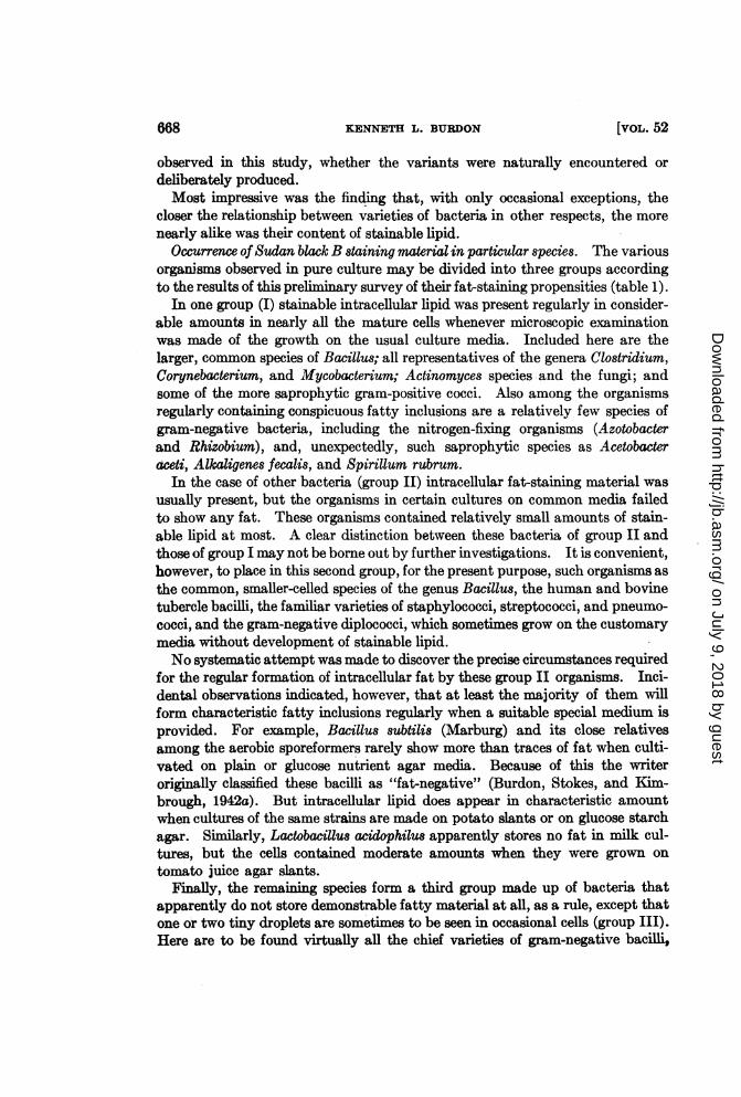
Characteristic intracellular distribution of the fatty material in important generaand species. The accompanying drawings (figures 1, 2, and 3) give some ideaof the usual picture when organisms of various kinds are fat-stained. Thesketches can convey only an inadequate conception of the striking appearancesactually seen under the microscope, since they lack completeness and full pre-cision in details, and especially since there is an absence of color.
It seems evident that Sudan black B stains more than neutral fat. It notonly imparts a dark blue-black color to distinctly outlined intracellular fatdroplets, but also gives a more or less intense bluish-gray tint to relatively large,ill-defined areas of cytoplasm in various bacteria and fungi. Moreover, incertain bacteria and most fungi, it stains intensely a thin, irregular, peripheralline, which is clearly part of the cellular structure, and presumably is the cyto-plasmic membrane.
Detailed descriptions will not be attempted here, but certain features of generalinterest will be pointed out.
Bacillus-MAl members of this genus that have been studied, including virulentstrains of B. anthracis, store fat (figure 1). Certain small-celled varieties, how-
r
on July 9, 2018 by guesthttp://jb.asm
.org/D
ownloaded from

FATTY MATERIAL IN BACTERIA AND FUNGI
ever, do not do so in every culture, and, moreover, even when stainable lipid ispresent in these bacilli, it is often difficult to discern. Most commonly the fatoccurs in these species at the ends of the rods.In the larger spore bearers, however, fat forms regularly and early as tiny drop-
lets along the periphery, which soon coalesce into larger drops and move into themiddle portions of the cell. The species that are otherwise closely similar (B.anthracis, B. cereus, and B. mycoides) all show large central droplets. B. megather-ium, on the other hand, presents a distinctly different picture. In this case rela-tively enormous amounts of lipid are present, even in cultures only a few hours old,and the deeply stained fatty material occurs almost entirely in the form of numer-ous relatively small droplets, which crowd the cytoplasm but show little tendencyto coalesce. The distinctive appearance makes this species easily recognizable.Clostridium-Considerable intracellular lipid is present in all the anaerobic spore
a~c.iIus
C1os+rridivym L orvyieb&c+er.w In
FIG. 1
bearers (figure 1). Characteristically, fat droplets occur at the extreme peripheryof the counterstained rod, or even appear to be just outside the cells.Corynebacterium-Abundant fatty material is revealed in all the diphtheria
bacillus group; it is especially conspicuous in C. xerose (figure 1). In this genus thefat stain outlines with bluish black the cell periphery and the cross septa so charac-teristic of the barred types of these bacilli. Lipid matter also appears as diffusemasses at the ends or edges of the cells.Mycobacterium-The general picture in the case of all varieties of the acid-fast
bacilli is essentially the same There are differences, however, in the number andsize of distinctly staining, intracellular fat droplets, and in the regularity withwhich these appear, among different varieties of these organisms (figure 3). Acharacteristic feature of the whole group is the tendency of the cells to take a lightbluish-gray color throughout. This is in addition to the appearance of distinct,dark-blue droplets of fat within many of the rods.The human and bovine tubercle bacilli often contain many fat granules, but since
1946] 671
on July 9, 2018 by guesthttp://jb.asm
.org/D
ownloaded from
KENNETH L. BURDON
we have observed a number of cultures showing only traces of fat, we have listedthese organisms in group II (table 1). Unfortunately, no features have been de-
&r&m Positive Cocc i
G Q9
C1s CO ac,.
Ne.isstri.
S /7f *^aQ d.
GxronNew. In+esfinal B&cilUi AIK. tecalis k1r ovsOUSci2Ik i
FIG. 2
IMycobac+eriuTn
r A
Acti-tiormyces
!;i.o 0N
40 Vol* 100e,rj) ft
Azo*ob x-e.+e
*
Rhizob
._.. _ .
V&riovs Fu vq OKI.fKL
FIG. 3
tected which would assist in differentiation of the human from the bovine type.The fat-containing rods have no relation to the "beaded" forms of tubercle bacilli(not infrequently seen after carbol fuchsin staining), whose significance was so long
672 [VOL. 52
on July 9, 2018 by guesthttp://jb.asm
.org/D
ownloaded from
FATTY MATERIAL IN BACTERIA AND FUNGI
debated. The "beads" have recently been shown to be artifacts (Porter andYegian, 1945; Lamanna, 1946).The most conspicuous fatty inclusions are seen in the frankly saprophytic acid-
fast strains, and in cultures labeled Mycobacterium leprae. (The bacilli in directpreparations from leprous lesions apparently contain no stainable fat. Thisdisparity in the appearance of the true Hansen's bacilli and the cultured, so-called"leprosy bacilli" when fat-stained is pointed out in a separate communication(Burdon, 1946).Azotobacter and Rhizobium-The extraordinary amount of lipid in the mature
cells of the nitrogen-fixing bacteria is revealed in a striking way (figure 3). Allstages in the process of accumulating the fat, from the first appearance of almostinvisible droplets at the edges of the cells to the final stage in which the entireorganism is solidly ffiled with fat-staining material, may be followed easily.
Cocci, Actinomyces, and Fungi-In cocci the Sudan black B staining materialmost frequently occurs as a dark bluish mantle around the circumference of thecells (figure 2). Some of it also appears as separate, small, internal droplets.In Actinomyces and the filamentous fungi (figure 3) thin strips of lipoid matter or
innumerable tiny fat droplets seem to line the hyphae throughout their length. Inaddition, sizeable round masses and irregular areas within the cytoplasm take thefat stain.In the yeastlike fungi much fat is also revealed, although it is apparently not as
abundant, as a rule, as it is in the mycelium-forming fungi.
Fat in ghost forms; "fatty degeneration." An observation of general interestis the frequent occurrence of fat globules within the "ghost forms" of bacteria,from which stainable cytoplasm is largely or wholly lost. Often these de-generated organisms are packed full of material that takes a dark, blue-blackcolor. These forms appear not only in those species that regularly store lipid,but also in varieties containing no demonstrable fatty inclusions when in theiractive, growing state. They have been seen, for example, in cultures of Pas-teurella and Klebsiella.
This appears to be a kind of "fatty degeneration." It is probably a phe-nomenon basically different from the storage of intracellular fat by activelymetabolizing cells. Further study is needed before the mechanisms involvedcan be understood.
Results of staining filmsfrom primary cultures andfrom body secretions. Micro-scopic examination of colonies of various kinds developing on agar plates ex-posed to dust showed that the sporulating bacilli and common fungi in suchprimary mixed cultures contain fat in a characteristic pattern, and that variousother fat-positive organisms are likely to be present. Many, but not all, of thebacteria in ordinary hay infusions, and similar mixed growths, were found tocontain fat.
Films made from the growth on blood agar plates inoculated with swabbingsfrom the human throat or nose often showed fatty material present in the usualform in various cocci, and in special abundance in diphtheroids. A few of thetubercle bacilli in films of tuberculous sputum were found to contain stainablelipid, and also fat droplets have been seen in the fusiform bacilli in direct prep-arations from patients with Vincent's angina. On the other hand, no fat hasbeen detected in gonococci in the several films of gonorrheal pus examined.Capsulated anthrax bacilli in direct preparations from the spleen or blood of
1946] 673
on July 9, 2018 by guesthttp://jb.asm
.org/D
ownloaded from
4KENNETH L. BURDON
animnals dying of natural or experimental infection were observed to contain onlya few very tiny fat droplets along their outer edges. In their very first growthon common media, however, these same organisms always develop character-istic large, centrally located fat granules.
EXTRACTION EXPERIMENTS WITH LEPOID SOLVENTS
As a preliminary step toward some understanding of the nature of the materialstained, experiments were carried out to test the loss of Sudan black B stainingmatter when bacteria are exposed to recognized fat solvents.
Method&. A majority of the extraction tests were carried out by exposingordinary fixed, but unstained, films to the solvents for varying lengths of time.A number of films were prepared at the same time on separate slides from cul-tures of various bacteria. These films were fixed by heat as usual, but leftunstained, except for one slide of each organism, which was fat-stained to serveas a control. The slides having unstained films were then immersed in thevarious solvents in Coplin jars. After certain time intervals a slide representingeach culture was removed from each of the solvents, stained by the routine pro-cedure, and examined for comparison with the controls. The principal solventsused were glacial acetic acid, 95 per cent ethyl alcohol, acetone, chloroform, andcarbon tetrachloride.A few tests were made by emulsifying the organisms in the solvents directly
from cultures. Films were then prepared and stained after varying timeintervals.
Results. A summary of the findings in several representative experimentsis presented in table 2. It is evident that the Sudan black B staining materialwas removed in large part from all the organisms by most of the solvents within72 hours or less. No other change in the microscopic appearance of the bacteriaoccurred. The rate of extraction varied, however, with different species, aswell as with different solvents.Carbon tetrachloride had scarcely any effect on the fat-staining material in the
six varieties of acid-fast bacilli tested, or in Corynebacterium diphtheriae, but itremoved the lipid matter from five species of aerobic sporeforming bacilli aboutas readily as the other solvents did. Chloroform also extracted the fatty ma-terial from the acid-fast bacilli and from the diphtheria bacillus with relativeslowness. The latter organism was the only species not cleared, or nearlycleared, of lipid by acetone within 72 hours. Most rapid extraction was broughtabout by alcohol, with glacial acetic acid a close second. These two solventsacted only slowly, however, on the closely related, large, chained bacilli B.anthracis and B. cereus.The most important influence in determining the extraction rate seemed to be
the relative ease with which the solvent was able to enter the bacterial cytoplasm.(It is supposed that this may depend, in part, upon the miscibility of the solventwith water and, in part, upon some peculiarity in the chemical nature of thebacterial cell membranes.) In the case of the slow removal of the lipid from thelarge bacilli mentioned above, the successive films showed clearly that the delay
[voL.52674
on July 9, 2018 by guesthttp://jb.asm
.org/D
ownloaded from

FAW2Y MATERIAL IN BACTERIA AND FUNGI
.0Ito
0
0
Ia
1 1I1 1 1
+ +++11-H++1 , I , , ,I I -,-1.1 ''''' 1111: III++++++1++++[ +j4 +
+ + - _ _ _ _ _+4+++ I+ +++
++
+++++++++- + +1 1111+-Hl4 + I+1- I+I I ++I____
+I+++++ . ++++++ +++~~~~~~~++
+1+-H+IH+ +++ +++-I I I I I II11+11 11+114
+++ +++ ++++ 1 ++ +++11+ +++11 ++ +++ +
+++
+ + + + +
1 1I++ 1 ++++ 11+
+ + ++1++ ~~~~++
1+0..
4._4. . . .
_ *K.@....
....¢0 0 0.
I I I I I +,
-^ ...s-. .
Z1 * - - - - .
1946]
+11
0
00
:-)
aS
014
1la
'0
4.
8
.I
0
I.
0
o.
0
¢Pe.qa
es
E-4.4.4.
-4.
cq P0
.t4)
675
*
++
ElbO
0@5
@5
ni
0
0
bO
5-
0_
++
-Ii0
00
H0
1++1+1 +
0 .
0.
*0.
. %.) -
* .eb % i
t.*., "~;.2-S--* X&
* *
W4
C4 + + + +
al. + + + + +
on July 9, 2018 by guesthttp://jb.asm
.org/D
ownloaded from

66KENNETH L. BURDON [
was due chiefly to the slowness with which the solvents were able to penetrateinto the cells so as to reach the fatty material. Of course, the relatively largeamount of lipid to be dissolved away from these organisms was a factor also.
DISCUSSION
The observations reported here serve chiefly to suggest the potential futureusefulness of the staining technique described. More detailed studies withparticular groups of organisms will have to be carried out with the aid of thestain before full answers will be obtained to the numerous questions about theintracellular lipid that come to mind.The origin and fate of fatty inclusions when they occur regularly in actively
metabolizing cells, and when they appear in degenerated forms, and the distinc-tion between fat storage as a useful accompaniment of cellular life and the mereaccumulation of lipid deposits ("fatty degeneration") are among the matters thatshould be investigated. From the limited study so far made it seems likelythat fat droplets in the cytoplasm originate at the cell periphery, and presumablyin some relationship to the cytoplasmic membrane, which itself is apparentlycolored by Sudan black B in many fungi and in some bacteria. This is in accordwith the views of Knaysi (1945, 1946), who has observed the formation of fatdroplets from the cytoplasmic membrane in living cells of Bacillus cereus. Weagree with this investigator that the function (if any) of the fatty inclusions isnot clear, and share his opinion that the usual conception of them as droplets of"reserve food material" is inadequate. Although we have noticed indicationsthat some of the intracellular fat is utilized in the late phases of cell growth, mostof it (under ordinary cultural conditions, at least) appears to remain unchanged,in situ, while the cytoplasm containing it disintegrates.The precise chemical nature of all the material stained in dried films by Sudan
black B is as yet unknown. The dye evidently colors not merely free fat butalso lipoid complexes. The extraction experiments demonstrate that recognizedlipid solvents will eventually remove all this material from intact bacterial cells,although the rate of extraction differs with the species as well as with the solvent.Study of successive preparations exposed for increasing lengths of time to fatsolvents shows that the Sudan black B staining material is truly within the cell,and is gradually reiaoved as the solvent penetrates the cell body. These ob-servations reinforce the conclusion that the substances that color with Sudanblack B are not merely surface deposits of stain or other artifacts, but are truecytoplasmic inclusions or integral parts of genuine cell structures. They affordnew evidence of the complexity of the bacterial cell (Dubos, 1945).Of special interest is the finding of definite fat droplets in anaerobic bacteria,
both sporeforming and nonsporeforming; this is contrary to the expectations ofsome authors (ImAenecke, 1945; Meyer, 1912).An obvious advantage of a fat-staining technique for ordinary dried films is the
unlimited opportunity afforded for direct comparisons, at leisure, with theappearance of parallel films of the same organisms treated with other dyes thatleave the intracellular lipid unstained. Such comparisons make very clear the
676 [VOiL. 52
on July 9, 2018 by guesthttp://jb.asm
.org/D
ownloaded from

FATTY MATERIAL IN BACTERIA AND FUNGI
important role of fatty inclusions in causing "vacuolation," distortion of thestainable cytoplasm pushed aside by the fat, and "irregular staining" generally.(For a recent study of such staining on the part of the glanders organism, see thearticle by Worley and Young, 1945.) Lewis (1941) and others have pointedout how the bizarre appearances of fat-containing organisms when colored withmethylene blue or other simple stains have led in the past to claims for the pres-ence in various bacteria of "nuclei," "gonidia," and other special structures,and to the idea that such organisms go through complex "life cycles." It issurely no accident that such claims have often been concerned with fat-richforms (e.g., Azotobacter, Rhizobium, and Bacitlus megatherium).
SUMfMARY
An improved technique for demonstrating intracellular lipid in microorganismsby staining dried, fixed preparations with Sudan black B and counterstains isdescribed.The application of this staining method to films of the principal species culti-
vated on common media revealed that stainable fatty material in the form ofcytoplasmic inclusions, or such material associated with structural elements ofthe cells, is present in all fungi and in the great majority of bacteria, whetherthese are aerobic or anaerobic, saprophytic or parasitic, pathogenic or non-pathogenic.A list is given in which the organisms studied are divided arbitrarily into
three groups according to their content of demonstrable lipid matter when grownon the media customarily employed for each kind of organism.With certain notable exceptions (e.g., Alcaligenes fecalis and Azotobacter),
fatty material was found to be more abundant and to occur with greater re-gularity in gram-positive bacteria than in gram-negative bacteria. An apparenttendency for saprophytic species to contain more fat than parasitic species wasnoted. Acid-fast bacteria were found to take a bluish-gray color throughout inmany instances; the rods were seen to contain distinct, deep-staining fat dropletsas well.
Intracellular lipid was discovered in organisms in primary (mixed) cultures,as well as in pure stock cultures, and a number of bacteria and fungi in directsmears from body surfaces and excretions were found to contain fatty material.Although media of special composition are required for the appearance of
stainable lipid in the case of certain bacteria, the formation of fatty inclusionswas shown not to be dependent upon the presence of glucose, glycerol, or otherfermentable carbohydrate in the medium.
Of greatest interest was the unexpected finding that the relative amount ofstainable fatty material and its form and location within the cells of bacteria areremarkably constant for any one kind of organism. Definite differences occuramong different kinds, however. Thus, the pattern of intracellular lipid ex-hibited in the stained films is to a considerable degree characteristic for thebacteria of a particular genus, and in some cases for those of a particular species.That this is not a mere happenstance is indicated by the further fact that,
1946] 677
on July 9, 2018 by guesthttp://jb.asm
.org/D
ownloaded from
KENNETH L. BURDON
throughout the whole phylum, bacteria of species or genera known to be closelyinterrelated show a marked similarity in their appearance when fat-stained.Indeed, with only occasional exceptions, the closer the relationship betweenvarieties of bacteria in other respects, the more nearly alike is their usual contentof stainable lipid. The formation of intracellular fatty material in a particularpattern is evidently a fixed habit in many bacteria, and one that must be associ-ated with something fundamental in the enzymatic or structural make-up of theorganisms.The simplicity of the staining method suggests its routine use in the character-
ization of microorganisms. -Its value as an aid in the differentiation of speciesand genera will be more fully established by further trials. Its possible useful-ness in practical diagnostic work is largely unexplored. The stain should proveespecially helpful in future investigations of bacterial cytology and metabolism.
REFERENCES
BuWoDN, K. L. 1946 Disparity in appearance of true Hansen's bacilli and cultured"leprosy bacilli" when stained for fat. J. Bact., 52, 679-680.
BuEDON, K. L., STOKES, J. C., AND KIMBROUGH, C. E. 1942a Studies of the commonaerobic spore-forming bacilli. I. Staining for fat with Sudan black B-safranin. J.Bact., 43, 717-724.
BURDON, K. L., STOKES, J. C., AND KImBROUGH, C. E. 1942b Studies of the commonaerobic spore-forming bacilli. II. Fermentation reactions in brom-cresol-purple agarbutt-slants. J. Bact., 44, 163-168.
DuBos, R. J. 1945 The bacterial cell. Harvard Univ. Press, Cambridge, Mass.HARTMAN, T. L. 1940 The use of Sudan black B as a bacterial fat stain. Stain Tech., 15,
23-28.IMBENECKI, A. 1945 On the structure of anaerobic bacteria. J. Bact., 49, 1-5.KNAYSI, G. 1945 Onthe origin and fate of the fattyinclusions in a strain of Bacillu8cereus.
Science, 102, 424.ENAYSI, G. 1946 On the existence, morphology, nature, and functions of the cytoplasCic
membrane in the bacterial cell. J. Bact., 51, 113-121.LAMANNA, C. 1946 The nature of the acid-fast stain. J. Bact., 52, 99-103.LEWIS, I. M. 1941 The cytology of bacteria. Bact. Revs., 5, 181-236.MEYER, A. 1912 Die Zelle der Bakterien. G. Fischer, Jena.PORTER, K. R., AND YEGIAN, D. 1945 Some artifacts encountered in stained preparations
of tubercle bacilli. III. Much granules and beads. J. Bact., 50, 563-575.WORLEY, G., JR., AND YOUNG, G. 1945 The glanders organism with reference to its cell
inclusions. J. Bact.. 49. 97-100.
678 [VOL. 52
on July 9, 2018 by guesthttp://jb.asm
.org/D
ownloaded from

![ProductDataSheet FullSize v4 - Novus Biologicalsamino acids 530 - 825 of the mouse HIF-1 alpha protein [Uniprot# Q61221]. ... Rinse the blot in water to remove excess stain and mark](https://img.pdfslide.us/doc/110x75/60c63cd9fffccc64a650c960/productdatasheet-fullsize-v4-novus-biologicals-amino-acids-530-825-of-the-mouse.jpg)