Embed Size (px)
Citation preview

Liver Micronucleus Test (LMNT) Subgroup
6th IWGT
Iguassu, Brazil
Chair: Yoshifumi Uno (Mitsubishi Tanabe Pharma)
Co-chair: Takeshi Morita (NIHS)
Rapporteur: Mirjam Luijten (RIVM)
Members:
Carol Beevers (Covance)
Shuichi Hamada (Mitsubishi Chemical Medience)
Satoru Itoh (Daiichi Sankyo)
Wakako Ohyama (Yakult)
Hironao Takasawa (Mitsubishi Chemical Medience)
(Makoto Hayashi (BSRC): absent, but joined in our
pre-meeting discussion)

MNT was originally established in bone marrow (BM), because the
cell turnover is quick, and this property enables us to sensitively
detect clastogens and aneugens with BMMNT.
Hepatocyte (HEP) turnover is slow. Thus the history of LMNT
development is how HEP proliferation is accelerated.
1980-:
Partial hepatectomy (PH) method: stimulation of HEP proliferation
Mitogen method (now not used): stimulation of HEP proliferation
1996-:
Juvenile/young rodent (JR) method: testing in liver growth phase
2012-:
Repeated dose (RD) method: waiting for MN-HEP accumulation
History of LMNT Development
6th IWGT
Iguassu, Brazil
6th IWGT
Iguassu, Brazil
Summary of LMNT Discussion in Previous IWGT
In 2nd IWGTP at Washington DC, 1999 (Hayashi et al., 2000), • Rodent LMNT with partial hepatectomy (PH) or mitogen treatment (e.g.,
4-AAF, CCl4) is mainly used and reported.
• LMNT with juvenile/young rats is currently reported.
• LMNT is one of the useful method to detect or to evaluate genotoxic
carcinogens that specially induce liver tumors.
In 4th IWGT at San Francisco, 2005 (Hayashi et al., 2007), • Young (4-week-old) rat liver can metabolize important xenobiotic
chemicals, e.g., heterocyclic hydrocarbons/ amines, nitrosamines,
almost as effectively as in adult rats.
• Thus LMNT with young rats offers an alternative methodology to the use
of PH or mitogenic stimulation.
• But not many articles have been published after the last survey in 2nd
IWGTP. One of the possible reasons might be that identification of
aneugenesis detected by the FISH method or the use of simpler methods
that determine DNA damage may be being used more frequently than
cytogenetic analysis or measurement of mutation per se.

6th IWGT
Iguassu, Brazil
More Current Activity of LMNT after 4th IWGT
Many new data have been published/presented after
4th IWGT.
One of the reasons of this trend was that 2nd in vivo
tests were focused in ICH-S2(R1) discussion. The
2nd tests have to be done with tissues other than
bone marrow (BM)/peripheral blood (PB).
JEMS/MMS organized a collaborative study of LMNT
with jevunile rats, and the results were published.
A collaborative study of LMNT in repeated dosing of
chemicals to rats is on-going in JEMS/MMS, and the
data were reported at some (J/E)EMS meetings.
New LMNT data with PH were also published after
4th IWGT.

6th IWGT
Iguassu, Brazil
1. To reach recommended protocol(s) for LMNT
with PH, JR and RD methods.
2. To clarify strengths and limitations of each
method.
3. To identify promising micronucleus tests in
tissues other than liver, for example GI tracts.
Purpose of LMNT Subgroup at 6th IWGT - The First Discussion about Practical Aspects -

1. PH method Rodents are administered once or twice before and/or after PH,
and MN-HEPs are examined on 4 days after PH in once dosing or
2 and 3 days after PH in twice dosing.
2. JR method JR (e.g., about 4-week-old rats) are administered once with a test
chemical, and MN-HEPs are examined on 4 days after the dosing.
3. RD method Young adult rodent (e.g., 6-week old rats) are administered for 2
or more weeks (e.g., 4 weeks) with a test chemical, and MN-HEPs
are examined in the next day of final dosing. This method is
intended to be integrated into general toxicity study.
Outlines of LMNT Methods
6th IWGT
Iguassu, Brazil

Genotoxicity: Positive
Genotoxicity: Negative
Rat Hepato-
Carcinogenicity
Negative
11/12 = 91%
Positive
Rat Hepato-
Carcinogenicity
Positive
Concordance of JR-LMNT: Takasawa, et al.
“Genotoxic” is defined as “Ames-Positive” or
“Bone Marrow MN-Positive” in this presentation.
Genotoxic Hepatocarcinogen
in Rats
MMC (i.p.): positive
Genotoxic Non-Liver
Carcinogen in Rats
9/10 = 90% Negative
Bromobenzene is equivocal: this
chemical is reported to produce
DNA adducts in liver.
4/5 = 80% Negative
Non-Genotoxic Non-Liver
Carcinogens in Rats
Clofibrate, DEHP, Thioacetamide.
3/3 = 100% Negative
Non-Genotoxic Rat
Hepatocarcinogens

Rat
Carcinogenecity
Rat Liver
Carcinogenecity
Repeated Dose
Liver
MN Assay
Repeated Dose
Bone marrow
MN Assay
Bone marrow
+
Liver MN Assay
DMN + + + - +
NPYR + + + - +
MDA + + + + +
NDPA + + + - +
2,4-DNT + + + - +
2,6-DNT + + + - +
QUN + + + - +
DAB + + + + +
2-NP + + + - +
MCT + + + - +
NMOR + + + - +
2-AAF + + + + +
Sudan I + + - + +
TAA + + - - -
CFB + + + - +
MP + + + - +
MMC + per + + +
CP + ub, lym, ner - + +
KBrO3 + kid, per, thy - + +
MNNG + eso, smi, sto - + +
MMS + nas, ner + + +
KA + thy - - -
Chemicals
A-1
B
A-2
C
RD-LMNT results are closely consistent with hepatocarcinogenicity, while
RD-BMMNT gave negative results for almost all hepatocarcinogens.
Almost all carcinogens were detectable with combination of RD-BM/LMNT.
RD-LMNT: Results of 22 Test Chemicals - Collaborative Study in JEMS/MMS: Hamada, et al. -

6th IWGT
Iguassu, Brazil
Usefulness of LMNT: Consensus in IWGT
The end-point is mutagenicity in chromosomes.
It is different from DNA damage in Comet/alkaline
elution/UDS tests and gene mutation in TG rodents.
LMNT is expected to detect mutagens that require
metabolic activation, e.g., 2,4-diaminotoluene was
mostly reported as negative in BM- and/or PB-MNT,
but as positive in LMNT.
In the practical aspects, LMNT has an advantage of
flexibility in tissue-sampling time compared with
BM/PB-MNT, because MN hepatocytes seem to
remain for a long time although BM/PB-MN cells
decrease in a short period.

Method Strengths Weaknesses
Partial hepatectomy
(PH)
22 substances
tested
• Aneugens detected
(provided dosing occurs
after PH)
• Combine with PB-MN
• Sensitivity/specificity
seems high
• Technical challenge
• Animal welfare
considerations
• Method may affect
toxicity other organs:
integration difficult
Juvenile rat
(JR)
30 substances
tested
• Combine with PB-MN
• Relatively easy method
• Sensitivity/specificity
seems high
• Metabolism perhaps
not fully comparable to
adult animals?
• Insensitive to aneugens
using ‘short’ protocol
Repeated dose
(RD)
22 substances
tested in
collaborative study
• Integration with 28-day
toxicity study
• Combine with BM-MN
• Usefulness of analysis
later when necessary
• Sensitivity/specificity
seems high
• Questionable whether
method is suitable to
detect aneugens
(tolerance)
LMNT Methods: Pros & Cons 6th IWGT
Iguassu, Brazil
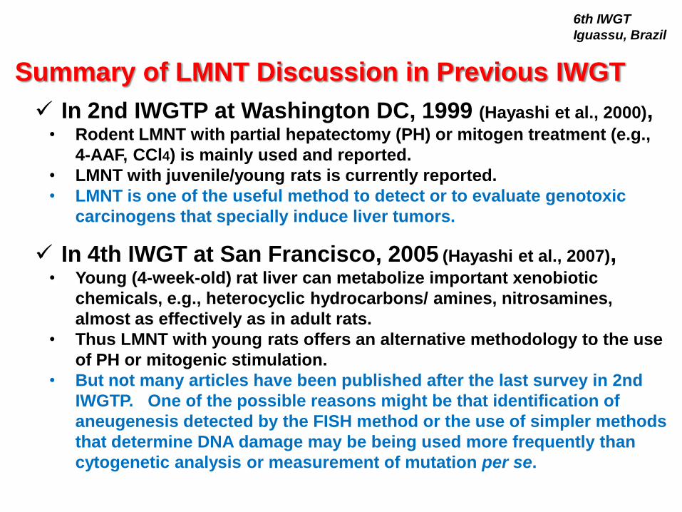
Protocol Discussion
1. The following items were discussed and reached to consensus
i. Test substance
ii. Dose level
iii. Positive control (except for a few issues: see next)
iv. Test animal (except for a few issues: see next)
v. Dosing procedure (except for a few issues: see next)
vi. Sampling time (except for a few issues: see next)
vii. Cell preparation
viii. Slide preparation
2. The following could not be fully discussed due to limited time
but will be noted in the IWGT meeting report to show methods
already used.
i. Slide observation
ii. Statistics and Judgment
6th IWGT
Iguassu, Brazil

General
Need data on toxic, non-carcinogenic, substances
Need data on other routes of administration:
dietary exposure, drinking water
Need data to decide on number of hepatocytes to be counted
PH method
Sampling time (in twice dosing): need for 1 or 2 timepoints?
JR method
Suitable age range animals
Need data for sampling time after 7 days
RD method
Positive control: need for concurrent control, duration of dosing
positive control?
Suitable age range animals
Focus Further Research
6th IWGT
Iguassu, Brazil

Most likely situations to select and use LMN test:
1. As 2nd in vivo test in case:
• relevant positive result in in vitro mammalian cell
genotoxicity test (especially under the condition of
metabolic activation)
• negative result in in vivo BM/PB-MN test
2. Suitable in vivo follow-up test when suspecting aneugen
(based on in vitro MN test)??
When to Use LMNT?
RD-LMNT may be the easiest method to integrate into a standard
general toxicity study
6th IWGT
Iguassu, Brazil

Performance (especially specificity) of the LMNT?
How to assess systemic exposure? Plasma levels
sufficient?
How to assess cytotoxicity upon exposure?
How to demonstrate proliferation of the hepatocytes?
In addition,
MN tests in other tissues, especially in GI tract (glandular
stomach & colon), seem promising. Further research
needed.
Discussion Points
6th IWGT
Iguassu, Brazil

Back-up slides
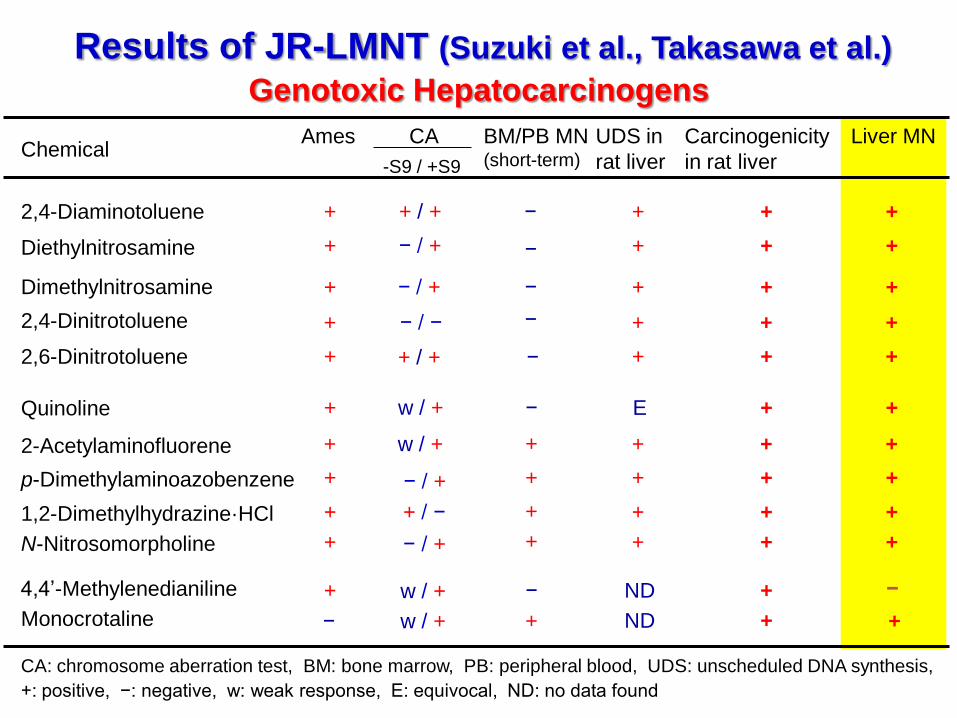
Results of JR-LMNT (Suzuki et al., Takasawa et al.)
Genotoxic Hepatocarcinogens
2-Acetylaminofluorene
2,4-Diaminotoluene
Diethylnitrosamine
p-Dimethylaminoazobenzene
Dimethylnitrosamine
2,4-Dinitrotoluene
4,4’-Methylenedianiline
Quinoline
2,6-Dinitrotoluene
Chemical BM/PB MN (short-term)
UDS in
rat liver
Liver MN Carcinogenicity
in rat liver
Ames
+ + +
+ + − +
+ + +
+ + + +
+ + +
+ + +
+ + +
+ E − +
+ − ND − +
1,2-Dimethylhydrazine·HCl + + + +
CA
w / +
+ / +
− / +
− / +
− / −
w / +
+ / −
-S9 / +S9
+
CA: chromosome aberration test, BM: bone marrow, PB: peripheral blood, UDS: unscheduled DNA synthesis,
+: positive, −: negative, w: weak response, E: equivocal, ND: no data found
N-Nitrosomorpholine + + + + − / +
+ / +
−
−
−
−
− / +
w / +
Monocrotaline + ND w / + − + +
+
+
+
+
+
+
+
+
+
+

RD-LMNT: Profile of 22 Test Chemicals - Collaborative Study in JEMS/MMS: Hamada, et al. -
Ames CA Single 4W Liver Others
Dimethylnitrosoamine (DMN) + + - ND + kid, lun, vsc, tes
N-Nitrosopyrrolidine (NPYR) + ND - ND + kid, vsc, tes
4,4'-Methylenedianiline (MDA) + + - ND + thy
N-Nitrosodipropylamine (NDPA) + + - ND + eso, nas
2,4-Dinitrotoluene (2,4-DNT) + - - ND + ski, mgl
2,6-Dinitrotoluene (2,6-DNT) + + - ND + -
Quinoline (QUN) + + - ND + -
p-dimethylaminoazobenzene (DAB) + + - ND + -
2-Nitropropane (2-NP) + + - ND + -
Monocrotaline (MCT) - + + - + -
N-Nitrosomorpholine (NMOR) + + + ND + vsc
2-Acetylaminofluorene (2-AAF) + + + + + ski
C.I.solvent yellow 14 (Sudan I) + - + ND + -
Thioacetamide (TAA) - TC + ND + -
Mitomycin C (MMC) + + + - - per
Cyclophosphamide H2O (CP) + + + + - ub, lym, ner
Potassium bromate (KBrO3) + + + + - kid, per, thy
1-Methyl-3-nitro-1-nitrosoguanidine (MNNG) + + + ND - eso, smi, sto
Methyl methanesulfonate (MMS) + + + + - nas, ner
Kojic acid (KA) + E E ND - thy
Crofibrate (CFB) - + ND ND + pan
Methapyrilene HCl (MP) E + ND ND + -
Chemical (Abbreviation) Rat carcinogenecity
Literature
C
A-1
in vivo MN
(BM/PB)in vitro
B
A-2
mgl: mammary gland; eso: esophagus; kid: kidney; vsc: vascular system; orc: oral cavity; sto:stomach; lun: lung; tes: testes; thy:
thyloid gland; nas: nasal; ski: skin; col: colon; per: peritoneal cavity; ub: urinary bladder; lym: lymphocyte; ner: nervas

Protocol Discussion about Major Issues
i. Summary of LMNT protocols: consensus items in
pre-meeting discussion
ii. Major issues picked up in pre-meeting discussion
A) Protocol issues
a. Positive control
b. Sampling time (to detect aneugens)
c. Scoring number of hepatocytes per animal
B) Other concerns on strategy issues
6th IWGT
Iguassu, Brazil

A. Test substance
6th IWGT
Iguassu, Brazil
The following properties of test substance are mentioned in the study
protocol/report when available: identification, supplier, batch/lot,
physical nature, and purity.
A test substance is dissolved or suspended in an appropriate
solvent/vehicle, e.g., water, aqueous solution such as methylcellulose,
or corn/olive oil.
In RD method, it is recommended that, wherever possible, the use of
an aqueous solution/suspension should be considered first, followed
by consideration of a solution/suspension in oil (ref. The OECD
guidelines for the testing chemicals 407, repeated dose 28-day oral
toxicity study in rodents).
The solvent/vehicle is used as a negative control.

B. Dose level
6th IWGT
Iguassu, Brazil
PH and JR methods (ref., ICH-S2(R1) guideline): Typically three dose
levels are used. The top dose is a limit dose of 2000 mg/kg, if this is
tolerated, or a maximum tolerated dose defined as the dose
producing signs of toxicity such that higher dose levels, based on the
same dosing regimen, would be expected to produce lethality. Lower
doses are generally spaced at approximately two or three fold
intervals below this.
RD method: Typically three dose levels are used. The top dose is a
limit dose of 1000 mg/kg for studies of 14 days or longer, if this is
tolerated (OECD 407). Dose levels are set suitably in consideration of
general toxicity, but at least one dose level showing no hepatotoxicity
should be included because toxicity to the liver may decrease
micronuclei (e.g., monocrotaline).
Note: Blue letters indicate specific recommendation in LMNT.

C. Test animal 6th IWGT
Iguassu, Brazil
Species:
• Rodents are generally used (no data are available for non-rodents).
• PH method: Rats and mice can be used, but rats may be preferable
due to an easiness of PH.
• JR and RD methods: Rats are usually used, although there are a few
publications using juvenile mice.
Sex:
• PH and JR methods (ref., ICH-S2(R1)): If sex-specific drugs are to be
tested, then the assay can be done in the appropriate sex. In vivo
tests with the acute protocol can generally be carried out in only
one sex. For acute tests, both sexes should be considered only if
any existing toxicity, metabolism or exposure (Cmax or AUC) data
indicate a toxicologically meaningful sex difference in the species
being used. Otherwise, the use of males alone is considered
appropriate for acute genotoxicity tests.
• RD method: Males are used. No published data are available for
females, but it is assumed that females can be used if justified.
Strain: Any strains are acceptable if justified. The following are used
in published papers: Wistar, SD and F344 rats for PH method, SD and
F344 rats for JR method, and SD rats for RD method. In PH methods,
Wistar or SD rats may be preferable because of easiness of post-
surgical care compared with F344 rat due to the size of animals.

C. Test animal (continued) 6th IWGT
Iguassu, Brazil
Age of (initial) dosing:
• PH method: 7-12 weeks old.
• JR method: approximately 4 weeks old may be recommended.
Younger rats (e.g.. 5 days old) are reported to be used, but
justification of metabolism equivalency to adult rats may be needed
when used.
• RD method: 6 or more weeks old may be recommended, because
such aged animals are generally used in general toxicity studies as
RD method is intended to be integrated into general toxicity studies,
although data using 6-week-old rats are only available at the present
moment.
Number of animals per group: Five or more animals are usually used.
Power-calculation would be required to justify the number of animals.
In PH method, a few animals may be additionally provided for a
technical error, especially in PH.
Animal maintenance: Animals should be properly maintained in
ethical consideration of animal welfare.

D. Dosing procedure 6th IWGT
Iguassu, Brazil
Dosing route: Any routes are acceptable if justified. The following are
applied in published papers: oral gavage, s.c., i.v. and drinking water
dosing for PH methods, and oral gavage for JR and RD methods. I.p.
injection may not be recommended, especially in case of dosing after
PH.
Dosing schedule:
• PH method: single or twice dosing. Dosing of 1 day before PH is
preferable to detect clastogens than dosing of 1 day after PH.
Dosing of 1 day after PH is essential and recommended to detect
aneugens. Dose day 1 and day 3, and PH day 2 regimen can detect
both types (Sample groups on Day 4 and Day 5). These three
methodologies are equally recommended based on lab experiences.
In single dosing, dosing and PH schedules are selected in
consideration of the mode of actions of a test chemicals that is
estimated with the other in vitro/in vivo genotoxicity test results.
When both clastogenic and aneugenic effects are anticipated, both
dosing and PH regimens should be applied.
• JR method: Twice dosing in 24-hour-interval.
• RD method: Once daily for 14 or 28 days.
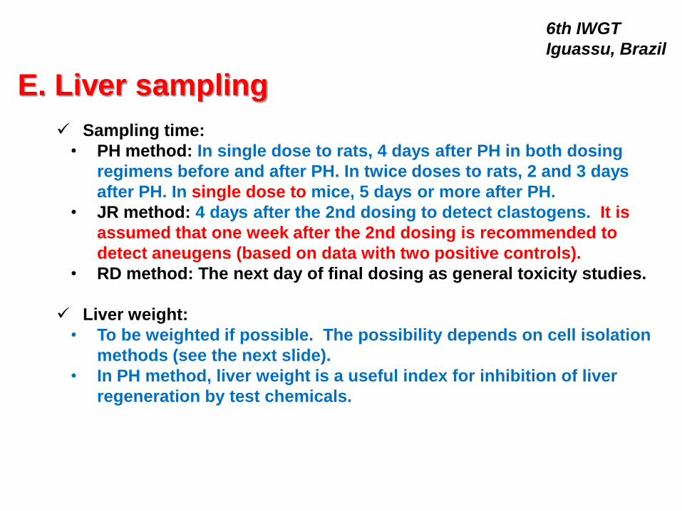
E. Liver sampling
6th IWGT
Iguassu, Brazil
Sampling time:
• PH method: In single dose to rats, 4 days after PH in both dosing
regimens before and after PH. In twice doses to rats, 2 and 3 days
after PH. In single dose to mice, 5 days or more after PH.
• JR method: 4 days after the 2nd dosing to detect clastogens. It is
assumed that one week after the 2nd dosing is recommended to
detect aneugens (based on data with two positive controls).
• RD method: The next day of final dosing as general toxicity studies.
Liver weight:
• To be weighted if possible. The possibility depends on cell isolation
methods (see the next slide).
• In PH method, liver weight is a useful index for inhibition of liver
regeneration by test chemicals.

F. Cell preparation 6th IWGT
Iguassu, Brazil
Cell preparation method: Three methods can be applied to LMNT, i.e.,
an in situ collagenase perfusion method via the portal vein (Seglen,
1976), a simplified method for liver perfusion (Igarashi & Shimada,
1997), or a method without in situ perfusion via the portal vein
(Narumi et al., 2012). The latter two methods are recommended
because the liver can be weighted.
Enzyme digestion: Type IV collagenase is used in all cell preparation
methods. Any supplier and unit are acceptable, if suitable specimens
can be prepared for MN observation. This is a key step for good
hepatocyte isolation, therefore, to examine and find a suitable
combination of supplier and unit is recommended before examining a
test chemical (100-125 units/mL may be used in general).
Hepatocyte suspension: Hepatocyte suspension is centrifuged at 50 x
g for 1-2 min. The sediment is suspended and fixed in a sufficient
volume of 10 vol% neutral buffered formalin (for.) and centrifuged at
50 x g for 1-2 min. This procedure is repeated once in Narumi’s
method or al least two times in Igarashi’s method. The sediment is
suspended in a small volume of for. (e.g., a volume ratio of 1:3-4).
This suspension can be stored for 1 year in room temp. and 3 years in
a refrigerator.

G. Slide preparation
6th IWGT
Iguassu, Brazil
Staining solution: A mixture of acridine orage (AO) and 4’,6-
diamidino-2-phenylindole dihydrochloride (DAPI) is used. AO and
DAPI are separately dissolved in purified water at 1 mg/mL and 20
µg/mL, respectively. These are mixed at the same volumes, and used
as a staining solution. The staining solution should be stored in a
refrigerator and used within one month.
Staining procedure: Just prior to the microscopic observation, the
hepatocyte suspension is mixed and stained with the same volume of
the AO-DAPI staining solution. The mixture is spread onto a clean
glass slide for microscopic observation. One or two slides per animal
are prepared. The slides should be randomized for observation.

H. Slide observation
6th IWGT
Iguassu, Brazil
Observation: The slides are observed with a fluorescence microscope
at magnification X400 or more. At least one slide per animal is scored.
At least 2000 hepatocytes (HEPs) excluding metaphase cells and
nuclear fragment cells are analyzed per animal: TO BE DISCUSSED
ABOUT THE NUMBER OF CELLS EXAMINED.
Parameter: The number of micronucleated hepatocytes (MNHEPs) is
examined, and the incidence of MNHEPs (%) is calculated. The
numbers of mononucleated hepatocyte, binucleated hepatocyte and
multinucleated hepatocyte (more than 3 nuclei) are also scored in PH
method, because those are a good indicator for inhibition of liver
regeneration. Mitotic index (MI) of HEPs is not recommended to
evaluate the inhibition of HEP proliferation based on lab experiences
in PH method and due to very low spontaneous MI in JR and RD
methods.

I. Statistics and Judgment
6th IWGT
Iguassu, Brazil
Statistics: There is no consensus method for LMNT at the present
moment. The following are reported to be applied to LMNT analysis.
• PH method: The incidence of MNHEPs and dose dependency across
all groups including vehicle control group are analyzed by two
tailed Fisher’s exact test and two-tailed Cochran-Armitage trend test,
respectively, with significance level at 5%.
• JR and RD methods: Differences in the incidences of MNHEPs
between the test compound groups and the negative control group
are analyzed using the conditional binomial test (Kastenbaum and
Bowman, 1970).
Judgment: A positive response is defined as a statistically significant
increase in the incidence of MNHEPs in comparison with the vehicle
control group (as well as a statistically significant linear Trend test in
PH method). A positive control group should produce a statistically
significant increase in the incidence of MNHEPs. When a significant
MNHEP increase is noted, the toxicological significance is carefully
examined in consideration of historical data (and histopathology if
necessary). When negative, it is needed to consider whether or not
inhibition of HEP proliferation affects the result.
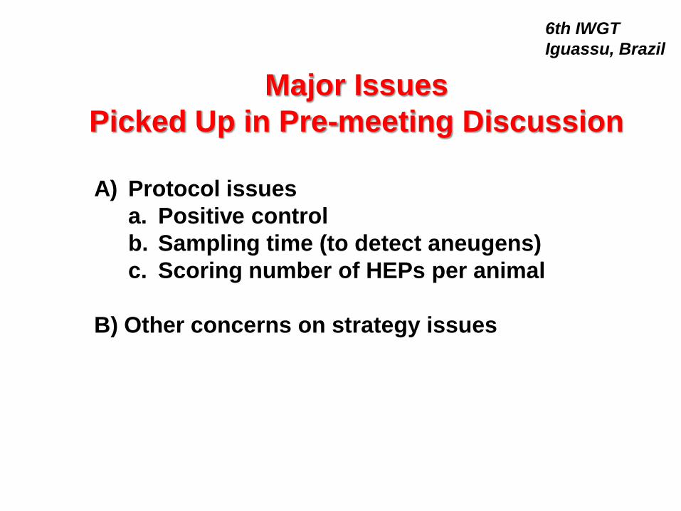
Major Issues
Picked Up in Pre-meeting Discussion
6th IWGT
Iguassu, Brazil
A) Protocol issues
a. Positive control
b. Sampling time (to detect aneugens)
c. Scoring number of HEPs per animal
B) Other concerns on strategy issues
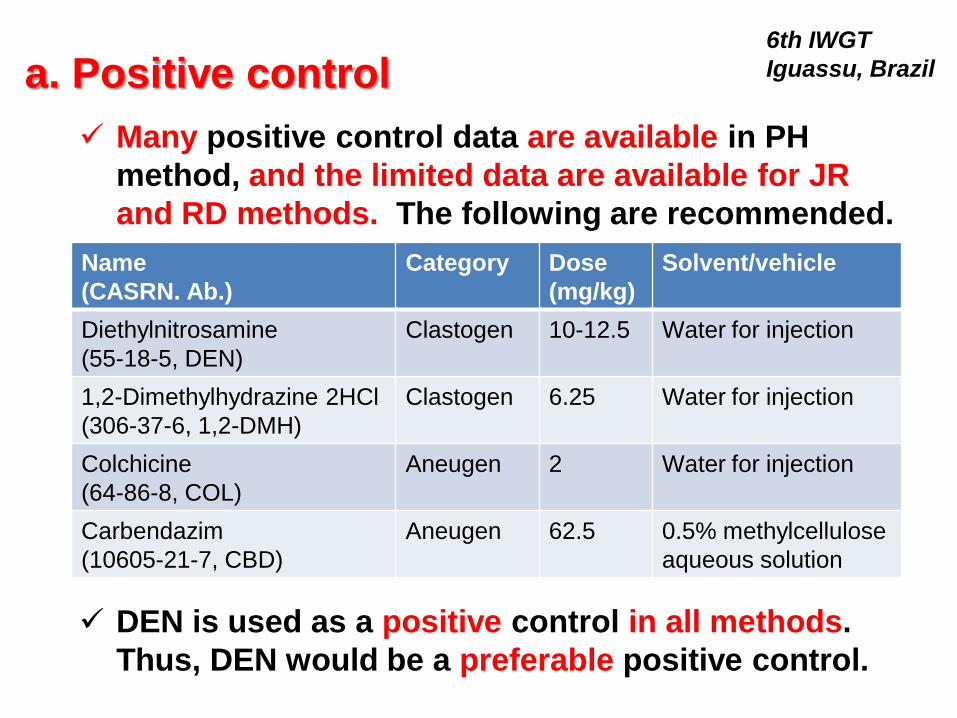
a. Positive control 6th IWGT
Iguassu, Brazil
Many positive control data are available in PH
method, and the limited data are available for JR
and RD methods. The following are recommended.
Name
(CASRN. Ab.)
Category Dose
(mg/kg)
Solvent/vehicle
Diethylnitrosamine
(55-18-5, DEN)
Clastogen 10-12.5 Water for injection
1,2-Dimethylhydrazine 2HCl
(306-37-6, 1,2-DMH)
Clastogen 6.25 Water for injection
Colchicine
(64-86-8, COL)
Aneugen 2 Water for injection
Carbendazim
(10605-21-7, CBD)
Aneugen 62.5 0.5% methylcellulose
aqueous solution
DEN is used as a positive control in all methods.
Thus, DEN would be a preferable positive control.

6th IWGT
Iguassu, Brazil
Dr. Hamada suggests that no concurrent positive control would
be needed in RD method due to low feasibility, because RD
method is intended to integrate into general toxicity study.
Instead, HEP suspension in formalin obtained from a periodic
study with a positive control can be used to assure the staining
and observation procedures.
The above consideration may be supported by ICH-S2(R1), i.e.,
after a laboratory has demonstrated that it can consistently
detect appropriate positive control compounds in multiple
independent experiments, carrying out positive control
experiments periodically is generally sufficient provided
experimental conditions are not changed.
However, many people suggested that a concurrent positive
control would be needed, because this method is still under
development.
No consensus at this meeting. One feasible suggestion is that a
few daily dosing of 1,2-DMH can show a positive response even
in adult rats. Discussion will continue after the meeting.
a. Positive control (continued)

b. Sampling time (to detect aneugens)
6th IWGT
Iguassu, Brazil
Points to be discussed.
• In JR method, when is the suitable
sampling time to detect aneugens?
Dr. Takasawa will show us some data
to discuss this point: see the slide 26.

c. Scoring number of HEPs per animal
6th IWGT
Iguassu, Brazil
Points to be discussed.
• Can we accept that 2000 HEPs are
sufficient to evaluate LMN induction?
• If not, how should we determine the
suitable number of HEPs scored?
• Not determined, because 5000 cells
may be required even in BM/PB-MN
tests.

c. Scoring number of HEPs (continued)
6th IWGT
Iguassu, Brazil
Dr. Itoh suggests that spontaneous incidence of MNHEPs in PH
method is around 0.20% in rats and this value is comparable to that in
rat BM-MNT (0.14±0.12, Environ Mol. Mutagen, 1998 (32), 84-100), and
thus 2000 HEPs would be acceptable as 2000 BM cells are acceptable.
He also commnets that more HEPs may be observed in JR method
due to lower spontaneous incidence of MNHEPs.
Dr. Hamada suggests that, as for negative control rats with no
MNHEPs after 2000-cell- observation, since there was a concern for a
lack of statistical sensitivity in the 2000-cell- observation, observation
was continued until one MNHEP was observed. As a result, at least
one MNHEP was noted by observation of 4000 cells in almost all
cases, and thus 4000 cells are considered appropriate for evaluation
in RD method. However, as there was no change of results from
negative to positive or vice versa by observation of 2000 or 4000 cells,
2000 cells were considered appropriate to evaluate LMN induction.
Can we accept the above suggestions? Are 2000 HEPs sufficient?

No. of rats No. of experiments
Mean ± SD (%) No. of rats with zero MN in 2000 HEPs
F344/DuCrlCrlj 110 26 0.23 ± 0.09 1
Crl: CD(SD) 25 6 0.19 ± 0.11 1
No. of rats
No. of experiments
Mean ± SD (%) No. of rats with zero MN in 2000 HEPs
F344/DuCrlCrlj 20 5 0.06 ± 0.06 7
Table 1. Historical control data of LMNT with PH in Daiichi Sankyo
Table 2. Historical control data of LMNT using juvenile rat in Daiichi Sankyo
Water or 0.5% methylcellulose was administered to male rats orally
Water or 0.5% methylcellulose was administered to male rats orally
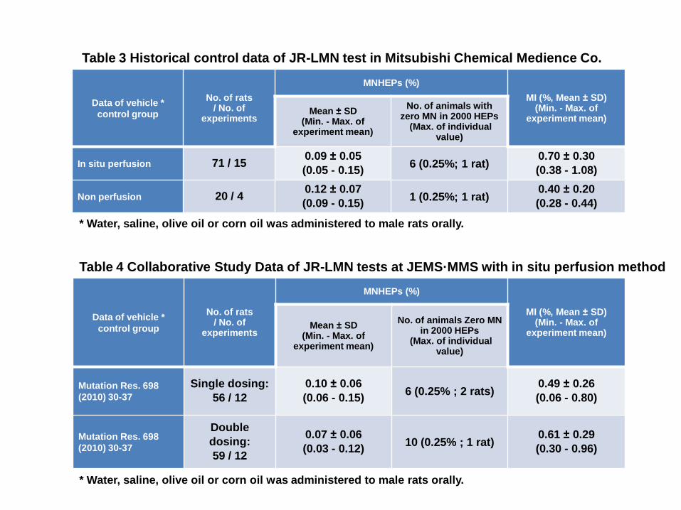
Data of vehicle *
control group
No. of rats / No. of
experiments
MNHEPs (%)
MI (%, Mean ± SD) (Min. - Max. of
experiment mean) Mean ± SD
(Min. - Max. of experiment mean)
No. of animals with zero MN in 2000 HEPs
(Max. of individual value)
In situ perfusion 71 / 15 0.09 ± 0.05
(0.05 - 0.15) 6 (0.25%; 1 rat)
0.70 ± 0.30
(0.38 - 1.08)
Non perfusion 20 / 4 0.12 ± 0.07
(0.09 - 0.15) 1 (0.25%; 1 rat)
0.40 ± 0.20
(0.28 - 0.44)
Data of vehicle *
control group
No. of rats / No. of
experiments
MNHEPs (%)
MI (%, Mean ± SD) (Min. - Max. of
experiment mean) Mean ± SD
(Min. - Max. of experiment mean)
No. of animals Zero MN in 2000 HEPs
(Max. of individual value)
Mutation Res. 698
(2010) 30-37
Single dosing:
56 / 12
0.10 ± 0.06
(0.06 - 0.15) 6 (0.25% ; 2 rats)
0.49 ± 0.26
(0.06 - 0.80)
Mutation Res. 698
(2010) 30-37
Double
dosing:
59 / 12
0.07 ± 0.06
(0.03 - 0.12) 10 (0.25% ; 1 rat)
0.61 ± 0.29
(0.30 - 0.96)
Table 4 Collaborative Study Data of JR-LMN tests at JEMS·MMS with in situ perfusion method
Table 3 Historical control data of JR-LMN test in Mitsubishi Chemical Medience Co.
* Water, saline, olive oil or corn oil was administered to male rats orally.
* Water, saline, olive oil or corn oil was administered to male rats orally.

Table 5 Negative control data of RD-LMN assay
The negative background values were remarkably low and stable, which
were 0.05 ± 0.05% for 8-week-old and 0.06 ± 0.06% for 10-week-old SD
rats. However, the number of MNHEPs in 2000 hepatocytes/animal was
found to be 0 in 80 of 210 8-week-old rats and 29 of 92 10-week-old rats in
the negative control group.

d. Other concerns on strategy issues
6th IWGT
Iguassu, Brazil
Points to be discussed.
• When, where and how to use LMNT?
• Judgment and implication of results?
• Hazard identification or risk
assessment?
• Combination/integration with other
genotoxicity/general toxicity tests?

Most likely situations to select and use LMN test may be:
1. Relevant chromosome aberration is observed with a test chemical
(especially under the condition of metabolic activation) in in vitro
mammalian cell genotoxicity tests, and a negative results is obtained in
the in vivo BM/PB MN test. LMN test may be selected as a 2nd in vivo
test.
2. RD-LMN test may be the easiest integration method into a general
toxicity study as well as BM/PB-MN test or Pig-a assay, because the
other in vivo genotoxicity test have some difficulty to be integrated into
general toxicity study, e.g., TG assay uses TG animals and needs 3
additional days, Comet assay requires an additional administration just
before sampling tissues.
3. It may be useful to obtain and store the liver-cell-samples in formalin
routinely when a general toxicity study is conducted, because it allows
to analyze LMN later whenever necessary.
When, where and how to use LMNT?

6th IWGT
Iguassu, Brazil
IV. MN test other than BM/PB and LMN tests
i. Review of MNT other than BM/PB and LMN
tests: Dr. Morita
ii. Update of MNT in GI tracts: Dr. Oyama