Embed Size (px)
Citation preview
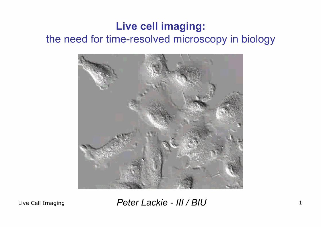
Live Cell Imaging 1
Live cell imaging:the need for time-resolved microscopy in biology
Peter Lackie - III / BIU
Live Cell Imaging 2
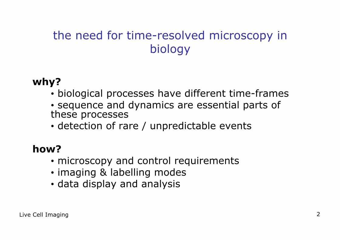
the need for time-resolved microscopy inbiology
why?• biological processes have different time-frames• sequence and dynamics are essential parts ofthese processes• detection of rare / unpredictable events
how?• microscopy and control requirements• imaging & labelling modes• data display and analysis
Live Cell Imaging 3
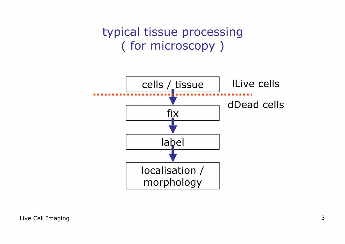
typical tissue processing( for microscopy )
cells / tissue
fix
label
localisation /morphology
lLive cells
dDead cells
Live Cell Imaging 4
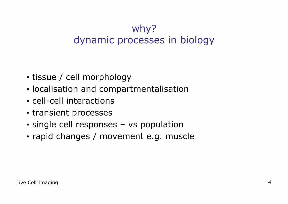
why?dynamic processes in biology
• tissue / cell morphology• localisation and compartmentalisation• cell-cell interactions• transient processes• single cell responses – vs population• rapid changes / movement e.g. muscle
Live Cell Imaging 5
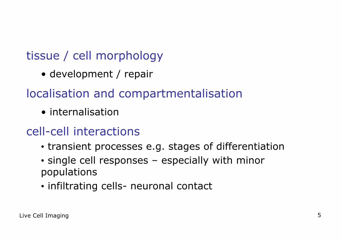
tissue / cell morphology
• development / repair
localisation and compartmentalisation
• internalisation
cell-cell interactions• transient processes e.g. stages of differentiation• single cell responses – especially with minorpopulations• infiltrating cells- neuronal contact
Live Cell Imaging 6
rapid changes /movement• transient processes > 1 Hz - rapid calcium flux• cilia - fast movement
slow changes / movement• transient processes - unpredictable & asynchronousevents• slow motility > minutes
Live Cell Imaging 7
live cell imaging- how is it done?
• requirements• imaging modes for live-cell imaging• slowing down• speeding up• analysis
Live Cell Imaging 8
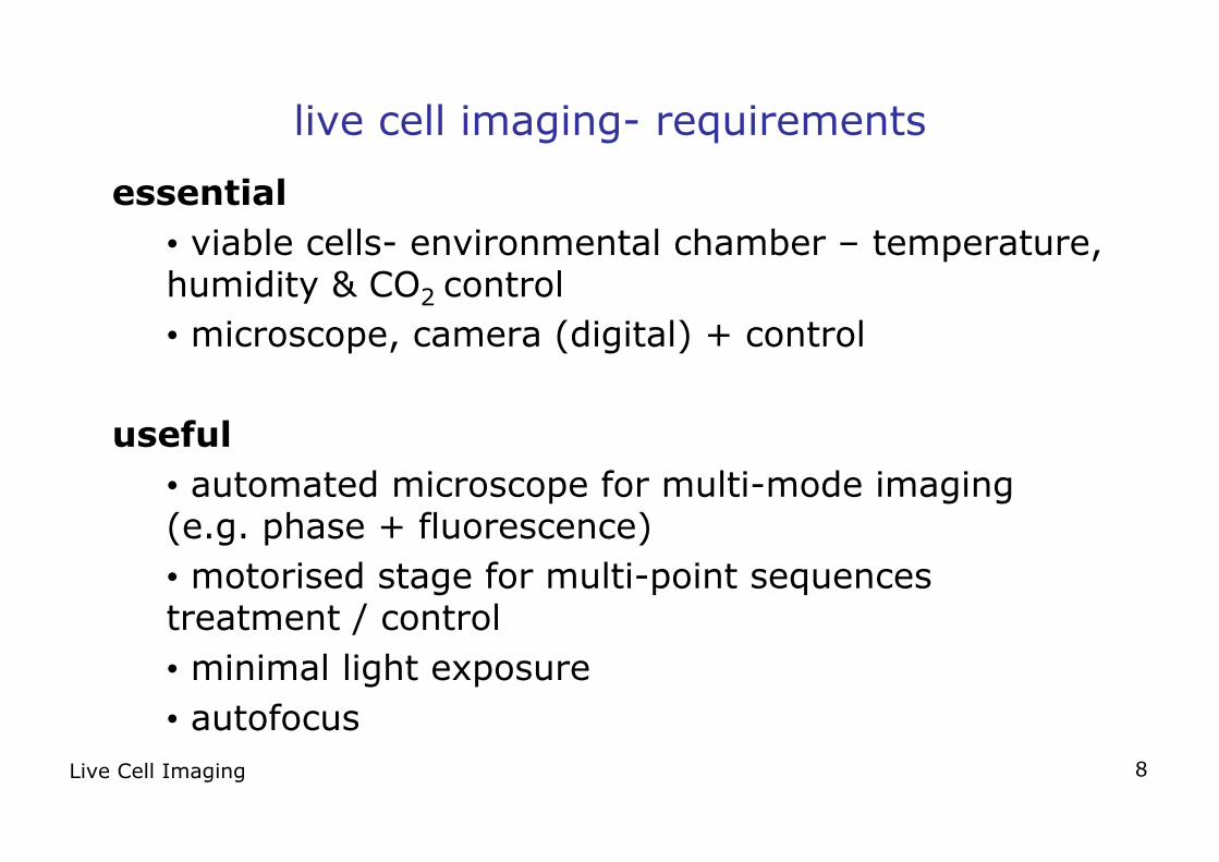
live cell imaging- requirements
essential• viable cells- environmental chamber – temperature,humidity & CO2 control• microscope, camera (digital) + control
useful• automated microscope for multi-mode imaging(e.g. phase + fluorescence)• motorised stage for multi-point sequencestreatment / control• minimal light exposure• autofocus
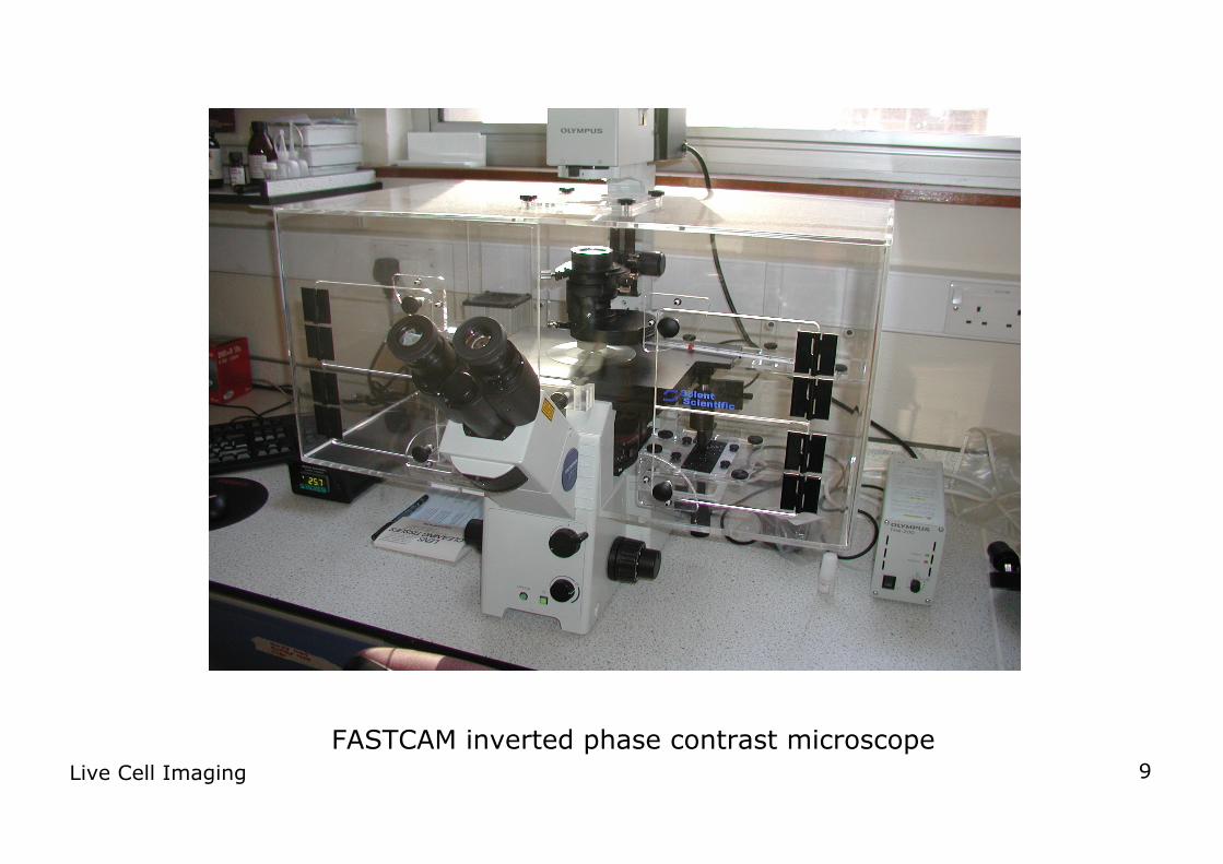
Live Cell Imaging 9FASTCAM inverted phase contrast microscope
Live Cell Imaging 10
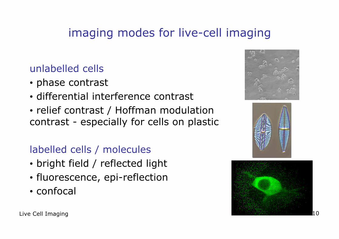
imaging modes for live-cell imaging
unlabelled cells• phase contrast• differential interference contrast• relief contrast / Hoffman modulationcontrast - especially for cells on plastic
labelled cells / molecules• bright field / reflected light• fluorescence, epi-reflection• confocal
Live Cell Imaging 11
visualisation/ labelling
• unstained cells- altered morphology / autofluorescence• viable dyes e.g. Hoechst 33258• labelled probes / beads e.g. phycoerythrin• tagged molecules e.g. GFP
Live Cell Imaging 12
live cell imaging - some cautions
• demonstrate that cell viability is maintained• light exposure (especially fluorescence)• cell behaviour can be altered by labelling e.g.antibodies / lectins
Live Cell Imaging 13
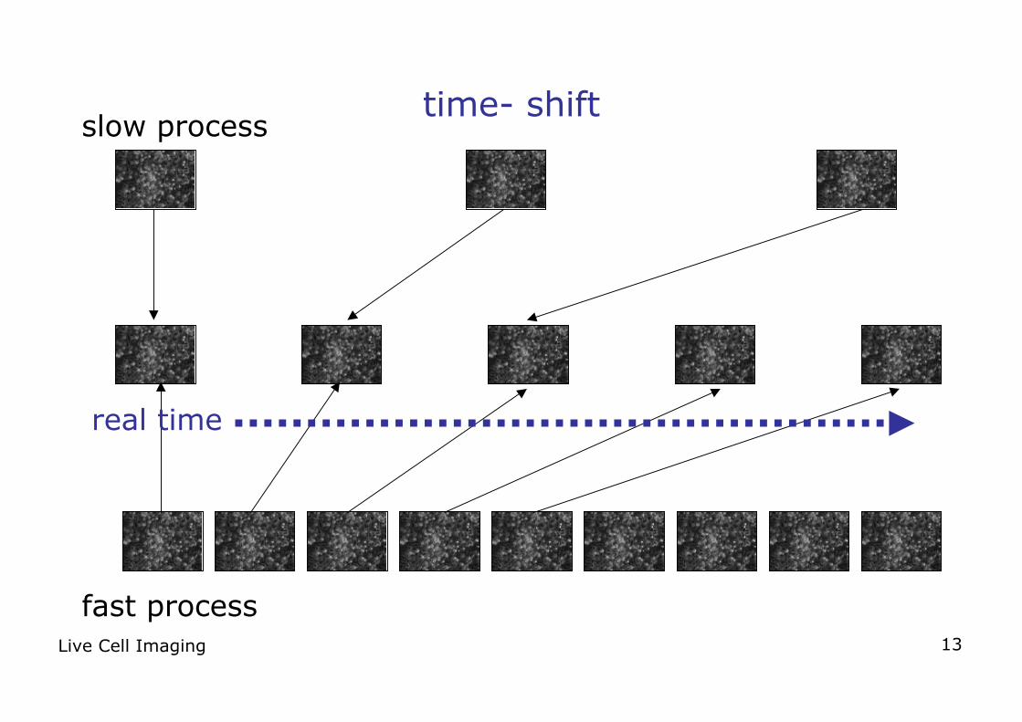
time- shift
fast process
slow process
real time
Live Cell Imaging 14
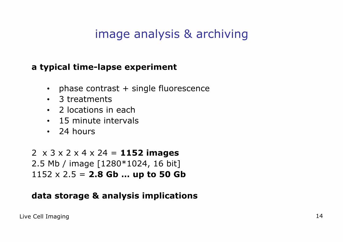
image analysis & archiving
a typical time-lapse experiment
• phase contrast + single fluorescence• 3 treatments• 2 locations in each• 15 minute intervals• 24 hours
2 x 3 x 2 x 4 x 24 = 1152 images2.5 Mb / image [1280*1024, 16 bit]1152 x 2.5 = 2.8 Gb … up to 50 Gb
data storage & analysis implications
Live Cell Imaging 15
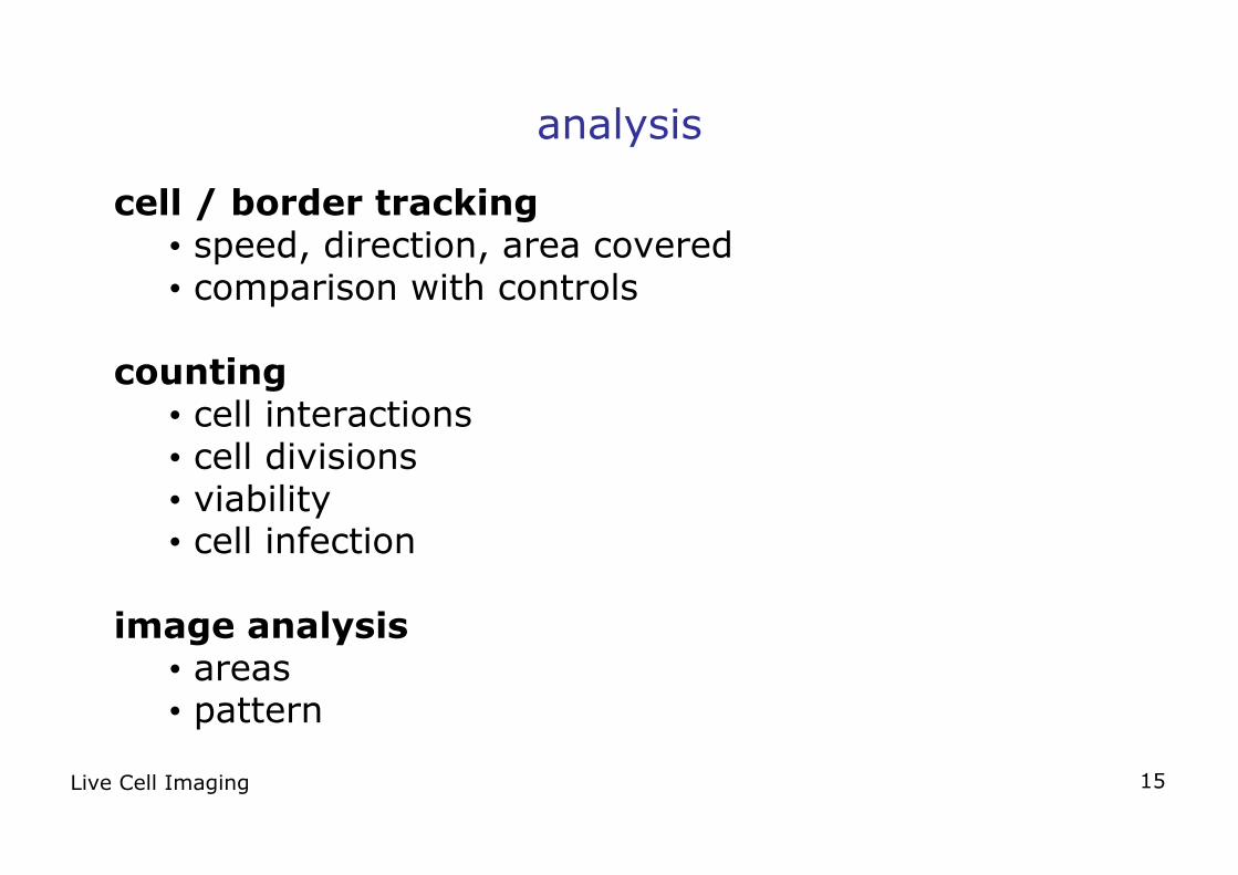
analysis
cell / border tracking• speed, direction, area covered• comparison with controls
counting• cell interactions• cell divisions• viability• cell infection
image analysis• areas• pattern
Live Cell Imaging 16
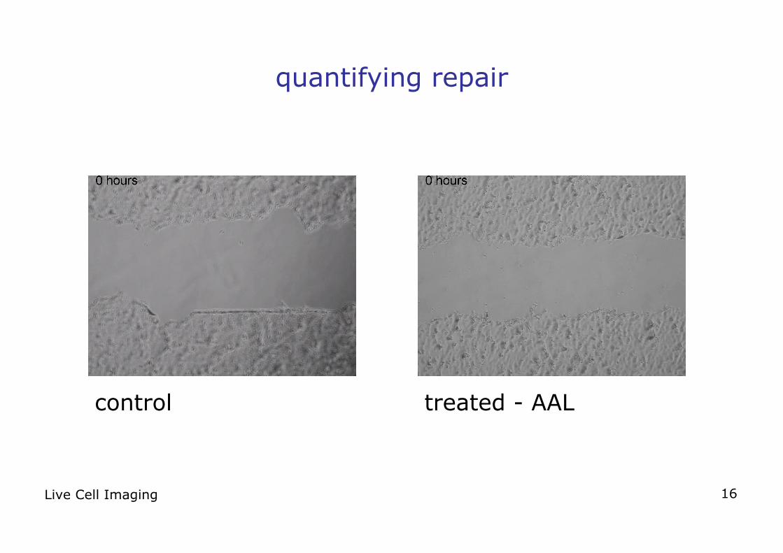
quantifying repair
control treated - AAL
Live Cell Imaging 17
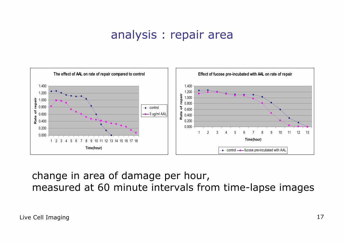
analysis : repair area
The effect of AAL on rate of repair compared to control
0.000
0.200
0.400
0.600
0.800
1.000
1.200
1.400
1 2 3 4 5 6 7 8 9 10 11 12 13 14 15 16 17 18
Time(hour)
Ra
te o
f re
pa
ir
control
3 ug/ml AAL
change in area of damage per hour, measured at 60 minute intervals from time-lapse images
Effect of fucose pre-incubated with AAL on rate of repair
0.000
0.200
0.400
0.600
0.800
1.000
1.200
1.400
1 2 3 4 5 6 7 8 9 10 11 12 13
Time(hour)
Ra
te o
f re
pa
ir
control fucose pre-incubated with AAL
Live Cell Imaging 18
conclusion
time-resolved live cell imaging provides information notavailable by other means
Live Cell Imaging 19
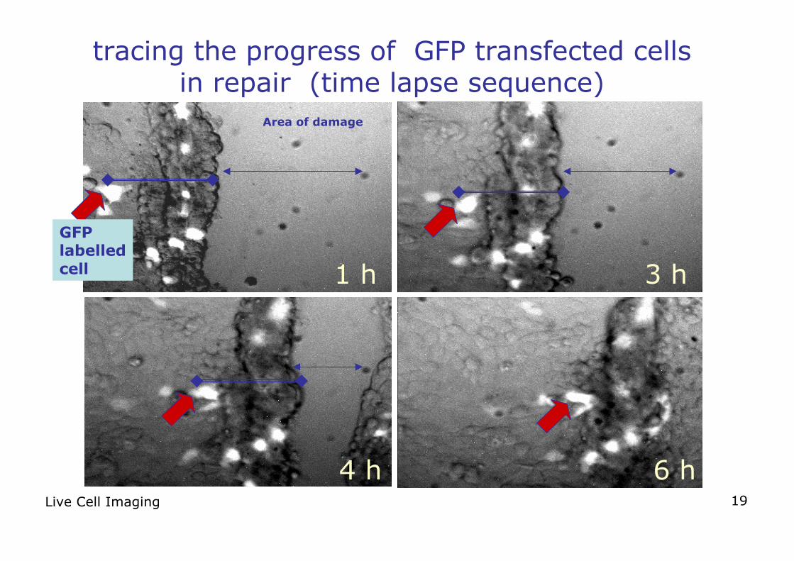
tracing the progress of GFP transfected cellsin repair (time lapse sequence)
1 h 3 h
4 h 6 h
Area of damage
GFP labelledcell