Embed Size (px)
Citation preview

28 I ENDOVASCULAR TODAY I JULY 2009
IMAGING & DIAGNOSTICS
Today’s endovascular interventionists must beskilled with the latest device and imaging tech-nology to best treat their patients. An accuratemorphological assessment of blood vessels and
their relationship with interventional devices is essentialto the management of vascular lesions, and digital sub-traction angiography (DSA) has had a central role in thedeployment of interventional procedures. High resolu-tion, the ability to selectively evaluate individual vessels,and the access to direct physiological information makeDSA a cornerstone technology in endovascular proce-dures. The addition of three-dimensional rotationalangiography (3D RA) and flat panel detector technologyhave further contributed to greater diagnostic accuracy,faster procedures, and improved outcomes.1-4 In particu-lar, 3D reconstructions from rotational angiograms haveovercome limitations of two-dimensional (2D) imagingsuch as false vascular foreshortening due to projectionangle and vessel obscuration due to overlying vessel orbone. These 3D reconstructions are now routinely usedto reveal complex vascular relationships. Major develop-ments in hardware and software of x-ray angiographicsystems have significantly increased the usability of thistechnique, and 3D reconstructions are now performedand available for analysis and manipulation in real-time.The availability of 3D reconstructions also carries along awealth of 3D tools that can improve current proceduralworkflows starting from disease assessment to treatmentevaluation through navigation and treatment planning.
The use of 3D RA and associated tools in endovascu-lar procedures is, however, not yet common practice.Although evidence of the clinical benefit and reductionin radiation exposure to patients and staff following theuse of 3D RA has been extensively reported for vascular
lesions of the neck and brain,5-10 there is not enoughinformation in the literature on how the use of 3D toolscan improve the (peripheral) vascular procedural work-flow with potentially no changes (or reduction) to radi-ation and contrast administration. This article providesa showcase of minimally invasive endovascular applica-tions in which the availability of 3D tools was distinc-tively beneficial to achieve effective disease manage-ment in a busy day-to-day clinical practice.
M ATERIAL S AND METHODSThe interventional radiology department at Paoli
Hospital performs approximately 5,400 procedures peryear, covering the full spectrum of vascular and nonvas-cular interventions. In order to provide a suitable rangeof endovascular procedures, 10 patients were selectedretrospectively from a data pool of more than 200 sub-jects treated between 2006 and 2007 with clinicallyindicated rotational angiography. The clinical indicationwas made by the interventional radiologist on a per-case basis based on his judgment as to whether the 3Dtools would help in problem solving or clarification ofanatomy, offer any potential time savings, possiblyreduce contrast or x-ray dose to the patient, or uncovera suspected hidden lesion. All rotational examinations
Live 3D Guidance in Endovascular
ProceduresA showcase of patient studies illustrating the benefits of advanced live
3D guidance tools in a busy day-to-day clinical practice.
BY ATUL GUPTA, MD, AND ALESSANDRO G. RADAELLI, PHD
“The addition of 3D RA and flat paneldetector technology have furthercontributed to greater diagnosticaccuracy, faster procedures, and
improved outcomes.”

JULY 2009 I ENDOVASCULAR TODAY I 29
IMAGING & DIAGNOSTICS
were performed on a Philips Allura Xper FD20 system(Philips Medical Systems, Best, The Netherlands) usingan automated “two-button” 3D acquisition process.Contrast material was injected during acquisitionthrough a catheter placed into the vascular territory ofinterest. A total of 120 frames were obtained during a240° rotation for a total scan time of 4 seconds. Theframes were automatically sent to a dedicated worksta-tion where 3D reconstructions with a field of view of25 X 19 X 25 cm3 and a matrix size of 1283 were generat-
ed in real time. These real-time 3D images were dis-played in the procedure room, alongside or evenembedded within the fluoroscopic images. All 3D analy-ses were performed live by the physician who carriedout the examination.
The 3D reconstructions were used to visualize thevascular anatomy from multiple angles, select the opti-mal working projection, and simultaneously synchro-nize the position of the C-arm. This is now possiblebecause modern workstations are fully integrated with
Figure 1. A renal artery stenosis in an 81-year-old woman.The patient was referred for treatment after failed medical manage-
ment of hypertension. Preprocedure contrast-enhanced MR angiography showing a tight ostial stenosis of the right renal
artery (arrows) and a patent left renal artery (dashed arrows) (A). Repeat anteroposterior (AP) arteriogram of descending aorta
and iliac arteries confirming the right renal artery stenosis (B). Selective DSA image of the right renal artery showing stenosis
(arrows) at the origin of the vessel (C). Selective DSA image of the right renal artery showing successful stenting (D). AP view of
the 3D RA reconstruction showing unremarkable left (dashed arrows) renal artery and the good poststent appearance of the
right renal artery (arrows) (E). Oblique view (right anterior oblique [RAO] = -6°, cranial [CRAN] = -52°) revealing the presence of
two overlapping vascular segments on the left side, which hide a very tight stenosis of the left renal artery (dashed arrow) (F).
The lesion was further confirmed with pullback pressure measurements. After stenting of the left renal artery, hypertension
was successfully controlled.
A
E F
B C
D

30 I ENDOVASCULAR TODAY I JULY 2009
IMAGING & DIAGNOSTICS
the acquisition system and directly coupled with the C-arm geometry. The synchronization could also beexploited to achieve live 3D navigation through thereal-time superimposition of live fluoroscopy images ona surface-rendered 3D reconstruction (Dynamic 3DRoadmap, Philips Medical Systems). Changes to the livefluoroscopy images after adjustment of x-ray/detectordistance, position, and/or magnification are transferredto the 3D reconstruction so that the matching is main-tained throughout the procedure. When available,preinterventional computed tomography (CT) andmagnetic resonance (MR) scans could also be trans-ferred to the workstation, matched with 2D live fluo-roscopy, and used for 3D navigation.
Three-dimensional reconstructions were furtherprocessed using quantitative analysis tools. Vessel size
and stenosis grading were measured in 3D usingadvanced vascular analysis and virtual stenting func-tionalities, which provide an accurate delineation oflocal vascular diameter and length and a simulation ofstent placement that facilitates the selection of com-mercially available stents. In the case of aneurysms, abuilt-in, computer-assisted aneurysm analysis tool wasused to automatically detect significant vascular dilata-tions and obtain quantitative information such asaneurysmal volume and neck size.
For procedures requiring intraoperative visualizationof soft tissues, CT-like 3D reconstructions were acquiredusing the XperCT (Philips Medical Systems) technique.XperCT is an additional rotational acquisition mode per-formed over a scan range of 240º resulting in 310 framesand a total scan duration of 10 seconds. When required,
Figure 2. An external iliac artery stenosis in a 79-year-old man. AP view of aortoiliac arteriogram (A). Lateral view (RAO = -38°,
CRAN = -4°) of a 3D RA reconstruction better showing the vascular morphology (B). Had the stenosis extended over a longer
segment, stent sizing based on 2D imaging would have been particularly unfavorable.The same view of the posttreatment 3D
RA reconstruction and results of virtual interventional tools, used in this case to analyze the stented vascular segment (C).
Quantitative information provided by the advanced vascular analysis tool (D).The good outcome of the stenting procedure is
shown by a satisfactory stent opening throughout the lesion and by a smooth transition from proximal to distal ends (blue
line) also when compared with an ideal linear transition provided by the software (yellow line).
A B
C D

XperCT and 3D RA were matched to visualizeinterventional devices and soft tissues along withcontrast-filled vessels and assess treatment com-pletion.
CLINICAL RE SULTSVascular Stenoses
Three-dimensional imaging is useful in theassessment and grading of vascular stenoses. Notonly can it provide a view of the anatomy fromangles not achievable with 2D imaging, but 3Dimaging also helps in the detection of hiddenstenoses not visible in 2D projections and otherdiagnostic images (Figure 1).
The benefits of 3D information also extend toa better estimation of the required stent charac-teristics. For instance, when performing iliacstenting, most interventional radiologists aretrained to acquire an anteroposterior (AP) aor-toiliac arteriogram and two additional obliqueangiographic projections. Due to the high tortu-osity of the external iliac arteries, the use of con-ventional 2D images and “eyeballing” may leadto underestimation of the length of the vesseland ultimately to incorrect stent sizing. By using3D RA and quantitative tools, the radiologist canaccurately evaluate the extension and grading ofthe stenosis and select the right stent from thestart, thus avoiding the need of overlappingstents with the associated extra costs and risks ofrecurrent stenosis (Figure 2).
TraumaThree-dimensional imaging provides detailed
anatomical information that assists decisionmaking for emergency and trauma patients.Three-dimensional information can be used toelucidate the relationship between foreign bodiesand vascular structures and support/exclude theneed for complex surgical procedures (Figure 3).In this case, 3D RA was superior to high-resolu-tion multiplanar CT visualization, film radiogra-phy, and conventional 2D venography in provid-ing quick, optimal anatomical viewing and facili-tating timely and confident decisions.
Visceral AneurysmsAccurate imaging information on volume, location,
neck size as well as the relationship with parent vesseland side branches is essential in the diagnostic assess-ment and treatment planning of aneurysms. Often, thistype of information cannot be obtained—neither from
2D DSA due to complex angles required to view theaneurysm neck nor from CT angiography (CTA) andmagnetic resonance angiography (MRA)—because ofinadequate spatial resolution, especially for small branchvessels. CTA and MRA typically provide a spatial resolu-tion greater than 0.4 to 0.5 mm, whereas a resolution of0.1 to 0.2 mm can be expected from 3D RA.
32 I ENDOVASCULAR TODAY I JULY 2009
IMAGING & DIAGNOSTICS
Figure 3. An accidental drill bit penetration in the pelvis of 75-year-old
woman during hip replacement. Radiography showing the location of
the broken drill bit (arrow) in a precarious area of the pelvis (A). Axial
view of subsequent CT scan (B).The head of the drill bit (arrow) appears
to have penetrated the common iliac vein (dashed arrow).Two-dimen-
sional venograms acquired at several different oblique angles further
suggest penetration of the drill bit into the iliac vein (C through E). A 3D
rotational venogram showing the relationship between the prosthesis’
acetabular head, drill bit, and common iliac vein (F). Magnified and rotat-
ed view (RAO = 60°, caudal = 57°) revealing a tissue plane between the
tip of the drill bit and the iliac vein (arrow), conclusively ruling out vessel
perforation, contrary to CT and 2D venogram findings (G).This obliquity
was impossible to achieve with 2D venography. Fluoroscopic image
acquired during subsequent intervention (H).The drill bit is simply
grabbed and pulled out with a hemostat without any resulting compli-
cation.Three-dimensional imaging saved the patient a complex
retroperitoneal dissection and vascular surgery.
A B
C D E
HGF

JULY 2009 I ENDOVASCULAR TODAY I 33
Renal arteries are common locations of vascularaneurysms, and appropriate anatomical information iscritical. For example, incorrect deployment of endovas-cular coils due to underestimation of neck size maylead to nontarget embolization with possible kidneyinfarction. Three-dimensional RA offers high spatial res-olution and can achieve a detailed 360° view of the ves-sels of interest. Computer-assisted aneurysm analysis
tools can also be used to automatically define theaneurysm sac, calculate its dimensions, and analyze itsrelationship with the surrounding vessels, facilitatingboth diagnostics and interventional planning (Figure 4).In this case, a video file of the 3D RA reconstructionwas supplied to transplant surgeons prior to surgery tohelp them plan the best method of vascular repair dur-ing bench surgery (autotransplantation). This resulted
IMAGING & DIAGNOSTICS
Figure 4. A renal artery aneurysm in a 55-year-old man (A through C). Selected views of diagnostic 3D MRA showing an aneurysm
of the right renal artery. Although spatial resolution is inadequate for confident neck localization and sizing, the image (C) shows
optimal viewing angle to best define the aneurysm neck. A similar view with 2D DSA would not be achievable because it would
have required a very steep craniocaudal oblique angle. Selective 3D RA reconstruction of vascular aneurysm after the injection of
8 mL of contrast using a catheter placed into the right renal artery (captured on the image).The relationship between the
aneurysm and feeding vessels could be visualized at any angle. Surface rendering of the 3D RA reconstruction (D).The aneurysm is
isolated and shaded in blue using an automatic detection algorithm implemented in the computer-assisted aneurysm analysis
tool.The visualization helps in the quantification of aneurysm size and shows how all three vessels feeding the kidney (arrows)
arise from the aneurysm sac. An additional view offering a better depiction of the posterior vessel branch (dashed arrow) (E).The
patient was referred, along with a 3D RA video file, to a transplant surgeon for bench surgery and autotransplantation as endovas-
cular repair with coils/covered stents was not a good option.
A B
D E
C

in minimizing the ischemia time of the explantednative kidney during repair, because much of the repairplanning could be done prior to surgery.
Inferior Vena Cava Filter RemovalRetrievable inferior vena cava (IVC) filters are increas-
ingly being used to prevent pulmonary embolism.Several weeks or months after implantation, patientsare typically brought back for filter removal. Mostretrievable IVC filters incorporate a “retrieval hook”that is used to capture the filter, and these filters aredesigned in a manner to avoid tilting and ensure cor-rect positioning of the hook in the middle of the IVC.
When properly placed, the filter is typically removedwithin minutes with a snare and sheath.
Often, the removal process requires several snareattempts and lengthy fluoroscopic exposures. Evensymmetric filters, while appearing untilted in standard2D venograms, may hide a significant degree of tilt,which is often revealed with unusual projectionangles.11 Our approach is therefore to routinely per-form a rapid, 4-second 3D RA at the start of the proce-dure to achieve a fast and confident removal, even incomplex cases. A single acquisition provides all thenecessary information to quickly and accurately visual-ize the filter and the IVC, plan the appropriate removal
34 I ENDOVASCULAR TODAY I JULY 2009
IMAGING & DIAGNOSTICS
Figure 5. An IVC filter removal in a 44-year-old man.Two-dimensional AP venography showing the IVC and OptEase IVC filter
(Cordis Corporation, Warren, NJ) (A). A magnified view of the filter suggests the correct positioning of the hook (arrow) in the
middle of the vein’s lumen (B).Two-dimensional AP fluoroscopy showing an unsuccessful attempt to capture the filter’s hook
using a snare (C). A 4-second 3D RA reconstruction showing the spine, tilted IVC filter, and its relationship with the snare used
to capture the hook (D). Lateral (84°) 2D fluoroscopy optimally showing filter tilting (E).The exact lateral angle was selected
from the 3D image. Lateral (84°) 2D digital subtraction venography further exposing the relationship between the filter and the
IVC (F).The filter is not only tilted, but the hook (arrow) is also embedded into the vessel wall. Snare retrieval would have been
physically impossible and would have inevitably led to numerous unsuccessful attempts. An endovascular forceps (shown
inset) is advanced and grasps the filter’s struts (G).The hook is gently brought back into the vessel lumen. Fluoroscopic
sequence showing subsequent filter removal (H though J).
A
E F G H I J
B C D

JULY 2009 I ENDOVASCULAR TODAY I 35
approach, and select the optimal working projection,often becoming a significant time saver (Figure 5).
The possibility of visualizing the rotational acquisi-tion as a “CT-like” multiplanar reconstruction furtherfacilitates the analysis of the hook positioning withrespect to the IVC lumen, thus making the removalprocess simple and fast (Figure 6).
Vascular MalformationsEndovascular treatment of arteriovenous malforma-
tions (AVMs) can be quite complex. Although diagnos-tic multiplanar 3D CTA or MRA images are valuable forAVM detection, they do not offer adequate spatial reso-lution to precisely analyze the “spaghetti-like” arterialsupply and determine the optimal angle of approachfor each feeding vessel. Three-dimensional RA offersmore detailed anatomical information that enables theradiologist to untangle the tangled web of vessels sur-rounding the AVM, choose the angles to selectivelyshow the feeders, and define treatment approach andchronology.
A comprehensive treatment plan based on 3D RA isachieved with a single run and helps to minimize thenumber of 2D DSA and fluoroscopic images requiredduring treatment. The minimization of iodinated mate-rial is particularly important for patients affected byrenal AVMs because renal function may be impaireddue to preparenchymal shunting of blood, with result-ant bypassing of renal filtration. Additionally, furtherkidney injury may result from imprecise or nontargetembolization. Thus, optimal viewing angles are critical.The use of 3D imaging is particularly beneficial in thesepatients and helps us to achieve a favorable balance
between contrast minimization, optimal viewing, and aprompt recovery of kidney function (Figure 7).
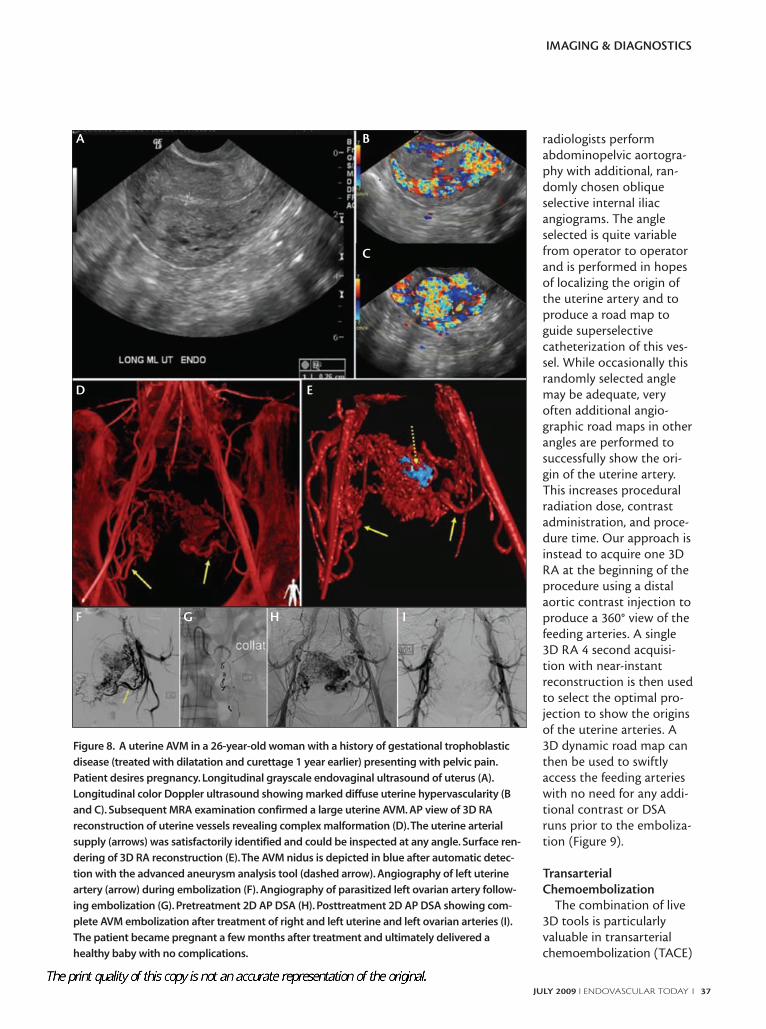
Accurate 3D imaging plays a significant role for mal-formations affecting the uterine arteries. Uterine AVMsare particularly complex and tortuous, and, even if theproximal uterine artery is correctly embolized, parasiti-zation and collateral vessels such as the ovarian arterymay rapidly take over and continue feeding the malfor-mation. Should the malformation be inadequatelyembolized, uterine expansion during pregnancy maylead to vessel rupture and to life-threatening hemor-rhage for the mother. The availability of 3D RA recon-structions is therefore essential to quickly identify thenidus, unravel the intricate vascular configuration of thefeeding vessels, select the optimal working projectionsto access all feeding vessels, and achieve a completeembolization (Figure 8).
Uterine Fibroid EmbolizationUterine fibroid embolization (UFE) is an important
application making use of 3D imaging.12 UFE is per-formed by introducing small embolic particles intouterine arteries feeding the fibroid, resulting in fibroidshrinkage. The imaging technique of choice for this pro-cedure is conventional angiography. Most interventional
IMAGING & DIAGNOSTICS
Figure 6. Dual retrieval of bilateral IVC Celect filters (Cook Medical, Bloomington, IN) (first reported worldwide) in a 58-
year-old man with duplicated IVC. Two-dimensional AP fluoroscopy of the patient’s abdomen showing IVC filters (A).
Volume rendering of pretreatment 3D rotational venography (B). The intensity threshold value was selected so as to opti-
mally visualize the filters only. The virtual plane (in yellow) corresponds to the location of the axial slice (C). Selected axial
slice of real-time CT-like reconstruction from 3D venography showing the location of the left filter’s hook (arrow) in the
middle of the IVC (C). Two-dimensional fluoroscopy during removal of the right filter (D). Two-dimensional fluoroscopy
during removal of the left filter (E).
A B C D E
“Three-dimensional imaging providesdetailed anatomical information that
assists decision making for emergency and trauma patients.”

36 I ENDOVASCULAR TODAY I JULY 2009
IMAGING & DIAGNOSTICS
Figure 7. Bilateral renal AVMs discovered in a 69-year-old woman with congestive heart failure (CHF) who presented with severe
shortness of breath and suspected pulmonary embolism (PE).An AP view of chest showing severe cardiomegaly (A).The last axial
slice obtained from the CT PE study (dashed line [A]) shows abnormal appearance of the renal arteries,suggesting the presence of
bilateral AVMs (B).MRA of renal arteries confirming AVMs with early enhancement of the renal veins and IVC (arrow) suggesting high-
flow arteriovenous shunting (C).AP view of selective 3D RA surface rendering of the left renal artery (LRA) showing complex AVM
with outpouching aneurysms prior to embolization (D).Location and feeding vessels of the AVM (arrows) are nicely exposed.Oblique
view of 3D RA grayscale rendering used for aneurysm measurement and for selection of angles needed to access the feeding vessels
(E).Embolization performed with angles selected from 3D RA (F).Posttreatment 2D AP DSA (G).Patient’s fatigue and shortness of
breath resolved after treatment,with an improvement in the kidney function (creatinine dropped from 1.9 to 1.5 mg/dL) due to
improved renal parenchymal perfusion.
A
D E
F G
B C
JULY 2009 I ENDOVASCULAR TODAY I 37
radiologists performabdominopelvic aortogra-phy with additional, ran-domly chosen obliqueselective internal iliacangiograms. The angleselected is quite variablefrom operator to operatorand is performed in hopesof localizing the origin ofthe uterine artery and toproduce a road map toguide superselectivecatheterization of this ves-sel. While occasionally thisrandomly selected anglemay be adequate, veryoften additional angio-graphic road maps in otherangles are performed tosuccessfully show the ori-gin of the uterine artery.This increases proceduralradiation dose, contrastadministration, and proce-dure time. Our approach isinstead to acquire one 3DRA at the beginning of theprocedure using a distalaortic contrast injection toproduce a 360° view of thefeeding arteries. A single3D RA 4 second acquisi-tion with near-instantreconstruction is then usedto select the optimal pro-jection to show the originsof the uterine arteries. A3D dynamic road map canthen be used to swiftlyaccess the feeding arterieswith no need for any addi-tional contrast or DSAruns prior to the emboliza-tion (Figure 9).
TransarterialChemoembolization
The combination of live3D tools is particularlyvaluable in transarterialchemoembolization (TACE)
IMAGING & DIAGNOSTICS
Figure 8. A uterine AVM in a 26-year-old woman with a history of gestational trophoblastic
disease (treated with dilatation and curettage 1 year earlier) presenting with pelvic pain.
Patient desires pregnancy. Longitudinal grayscale endovaginal ultrasound of uterus (A).
Longitudinal color Doppler ultrasound showing marked diffuse uterine hypervascularity (B
and C). Subsequent MRA examination confirmed a large uterine AVM. AP view of 3D RA
reconstruction of uterine vessels revealing complex malformation (D).The uterine arterial
supply (arrows) was satisfactorily identified and could be inspected at any angle. Surface ren-
dering of 3D RA reconstruction (E).The AVM nidus is depicted in blue after automatic detec-
tion with the advanced aneurysm analysis tool (dashed arrow). Angiography of left uterine
artery (arrow) during embolization (F). Angiography of parasitized left ovarian artery follow-
ing embolization (G). Pretreatment 2D AP DSA (H). Posttreatment 2D AP DSA showing com-
plete AVM embolization after treatment of right and left uterine and left ovarian arteries (I).
The patient became pregnant a few months after treatment and ultimately delivered a
healthy baby with no complications.
A
D
F G H I
E
B
C

procedures. Starting from a 3D RA scan, accurateanatomical information is available to identify the ves-sels feeding the tumor and plan the treatmentapproach. The 3D RA volume can also be registeredand fused with diagnostic CTA or MRA to obtainvaluable multimodal visualizations of feeding vesselsand tumors. During navigation, the 3D RA volume canthen be matched with live 2D fluoroscopy to achieve aDynamic 3D Roadmap and a smooth catheter place-ment into the feeding vessels. Finally, soft tissue infor-mation obtained with the XperCT protocol can beused to confirm treatment completion by comparisonwith pretreatment CTA or MRA. The availability ofCT-like imaging in the interventional suite enables theinterventionist to promptly tackle possible tumorresiduals and improves the clinical workflow eliminat-ing the need of moving the patient to a CT unit(Figure 10).
DISCUSSIONThe patient studies in this article demonstrate the
utility of intraprocedure 3D guidance and imaging dur-ing endovascular procedures. Three-dimensional RAacquisitions provide accurate anatomical informationand advanced quantitative analysis tools, multimodalitymatching, and 3D navigation; soft tissue imaging canthen be routinely used in synergy with 2D imaging toachieve improved clinical outcome and faster procedur-al workflow for a variety of applications. Our resultssuggest that 3D tools have clinical scope, particularly inoptimal 3D viewing and navigation of tortuous andhighly bifurcating vascular networks such as renal,hepatic, uterine, and external iliac arteries and veins.Three-dimensional runs also allow fast selection offavorable working projections during the embolizationof aneurysms, AVMs, tumors, and uterine fibroids, andin a comprehensive treatment planning and evaluation
38 I ENDOVASCULAR TODAY I JULY 2009
IMAGING & DIAGNOSTICS
Figure 9. A uterine fibroid embolization in a 41-year-old woman.Two-dimensional aortoiliac angiogram showing bilateral sup-
ply of the uterine arteries feeding the fibroid (A). AP view of 3D RA surface rendering obtained after aortic injection of 30 mL of
iodinated contrast (B). Oblique view of cropped 3D RA surface rendering showing branches of the right external and internal
iliac arteries (REIA and RIIA, respectively) used to identify the origin (arrow) of the right uterine artery (RUA) (C). Oblique view of
cropped 3D RA surface rendering showing branches of the left external and internal iliac arteries (LEIA and LIIA, respectively)
used to identify the origin (dashed arrow) of the left uterine artery (LUA) (D). Snapshots recorded during live 3D navigation
with the Dynamic 3D Roadmap (E through H). A single 3D reconstruction was used throughout the embolization procedure.
The magnification and projection angle of the live fluoroscopy were automatically transferred to the 3D reconstruction so as to
preserve their matching.The blending of the two types of data could also be controlled to, for example, highlight vascular land-
marks or boost catheter visualization.
A B C D
E F G H

JULY 2009 I ENDOVASCULAR TODAY I 39
with detailed visualization of devices and soft tissue inCT-like imaging (which is particularly useful in oncolog-ic TACE procedures). In addition, the fact that a full 3Dacquisition can now be obtained in a matter of seconds(approximately 4 seconds with the system and settingsused in our institution) makes 3D RA readily availablefor all cases involving a number of initial unsuccessfulattempts during navigation and/or embolization basedon 2D imaging. Other potential endovascular applica-tions not considered here but reported in the literatureinclude anatomical visualization during aortic endograftprocedures, evaluation and follow-up imaging of trans-
planted kidneys, and the evaluation of pancreas allo-grafts.13-15
An optimal combination of 2D and 3D imaging mayalso lead to a reduction of radiation dose and contrastadministration during endovascular procedures.Although quantitative studies should be pursued toevaluate procedure-based benefits of 3D RA, the use ofa single 3D RA for 3D road mapping and for pretreat-ment selection of optimal working projections showedclear potentials for cases requiring multiple oblique 2DDSA runs and injections. It should be noted that,although 3D RA series involve a larger number of
IMAGING & DIAGNOSTICS
Figure 10. A TACE in a 67-year-old man affected by hepatocellular carcinoma. Multimodal reconstruction of common hepatic
artery supply (arrow) from 3D RA surface rendering superimposed upon CT acquired before treatment. A globe-like tumor
(dashed arrow) is shown in the right lobe of the patient’s liver (A and B). Snapshot recorded during live navigation with
Dynamic 3D Roadmap. Although breathing artifacts can make the use of 3D information difficult, the Dynamic 3D Roadmap
still provided enough detail to achieve an adequate positioning of the catheter at the origin of the arterial supply prior to
embolization (C). Posttreatment XperCT showing complete embolization of liver tumor (dashed arrow) (D and E).The intratu-
mor enhancement due to the iodinated embolic material was validated throughout the entire tumor volume, thus confirming a
technically successful TACE procedure.
A
C D E
B

images, depending on the exposed anatomical region, asingle 3D RA image requires a dose that is 10 to 80 timeslower than a single 2D DSA image.9 This means that a3D RA scan is equivalent to less than 4 seconds of a 2DDSA acquisition at 3 frames per second. A recent multi-center comparison has shown that the use of 3D RA forcarotid artery treatment involves a cumulative radiationdose three times lower than for procedures based on2D angiography only, mainly due to quicker selection ofworking projections and consequent reduction in fluo-roscopy time and number of 2D DSA series.10
A correct combination of 2D and 3D information isalso essential to support the interventionist in thedeployment of new minimally invasive proceduralapproaches. For example, the recent combination offluoroscopy and 3D imaging in an integrated trackingand navigation system has allowed interventional radi-ologists to achieve live 3D guidance in the intervention-al suite and to open new application areas for deepinjections, biopsies, and drainages.16 Intraprocedure 3Dinformation also allows for a smooth integration of CTAand MRA diagnostic data in the interventional work-flow and is essential for the introduction of advancedmodeling and simulation technologies.17 Further tech-nology development should focus on the compensa-tion of motion artifacts affecting the integration of live2D and 3D imaging, with the potential of extending theusability and application range of 3D tools in endovas-cular procedures.
Vendors of angiography systems offer competing ver-sions of the 3D interventional tools mentioned in thisarticle, which go by names like Innova 3D and InnovaCT (GE Healthcare, Chalfont St. Giles, UnitedKingdom); syngo InSpace 3D, DynaCT, iPilot, andiGuide (Siemens Healthcare, Erlangen, Germany); Allura3D RA, XperCT, Dynamic 3D Roadmap, and XperGuide(Philips Medical Systems); and 3D Angio (ToshibaMedical Systems Corporation, Tokyo, Japan). Thesevendors’ 3D packages do vary significantly in theirspeed of acquisition, image quality, feature set, recon-struction times, and ease of use. We encourage theinterventionist to carefully compare the various ven-dors’ 3D technologies when choosing an angiographysystem, because we expect the use of these 3D tools tosignificantly increase and be an integral part of futureinterventions.
CONCLUSIONThe availability of 3D tools is indispensable in a busy
day-to-day endovascular practice and promotes goodclinical outcome and efficient procedural workflow. Weencourage the use of 3D tools to achieve fast, accurate,
and confident decisions with a potential reduction incontrast administration and radiation dose to patientsand staff. ■
For videos associated with this article, please visit theJuly 2009 issue of Endovascular Today at evtoday.com.
Atul Gupta, MD, is the Director of InterventionalRadiology at Paoli Hospital, Main Line Health, in Paoli,Pennsylvania. He has disclosed that he is a member of theMedical Advisory Board for Philips Healthcare. Dr. Guptamay be reached at (610) 648-1255; [email protected].
Alessandro G. Radaelli, PhD, is a clinical scientist (cardio-vascular x-ray) employed by Philips Healthcare in Best, TheNetherlands. Dr. Radaelli may be reached at +31 40 2766685; [email protected].
1. Kakeda S, Korogi Y, Ohnari N, et al. Usefulness of cone-beam volume CT with flat paneldetectors in conjunction with catheter angiography for transcatheter arterial embolization. JVasc Interv Radiol. 2007;18:1508-1516.2. Klucznik RP. Current technology and clinical applications of three-dimensional angiogra-phy. Radiol Clin North Am. 2002;40:711-728.3. Seibert JA. Flat-panel detectors: how much better are they? Pediatr Radiol. 2006;36:173-181.4. Willhelm K, Babic D. 3D angiography in the interventional clinical routine. MedicaMundi.2006;50:24-31.5. Anxionnat R, Bracard S, Ducrocq X, et al. Intracranial aneurysms: clinical value of 3D dig-ital subtraction angiography in the therapeutic decision and endovascular treatment.Radiology. 2001;218:799-808.6. Abe T, Hirohata M, Tanaka N, et al. Clinical benefits of rotational 3D angiography inendovascular treatment of ruptured cerebral aneurysm. AJNR Am J Neuroradiol.2002;23:686-688.7. Hochmuth A, Spetzger U, Schumacher M. Comparison of three-dimensional rotationalangiography with digital subtraction angiography in the assessment of ruptured cerebralaneurysms. AJNR Am J Neuroradiol. 2002;23:1199-1205.8. Racadio JM, Fricke BL, Jones B, et al. Three-dimensional rotational angiography of neu-rovascular lesions in pediatric patients. AJR Am J Roentgenol. 2006;186:75-84.9. Schueler BA, Kallmes DF, Cloft HJ. 3D cerebral angiography: radiation dose comparisonwith digital subtraction angiography. AJNR Am J Neuroradiol. 2005;26:1898-1901.10. Tsapaki V, Vano E, Muavrikou I, et al. Comparison of patient dose in two-dimensionalcarotid arteriography and three-dimensional rotational angiography. Cardiovasc InterventRadiol. 2008;31:477-482.11. Bozlar U, Edmunds JS, Turba UC, et al. Three-dimensional rotational angiography ofthe inferior vena cava as an adjunct to inferior vena cava filter retrieval. Cardiovasc InterventRadiol. 2009;32:86-92.12. Naguib NNN, Nour-Eldin NEA, Hammerstingl RM, et al. Three-dimensional recon-structed contrast-enhanced MR angiography for internal iliac artery branch visualizationbefore uterine artery embolization. J Vasc Interv Radiol. 2008;19:1569-1575. 13. van den Berg, JC. Radio-anatomy of the thoracic aorta: 3D imaging of the aorta (CT, MRIand 3D rotational angiography). In: Rousseau H, Verhoye J-P, Heautot J-F, eds. ThoracicAortic Diseases. New York, NY: Springer Berlin Heidelberg; 2006:3-19.14. Hagen G, Wadstrom J, Magnusson A. 3D rotational angiography of transplanted kidneys.Acta Radiol. 2003;44:193-198.15. Bozlar U, Brayman K, Hagspiel K. Pancreas allografts: comparison of three-dimensionalrotational angiography with standard digital subtraction angiography. J Vasc Interv Radiol.2008;19:239-244.16. Racadio JM, Babic D, Homan R, et al. Live 3D guidance in the interventional radiologysuite. AJR Am J Roentgenol. 2007;189:357-364.17. Bullitt E, Aylward S. Visualizing blood vessel trees in three dimensions: clinical applica-tions. Presented at: Medical Imaging 2005: Image Perception, Observer Performance, andTechnology Assessment; February 15, 2005. doi: 10.1117/12.604746.
40 I ENDOVASCULAR TODAY I JULY 2009
IMAGING & DIAGNOSTICS


![Rotational angiography and 3D overlay in transcatheter ... · catheterization with angiocardiography has long been the gold standard for making and ... of 3D CTA data [15].The use](https://img.pdfslide.us/doc/110x75/5ec1839996670e544b361c93/rotational-angiography-and-3d-overlay-in-transcatheter-catheterization-with.jpg)


![Computed Tomography Angiography as a Non-Invasive … · 2017-04-27 · and dialysis access, and for follow-up evaluation of endovascular or surgical procedures [1]. CTA imaging in](https://img.pdfslide.us/doc/110x75/5fa13341a7b1d613ce54e2fe/computed-tomography-angiography-as-a-non-invasive-2017-04-27-and-dialysis-access.jpg)