Embed Size (px)
Citation preview
Proc. Natl. Acad. Sci. USAVol. 90, pp. 5438-5442, June 1993Biophysics
Lithium phthalocyanine: A probe for electron paramagneticresonance oximetry in viable biological systems
(in Wvo electron paramagnedc resonance/oxygen)
K. J. LIU*t, P. GAST*, M. MOUSSAVIt, S. W. NoRBY*, N. VAHIDI§, T. WALCZAK*t, M. Wu*,AND H. M. SWARTZ*t§¶Departments of *Medicine, and §Physiology & Biophysics, University of Illinois, Urbana, IL 61801; *LeTi, Centre D'Etudes Nucleaires De Grenoble, 85 X,38041 Grenoble, France; and tDepartment of Radiology, Dartmouth Medical School, Hanover, NH 03755
Communicated by George Feher, December 30, 1992 (receivedfor review June 24, 1992)
ABSTRACT Lithium phthalocyanine (LiPc) is a prototypeof another generation of synthetic, metallic-organic, paramag-netic crystallites that appear very useful for in vitro and in vivoelectron paramagnetic resonance oximetry. The peak-to-peakline width of the electron paramagnetic resonance spectrum ofLiPc is a linear function ofthe partial pressure ofoxygen (PO2);this linear relation is independent of the medium surroundingthe LiPc. It has an extremely exchange-narrowed spectrum(peak-to-peak line width = 14 mG in the absence of 02).Physicochemically LiPc is very stable; its response topO2 doesnot change with conditions and environments (e.g., pH, tem-perature, redox conditions) likely to occur in viable biologicalsystems. These characteristics provide the sensitivity, accu-racy, and range to measure physiologically and pathologicallypertinent 02 tensions (0.1-50 mmHg; 1 mmHg = 133 Pa). Theapplication of LiPc in biological systems is demonstrated inmeasurements of PO2 in vivo in the heart, brain, and kidney ofrats.
The purpose of this article is to describe a technology basedon electron paramagnetic resonance (EPR or equivalently,electron spin resonance, ESR) that can significantly improvethe ability to measure the partial pressure of 02 (PO2) underbiologically pertinent conditions in vitro, in vivo, and poten-tially in human subjects. This article focuses on a prototypeofa class of crystalline paramagnetic probes, lithium phthalo-cyanine (LiPc), and aims at providing sufficient detail tofacilitate the use of these probes in viable biological systems.The critical capabilities of this technology are the ability tomeasure PO2 at the levels (usually <40 mmHg and can be aslow as 0.1 mmHg; 1 mmHg = 133 Pa) and sites (e.g., in tissuesin vivo and inside cells) needed to understand biologicalprocesses.P02 is one of the most important variables in many phys-
iological, pathological, and therapeutic processes. As theterminal acceptor in the electron transport chain, 02 plays acritical role in cellular metabolism. Reactions of 02 withbiological substrates such as autoxidizable metabolites andxenobiotics, unsaturated lipids, etc. lead to products (e.g.,02, H202, OH', organic peroxides, aldehydes) that are in-volved in many pathological processes, such as lipid perox-idation, ischemia-reperfusion injury, generation of cytotoxicproducts by leukocytes during inflammatory response, car-cinogenesis, freezing damage, and aging (1-5). PO2 is anespecially important variable affecting the response ofcells toionizing radiation and cytotoxic chemotherapeutic agents.The existing methods for measuring P02 in cells and
tissues-e.g., the Clark electrode (6), fluorescence quench-ing (7), 02 binding to myoglobin and hemoglobin (8), chemilu-
The publication costs of this article were defrayed in part by page chargepayment. This article must therefore be hereby marked "advertisement"in accordance with 18 U.S.C. §1734 solely to indicate this fact.
minescence (9), phosphorescence quenching (10), and spinlabel oximetry (11), are useful but have certain limitations,especially when used in vivo. Measurement of P02 by theClark electrode is the most frequently used method, but it istechnically difficult to use in tissue because (i) it may damagethe tissue during insertion; (ii) accurate measurements arevery difficult below 10 mmHg of 02; and (iii) it may changeP02 in the region of the electrode because it consumes O2.Myoglobin is present only in muscle tissue and is a usefulprobe for P02 only in a limited concentration range. Fluo-rescence quenching also is limited due to technical difficul-ties. Although the phosphorescence technique can detect P02as low as 1.5 x 10⁻² mmHg, it is restricted to measurementOf02 at the surface, with a maximum sampling depth of 1 mmunder normal conditions (12). Spin label oximetry with EPR,especially when combined with the capabilities to use EPR invivo also is very promising, but it is not sensitive at P02 <10mmHg, and the nitroxides can undergo bioreduction toEPR-inactive species (13).The paramagnetic metallophthalocyanines are organic
compounds with semiconductor properties and can haveEPR signals that are extraordinarily sensitive to oxygen (14,15). Metallophthalocyanines can have very favorable biolog-ical properties, including stability, very low toxicity, andrapid response to P02. We have described some of theproperties of LiPc in a preliminary report (16).
MATERIALS AND METHODSMaterials. LiPc was synthesized by a reported method (17).
2,2,6,6-Tetramethylpiperidine-dl6-1-15N-oxyl-4-one (15N-PDT) was purchased from MSD Isotopes. Other chemicalswere purchased from Sigma. Cell culture medium and serumwere purchased from GIBCO.EPR Measurements. The spectra in tissues were obtained
by using a modified Varian E-15 EPR spectrometer, equippedwith a home-made low-frequency (1.2 GHz, L-band) micro-wave bridge (18). Some calibrations and studies of physio-chemical properties ofLiPc were done by using a Varian E-12EPR spectrometer (9.2 GHz, X-band). Typical settings forthe spectrometers were as follows: magnetic field, 3210 G(X-band)/392 G (L-band); microwave power, 15-decibel at-tenuation; modulation frequency, 10 kHz or 100 kHz, de-pending on the line width of the spectrum. Modulationamplitude was set at less than one-third ofthe EPR line width.Temperature control at X-band was achieved by a Varian gasflow system using a N2/02 mixture. At L-band, the temper-ature of anesthetized rats and mice (37 ± 2°C, measured by
Abbreviations: LiPc, lithium phthalocyanine; EPR, electron para-magnetic resonance; LW, peak-to-peak line width.1To whom reprint requests should be addressed at: DartmouthMedical School, HB 7252, 308 Strasenburgh Hall, Hanover, NH03755.
5438
Proc. Natl. Acad. Sci. USA 90 (1993) 5439
a rectal probe) was regulated by an infrared lamp. The EPRspectra on both spectrometers were collected by using thesoftware EW (Scientific Software, Normal, IL) installed onan IBM AT computer. For measurements of spin density, theEPR spectrum was recorded at X-band and double inte-grated. a-a-Diphenyl-f-picrylhydrazyl was used as a second-ary standard (19).
Calibration of Line Width with P02. A single crystal ofLiPcin various media was drawn into an 8-cm-long gas permeableTeflon tube (Zeus Industrial Products, Raritan, NJ,0.623-mm i.d.; wall thickness, 0.038 ± 0.004 mm). ThisTeflon tube was folded twice and inserted into a quartz EPRtube open at both ends. Samples were maintained in thecavity (Varian TE102) at 37 ± 0.2°C. P02 in the perfusing gaswas monitored and measured by a Clark electrode (Micro-electrodes, Londonderry, NH), calibrated by vigorouslystirring 100 ml of distilled water equilibrated with O2 and/orN2. The quantitative dependence on P02 of the EPR spec-trum was obtained by measuring the peak-to-peak line width(LW) as a function of PO2 in the perfusing gas, with LWdefined as the difference in magnetic field between themaximum and minimum of the first-derivative recording ofthe signal. The resulting calibration curve was fitted to afirst-order regression equation, which then was used toconvert values of LW measured in biological systems intoappropriate values of P02.
Relationship Between %O, P02, and [021. At least threedifferent units are used in the literature for the expression ofoxygenation, %02, P02, and [021. Each may be appropriatefor specific conditions, and often they are interconvertible.For example, one atmosphere (760 mmHg) of air (20.95%02/78.08% N2) is equivalent to a PO2 = 159/pN2 = 593mmHg gas mixture. At 37°C, air in equilibrium with puredistilled water or olive oil will result in solutions containing224 or 1074 ,uM [021, respectively. Therefore in water at 37°C,1%, 7.6 mmHg, and 11.2 ,uM of 02 are equivalent. The P02will be used in this paper.
Preparation of Sonicated LiPc. LiPc was sonicated indistilled water for variable times (0.1-3 hr). The final size ofthe LiPc crystals depends on the length of sonication. Forexample, 1-,um-diameter crystals were obtained after 2 hr ofsonication at room temperature in an ultrasonic bath (model8854, Cole-Parmer).
Implantation of LiPc in Tissues. The LiPc was introducedinto tissue through a 26-gauge needle with a fine glass rod asplunger.Measurement of pO2 in Rat Heart. Adult rats were anes-
thetized (sodium pentobarbital at 40 mg/kg of body weight,i.p.), intubated, and subjected to a left thoracotomy. Crystalsof LiPc (=100 um) were implanted into the left ventricularwall. The animal was placed in the gap of the magnet with theL-band detector (coupled loop) positioned over the heart. Inthe experiment with isolated hearts, LiPc crystals wereplaced into the wall of the left ventricle immediately afterremoval, and the still-beating heart was placed directly on thesurface of a loop gap detector (18).Measurement of P02 in Skeletal Muscles of Mice. A single
crystal of LiPc was inserted in the gastrocnemius muscle ofan anesthetized (ketamine HCI, 0.125 mg/g of body weight,i.p.) adult mouse. The leg of the animal was then centered onthe detector. When required, blood flow was restricted by aligature around the upper leg.
Cell Preparation. Chinese hamster ovary (CHO) cell cul-tures were maintained as monolayers and subcultured threetimes a week. Monolayers of the cells were grown to con-fluence in McCoy's 5A medium/10% of fetal bovine se-rum/1% of penicillin/streptomycin a 37°C incubator with5% CO2. Twenty-four hours before the experiment, the cellswere transferred to a flask at a density of 105 cell per ml. Cellswere washed with serum-free medium before use.
RESULTSCalibration Curve of LiPc. LW and spin density ofthe LiPc
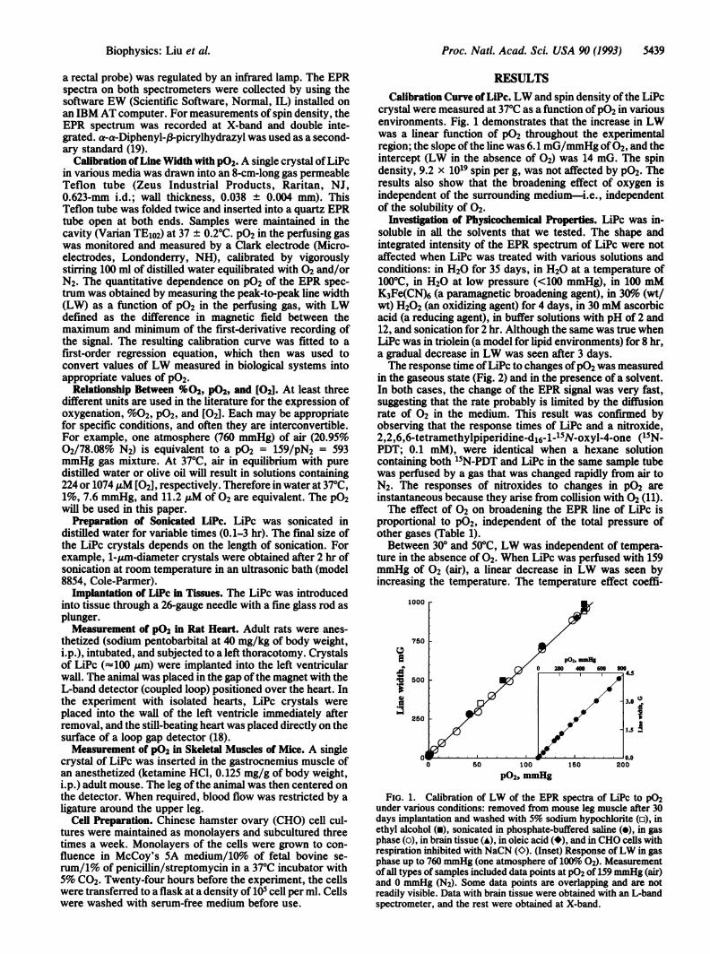
crystal were measured at 37°C as a function ofP02 in variousenvironments. Fig. 1 demonstrates that the increase in LWwas a linear function of P02 throughout the experimentalregion; the slope ofthe line was 6.1 mG/mmHg of02, and theintercept (LW in the absence of 02) was 14 mG. The spindensity, 9.2 x 10¹⁹ spin per g, was not affected by P02. Theresults also show that the broadening effect of oxygen isindependent of the surrounding medium-i.e., independentof the solubility of 02.
Investigation of Physicochemical Properties. LiPc was in-soluble in all the solvents that we tested. The shape andintegrated intensity of the EPR spectrum of LiPc were notaffected when LiPc was treated with various solutions andconditions: in H20 for 35 days, in H20 at a temperature of100°C, in H20 at low pressure (<100 mmHg), in 100 mMK3Fe(CN)6 (a paramagnetic broadening agent), in 30%o (wt/wt) H202 (an oxidizing agent) for 4 days, in 30 mM ascorbicacid (a reducing agent), in buffer solutions with pH of 2 and12, and sonication for 2 hr. Although the same was true whenLiPc was in triolein (a model for lipid environments) for 8 hr,a gradual decrease in LW was seen after 3 days.The response time ofLiPc to changes ofP02 was measured
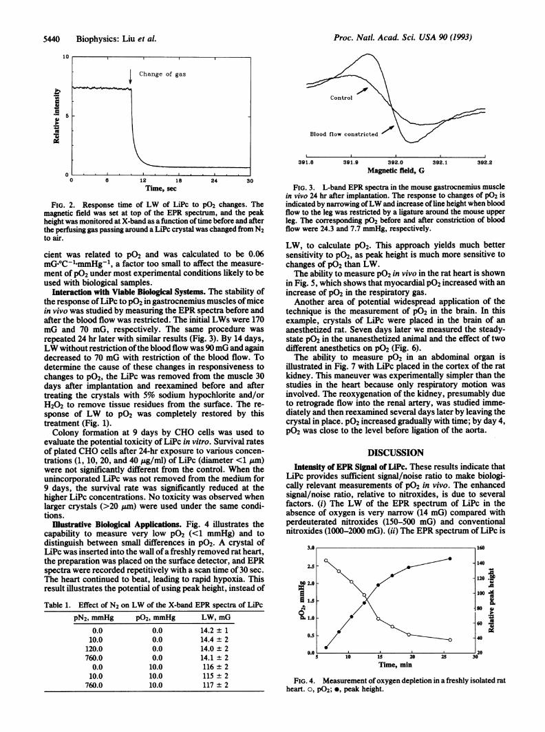
in the gaseous state (Fig. 2) and in the presence of a solvent.In both cases, the change of the EPR signal was very fast,suggesting that the rate probably is limited by the diffusionrate of 02 in the medium. This result was confirmed byobserving that the response times of LiPc and a nitroxide,2,2,6,6-tetramethylpiperidine-dl6-1-15N-oxyl-4-one (15N-PDT; 0.1 mM), were identical when a hexane solutioncontaining both 15N-PDT and LiPc in the same sample tubewas perfused by a gas that was changed rapidly from air toN2. The responses of nitroxides to changes in P02 areinstantaneous because they arise from collision with 02 (11).The effect of 02 on broadening the EPR line of LiPc is
proportional to P02, independent of the total pressure ofother gases (Table 1).Between 300 and 50°C, LW was independent of tempera-
ture in the absence of 02. When LiPc was perfused with 159mmHg of 02 (air), a linear decrease in LW was seen byincreasing the temperature. The temperature effect coeffi-
1000
750
P02 iuuuH2020 400 600 04
P 600
3.0
250
0 0.00 50 100 150 200
P02, mmHg
FIG. 1. Calibration of LW of the EPR spectra of LiPc to pO2under various conditions: removed from mouse leg muscle after 30days implantation and washed with 5% sodium hypochlorite (o), inethyl alcohol (m), sonicated in phosphate-buffered saline (*), in gasphase (o), in brain tissue (A), in oleic acid (*), and in CHO cells withrespiration inhibited with NaCN (>). (Inset) Response ofLW in gasphase up to 760 mmHg (one atmosphere of 100%o 02). Measurementof all types of samples included data points at P02 of 159 mmHg (air)and 0 mmHg (N2). Some data points are overlapping and are notreadily visible. Data with brain tissue were obtained with an L-bandspectrometer, and the rest were obtained at X-band.
Biophysics: Liu et al.
Proc. Natl. Acad. Sci. USA 90 (1993)
12 18Time, sec
24 30
FIG. 2. Response time of LW of LiPc to P02 changes. Themagnetic field was set at top of the EPR spectrum, and the peakheight was monitored at X-band as a function oftime before and afterthe perfusing gas passing around a LiPc crystal was changed from N2to air.
cient was related to P02 and was calculated to be 0.06mG.oC-1-mmHg-l, a factor too small to affect the measure-ment of P02 under most experimental conditions likely to beused with biological samples.
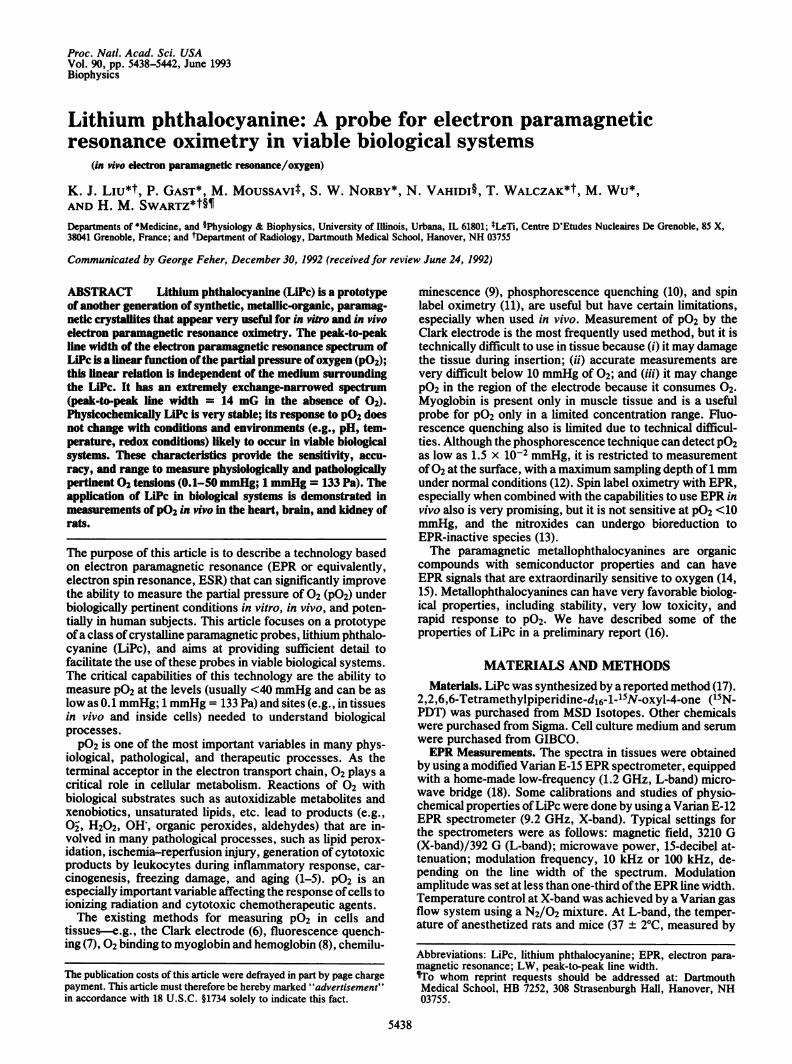
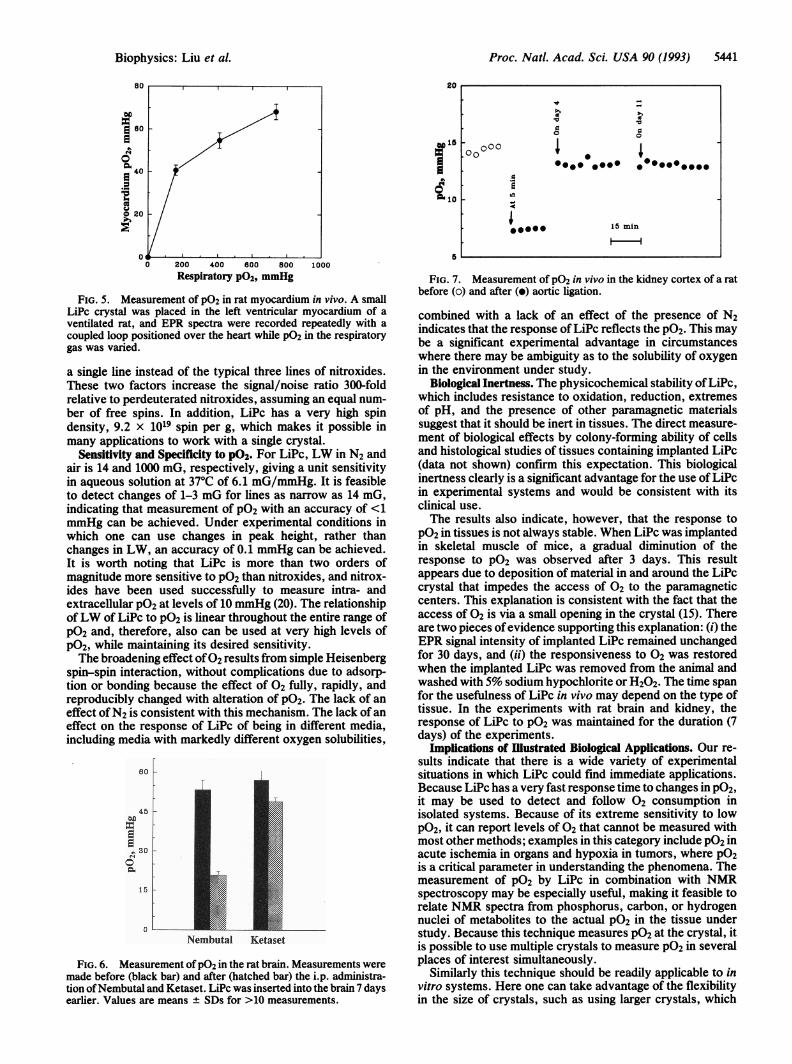
Interaction with Viable Biological Systems. The stability ofthe response ofLiPc to PO2 in gastrocnemius muscles ofmicein vivo was studied by measuring the EPR spectra before andafter the blood flow was restricted. The initial LWs were 170mG and 70 mG, respectively. The same procedure wasrepeated 24 hr later with similar results (Fig. 3). By 14 days,LW without restriction ofthe blood flow was 90 mG and againdecreased to 70 mG with restriction of the blood flow. Todetermine the cause of these changes in responsiveness tochanges to P02, the LiPc was removed from the muscle 30days after implantation and reexamined before and aftertreating the crystals with 5% sodium hypochlorite and/orH202 to remove tissue residues from the surface. The re-sponse of LW to P02 was completely restored by thistreatment (Fig. 1).Colony formation at 9 days by CHO cells was used to
evaluate the potential toxicity of LiPc in vitro. Survival ratesof plated CHO cells after 24-hr exposure to various concen-trations (1, 10, 20, and 40 pg/ml) of LiPc (diameter <1 ,um)were not significantly different from the control. When theunincorporated LiPc was not removed from the medium for9 days, the survival rate was significantly reduced at thehigher LiPc concentrations. No toxicity was observed whenlarger crystals (>20 um) were used under the same condi-tions.
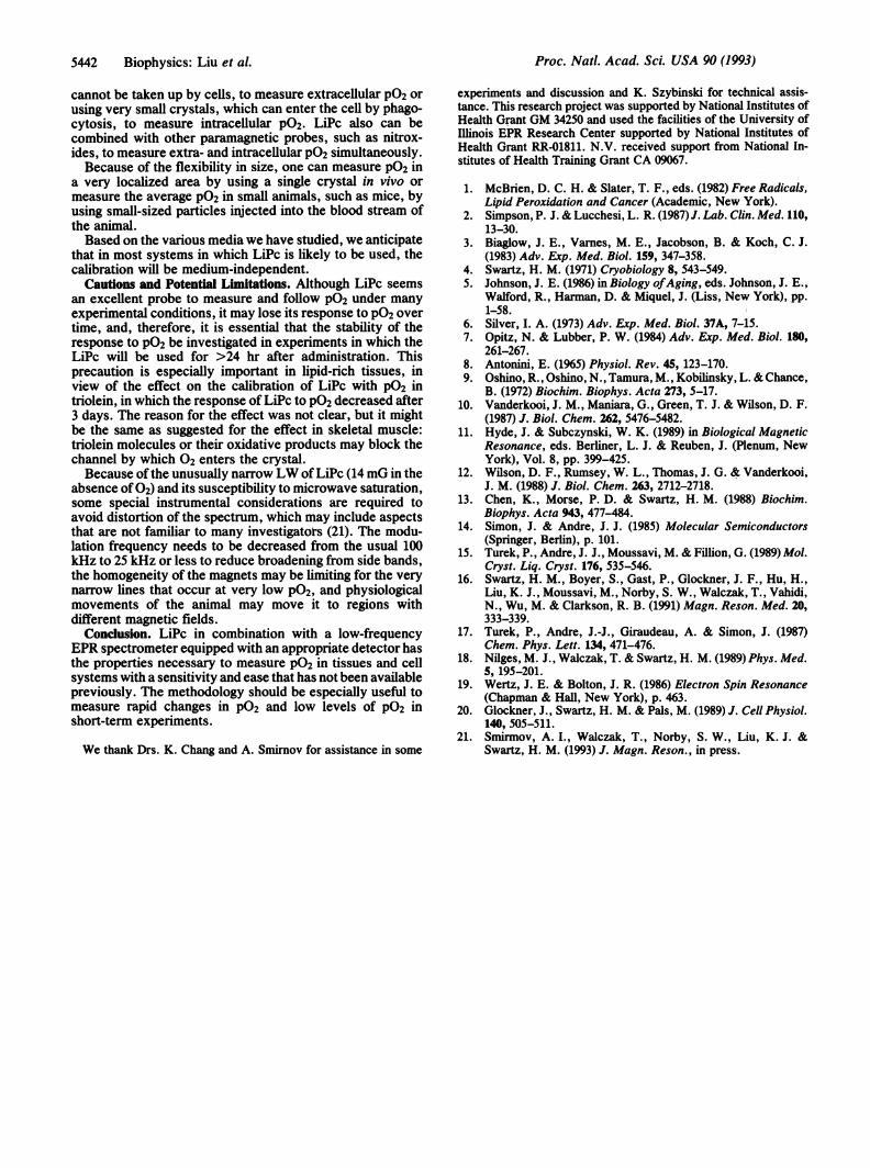
Illustrative Biological Applications. Fig. 4 illustrates thecapability to measure very low PO2 (<1 mmHg) and todistinguish between small differences in PO2. A crystal ofLiPc was inserted into the wall ofa freshly removed rat heart,the preparation was placed on the surface detector, and EPRspectra were recorded repetitively with a scan time of 30 sec.The heart continued to beat, leading to rapid hypoxia. Thisresult illustrates the potential of using peak height, instead of
Table 1. Effect of N2 on LW of the X-band EPR spectra of LiPc
pN2, mmHg P02, mmHg LW, mG
0.0 0.0 14.2 ± 110.0 0.0 14.4 ± 2
120.0 0.0 14.0 ± 2760.0 0.0 14.1 ± 2
0.0 10.0 116 ± 210.0 10.0 115 ± 2
760.0 10.0 117 + 2
Controlsrt
Blood flow constricted 'OOO/
391.8 391.9 392.0Magnetic field, G
392.1 392.2
FIG. 3. L-band EPR spectra in the mouse gastrocnemius musclein vivo 24 hr after implantation. The response to changes of P02 isindicated by narrowing ofLW and increase of line height when bloodflow to the leg was restricted by a ligature around the mouse upperleg. The corresponding P02 before and after constriction of bloodflow were 24.3 and 7.7 mmHg, respectively.
LW, to calculate P02. This approach yields much bettersensitivity to PO2, as peak height is much more sensitive tochanges of P02 than LW.The ability to measure P02 in vivo in the rat heart is shown
in Fig. 5, which shows that myocardial PO2 increased with anincrease of P02 in the respiratory gas.Another area of potential widespread application of the
technique is the measurement of P02 in the brain. In thisexample, crystals of LiPc were placed in the brain of ananesthetized rat. Seven days later we measured the steady-state PO2 in the unanesthetized animal and the effect of twodifferent anesthetics on PO2 (Fig. 6).The ability to measure P02 in an abdominal organ is
illustrated in Fig. 7 with LiPc placed in the cortex of the ratkidney. This maneuver was experimentally simpler than thestudies in the heart because only respiratory motion wasinvolved. The reoxygenation of the kidney, presumably dueto retrograde flow into the renal artery, was studied imme-diately and then reexamined several days later by leaving thecrystal in place. PO2 increased gradually with time; by day 4,PO2 was close to the level before ligation of the aorta.
DISCUSSIONIntensity of EPR Signal of LiPc. These results indicate that
LiPc provides sufficient signal/noise ratio to make biologi-cally relevant measurements of P02 in vivo. The enhancedsignal/noise ratio, relative to nitroxides, is due to severalfactors. (i) The LW of the EPR spectrum of LiPc in theabsence of oxygen is very narrow (14 mG) compared withperdeuterated nitroxides (150-500 mG) and conventionalnitroxides (1000-2000 mG). (ii) The EPR spectrum of LiPc is
2.5F
MO 2.0
1::Is 1.5
1.0
0.5
0.0
5 10 15 20
Time, min
140.5-
120 .T
ioo-100jX
80 w
60-
40
Iz e
Ii120
FIG. 4. Measurement of oxygen depletion in a freshly isolated ratheart. 0, P02; *, peak height.
0
0
0
0
0
-
J-V, lXW
5440 Biophysics: Liu et al.
25 30-
Proc. Natl. Acad. Sci. USA 90 (1993) 5441
80
40
2020
0 _0 200 400 600 800 1000
Respiratory P02, mmHg
FIG. 5. Measurement of P02 in rat myocardium in vivo. A small
LiPc crystal was placed in the left ventricular myocardium of aventilated rat, and EPR spectra were recorded repeatedly with a
coupled loop positioned over the heart while P02 in the respiratorygas was varied.
a single line instead of the typical three lines of nitroxides.These two factors increase the signal/noise ratio 300-foldrelative to perdeuterated nitroxides, assuming an equal num-ber of free spins. In addition, LiPc has a very high spindensity, 9.2 x 10¹⁹ spin per g, which makes it possible inmany applications to work with a single crystal.
Sensitivity and Specificity to P02. For LiPc, LW in N2 andair is 14 and 1000 mG, respectively, giving a unit sensitivityin aqueous solution at 37°C of 6.1 mG/mmHg. It is feasibleto detect changes of 1-3 mG for lines as narrow as 14 mG,indicating that measurement of PO2 with an accuracy of <1mmHg can be achieved. Under experimental conditions inwhich one can use changes in peak height, rather thanchanges in LW, an accuracy of 0.1 mmHg can be achieved.It is worth noting that LiPc is more than two orders ofmagnitude more sensitive to PO2 than nitroxides, and nitrox-ides have been used successfully to measure intra- andextracellular P02 at levels of 10 mmHg (20). The relationshipofLW of LiPc to P02 is linear throughout the entire range ofPO2 and, therefore, also can be used at very high levels ofP02, while maintaining its desired sensitivity.The broadening effect of02 results from simple Heisenberg
spin-spin interaction, without complications due to adsorp-tion or bonding because the effect of 02 fully, rapidly, andreproducibly changed with alteration of PO2. The lack of aneffect ofN2 is consistent with this mechanism. The lack of aneffect on the response of LiPc of being in different media,including media with markedly different oxygen solubilities,
60
45u
30
15
0
Nembutal Ketaset
FIG. 6. Measurement ofP02 in the rat brain. Measurements weremade before (black bar) and after (hatched bar) the i.p. administra-tion ofNembutal and Ketaset. LiPc was inserted into the brain 7 daysearlier. Values are means ± SDs for >10 measurements.
20
gf~~~~~00 0050
10 _ e _
15 *O 15min
5
FIG. 7. Measurement of PO2 in vivo in the kidney cortex of a ratbefore (o) and after (o) aortic ligation.
combined with a lack of an effect of the presence of N2indicates that the response ofLiPc reflects the PO2. This maybe a significant experimental advantage in circumstanceswhere there may be ambiguity as to the solubility of oxygenin the environment under study.
Biological Inertness. The physicochemical stability ofLiPc,which includes resistance to oxidation, reduction, extremesof pH, and the presence of other paramagnetic materialssuggest that it should be inert in tissues. The direct measure-ment of biological effects by colony-forming ability of cellsand histological studies of tissues containing implanted LiPc(data not shown) confirm this expectation. This biologicalinertness clearly is a significant advantage for the use of LiPcin experimental systems and would be consistent with itsclinical use.The results also indicate, however, that the response to
P02 in tissues is not always stable. When LiPc was implantedin skeletal muscle of mice, a gradual diminution of theresponse to PO2 was observed after 3 days. This resultappears due to deposition of material in and around the LiPccrystal that impedes the access of 02 to the paramagneticcenters. This explanation is consistent with the fact that theaccess of 02 is via a small opening in the crystal (15). Thereare two pieces ofevidence supporting this explanation: (i) theEPR signal intensity of implanted LiPc remained unchangedfor 30 days, and (ii) the responsiveness to 02 was restoredwhen the implanted LiPc was removed from the animal andwashed with 5% sodium hypochlorite or H202. The time spanfor the usefulness of LiPc in vivo may depend on the type oftissue. In the experiments with rat brain and kidney, theresponse of LiPc to PO2 was maintained for the duration (7days) of the experiments.
Implications of Illustrated Biological Applications. Our re-sults indicate that there is a wide variety of experimentalsituations in which LiPc could find immediate applications.Because LiPc has a very fast response time to changes in P02,it may be used to detect and follow 02 consumption inisolated systems. Because of its extreme sensitivity to lowPO2, it can report levels of 02 that cannot be measured withmost other methods; examples in this category include PO2 inacute ischemia in organs and hypoxia in tumors, where PO2is a critical parameter in understanding the phenomena. Themeasurement of PO2 by LiPc in combination with NMRspectroscopy may be especially useful, making it feasible torelate NMR spectra from phosphorus, carbon, or hydrogennuclei of metabolites to the actual P02 in the tissue understudy. Because this technique measures PO2 at the crystal, itis possible to use multiple crystals to measure PO2 in severalplaces of interest simultaneously.
Similarly this technique should be readily applicable to invitro systems. Here one can take advantage of the flexibilityin the size of crystals, such as using larger crystals, which
Biophysics: Liu et al.
Proc. Natl. Acad. Sci. USA 90 (1993)
cannot be taken up by cells, to measure extracellular P02 orusing very small crystals, which can enter the cell by phago-cytosis, to measure intracellular P02. LiPc also can becombined with other paramagnetic probes, such as nitrox-ides, to measure extra- and intracellular P02 simultaneously.Because of the flexibility in size, one can measure PO2 in
a very localized area by using a single crystal in vivo ormeasure the average PO2 in small animals, such as mice, byusing small-sized particles injected into the blood stream ofthe animal.Based on the various media we have studied, we anticipate
that in most systems in which LiPc is likely to be used, thecalibration will be medium-independent.
Cautions and Potential Limitations. Although LiPc seemsan excellent probe to measure and follow P02 under manyexperimental conditions, it may lose its response to P02 overtime, and, therefore, it is essential that the stability of theresponse to PO2 be investigated in experiments in which theLiPc will be used for >24 hr after administration. Thisprecaution is especially important in lipid-rich tissues, inview of the effect on the calibration of LiPc with P02 intriolein, in which the response of LiPc to P02 decreased after3 days. The reason for the effect was not clear, but it mightbe the same as suggested for the effect in skeletal muscle:triolein molecules or their oxidative products may block thechannel by which 02 enters the crystal.Because ofthe unusually narrowLW of LiPc (14 mG in the
absence of 02) and its susceptibility to microwave saturation,some special instrumental considerations are required toavoid distortion of the spectrum, which may include aspectsthat are not familiar to many investigators (21). The modu-lation frequency needs to be decreased from the usual 100kH{z to 25 kHz or less to reduce broadening from side bands,the homogeneity of the magnets may be limiting for the verynarrow lines that occur at very low PO2, and physiologicalmovements of the animal may move it to regions withdifferent magnetic fields.Condusion. LiPc in combination with a low-frequency
EPR spectrometer equipped with an appropriate detector hasthe properties necessary to measure PO2 in tissues and cellsystems with a sensitivity and ease that has not been availablepreviously. The methodology should be especially useful tomeasure rapid changes in P02 and low levels of P02 inshort-term experiments.
We thank Drs. K. Chang and A. Smirnov for assistance in some
experiments and discussion and K. Szybinski for technical assis-tance. This research project was supported by National Institutes ofHealth Grant GM 34250 and used the facilities of the University ofIllinois EPR Research Center supported by National Institutes ofHealth Grant RR-01811. N.V. received support from National In-stitutes of Health Training Grant CA 09067.
1. McBrien, D. C. H. & Slater, T. F., eds. (1982) Free Radicals,Lipid Peroxidation and Cancer (Academic, New York).
2. Simpson, P. J. & Lucchesi, L. R. (1987)J. Lab. Clin. Med. 110,13-30.
3. Biaglow, J. E., Varnes, M. E., Jacobson, B. & Koch, C. J.(1983) Adv. Exp. Med. Biol. 159, 347-358.
4. Swartz, H. M. (1971) Cryobiology 8, 543-549.5. Johnson, J. E. (1986) in Biology ofAging, eds. Johnson, J. E.,
Walford, R., Harman, D. & Miquel, J. (Liss, New York), pp.1-58.
6. Silver, I. A. (1973) Adv. Exp. Med. Biol. 37A, 7-15.7. Opitz, N. & Lubber, P. W. (1984) Adv. Exp. Med. Biol. 180,
261-267.8. Antonini, E. (1965) Physiol. Rev. 45, 123-170.9. Oshino, R., Oshino, N., Tamura, M., Kobilinsky, L. & Chance,
B. (1972) Biochim. Biophys. Acta 273, 5-17.10. Vanderkooi, J. M., Maniara, G., Green, T. J. & Wilson, D. F.
(1987) J. Biol. Chem. 262, 5476-5482.11. Hyde, J. & Subczynski, W. K. (1989) in Biological Magnetic
Resonance, eds. Berliner, L. J. & Reuben, J. (Plenum, NewYork), Vol. 8, pp. 399-425.
12. Wilson, D. F., Rumsey, W. L., Thomas, J. G. & Vanderkooi,J. M. (1988) J. Biol. Chem. 263, 2712-2718.
13. Chen, K., Morse, P. D. & Swartz, H. M. (1988) Biochim.Biophys. Acta 943, 477-484.
14. Simon, J. & Andre, J. J. (1985) Molecular Semiconductors(Springer, Berlin), p. 101.
15. Turek, P., Andre, J. J., Moussavi, M. & Fillion, G. (1989) Mol.Cryst. Liq. Cryst. 176, 535-546.
16. Swartz, H. M., Boyer, S., Gast, P., Glockner, J. F., Hu, H.,Liu, K. J., Moussavi, M., Norby, S. W., Walczak, T., Vahidi,N., Wu, M. & Clarkson, R. B. (1991) Magn. Reson. Med. 20,333-339.
17. Turek, P., Andre, J.-J., Giraudeau, A. & Simon, J. (1987)Chem. Phys. Lett. 134, 471-476.
18. Nilges, M. J., Walczak, T. & Swartz, H. M. (1989) Phys. Med.5, 195-201.
19. Wertz, J. E. & Bolton, J. R. (1986) Electron Spin Resonance(Chapman & Hall, New York), p. 463.
20. Glockner, J., Swartz, H. M. & Pals, M. (1989) J. Cell Physiol.140, 505-511.
21. Smirmov, A. I., Walczak, T., Norby, S. W., Liu, K. J. &Swartz, H. M. (1993) J. Magn. Reson., in press.
5442 Biophysics: Liu et al.