Embed Size (px)
Citation preview

Literature review
A novel approach for biotransformation of L- tyrosine to melanin by microbial system
8
“An expert is a man who has made all the mistakes
which can be made in a very narrow field”
- Edward Teller
II
Literature review
Literature review
A novel approach for biotransformation of L- tyrosine to melanin by microbial system
9
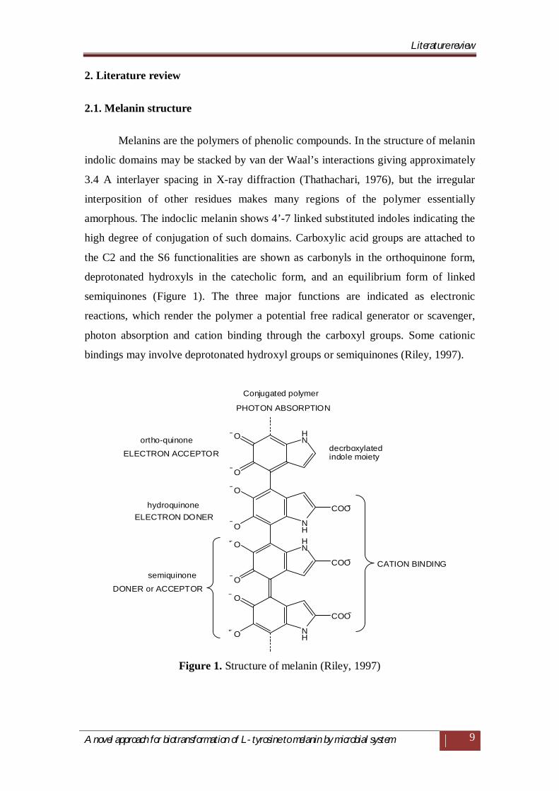
2. Literature review
2.1. Melanin structure
Melanins are the polymers of phenolic compounds. In the structure of melanin
indolic domains may be stacked by van der Waal’s interactions giving approximately
3.4 A interlayer spacing in X-ray diffraction (Thathachari, 1976), but the irregular
interposition of other residues makes many regions of the polymer essentially
amorphous. The indoclic melanin shows 4’-7 linked substituted indoles indicating the
high degree of conjugation of such domains. Carboxylic acid groups are attached to
the C2 and the S6 functionalities are shown as carbonyls in the orthoquinone form,
deprotonated hydroxyls in the catecholic form, and an equilibrium form of linked
semiquinones (Figure 1). The three major functions are indicated as electronic
reactions, which render the polymer a potential free radical generator or scavenger,
photon absorption and cation binding through the carboxyl groups. Some cationic
bindings may involve deprotonated hydroxyl groups or semiquinones (Riley, 1997).
HN
NHHN
NH
O
O
O
O
O
O
O
O
COO
COO
COO
PHOTON ABSORPTION
ELECTRON ACCEPTOR
ELECTRON DONER
DONER or ACCEPTOR
CATION BINDING
ortho-quinone
hydroquinone
semiquinone
decrboxylatedindole moiety
Conjugated polymer
Figure 1. Structure of melanin (Riley, 1997)
Literature review
A novel approach for biotransformation of L- tyrosine to melanin by microbial system
10
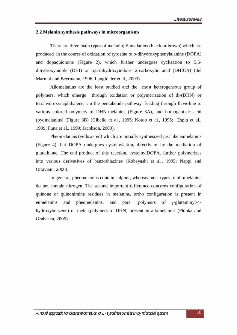
2.2 Melanin synthesis pathways in microorganisms
There are three main types of melanin; Eumelanins (black or brown) which are
produced in the course of oxidation of tyrosine to o-dihydroxyphenylalanine (DOPA)
and dopaquionone (Figure 2), which further undergoes cyclization to 5,6-
dihydroxyindole (DHI) or 5,6-dihydroxyindole- 2-carboxylic acid (DHICA) (del
Marmol and Beermann, 1996; Langfelder et al., 2003).
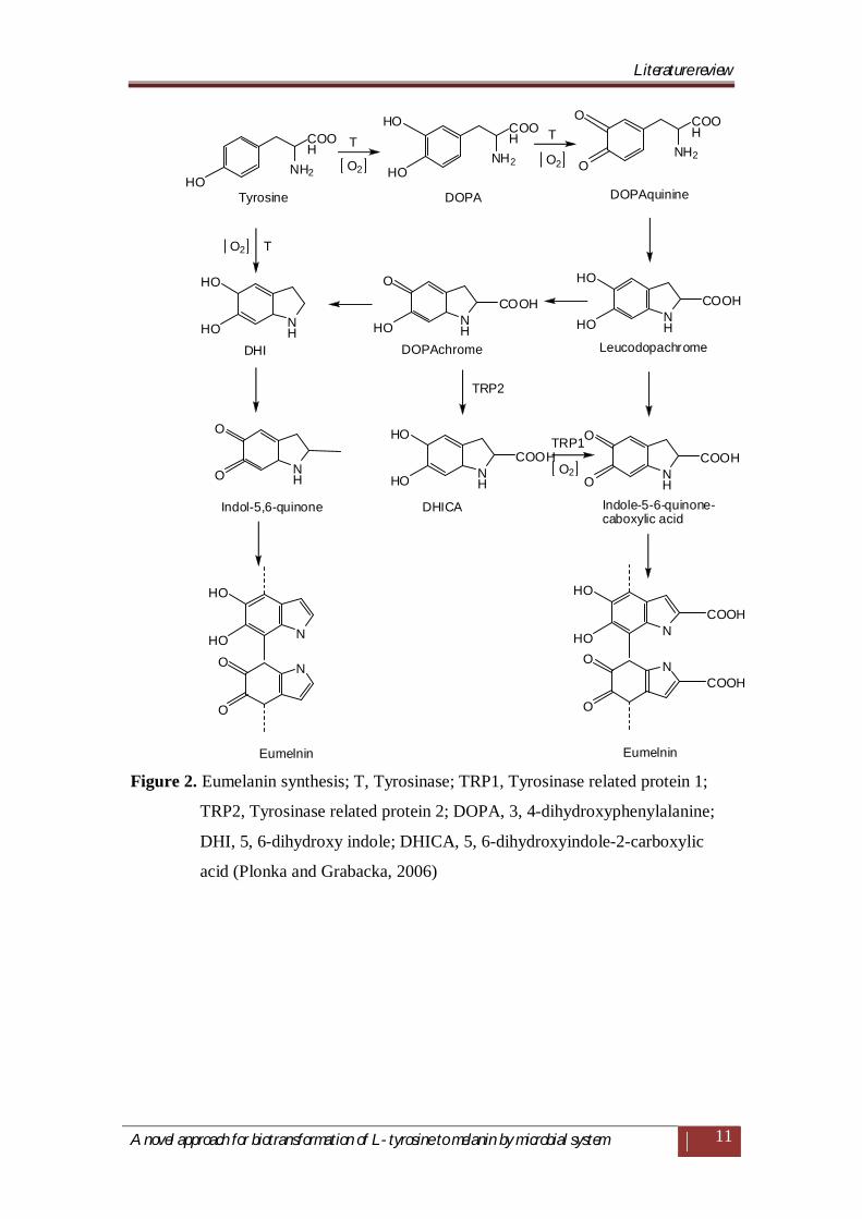
Allomelanins are the least studied and the most heterogeneous group of
polymers, which emerge through oxidation or polymerization of di-(DHN) or
tetrahydroxynaphthalene, via the pentaketide pathway leading through flavioline to
various colored polymers of DHN-melanins (Figure 3A), and homogentisic acid
(pyomelanins) (Figure 3B) (Gibello et al., 1995; Kotob et al., 1995; Espin et al.,
1999; Funa et al., 1999; Jacobson, 2000).
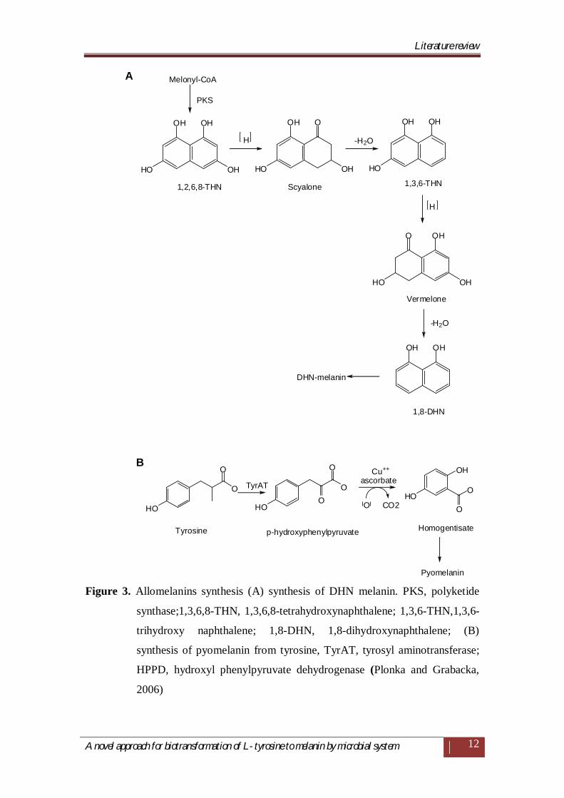
Pheomelanins (yellow-red) which are initially synthesized just like eumelanins
(Figure 4), but DOPA undergoes cysteinylation, directly or by the mediation of
glutathione. The end product of this reaction, cysteinylDOPA, further polymerizes
into various derivatives of benzothiazines (Kobayashi et al., 1995; Nappi and
Ottaviani, 2000).
In general, pheomelanins contain sulphur, whereas most types of allomelanins
do not contain nitrogen. The second important difference concerns configuration of
quinone or quinonimine residues in melanins, ortho configuration is present in
eumelanins and pheomelanins, and para (polymers of γ-glutaminyl-4-
hydroxybenzene) or meta (polymers of DHN) present in allomelanins (Plonka and
Grabacka, 2006).
Literature review
A novel approach for biotransformation of L- tyrosine to melanin by microbial system
11
NH2
COOH
NH
N
N
O
O
O
HO
HO
HO
HO
COOHNH
HO
HO
COOHNH
HO
HO
NH2
COOH
HONH2
COOH
O
NH
O
O NH
HO
HO
COOHNH
O
O
COOH
N
N
O
O
HO
HO
COOH
COOH
HO O
Tyrosine DOPA DOPAquinine
DHI DOPAchrome Leucodopachrome
Indol-5,6-quinone DHICA Indole-5-6-quinone-caboxylic acid
Eumelnin Eumelnin
T T
O2O2
T
TRP2
TRP1
O2
O2
Figure 2. Eumelanin synthesis; T, Tyrosinase; TRP1, Tyrosinase related protein 1;
TRP2, Tyrosinase related protein 2; DOPA, 3, 4-dihydroxyphenylalanine;
DHI, 5, 6-dihydroxy indole; DHICA, 5, 6-dihydroxyindole-2-carboxylic
acid (Plonka and Grabacka, 2006)
Literature review
A novel approach for biotransformation of L- tyrosine to melanin by microbial system
12
OO
O
O
O
O
OH OH
HO
OH
HO OHOH HO
OH OH
OH
OHOH
OHHO
HO HOHO
OH
O
O
O
Melonyl-CoA
PKS
H -H2O
-H2O
H
TyrAT ascorbateCu++
O CO2
1,2,6,8-THN Scyalone 1,3,6-THN
Vermelone
DHN-melanin
Tyrosine p-hydroxyphenylpyruvate Homogentisate
Pyomelanin
A
B
1,8-DHN
Figure 3. Allomelanins synthesis (A) synthesis of DHN melanin. PKS, polyketide
synthase;1,3,6,8-THN, 1,3,6,8-tetrahydroxynaphthalene; 1,3,6-THN,1,3,6-
trihydroxy naphthalene; 1,8-DHN, 1,8-dihydroxynaphthalene; (B)
synthesis of pyomelanin from tyrosine, TyrAT, tyrosyl aminotransferase;
HPPD, hydroxyl phenylpyruvate dehydrogenase (Plonka and Grabacka,
2006)
Literature review
A novel approach for biotransformation of L- tyrosine to melanin by microbial system
13
NH2NH2
S
NNH2
HO
H2N S
NH
S
NH2
NH2N
S
COOHCOOH
OH
COOH
COOH COOH
COOH
OH
O
O
HO
HO
DOPAquinone
CysteinlyDOPA
1,4-Benzothiazinylalanine
Pheomelanin
Cystine
glutathione
Figure 4. Pheomelanin synthesis (Plonka and Grabacka, 2006)
2.3 Microbial enzymes of melanogenesis
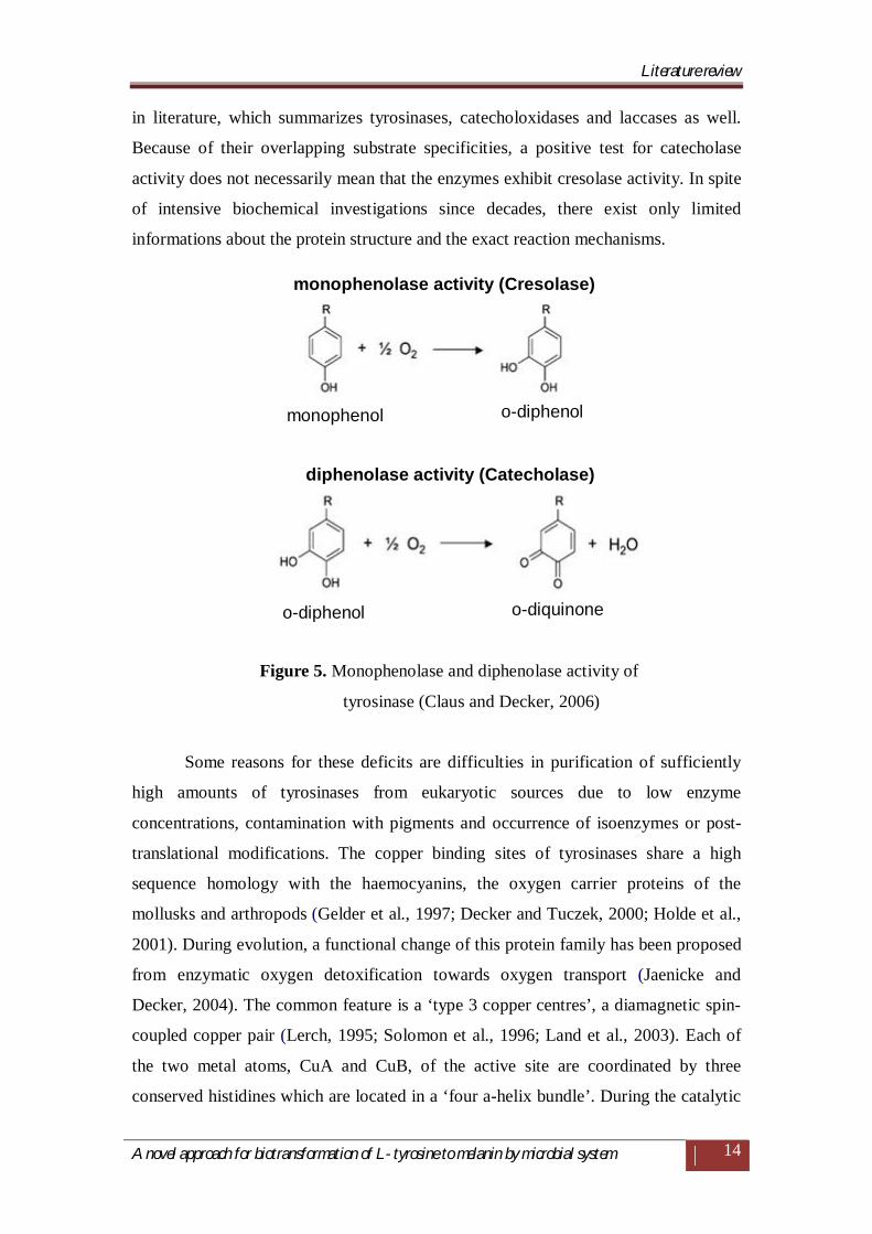
2.3.1 Tyrosinase
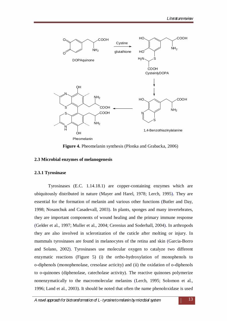
Tyrosinases (E.C. 1.14.18.1) are copper-containing enzymes which are
ubiquitously distributed in nature (Mayer and Harel, 1978; Lerch, 1995). They are
essential for the formation of melanin and various other functions (Butler and Day,
1998; Nosanchuk and Casadevall, 2003). In plants, sponges and many invertebrates,
they are important components of wound healing and the primary immune response
(Gelder et al., 1997; Muller et al., 2004; Cerenius and Soderhall, 2004). In arthropods
they are also involved in sclerotization of the cuticle after molting or injury. In
mammals tyrosinases are found in melanocytes of the retina and skin (Garcia-Borro
and Solano, 2002). Tyrosinases use molecular oxygen to catalyze two different
enzymatic reactions (Figure 5) (i) the ortho-hydroxylation of monophenols to
o-diphenols (monophenolase, cresolase acticity) and (ii) the oxidation of o-diphenols
to o-quinones (diphenolase, catecholase activity). The reactive quinones polymerize
nonenzymatically to the macromolecular melanins (Lerch, 1995; Solomon et al.,
1996; Land et al., 2003). It should be noted that often the name phenoloxidase is used
Literature review
A novel approach for biotransformation of L- tyrosine to melanin by microbial system
14
in literature, which summarizes tyrosinases, catecholoxidases and laccases as well.
Because of their overlapping substrate specificities, a positive test for catecholase
activity does not necessarily mean that the enzymes exhibit cresolase activity. In spite
of intensive biochemical investigations since decades, there exist only limited
informations about the protein structure and the exact reaction mechanisms.
Figure 5. Monophenolase and diphenolase activity of
tyrosinase (Claus and Decker, 2006)
Some reasons for these deficits are difficulties in purification of sufficiently
high amounts of tyrosinases from eukaryotic sources due to low enzyme
concentrations, contamination with pigments and occurrence of isoenzymes or post-
translational modifications. The copper binding sites of tyrosinases share a high
sequence homology with the haemocyanins, the oxygen carrier proteins of the
mollusks and arthropods (Gelder et al., 1997; Decker and Tuczek, 2000; Holde et al.,
2001). During evolution, a functional change of this protein family has been proposed
from enzymatic oxygen detoxification towards oxygen transport (Jaenicke and
Decker, 2004). The common feature is a ‘type 3 copper centres’, a diamagnetic spin-
coupled copper pair (Lerch, 1995; Solomon et al., 1996; Land et al., 2003). Each of
the two metal atoms, CuA and CuB, of the active site are coordinated by three
conserved histidines which are located in a ‘four a-helix bundle’. During the catalytic
monophenolase activity (Cresolase)
monophenol o-diphenol
o-diphenol o-diquinone
diphenolase activity (Catecholase)
Literature review
A novel approach for biotransformation of L- tyrosine to melanin by microbial system
15
cycle the ‘type 3 copper centre’ can adopt different functional forms: the oxy-state
[Cu (II)–O2 2-Cu (II)], deoxy-state [Cu (I) Cu (I) ], half-met state [Cu(I)Cu(II)] and
the met state [Cu(II)–OH-Cu(II)]. In the latter case the two copper atoms are bridged
by hydroxy ions. The valences of the two copper atoms change from Cu (I) to Cu (II),
which can be followed spectroscopically. In the oxy-state the molecular oxygen is
reversibly bound as peroxide between the two copper atoms in a ‘side-on’
conformation. In the absence of any substrate more than 85% of the enzyme is in the
met state, which can be regarded as the resting form of tyrosinase. According to
current conceptions, both, the met and the oxy state of tyrosinases enable the
diphenoloxidase activity, whereas the monohydroxylase reaction requires the oxy
state (Claus and Decker, 2006).
2.3.2 Laccase
Melanization in Streptomyces species and other microorganisms not only
catalyzed by tyrosinases but also via different pathways and enzymes which include
laccase (Butler and Day, 1998; Castro-Sowinski et al., 2002). Laccases (EC1.10.3.2,
p-diphenol: dioxygen oxidoreductases) are multi-copper proteins that use molecular
oxygen to oxidize various aromatic and non-aromatic compounds. (Solomon et al.,
1996; Claus, 2003). Until recently, laccases have been only found in eukaryotes
(fungi, higher plants and insects) but now there is strong evidence for their wide
distribution in prokaryotes (Claus, 2003). For the catalytic activity a minimum of four
copper atoms (type 1, type 2 and type 3) per active protein unit is needed (Solomon et
al., 1996; Claus, 2003). The occurrence of a spin coupled copper pair (type 3 copper)
is the common feature of the multi-copper oxidases and the protein super family of
tyrosinases and haemocyanins (Lerch, 1995; Decker and Tuczek, 2000). In contrast
to tyrosinases, laccases exhibit no monophenol hydroxylase activity, but oxidize
phenols by a radical-generating reaction mechanism (Claus, 2003). Some
microorganisms show both tyrosinase and laccase activities, and care must be taken to
avoid confusion because of overlapping substrate specificities. In Sinorhizobium
meliloti, a plasmid-encoded tyrosinase and a laccase have been demonstrated
(Mercado-Blanco et al., 1993; Castro-Sowinski et al., 2002). In addition to a typical
tyrosinase, a ‘multipotent’ phenoloxidase with both tyrosinase and laccase activities
has been purified from the marine bacterium Marinomanas mediterranea. The protein
Literature review
A novel approach for biotransformation of L- tyrosine to melanin by microbial system
16
shows some extra histidine-rich copper-binding domains that are very likely related to
its unique enzymatic properties (Sanchez-Amat et al., 2001). The phenoxazinone
synthase of Streptomyces antibioticus which is involved in the biosynthesis of
actinomycin is a multi-copper enzyme with laccase activity (Freeman et al., 1993). In
the meanwhile, more laccases have been isolated and characterized from
Streptomyces cyaneus (Arias et al., 2003), Streptomyces griseus (Endo et al., 2003)
and Streptomyces lavendulae (Suzuki et al., 2003).
2.3.3 Polyketide synthases (PKS)
It produces DHN melanins, and belongs to an old family of multidomain
proteins related to the animal fatty acid synthases (Kroken et al., 2003). The PKS
family is diversified and plentiful. Numerous microorganisms employ these enzymes
to produce pigments, antibiotics, toxins and other products of intermediate
metabolism (Hutchinson, 2003; Snyder et al., 2003). There are only few DHN-
melanin-producing PKS enzymes, which belong to the PKS-type-I group producing
aromatic, not reduced polyketides (Kroken et al., 2003).
2.4 Properties of Melanin
2.4.1 Light absorbance
The melanin polymer has many interesting properties, among which the most
conspicuous is the wide spectral absorbance due to high degree of conjugation in the
molecule. The darkness of the pigment is a result of the fact that much of the visible
spectrum is absorbed, including radiation with low quanta1 energy. The lowest energy
transitions are from nonbonding to anti-bonding pi-orbitals that occur predominantly
in carbonyl bonds, which are abundant in most melanins. Melanins also absorb in the
ultra-violet (UV) region of the spectrum, involving transitions from bonding to anti-
bonding pi-orbitals, which occur in unsaturated carbon bonds. Transitions from
bonding to anti-bonding orbitals are facilitated by conjugation permitting electronic
delocalization. As the degree of conjugation increases, lower quanta energies are
required for absorption; an effect termed bathochromicity. The majority of the visible
spectral energy absorbed by melanin is converted into heat through photon-phonon
coupling. Melanins with high levels of indole quinones (eumelanins) appear darker
Literature review
A novel approach for biotransformation of L- tyrosine to melanin by microbial system
17
because of the strong absorbance in the red part of the spectrum. This low frequency
light absorption is largely through the carbonyls (Riley 1997).
2.4.2 Redox properties
Melanins, especially eumelanins, exhibit marked redox properties, and
electron delocalization between orthoquinone and catecholic moieties of the polymer
give rise to semiquinone free radicals, which can be detected by electron spin
resonance spectroscopy (Sealy et al., 1980). Melanins can take part in one-electron
and two-electron redox reactions and one of the effects of light absorption is photo-
oxidation of the pigment, which, by increasing the carbonyl content, changes the
absorbance properties of melanin, the so called immediate pigment darkening (IPD)
reaction. This photo-oxidation process generates super oxide radicals (Sarna and
Sealy, 1984). Melanins also have powerful cation chelating properties (Sarna et al.,
1976) through the anionic functions such as the carboxyl and the deprotonated
hydroxyl groups.
2.5 Roles of melanin synthesis in microorganisms
2.5.1 Electron acceptor
In the process of anaerobic respiration, reducing, dissimilating bacteria use a
wide spectrum of electron acceptors replacing dioxygen in the last step of the
respiratory chain. Among the substances utilized as alternative electron acceptors,
there are mainly hydrated ferrous oxide, nitrates, sulphates, as well as organic
compounds, in particular the ones containing quinone groups, e.g. related to melanin
humic substances (Coates et al., 2002). Melanin, similarly to humic substances, is a
polymer of various groups able to donate or to accept an electron. Therefore it can act
as a final acceptor or a shuttle in the electron exchange with insoluble compounds of
iron (Menter and Willis, 1997).
The facultatively anaerobic bacterium Shewanella algae produce pyomelanin
and reduce it simultaneously with the oxidation of gaseous hydrogen. As Shewanella
algae is unable to carry out fermentation, its survival in the conditions of variable
oxygen concentration strongly depends on the presence of appropriate electron
acceptors. In the mineralized marine deposits the availability of such soluble
Literature review
A novel approach for biotransformation of L- tyrosine to melanin by microbial system
18
compounds is limited; therefore production of melanin is an important evolutionary
adaptation. Moreover, like other organisms, Shewanella algae produces melanin also
for the protection from ultraviolet irradiation (Turick et al., 2002).
2.5.2 Virulence factors
2.5.2.1 Escaping respiratory burst
The pathogenic bacterium Burkholderia cepacia serves as an example of how
melanin production increases virulence (Zughaier et al., 1999). This microorganism
causes serious lung infections, which often develop to sepsis, mainly due to the
presence of lipo-polysaccharide (LPS) which strongly enhances production and
release of pro inflammatory cytokines. Although LPS does not trigger an oxygen
burst directly, it stimulates the immunological system to an accelerated oxidative
response to other stimuli. Melanin isolated from Burkholderia cepacia reveals a dose-
dependent ability to sweep oxygen radical O2¯ produced by leukocytes during oxygen
burst. However, melanin does not influence the release or kinetics of the production
of reactive oxygen species (ROS). The ability to remove superoxide anion allows
Burkholderia cepacia to survive phagocytosis, so the host phagocytes are not able to
eliminate the pathogen. They remain in the state of a permanent stimulation by the
bacterial LPS, which causes a chronic inflammation. Similarly to Legionella
pneumoniae, Burkholderia cepacia is able not only to survive in the phagocytes
(alveolar macrophages), but also to proliferate intracellularly, which leads to cell
destruction and the infection of secondary macrophages (Saini et al., 1999; Abu-Zant
et al., 2005).
2.5.2.2 Escaping phagocytosis
The ability to produce melanin as an important factor of virulence is well
documented and confirmed in vivo for Cryptococcus neoformans. The presence of
melanin in the cell wall of C. neoformans is correlated with less efficient
phagocytosis, both in the case of C. neoformans, and S. schenckii (Nosanchuk and
Casadevall, 1997; Romero-Martinez et al., 2000). A likely explanation is that
phagocytosis is impaired by the decrease of the negative electric charge of the cell
Literature review
A novel approach for biotransformation of L- tyrosine to melanin by microbial system
19
wall, which is caused by melanin deposition (Wang et al., 1995; Nosanchuk and
Casadevall, 1997).
2.5.2.3 Antibiotic resistance
The melanin can increase the resistance of pathogenic microbes though
binding with potential antibiotics such as tetracycline and vancomyin (Ikeda et al.,
2003; Lin et al., 2005; Nosanchuk and Casadevall, 2006).
2.5.2.4 Resistance against UV and gamma radiations
Melanins confer resistance to UV light by absorbing a broad range of the
electromagnetic spectrum and preventing photo induced damage (Hill, 1992).
Melanin protects several fungal and bacterial species from UV, solar or gamma
radiation. Increased melanin production is associated with the greater resistance of
pigmented fungi to radiation (Vasilevskaya, 1970; Zhdanova et al., 1973). The
protective properties of melanin against radiation injury could account for the growth
of black fungi in the highly contaminated atomic reactors (Nosanchuk and Casadevall,
2006)
2.5.2.5 Heavy metal resistance
Melanins are able to bind with the heavy metals that are routinely found in the
environment (Zunino and Martin, 1977; Rizzo et al., 1992; Fogarty and Tobin, 1996).
The carboxyl, phenolic, hydroxyl, and amine groups on melanin provide numerous
potential binding sites for metal ions (Fogarty and Tobin, 1996). Melanized
Cryptococcus neoformans cells are more resistant to killing by silver nitrate, a
compound highly toxic to bacteria and fungi, than nonmelanized cells (Garcia-Rivera
and Casadevall, 2001).
2.6 Applications of melanin
2.6.1 Cosmetics
The melanin is used in Sun screen creams and other skin whitening creams
basically for its UV radiations protecting and free radical scavenging properties. It is
Literature review
A novel approach for biotransformation of L- tyrosine to melanin by microbial system
20
also used in photoprotective eye glasses (Riley 1997; Nosanchuk and Casadevall,
2006). The inhibitory effect of skin whitening creams was studied earlier on L-DOPA
oxidation and tyrosinase activity. The melanin producing microorganisms can be used
as test organism to find out the skin whitening agents (Jeon et al., 2005).
2.6.2 Bioinsecticide protection from UV radiations
The bioisecticidal endotoxin produced from Bacillus thuringiensis is easily
gets inactivated by solar radiation in nature. Natural sunlight, especially the UV-A
(400 nm to 320 nm) and UV-B (320 nm to 290 nm) portions of the spectrum, is
responsible for the inactivation of microbial insecticides (Pusztai et al., 1991). The
melanin produced from Aeromonas media and Bacillus cereus 58 when added with
bioinsecticidal preparations the result indicates that the melanin can protect the
insecticidal crystal proteins from degradation caused by UV radiation (Wan et al.,
2007; Zhang et al., 2007)
2.6.3 Vaccine against human melanocyte cancer (Melanoma)
The melanin can be used as vaccine against melanoma (Human melanocyte
cancer). Melanoma is a malignant tumor of melanocytes which are found
predominantly in skin but also in the bowel and the eye. It is one of the less common
types of skin cancer but causes the majority (75%) of skin cancer related deaths.
Malignant melanoma of skin accounts for 1,60,000 new cases annually and in the
United States, about one in 150 people will develop a malignant melanoma during
their lifetime (Parkin et al., 2005; Jemal et al., 2007). The lymphocytes of melanoma
patients can be restimulated in vitro with autologous tumor cells to generate antitumor
cytolytic T lymphocytes (CTL) such antitumor CTL clones which appear to recognize
melanin as antigen. Also when blood lymphocytes of melanoma patients are
stimulated in vitro with tumor cells of the same patient, one often observes the
proliferation of T lymphocytes that exert cytolytic activity on the autologous
melanoma cells (Brichard et al., 1993). The antibody response to fungal melanin was
showed that melanin can be immunogenic, and the humoral immune response is T cell
independent. The melanin antigen may therefore constitute a useful target for specific
immunotherapy of melanoma. This clearly indicates that melanin can be used as
vaccine against the human melanocyte cancer (Nosanchuk et al., 1998).
Literature review
A novel approach for biotransformation of L- tyrosine to melanin by microbial system
21
2.6.4 Anti-HIV property
The melanin in solution inhibits the HIV (Human immunodeficiency virus) by
blocking syncyticum formation between viral surface glycoprotein gp120 and surface
of uninfected cell. Also the anti-HIV property was found due to the gp120-CD4
binding, thus melanin could prove to be a new class of pharmacologically active
substances with possible utility as anti-HIV therapeutics (Montefiori and Zhou 1991).
2.6.5 Bioremediation of radioactive waste
Microbial melanin production by bacteria was used to increased uranium
immobilization in uranium contaminated soil. In order to develop an in situ, uranium
bio-immobilization technology the one-time addition of tyrosine to soil exploited the
ability of indigenous microbes to produce pyomelanin, resulting in uranium
immobilization. Thus melanin producing bacteria can be used for bioremediation of
radioactive waste (Turick et al., 2008).
2.6.6 Reporter gene
The genes responsible for the melanin synthesis from bacteria were used as a
reporter gene to screen the recombination in host bacteria. The production of melanin
on tyrosine agar indicates the wild type while the absence of melanin indicates the
desired gene was transferred. Thus melanin producing genes can be a best alternative
to generally used blue white screening method in E. coli. (Tseng et al., 1990, Adham
et al., 2003).
2.7 Melanin producing microorganisms
Melanin synthesis ability is generally found in pathogenic strains of bacteria.
The melanin synthesis using homogentisic acid as a precursor of was first reported in
Vibrio cholerae, Hyphomonas species and Shewanella colwelliana (Kotob et al.,
1995). The synthesis of melanin and its characterization such as solubility, free radical
nature was initially studied in Proteus mirabilis (Agodi et al., 1996). A novel marine
bacterium Alteromonas strain MMB-1, was isolated from the Mediterranean Sea and
its melanin synthesis ability was studied using L-tyrosine as a precursor previously
(Solano et al., 1997). The melanin pigment from Burkholderia cepacia was formerly
Literature review
A novel approach for biotransformation of L- tyrosine to melanin by microbial system
22
reported for escaping monocyte respiratory burst activity by scavenging superoxide
anion (Zughaier et al., 1999).
The extra cellular melanin from Shewanella algae BrY was reported
previously to serve as the sole terminal electron acceptor. Upon reduction the reduced,
soluble melanin reduced insoluble hydrous ferric oxide in the absence of bacteria, and
melanin was proved as a soluble Fe (III)-reducing compound (Turick et al. 2002).
Melanin production was earlier studied by UV-resistant mutant of Bacillus
thuringiensis subsp. Kurstaki and its UV-protection ability for insecticidal crystals
was tested (Saxena et al., 2002). The thermo tolerant strains of Bacillus thuringiensis
were also reported for melanin production (Ruan et al., 2004). A wild strain of
Bacillus thuringiensis subsp. dendrolimus L-7601 producing melanin was reported
and the UV-protection efficacy of melanin on insecticide formulations following UV
irradiation was studied formerly (Chen et al., 2004). A hexahydroxy perylenequinone
melanin was produced earlier from Streptomyces griseus by employing biosynthetic
pathway involving a type III polyketide synthase (PKS), RppA, and a cytochrome P-
450 enzyme, P-450mel (Funa et al., 2005).
The purification of water-soluble melanin was reported first time from
Bacillus thuringiensis subsp. galleriae strain K1 which was carried out using different
sorbants such as activated charcoal, CM cellulose, silica gel C-25I and Dowex
(Aghajanyan et al. 2005). The induction of melanin synthesis in Cryptococcus
neoformans by Klebsiella aerogenes was found in which colorless colonies of C.
neoformans grown near to K. aerogenes colonies were observed to produce melanin.
This study concluded that precursor for melanin synthesis was produced by C.
neoformans (Frases et al., 2006). The optimization of physico-chemical parameters
for the melanin production was studied formerly in E. coli W3110 (Lagunas-Munoz et
al., 2006). The melanin synthesis was also reported earlier from Streptomyces species
(Dastager et al., 2006). The synthesis of pyomelanin was reported previously from
Burkholderia cenocepacia C5424 by using a homogentisate intermediate and the
antioxidant properties of melanin were also studied in this strain (Keith et al., 2007).
The high level of melanin was produced earlier from novel species of Aeromonas
media and offers effective photo protection of a commercial bioinsecticide against
UV radiation (Wan et al., 2007). The production of water-soluble melanin was
reported recently from recombinant deep-Sea sediment meta-genomic clone of E. coli
Literature review
A novel approach for biotransformation of L- tyrosine to melanin by microbial system
23
(Huang et al., 2008). The production of melanin was reported recently from Klebsiella
sp. GSK (Shrishailnath et al., 2010)
The pathogenic fungal species reported earlier to produce melanin includes
Cryptococcus neoformans which indicates that melanin production was virulence
associated (Nosanchuk and Casadevall, 1997). The melanin synthesis was detected
previously in the dimorphic fungal pathogen Paracoccidioides brasiliensis in vitro
and during infection (Gomez et al. 2001). The melanin associated with cell wall was
first demonstrated and its characterization was carried out in Pneumocystis carinii.
(Icenhour et al., 2003).
The production of melanin in vitro and in vivo was studied previously from
Aspergillus fumigatus conidia. (Youngchim et al., 2004). The melanin synthesis from
homogentisic acid was studied earlier by using Cryptococcus neoformans and
characterization of melanin was carried out (Frases et al. 2007). A comparative
studies of fungal melanin and humin like substances was recently carried out from
Cerrena maxima 0275 (Koroleva et al., 2007). The synthesis of melanin was recently
demonstrated in Coccidioides posadasii arthroconidia, spherules, and endospores
produce in vitro and tissue forms (Nosanchuk et al., 2007). The in vivo melanin
biosynthesis in Madurella mycetomatis and its effect on susceptibility to itaconazole
and ketoconazole were studied formerly in brief (Sande et al., 2007).
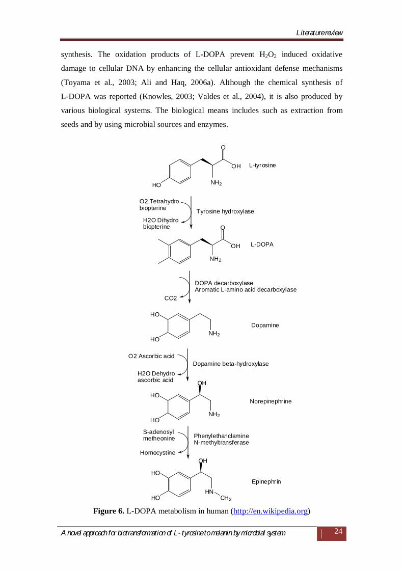
2.8 L-DOPA production
L-DOPA is an intermediate produced during initial step of melanin synthesis
from L-tyrosine. It is an amino acid analogue produced by one step reaction from
L-tyrosine. L-DOPA is widely used as drug for the treatment of Parkinson’s disease
(PD). The PD is associated with diminished level of dopamine in brain. L-DOPA is a
metabolic precursor of dopamine, can easily cross the blood brain barrier and finally
converted in to dopamine. L-DOPA can be orally administered to relive symptoms of
Parkinson's disease.
2.8.1 Function of L-DOPA in human body
The functions of L-DOPA in humans include a precursor of the catecholamine
neurotransmitters dopamine, nor-epinephrine and epinephrine (Figure 6) (Fling and
Paul, 2001; Ali and Haq, 2006a). It acts as an important modulator of renal dopamine
Literature review
A novel approach for biotransformation of L- tyrosine to melanin by microbial system
24
synthesis. The oxidation products of L-DOPA prevent H2O2 induced oxidative
damage to cellular DNA by enhancing the cellular antioxidant defense mechanisms
(Toyama et al., 2003; Ali and Haq, 2006a). Although the chemical synthesis of
L-DOPA was reported (Knowles, 2003; Valdes et al., 2004), it is also produced by
various biological systems. The biological means includes such as extraction from
seeds and by using microbial sources and enzymes.
OH
NH2
OH
NH2
HO
NH2
HN
O
O
OH
OH
NH2
HO
HO
HO
HO
HO
HO CH3
L-tyrosine
Tyrosine hydroxylase
DOPA decarboxylaseAromatic L-amino acid decarboxylase
L-DOPA
Dopamine
Dopamine beta-hydroxylase
Norepinephrine
PhenylethanclamineN-methyltransferase
Epinephrin
O2 Tetrahydrobiopterine
H2O Dihydrobiopterine
CO2
O2 Ascorbic acid
H2O Dehydroascorbic acid
S-adenosylmetheonine
Homocystine
Figure 6. L-DOPA metabolism in human (http://en.wikipedia.org)
Literature review
A novel approach for biotransformation of L- tyrosine to melanin by microbial system
25
2.8.2 Plant sources and cell suspension cultures
The L-DOPA is conventionally extracted from the seeds of Mucuna pruriens
seeds and Vicia faba beans (Chattopadhyay et al., 1994, Shetty et al., 2001). The
L-DOPA content from Vicia faba beans was reported previously to be enhanced by
using food grade elicitors, gellan gum, and a polysaccharide from Pseudomonas
elodea and xanthan gum from Xanthomonas campestris (Shetty et al., 2001). The cell
suspension culture Mucuna pruriens was also studied earlier for L-DOPA production
in buffer containing L-tyrosine (Chattopadhyay et al., 1994). The Portulaca
grandiflora cell suspension culture was formerly reported for L-DOPA production.
This callus cultures were induced in MS medium provided with growth regulators
such as benzylaminopurine and 2, 4-dichloro-phenoxyacetic acid (Rani et al., 2007).
Also banana cell suspension cultures were reported earlier for potential L-DOPA
synthesis (Bapat et al., 2000).
2.8.3 Microbial sources
The production of L-DOPA by microbiological methods has been reported by
two processes one is production in complex synthetic medium and other is in the
reaction mixture containing L-tyrosine and biomass. The two bacteria Vibrio
tyrosinaticus and Pseudomonas melanogenum were reported earlier for producing
good yield of L-DOPA in complex medium with intermittent addition of L-tyrosine.
These reports also describe the effects of various media components and substrates of
L-DOPA (Yoshida et al., 1973; Yoshida et al. 1974).
An actinomycete species was formerly isolated from soil after subjecting to
the chemical mutagenesis by N-methyl-N-nitro-N' nitrosoguanidine (NTG), the
resultant mutant strain was reported for potential L-DOPA production (Sukumaram et
al., 1979).
The novel fungal species Acremonium rutilum was reported earlier for
optimization of media conditions using potato dextrose broth for L-DOPA production.
(Krishnaveni et al., 2009). The Egyptian halophilic black yeast was reported recently
for L-DOPA production in a broth containing nitrogen source like ram horn
hydrolysate (Mahmoud and Bendary, 2010).
Literature review
A novel approach for biotransformation of L- tyrosine to melanin by microbial system
26
The other method for L-DOPA production was, employing grown biomass in
a reaction buffer containing L-tyrosine. This L-DOPA production by using this
method first reported by mutant fungal strain Aspergillus oryzae was (Haq et al.,
2002). L-DOPA yield was further improved by adding the reaction mixture with
specific enhancer cresoquinone (Haq et al., 2003). The double mutation of Aspergillus
oryzae by using UV radiations and chemical mutagen NTG were reported previously
for enhanced production of L-DOPA (Ali et al., 2005). The effect of a clay mineral
illite on conversion of L-tyrosine to L- DOPA by Aspergillus oryzae ME2 under
acidic reaction conditions was formerly observed (Ali and Haq, 2006a). The enhanced
L-DOPA production and kinetic basis of another clay mineral celite was studied
earlier using A. oryzae ME2 (Ali and Haq, 2006b). The yeast Yarrowia lipolytica cell
mass in tyrosine containing reaction buffer was reported recently for bioconversion of
L-tyrosine to L-DOPA with addition of diatomite (Ali et al., 2007). The L-DOPA
production from catechol and pyrochatechol in reaction mixture using Erwinia
herbicola cell mass was studied (Koyanagi et al., 2005).
2.8.4 Enzyme immobilization
The Escherichia intermedia cells were immobilized previously by entrapment
in carrageenan to produce L-DOPA from catechol, pyruvate and ammonia (Para and
Baratti, 1988). The production of L-DOPA from mushroom tyrosinase immobilized
zeolite was earlier studied by using glutaraldehyde as a cross-linking agent
(Seetharam and Saville, 2002). The mushroom tyrosinase was immobilized formerly
on modified polystyrene-polyamino styrene (PSNH) and polymethylchloride styrene
(PSCL) with glutaraldehyde as an activating agent for L-DOPA production from L-
tyrosine (Ho et al., 2003). The production of L-DOPA was studied recently by using
tyrosinase immobilized in copper-alginate gels by using batch and packed bed
reactors (Ates et al., 2007).
Literature review
A novel approach for biotransformation of L- tyrosine to melanin by microbial system
27
2.10 Research objectives and thesis outline
Isolation, screening and identification of bacterial species for production of
melanin and L-DOPA
Standardization of nutritional parameters for melanin production from isolated
high yielding bacterial species.
Purification and characterization of melanin by using analytical technique viz.
UV- Vis spectroscopy, FTIR and EPR.
Comparison of melanin yields from isolated bacterial species.
Microbial transformation of L-tyrosine to melanin by using cells of bacterial
species.
Standardization of nutritional parameters for L-DOPA production from
isolated high yielding bacterial species.
Comparison of L-DOPA yields from isolated bacterial species.
Bioconversion of L-tyrosine to L-DOPA by using cells of bacterial species.
Analysis of produced L-DOPA by using techniques viz. HPTLC, HPLC and
GCMS.
Purification and primary characterization of tyrosinase from melanogenic
bacterial species.