Embed Size (px)
Citation preview

CHAPTER II
LITERATURE REVIEW
1. Mangosteen Pericarp Extract and Its Therapeutic Potentials
Garcinia mangostana Linn, (family Guttiferae) or mangosteen, the queen of fruit, is
commomly encountered in Southeast Asia. It is one of the most interesting medicinal plants
because of its distinguished pharmacological activities. The fruit hull or pericarp of this
plant has been used in as a Thai traditional medicine for the treatment of diarrhea, amoebic
dysentery, skin infection, suppuration, chronic wound and Hongkong foot.
(Mahabusarakum, et al., 1983)
Phytochemical studies have shown that the active ingredients from mangosteen
pericarp extract belong to a group of xanthones such as Ot-mangostin (formerly
mangostin), (3-mangostin and y-mangostin, isomangostin, garcinone A, B, c and D,
gartanin, rubraxa'nthone, etc. (Mahabusarakam, et al., 1987) The structure of xanthone ring
and Its derivatives (Budavari, et al., 1996) are shown in Figure 1. Among these xanthones,
Ot-mangostin is the major component of mangosteen pericarp. (Bennett and Lee, 1989)
However, the differences in solvent and methods of extraction and purification may yield
different combinations of xanthones in the extract. (Nakatani, et al., 2002b)
Extract from mangosteen pericarp has been shown to exert antimicrobial activities
against several microorganisms including bacteria, (linuma, et al., 1996; Sundaram, et al.,
1983) fungus (Gopalakrishnan, Banumathi and Suresh, 1997; Sundaram, et al., 1983) and
virus. (Chen, Wan and Loh, 1996) It also possesses other biological activities such as anti
inflammatory, (Nakatani, et al., 2002b, Nakatani, et al., 2002a) anti-histamine,
(Chairungsrilerd, et al., 1996, Nakatani, et'al., 2002b) anti-oxidant, (Williams, et al., 1995)
and anti-tumor (Moongkarndi, et al., 2004) properties.

A. Xanthone ring
Chemical name
Molecule formula
Molecular weight
: Diphenylene ketone oxide
: C18H80 2
: 196.26 gram
B. Xanthone derivatives
(X-mangostin : r \ r 4 = A and R 2, R 3 = H
(3-mangostin : r \ r 4 = A and R 2 = Me, R 3
y-mangostin R 1 = A and R 2 = R 3 = R 4
R4 0 OH
MeO.ySrV r Rlr 3o '
■O R 2
= H
= H and Me is substituted by H
A =
OC-manaostin
Chemical name
Molecule formula
Molecular weight
Melting point
: Tetraoxygenated diprenylated xanthone
: C24H260 6
: 410.46 gram
: 181.6-182.6 ° c
Figure 1. Chemical structures of (A) xanthone ring and (ธ) xanthone derivatives: (X-
mangostin, (3-mangostin and y-mangostin. (Budavari et al., 1996)

7
Xanthones from ทาangosteen pericarp extract has broad-spectrum anti-bacterial
activities against a variety of gram-positive and gram-negative bacteria including enteric
pathogens, (Sindermsuk and Deekijsermphong, 1989; Sundaram, et al., 1983) both
methicillin-sensitive and ทาethicillin-resistant ร. aureus, (Mahabusarakum, et al., 1983, 1986)
and mycobacterium, (Suksamrarn, et al., 2003) etc. (Table 1). Among these xanthones, ot-
mangostin appears to exert the strongest anti-bacterial activity with MIC values ranging
from 1 to 50 |Ug/ml. However, the antimicrobial effect of the extract against oral pathogens
has never been demonstrated.
Several studies have demonstrated low toxicity of ทาangosteen pericarp extract.
Xanthones isolated from mangosteen pericarps were not toxic to rats when given orally at a
dose of 100 mg/kg body weight/day for up to 7 days. (Sapwarobol, 1997) When OC-
ทาangostin, a major component of the extract, was administered orally to rats at a high dose
(1.5 g/kg body weight). The finding indicated the low hepatotoxicity by slightly increases in
serum glutamate oxaloacetate transaminase (SGOT) and serum glutamate pyruvate
transaminase (SGPT) activities after 12 hours. These increases were much less than those
of paracetamol given at the same dose, and no change in total liver proteins was observed.
(Sornprasit, et al., 1987) เท a human clinical trial, 1.5% OC-mangostin cream was locally
applied on skin of patients with chronic ulcers for up to 3 weeks. No local irritation or side
effects were observed. (Kusuma, 2003)
Based on its various biological activities and low toxicity, mangosteen pericarp
extract has potentials for wide pharmacological applications. It has been developed in the
forms of topical cream or gel for chronic skin ulcers, (Kamjanachotdamrong, 2000; Kusuma,
2003) throat spray for upper respiratory tract infection, (Kongchunmitkul, 2002) and buccal
ทานcoadhesive film for oral ulcers. (Hiranras, 2001) This study will focus on its antimicrobial
activity against oral pathogens in dental plaque. Based on this knowledge, we can further
develop this extract in the forms of mouthrinse or local delivery drugs for chemical plaque
control.
8
Table 1. Antimicrobial activity of xanthones from mangosteen pericarp extract
Bacteria References Active components MICs
Pseudomonas aeruginosa Sundaram et al., OC-mangostin 12.5-50 JJ.g/ml.
Salmonella typhimurium 1983
Bacillus subtilis
Escherichia Coll Sindermsuketal., boiled crude 6.3-100 mg/ml
Salmonella species 1989 extract
Shigella species
Staphylococcus aureus Mahabusaraku ทา OC-mangostin 15.6 |ig/ml
et al., 1983; 1986 y-mangostin 31.2 |Tg/ml
1-isomangostin 62.5 |Llg/ml
3-isomangostin 125 |J.g/mlgartinin 250 (Tg/ml
Methicillin -resistance ร. aureus Mahabusarakum OC-mangostin 1.6-12.5 flg/ml
étal., 1983; 1986 1-isomangostin 125 |Tg/ml
y-mangostin 250 Jlg/ml
3-isomangostin 250 |ig/mlgartinin 250 (O-g/ml
linuma et al., OC-mangostin 1.6-12.5 (Tg/ml
1996
Mycobacterium tuberculosis Suksamrarn et al., OC-mangostin 6.3 (Tg/ml
2003 (3-mangostin 6.3 [ig/ml
y-mangostin 25 (Ig/ml

9
2. Cariogenic Bacteria
Dental caries development involves demineralization of tooth structure by a high
concentration of organic acids produced by specific bacteria in dental plaque. One of the
bacteria that has been implicated in the etiology of both coronal and root caries is
Streptococcus mutans. (Loesche, 1986; Zambon and Kasprzak, 1995) It is gram-positive,
facultative anaerobic cocci. Its colony has yellowish-white color, pinpoint to medium size,
irregular margin with rough and shiny surface on trypticase soy agar.
Streptococcus mutans possesses several cariogenic properties. It is able to
synthesize extracellular polysaccharides including insoluble glucan and fructan from dietary
sucrose. This process enhances plaque mass, promotes bacterial colonization and
changes diffusion properties of the plaque matrix. เท addition, this bacteria is acidogenic
and aciduric. It is capable of forming acids in the presence of sugars, and maintaining
sugar metabolism under extreme acidic environment such as in carious lesions, (van Houte,
1994; Zambon and Kasprzak, 1995)
3. Periodontopathic Bacteria
Periodontal disease is caused by a group of anaerobic bacteria in dental plaque,
which induce a release of inflammatory cytokines from periodontal tissues, leading to
periodontal destruction. The World Workshop on Clinical Periodontics in 1996 implicated 3
bacterial species including ActinobaciHus actinomycetemcomitans, Porphyromonas
gingivalis and Tannerella forsythia as étiologie agents for periodontal disease. (Zambon,
1996)
These bacteria fulfill the criteria for implicating them as the etiology of periodontal
disease. They are found in high numbers in the diseased sites, higher than in the healthy
sites of the same subjects. Subjects with periodontal disease make elevated antibody
levels to the antigens from these bacteria. They also produce a number of virulence factors,
which can directly damage periodontal tissues or immune cells, or indirectly

10
damage the tissues by cytokine induction. Eliminating or reducing the number of these
bacteria is important for the success of periodontal therapy. There is also data
demonstrating that these bacteria can induce periodontal disease in animal models.
(Zambon, 1996)
Actinobacillus actinomycetemcomitans is gram-negative, facultative anaerobic
bacteria. It is a small, non-motile, saccharolytic, round-end rod. It appears as a yellowish-
white small, smooth colony with a star-shaped inner structure. (Olsen, Shah and Gharbia,
1999) This bacterium produces a number of virulence factors such as leukotoxin and
lipopolysaccharide. Leukotoxin has been shown to kill polymorphonuclear leukocyte, T
cells and B cells. Lipopolysaccharide can stimulate macrophages to release inflammatory
cytokines including interleukin-1, prostaglandin E2 and tumor necrosis factor-OC. เท addition,
the ability of this microorganism to invade host epithelium and gingival connective tissue
makes it survive from host immune defense and mechanical periodontal therapy. (Fives-
Taylor, et al., 1999)
Porphyromonas gingivalis is gram-negative, obligate anaerobic bacteria. It is a
non-motile, asaccharolytic, short bacilli. It belongs to a black-pigmented Bacteroides
group, in which colonies are brown to black, smooth, shiny and exhibit complete hemolysis
on blood agar. (Olsen, et al., 1999) This organism has a large array of virulence factors
such as lipopolysaccharide, fimbriae and gingipain proteases. Lipopolysaccharide is
capable of inducing bone resorption in mice. Fimbriae mediates adherence to specific
receptors on host cells, and induces bacterial internalization. It is also capable of inducing
cytokine production, p. gingivalis proteinases have been shown to interact with the
cytokine networking systems, leading to dysrégulation of the local inflammatory reaction.
(Holt, et al., 1999)
4. Chlorhexidine
Chlorhexidine is considered one of the most effective antiseptics. It has broad-
spectrum antimicrobial activity against a variety of both gram-positive and gram-negative
11
bacteria including those causing dental caries and periodontal disease. Its
effectiveness also contributes to its high substantivity to oral tissues. The mechanism of
action is mainly on the rupturing of bacterial cell wall and precipitation of the cytoplasmic
content. (Ciancio, 2000; Jones, 1997)
Chlorhexidine has been incorporated into various formulations including mouthrinse
and local delivery drugs. Its efficacy in reducing plaque and dental caries and in
periodontal treatment has been clinically proven. (Overholser, 1988; Twetman, 2004)
However, it has some unwanted side effects including bad taste, tooth and tongue staining
and taste alteration. (Ciancio, 2000; Jones, 1997)
5. Anaerobic Bacterial Culture
There are two systems used to cultivate anaerobic bacteria: anaerobic chamber and
chemically generated anaerobic systems such as the BBL GasPak system (BBL
Microbiology Systems, Cockeysville, MD, USA). The anaerobic chamber provides a
convenient culture system for large-scale studies and exhibits good recovery for most
anaerobic organisms. However, it is expensive to purchase and maintain. The GasPak
system is limited to processing a few bacterial plates at a time but costs a lot less. (Doan, et
al., 1999) It consists of a polycarbonate anaerobic jar, a disposable hydrogen- and carbon
dioxide-generating envelope and a catalyst chamber containing 2.5 + 0.5 g of palladium
catalyst pellets. When water is added to the envelope, hydrogen gas and carbon dioxide
are generated. เท the presence of a catalyst, the resulting hydrogen combines with oxygen
inside the jar to produce water, thus establishing an anaerobic environment. Sixty minutes
after adding water, the atmosphere inside the jar contains approximately 4 to 7 % carbon
dioxide and 25 to 30% hydrogen, while the oxygen is decreased to less than 1%. (Seip and
Evans, 1980)
Anaerobic organisms differ in oxygen sensitivity. Anaerobic chamber is essential for
work with strict anaerobes, which are incapable of growing at an oxygen concentration of
greater than 0.5%. p gingivalis belongs to a group of moderate anaerobes, which are

12
capable of growth in the presence of oxygen levels as high as 2-8%. (Loesche, 1969)
When the Gaspak system was compared to the anaerobic chamber for the abilities to
support the growth of anaerobic periodontal pathogens, they were equally effective in
isolating p. gingivalis, but the GasPak system demonstrated higher proportional recoveries
of T. Forsythia . (Doan, et al., 1999) Comparison between the 2 systems was set up to test
11 antimicrobial agents regarding antimicrobial susceptibility against 38 anaerobesd. The
MIC results were comparable (94% aggreement) within 1 twofold dilution for organisms
incubated in both systems (Murray and Niles, 1982)
6. Measuring Bacterial Growth
Many methods are available. We’ll summarize the two methods used in this study.
6.1 Viable cell count using a spread plate method
The viable cell count is a measure of the concentration of cells present in a broth
culture that are alive and able to grow and produce colonies on the agar surface. A sample
of broth culture is serially diluted, usually ten fold at each dilution, and then a small amount
of each dilution is spread on a plate. After visible colonies have formed, plates from the
dilutions that contain separated colonies are selected. Choosing plates with between 30
and 300 colonies offers a good compromise between speed and accuracy. Plates with
fewer than 30 colonies may give exaggerated counts due to sampling errors, while plates
with more than 300 colonies may be too crowded to count and to allow all the bacteria to
form distinct colonies. The viable bacterial count is expressed in terms of colony-forming
units per ml (CFU/ml). CFU/ml is calculated by dividing the average number of colonies
with the plating volume (ml) and then multiplying with the dilution factor. (Ingraham and
Ingraham, 2004)
If the number of viable bacteria (CFU/ml) is plotted against time, the curve will get
steeper and steeper and finally shoots out of the range of the plot. Therefore, microbial
growth is conveniently graphed on a logarithmic scale. The growth curve is plotted as the
logarithm of the number of viable cells (log10CFU/ml). The growth curve of a typical

bacterial culture is divided into 4 phases: a lag phase, bacteria prepare to grow ; a log
or exponential growth phase, cell numbers double at regular intervals ; a stationary phase,
bacterial growth ceases ; and a death phase, number of live cells decline (Figure 2).
(Chynoweth, 2004)
6.2 Degree o f turbidity
The degree of turbidity or cloudiness by a broth culture can give an estimate of the
number of organisms present, both dead and live. Turbidity is measured using a
spectrophotometer. This device measures how much light can pass through bacterial
culture (Figure 3). As the mass of cells increases, turbidity increases, less light passes
through the culture, the reading on the spectrophotometer is higher. The degree of turbidity
is measured in terms of the absorbance (also called optical density or OD) at 600 nm. This
wavelength is selected to minimize the absorbance by the broth media because most
media are brown or brownish-yellow. (Ingraham and Ingraham, 2004) The growth curve
can be plotted between the absorbance at 600 nm and time in a similar fashion as
log 10CFU/ml (Figure 2).
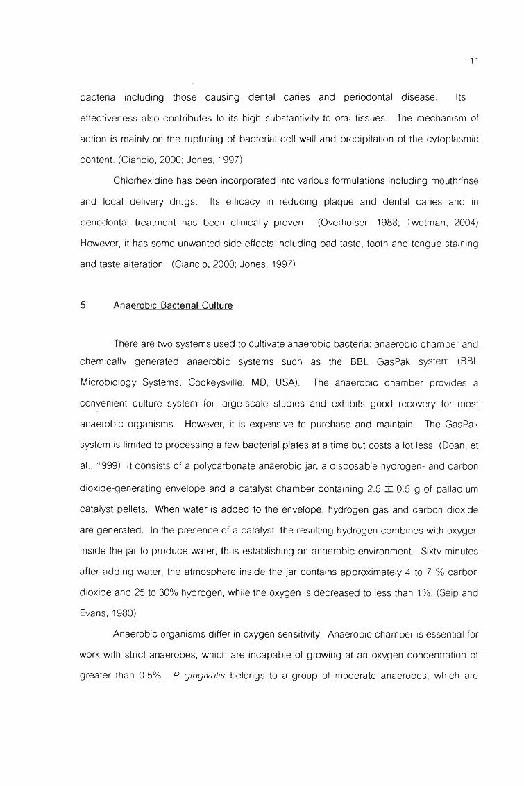
Because the mass of the culture and number of cells are related, turbidity can also
be used to determine the cell number. To convert spectrophotometer reading to the
number of bacteria, it is necessary to prepare a standard curve. The graph is drawn by
plotting logl0CFU/ml of bacteria against the absorbance at 600 nm (Figure 4). Only the
values from the exponential phase of growth (mostly live cells) are used to plot the standard
curve. (Ingraham and Ingraham, 2004) Linear regression analysis is used to fit a straight
line to the data. The equation for this straight line is :
Y = aX + b
Where Y = log10CFU/m! that corresponds to the absorbance value,
a = the slope of the line
X = the absorbance value
b = the Y-intercept of the line

14
EPLL๐O
c3Pบ ิ<Dบ ิO
ท ิท>
1.0
0.75
0.50
0.25
0.10
Figure 2. Growth curve of a typical bacterial culture. (Chynoweth, 2004)
Light source bacterial culture light-sensitive detector
absorbance value
Figure 3. Components of a spectrophotometer. The amount of light striking the light-
sensitive detector is inversely proportional to the number of bacteria. The higher
number of bacteria, the less light transmitted, the higher the absorbance reading.
(Ingraham and Ingraham, 2004)
Abso
rbanc
e
15
E3LL.๐Oç>O
OZพ>**-Oa>ท13
Absorbance at 600 ททา
Figure 4. Standard curve relating the absorbance at 600 nm to the number of viable
bacteria. Once the absorbance reading is obtained, the standard curve can be used to
determine the corresponding number of bacteria.
16
The regression line also gives a value called correlation coefficient (r). This
number ranges from 0 to 1. The closer r to 1, the more likely the data are to forming a
straight line. From this linear equation, an estimate of viable bacterial count can be predicted from the absorbance readings.