Embed Size (px)
Citation preview

Liposome Drug Delivery E. Sanarova*, A. Lantsova, N. Oborotova, O. Orlova, A. Polozkova, M. Dmitrieva, N. Nikolaeva
Research Institute of Experimental Diagnostics and Therapy of Tumors, N.N.Blokhin Russian Cancer Research Center, Moscow-115478, Russia
Abstract Liposomes are spherical vesicles with a phospholipid bilayer, were first described in 1965 by Alec Bangham and are extensively used in drug delivery. Liposomes are generally classified based upon structure, method of preparation, composition and application. Many methods have been reported in the literature for the preparation of liposomes. Here we considered basic methods such as the film method, extrusion, high pressure homogenization, ultrasonic method, Method based on replacement of organic solvents and others. After preparation and before use the liposomes are characterized by physical (size, shape, surface features, lamellarity phase behaviors and drug release profile), chemical (purity and potency of various liposomal constituents) and biological (safety and suitability of formulation for the in vivo use for therapeutic application) methods. Almost from the time of their discovery the demonstration of their entrapment potential, liposomal vesicles have drawn attention of researchers as potential carriers of various bioactive molecules that could be used for therapeutic applications. Encapsulation of a drug in liposomes prevents its early degradation and alters the biodistribution profile in the body. This enables higher concentrations of the drug in the desired site, leading to improved effectiveness, and reduced toxicity in the vital organs like heart, kidney etc. These advances have led to numerous clinical trials and studies in such diverse areas as the delivery of anti-cancer, anti-fungal and anti-biotic drugs, the delivery of gene medicines and drug delivery to site of action, long circulating PEGylated liposomes, triggered release liposomes and liposomes containing combinations of drugs. The liposomes have many applications which increase its importance over other formulations.
Keywords: Drug delivery, Liposomes, Phospholipid, Targeting
INTRODUCTION The name liposome is derived from two Greek words: 'Lipos' meaning fat and 'Soma' meaning body. A liposome is a vesicle, made out of the same material as a cell membrane. They are usually made of phospholipids, which are molecules that comprise a tail and a head group. The head is hydrophilic, whereas the tail which is made of a long hydrocarbon chain is hydrophobic. Normally, phospholipids can be found as a bilayer. [1-3]. This amphiphilic nature enables loading of hydrophilic and hydrophobic therapeutic agents in the core and the bilayer, respectively. The tiny size enables quick assimilation into the bloodstream and delivering at specific site, thus making them significant for modifying toxicity, solubility, stability and converting drugs into ideal candidates of improved pharmacokinetic and pharmacodynamic profiles. The issue with stability, high cost and limited shelf life due to the rancidification of lipids poses major limitations. An advance with drug delivery technology is a prospect to medicine and healthcare system. New inventions in materials chemistry have initially excited the advance of drug delivery systems, creating carriers that are biocompatible, biodegradable, targeting, and stimulus-responsive [4]. Liposomes were first described by British haematologist Dr Alec D Bangham FRS at the Babraham Institute, in Cambridge when they were discovered by him and R. W. Horne, who were testing the institute's new electron microscope by adding negative stain to dry phospholipids [5-7]. The main reason why research into liposomes advanced as it has, can be largely attributed to the fact that liposomes can mimic biological cells. This also means that liposomes are highly biocompatible, making them an ideal candidate
for a drug delivery system, with applications ranging from delivering enzymes, antibacterials, antiviral drugs, antiparasite drugs, fungicides, transdermal transporters, diagnostic tools and adjuvants for vaccines. To date liposomal formulations of anti-tumor drugs and antifungal agents have been commercialized [8].
COMPONENTS OF LIPOSOME STRUCTURE Liposomes are spherical vesicles composed of one or more lipid bilayers, involving an aqueous compartment (Figure 1). These are formed spontaneously when the lipids are dispersed in an aqueous medium by stirring. The basic components of liposomes are phospholipids which are stabilised by cholesterol, with other stabilisers sometimes added to the mixture depending on the specific use of the liposome. Phospholipids Phospholipids are the major structural component of biological membranes. The structure of the phospholipids are as follows: on the one end of the molecule are the hydrophobic acyl hydrocarbon chains. The other end of the molecule, which is also called the phosphate head group, is hydrophilic. Lipids all have a temperature at which their fluidity changes. This temperature is also known as transition temperature (TC). The TC is directly proportional to the length of the acyl chain; the longer the chain, the higher the TC and the more rigid the membrane. More rigid membranes keep entrapped drugs inside, or in other words, prevent leakage. The TC is very important, as it can affect the way the membrane reacts to fusing with other liposomes, stability, aggregation, permeability as well as contributing to the way the liposomes react in the presence of biological systems
E. Sanarova et al /J. Pharm. Sci. & Res. Vol. 11(3), 2019, 1148-1155
1148

Phospholipids containing the choline group are one of the most abundant lipids in nature. The phospholipid most often used for liposomes is the phospholipid known as phosphatidylcholine (PC). Molecule of phosphatidylcholine are not soluble in water and in aqueous media they align themselves closely in plannar bilayer sheets in order to minimize the unfavorable action between the bulk aqueous phase and long hydrocarbon fatty chain. This phospholipid is very popular because of its relative low cost and general tendency to be neutral. PC is procured from natural sources, plants with soybeans as an example, and mammalian sources such as bovine heart, spinal column or in some cases from egg yolk. Phosphatidylcholines are the most widely used due to their appropriate stability and their ability to act against changes in pH or salt concentrations in the product or/and biological environment [9] Sphingolipids are the membrane components containing sphingoid base [10]. Natural gangliosides class of sphingolipids are included in liposome formulations to provide a layer of surface charged groups, to prolong the lifetime of liposomes in the blood and to prevent their uptake by the reticuloendothelial system (RES). Sphingomyelins are important phospholipids useful in regulation of cholesterol distribution within membranes [11]. Cationic lipids are amphiphiles, analogous to natural phospholipids except for the presence of a cationic charge. It consists of long hydrocarbon chains (largely comprised of alkyl chains or cholesterol); hydrophilicity is by charged group (quaternary nitrogen) and linker bond (ester, carbamate etc). Due to their amphiphilicity nature upon hydration, selfassemble into lamellar vesicular structures with interior aqueous phase [12,13]. Cholesterol Cholesterol is one of the major components in liposomal formulations whose incorporation increases the rigidity of the lipid bilayer, improves fluidity of the membrane, improve stability, increases the time of circulation in the
blood stream [14,15]. Cholesterol dose not by itself form bilayer structure, but can be incorporated into phospholipid membranes in very high concentration upto 1:1 or even 2:1 molar ration of cholesterol to phosphatidylcholine. Cholesterol inserts into the membrane with its hydroxyl group oriented towards the aqueous surface and aliphatic chain aligent parallel to the acyl chains in the center of the bilayer. When cholesterol is added into the mixture the cholesterol stabilises the liposomes, or in other words, it increases the TC of the membrane. The addition of cholesterol decreases the permeability of the bilayer, thus helping to keep the liposome stable and to keep the intended drug entrapped.
CLASSIFICATION OF LIPOSOMES Liposomes are mainly classified in terms of size (small, intermediate, or large), number of bilayers (uni- and multi-lamellar), composition and mechanism of drug delivery [16]. Small unilamellar vesicles (SUV) consist of a single lipid bilayer with an average diameter ranging from 25 to 100 nm. Large unilamellar vesicles (LUV) also consist of one lipid bilayer and are greater than 100 nm, whereas multilamellar vesicles (MLV) are made up of several concentric lipid bilayers and measure of 1-5 µm (Figure 2) [17,18]. As regards the composition and mechanism of drug delivery, the liposomes can be classified as conventional liposomes, long-circulating liposomes, polymorphic liposomes (pH-sensitive, thermo-sensitive, and cationic liposomes), and decorated liposomes (surface-modified liposomes and immunoliposomes). Conventional liposomes – these liposomes are composed of natural phospholipids (which may be neutral or negatively charged) and cholesterol. These liposomes are often used for targeting of the RES. This shortens the circulation times of the liposomes substantially. Contents of these liposomes are most often destined for lysosomes.
Figure 1: Schematic presentation of the phospholipids, lipid bilayer and liposome
E. Sanarova et al /J. Pharm. Sci. & Res. Vol. 11(3), 2019, 1148-1155
1149

Figure 2: Classification of liposomes according to average diameter and number of bilayers
Figure 3: Film method and extrusion method preparation of liposomes
pH-sensitive liposomes – the membranes of these liposomes are composed of either cholesterol hemisuccinate, phosphatidyl ethanolamine , oleic acid or dioleoylphosphatidyl ethanolamine. These liposomes fuse with cells when the pH is low, thus releasing its content into the cell cytoplasm. These liposomes are ideal for the delivery of macromolecules and weak bases [19-23]. Cationic liposomes – cationic lipids make up the membrane of these liposomes with dimethyl-dioctadecyl ammonium bromide (DDAB), dioctadecyldimethyl ammonium chloride, 2,3dioleoyloxy- N - ( 2 (spermine carboxamido) - ethyl) - N, N-dimethyl – l - propanaminium fluoracetate, 1,2 dioleoyloxy-3-(trimethylammonio) propane, 1,2dimrystyloxypropyl-3-dimethyl-hydroxethyl ammonium bromide, and 1,2dioleyloxypropyl-3-dimethyl-hydroxyethyl ammonium bromide combined with dioleoylphosphatidyl ethanolamine. These liposomes tend to be toxic in high doses with a short lifespan, thus restricting them to local administration. They are most often used for the delivery of macro molecules that have a negative charge, this includes the delivery of DNA and RNA. Long-circulating Liposomes – the lipids used for this type of formulation are neutral lipids with a high TC. Cholesterol is also included in these formulations (normally
between 5 and 10 %). These liposomes have a very long circulation half life, of up to 40 hours. Immuno-liposomes – these liposomes are Conventional liposomes or Long Circulating Liposomes with antibody or other recognition sequences attached to the surface. These liposomes are formulated to bind to specific cells and to release the drug in that area, thus making it a targeted delivery system.
METHODS PREPARATION OF LIPOSOMES
Many methods have been reported in the literature for the preparation of liposomes. These are discussed here briefly. Film Method The original method of Bangham et al. [24,25] is still the simplest procedure for the liposome formation but having some limitation because of its low encapsulation efficiency [26-29]. In this technique liposome are prepared by hydrating the thin lipid film in an organic solvent and organic solvent is then removed by film deposition under vacuum. When all the solvent get removed, the solid lipid mixture is hydrated using aqueous buffer. The lipids spontaneously swell and hydrate to form liposome. This method yields a heterogeneous sized population of MLVs over 1 micro meter in diameter. To reduce the size of the
E. Sanarova et al /J. Pharm. Sci. & Res. Vol. 11(3), 2019, 1148-1155
1150
liposomes obtained, the most commonly used methods are extrusion or homogenization [30-32] Polycarbonate Membrane Extrusion Method (Figure 3) In this method lipid dissolved in chloroform is dried into thin film. The dried lipid film is then added to buffer solution containing the drug molecule of interest. The lipid solution is sonicated, freeze dried and subjected to extrusion through polycarbonate membrane to form liposomes. Uniform sized liposomes are formed by this method. High Pressure Homogenisation Homogenous blend of lipids is prepared by dissolving them in organic solvents, shock freezing in liquid nitrogen and freeze drying the blend. Freeze dried lipid is then dissolved in PBS and subjected to high pressure homogenisation to form liposomes [33]. Ultrasonic Method This method is used for the preparation of SUVs with diameter in the range of 15-25 µm. Ultrasonication of an aqueous dispersion of phospholipids is done by two types of sonicators i.e. either probe sonicators or bath sonicators. The probe sonicators are used for the small volume which requires high energy while the bath sonicators are employed for the large volume [34]. Method Based on Replacement of Organic Solvents In this method lipids are co-solvated in organic solution, which is then dispersed into aqueous phase containing material to be entrapped within the liposome. This method is of two types: I. Reverse Phase Evaporation The phospholipid mixture is added to a round bottom flask and the organic solvent (diethylether or isopropylether or mixture of isopropyl ether and chloroform) is removed under reduced pressure by a rotary evaporator. The system is purged with nitrogen and lipids are re-dissolved in the organic phase which is the phase in which the reverse phase vesicle will form. After the lipids are redissolved the emulsion are obtained and than the solvent is removed from an emulsion by evaporation to a viscous gel under reduced pressure. Non encapsulated material is then removed. This method is used for the preparation of large uni-lamellar and oligo-lamellar vesicles formulation and it has the ability to encapsulate large macromolecules with high efficiency [35]. With this method high encapsulation efficiency up to 65 % can be obtained in a medium of low ionic strength for example 0.01M NaCl. The method has been used to encapsulate small and large macromolecules. The main disadvantage of the method is the exposure of the materials to be encapsulated to organic solvents and to brief periods of sonication. II. Ether Vaporization Method [36] There are two method according to the solvent used: 1. Ethanol injection method. A lipid solution of ethanol is rapidly injected to a vast excess of buffer or other aqueous medium. The MLVs are immediately formed. The drawbacks of the method are that the population is heterogeneous (30-110 nm), liposomes are very dilute, it is difficult to remove all ethanol because it forms azeotrope with water and the possibility of various
biologically active macromolecules to inactivation in the presence of even low amounts of ethanol. 2. Ether injection method. A solution of lipids dissolved in diethyl ether or ether/methanol mixture is slowly injected to an aqueous solution of the material to be encapsulated at 55-65 °C or under reduced pressure. The subsequent removal of ether under vacuum leads to the formation of liposomes. The main drawbacks of the method are population is heterogeneous (70-190 nm) and the exposure of compounds to be encapsulated to organic solvents or high temperature. Freeze Thaw Extrusion Method Liposomes formed by the film method are vortexed with the solute to be entrapped until the entire film is suspended and the resulted MLVs are frozen in luke warm water and than vortexed again [37]. After two cycles of freeze thaw and vortexing the sample is extruded three times. This is followed by six freeze thaw cycle and addition eight extrusions. This process ruptures and defuses SUVs during which the solute equilibrates between inside and outside and liposome themselves fuse and increase in size to form large unilamellar vesicle by extrusion technique. For the encapsulation of protein this method is widely used [38]. The Dehydration- Rehydration Method In this method the empty buffer containing SUVs and rehydrating it with the aqueous fluid containing the material to be entrapped after which they are dried. This leads to a dispersion of solid lipids in finely subdivided form. Freeze drying is often the method of choice. The vesicles are than rehydrated. Liposomes obtained by this method are usually oligolamellar vesicle [39].
CHARACTERIZATION OF LIPOSOMES After preparation and before use the liposome must be characterized. Evaluation could be classified into three broad categories which are physical, chemical and biological methods (Table 1) [40-48]. The physical methods include various parameters, which are size, shape, surface features, lamellarity phase behaviors and drug release profile. Chemical characterization includes those studies which established the purity and potency of various liposomal constituents. Biological characterization is helpful in establishing the safety and suitability of formulation for the in vivo use for therapeutic application [49]. The characteristics of the carrier through appropriate choice of membrane components, size and charge determines the final behavior of liposomes both in vitro and in vivo as well. Mean Liposome Size and Size Distribution Liposome size is dependent on the preparation technique i.e. sonication times, extrusion pressures, lipid composition and measuring liposome-complement interactions. A number of methods are used to determine size and size distribution, among which light-scattering analysis is commonly used. The recently used methods are atomic force microscopy, ultracentrifugation, Coulter counter, gel exclusion chromatography, laser diffraction, and light microscopy [50].
E. Sanarova et al /J. Pharm. Sci. & Res. Vol. 11(3), 2019, 1148-1155
1151
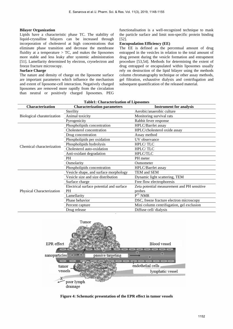
Bilayer Organization Lipids have a characteristic phase TC. The stability of liquid-crystalline bilayers can be increased through incorporation of cholesterol at high concentrations that eliminate phase transition and decrease the membrane fluidity at a temperature > TC, and makes the liposomes more stable and less leaky after systemic administration [51]. Lamellarity determined by electron, cryoelectron and freeze fracture microscopy. Surface Charge The nature and density of charge on the liposome surface are important parameters which influence the mechanism and extent of liposome-cell interaction. Negatively charged liposomes are removed more rapidly from the circulation than neutral or positively charged liposomes. PEG
functionalisation is a well-recognized technique to mask the particle surface and limit non-specific protein binding [52]. Encupsulation Efficiency (EE) The EE is defined as the percentual amount of drug entrapped in the vesicles in relation to the total amount of drug present during the vesicle formation and entrapment procedure [53,54]. Methods for determining the extent of drug entrapped or encapsulated within liposomes usually rely on destruction of the lipid bilayer using the methods column chromatography technique or other assay methods, gel filtration, exhaustive dialysis and centrifugation and subsequent quantification of the released material.
Table1: Characterization of Liposomes Characterization Characterization parameters Instrument for analysis
Biological charaterization Sterility Aerobic/anaerobic culture Animal toxicity Monitoring survival rats Pyrogenicity Rabbit fever response
Chemical characterization
Phospholipids concentration HPLC/Barrlet assay Cholesterol concentration HPLC/cholesterol oxide assay Drug concentration Assay method Phospholipids per oxidation UV observance Phospholipids hydrolysis HPLC/ TLC Cholesterol auto-oxidation HPLC/ TLC Anti-oxidant degradation HPLC/TLC PH PH meter Osmolarity Osmometer Phospholipids concentration HPLC/Barrlet assay
Physical Characterization
Vesicle shape, and surface morphology TEM and SEM Vesicle size and size distribution Dynamic light scattering, TEM Surface charge Free flow electrophoresis Electrical surface potential and surface PH
Zeta potential measurement and PH sensitive probes
Lamellarity P31 NMR Phase behavior DSC, freeze fracture electron microscopy Percent capture Mini column centrifugation, gel exclusion Drug release Diffuse cell/ dialysis
Figure 4: Schematic presentation of the EPR effect in tumor vessels
E. Sanarova et al /J. Pharm. Sci. & Res. Vol. 11(3), 2019, 1148-1155
1152

APPLICATIONS OF LIPOSOMES FOR DRUG DELIVERY Liposomes are used for drug delivery due to their unique properties. The encapsulation of drugs in vesicles (liposomes) prolongs their circulation in blood and enhances the efficiency of their accumulation in the diseased tissue (tumor, inflamed tissues), which protects the active substances from metabolic degradation and prevents changes in their tissue distribution, specifically, increased accumulation in organs rich in mononuclear phagocytes, liver, spleen, and bone marrow and reduced accumulation in kidney, myocardium, and brain [55]. Liposomes can be used to transport both hydrophilic and hydrophobic drugs [56,57]. The composition, surface charge, and size of lipids determine their physicochemical and biopharmaceutical characteristics, such as the rate of clearance from the injection site and blood plasma and the rate of delivery to a target organ. Liposomes with different lipid compositions can encapsulate different quantities of active substance, and, therewith, the encapsulation degree depends on the structure, size, charge, and lipid composition of lipo-somes, as well as on the intrinsic physicochemical characteristics of active substances. A liposome-encapsulated substance is protected from enzymes, which makes more efficient medicines susceptible to biodegradation in biological fluids. Liposomal formulations scarcely penetrate into myocardium and skeletal muscles, probably due to a peculiar structure of the endothelium of these organs. Liposomes do not come into the excretory system and, therefore, do not undergo glomerular filtration. The encapsulation in liposomes affects the pharmacokinetics of substances, specifically, the rates of their clearance from the injection site and blood, organ and tissue distribution and redistribution, and targeting efficiency. Since liposomes decrease toxicity of their encapsulated drug, the dose of the latter can be increased without evident side effects. As a result, cancer therapy could be brought to a qualitatively higher level [58]. The effort of many researchers is presently being focused on increasing the safety of efficient anticancer anthracycline antibiotics (doxorubicin [59], daunorubicin [60]), which cause severe side effects. The encapsulation in liposome carriers reduced the cardiotoxicity of these drugs and increased the survival rate of experimental animals compared to the control group administered free drugs [61,62]. The efficiency of anthracyclines is enhanced by their encapsulation in phospholipid vesicles with high cholesterol content in the lipid bilayer or in phospholipids with a hightemperature phase transition, which favor retention of drugs in liposomes when they enter the blood stream. Interesting property of liposomes is their natural ability to target cancer. The endothelial wall of all healthy human blood vessels is encapsulated by endothelial cells that are bound together by tight junctions. These tight junctions stop any large particle in the blood from leaking out of the vessel. Tumour vessels do not contain the same level of seal between cells and are diagnostically leaky. This ability is known as the Enhanced Permeability and Retention
effect (EPR-effect) (Figure 4). Liposomes of certain sizes, typically less than 400 nm, can rapidly enter tumour sites from the blood, but are kept in the bloodstream by the endothelial wall in healthy tissue vasculature. The approach for drug targeting via liposomes involves the use of ligands (e.g., antibodies, sugar residues, apoproteins or hormones), which are tagged on the lipid vesicles. The ligand recognises specific receptor sites and, thus, causes the lipid vesicles to concentrate at such target sites. By this approach the otherwise preferential distribution of liposomes into the RES is averted or minimized. The application of liposomes on the skin surface has been proven to be effective in drug delivery into the skin. Liposomes increase the permeability of skin for various entrapped drugs and at the same time diminish the side effect of these drugs because lower doses are now required. Antimicrobial agents have been encapsulated in liposomes for two reasons. First, they protect the entrapped drug against enzymatic degradation. For instance, the penicillins and cephalosporin are sensitive to the degradative action of β-lactamase, which is produced by certain microorganisms. Secondly, the lipid nature of the vesicles promotes enhanced cellular uptake of the antibiotics into the microorganisms, thus reducing the effective dose and the incidence of toxicity as exemplified by the liposomal formulation of amphotericin B.
Commercial products containing liposomes The products on the market which contain liposomes are few and far between, but this drug delivery system is proving its worth when applicated in the real world. Broadly, there are two separate classes of drugs that contain liposomes for treatment in humans, these classes being fungus infections and cancer. Currently amphotericin B is used with great success in severe fungal infections. Amphotericin is notoriously difficult to give parenterally because of the low tolerability, but liposome formulations have decreased the toxicity, which made it possible to considerably increase the dose as the therapeutic index was improved. This gave an increase in overall efficacy. Three different liposome-amphotericin products currently on the market, Abelcet™, AmBisome™ and Amphocil™ have completely different morphological and formulation setups, but still give this improvement [63]. When applied to anticancer therapy, liposomes were found to be very effective because of the specific targeting to cancer cells that is possible to achieve with targeted liposome systems. The liposomal formulations known as Doxil™ and DuanoXome™ are on the market, with research being done to expand the scope for these systems [64]. These liposomes are specific to tumours, and therefore, toxicity is reduced.
CONCLUSION After many years of search liposomes are considered as a good drug delivery vehicle. Since the discovery of liposomes, the field and applications thereof has broadened considerably. Liposomes may be composed of a whole host
E. Sanarova et al /J. Pharm. Sci. & Res. Vol. 11(3), 2019, 1148-1155
1153

of different lipids, naturally or manmade occurring, each having their own uses, advantages and disadvantages. The most used lipid component is phosphatidyl choline, because of its tendency to be neutral and relative low in cost. Another component usually added to the lipid mixture is cholesterol as cholesterol provides added stability. Liposomes can be classified according to production method, composition as well as size and shape. The advantages of using liposomes as a drug delivery system include: enhanced efficacy against pathogens and programmable target selectivity, decreased toxicity, improved pharmacokinetics and pharmacodynamics. Liposomes have been in use as drug delivery systems for a few years with a few formulations commercially available, which show great affectivity. Liposomes are used in sustain release, diagnostic purpose, intracellular delivery systems for proteins/peptides, antisense molecules, ribozymes and DNA. Liposomes have great promise as a drug delivery system.
ACKNOWLEDGMENT: The theoretical investigation has been made as a part of scientific program “Creation of innovative nanostructured hydrophobic formulation of domestic analogue of hypothalamic hormone somatostatin in the treatment of hormone-dependent tumors” under financial support of Russian President's Scholarship.
REFERENCES
1. Perche, F., Torchilin, V. P., J Drug Del. 2013, 2013, 1 – 32. 2. Fahr, A., Hoogevest, P., May, S., Bergstran, N., Leigh, M. L. S., Eur J
Pharm Sci. 2005, 26, 251 – 265. 3. Jone, A., J. Pharm. Sci. & Res. 2013, 5, 181 – 183. 4. Leong, K.W., Zhang, Y., Chan, H. F., Adv Drug Delivery Rev. 2013, 65,
104 – 120. 5. Immordino, M. L., Dosio, F., Cattel, L., Int J Namomedicine. 2006, 1,
297 – 315. 6. Zhang, J.X., Wang, K., Mao, Z.F., Fan, X., Jiang, D.L., Chen, M., Cui,
L., Sun, K., Dang, S.C., Int J Nanomedicine. 2013, 8, 1325 – 1334. 7. Torchilin, V. P., Nat. Rev. Drug Discov. 2005, 4, 145 – 160. 8. Samad, A., Sultana, Y., Aqil, M., Curr Drug Deliv. 2007, 4, 297 – 305. 9. Mahapatra, A. K., Murthy, P. N., Chandana, S., Swain, R. P., Polei, N.,
Der Pharm Lett. 2014, 6, 110 – 128. 10. Goni, F. M., Alonso, A., Biochim Biophys Acta. 2006, 1758, 1902 –
1921. 11. Allen, T. M., Adv Drug Delivery Rev. 1994, 13, 285 – 309. 12. Rao, N. M., Chem Phys Lipids. 2010, 163, 245 – 252. 13. Lonez, C., Vandenbranden, M., Ruysschaert, J. M., Adv Drug Delivery
Rev. 2012, 64, 1749 – 1758. 14. Plessis, J. D., Ramachandran, C., Weiner, N., Moiler, D. G., Int J
Pharm. 1996, 127, 273 – 278. 15. Popova, A.V., Dirk, K. H., Biophys J. 2007, 93, 1204 – 1214. 16. Hope, M.Y.; Bally, M.B.; Mayer, L.D., Chem. Phys. Lipids. 1986, 40,
89 – 107. 17. Lasic, D.D., Trends in Biotechnology. 1998, 16, 307 – 321. 18. Barista, C.M., Carvalho, C.M.B., Magalhaes, N.S.S., Braz J Pharm
Sci. 2007, 43, 167 – 179. 19. Drummond, D. C., Zignani, M., Leroux, J. C., Prog Lipid Res. 2000,
39, 409 – 460. 20. Ogawa, Y., Kodaka, M., Okuno, H., Chem Phys Lipids. 2002, 119, 51
– 68. 21. Ishida, T., Kirchmeier, M. J., Moase, E. H., Zalipsky, S., Allen, T. M.,
Biochim Biophys Acta. 2001, 1515, 144 – 158. 22. Felber, A. E., Dufresne, M. H., Leroux, J. C., Adv Drug Delivery Rev.
2012, 64, 979 – 992. 23. Roux, E., Francis, M., Winnik, M. F., Leroux J. C., Inter J Pharm.
2002, 242, 25 – 36.
24. Bangham, A.D., Standish, M.M., Watkens, J.C. J., Mol. Biol., 1965, 13, 238 – 252.
25. Bangham, A.D., Liposomes in Biological Systems, John Wiley and Sons, Chichester 1980.
26. Lantsova, A, Кotova, E., Sanarova, K., Oborotova, N., Poloskova, A., Barushnikov, A., Orlova, O., Krasnov, V., J Drug Del Sci Tech. 2012, 22, 469 – 472.
27. Sanarova, E.V., Lantsova, A.V., Polozkova, A.P., Orlova, O. L., Meerovich, I. G., Borisova, L. M., Kiseleva, M. P., Smirnova, Z. S., Kul’bachevskaya, N. Yu., Konyaeva, O. I., Oborotova, N. A., Nanotechnologies in Russia. 2015, 10, 492 – 500.
28. Meerovich, I.G, Sanarova, E.V., Meerovich, G.A., Derkacheva, V. M., Volkov, K. A., Negrimovsky, V.M., Barkanova, S.V., Lukyanets, E. A., Oborotova, N. A., Smirnova, Z. S., Borisova, L. M., Lantsova, A. V., Polozkova, A. P., Orlova, O. L.,. Loschenov, V. B, Umnova, L. V., Baryshnikov, A. Yu., Vorozhtsov, G. N., Russ J Gen Chem. 2015, 85, 280 – 288.
29. Nikolaeva, L., Oborotova, N., Bunyatyan, N., Zhang, X., Sanarova, E., Lantsova, A., Orlova, O., Polozkova, A., Pharmaceuticals. 2016, 9, 1 – 9.
30. Sanarova, E.V., Polozkova, A.P., Meerovich, I.G., Lantsova, A.V., Ignat’eva, E.V., Orlova, O.L., Oborotova, N.A., Pharm Chem J. 2012, 45, 741 – 745.
31. Sanarova, E., Meerovich, I., Lantsova, A., Kotova E., Shprakh, Z., Polozkova, A., Orlova, O., Meerovich, G., Borisova, L., Lukyanets, E., Smirnova, Z., Oborotova, N., Baryshnikov, A., J Drug Del Sci Tech. 2014, 24, 315 – 319.
32. Sanarova, E.V., Kotova, E.A., Lantsova, A.V., Journal of Physics. 2012, 345, 012045.
33. Kumari, A., Singla, R., Guliani, A., Yadav, S.K., EXCLI Journal. 2014, 13, 265 – 286.
34. Hwang, K.J., Padki, M.M., Chow, D.D., Biochim Biophys Acta. 1987, 901, 88 – 96.
35. Papahadjopoulos, D., Vali, W. J., Jacobson, K., Poste, G., Biochim Biophys Acta. 1975, 394, 483 – 491.
36. Dua, J.S., Rana, A.C., Bhandari, A. K., IJPSR. 2012, III, 14 – 20. 37. Liu, L., Yonetaini T., J Microencapsulation. 1994, 11, 409 – 421. 38. Mayer, L.D., Hope, M., Cullis, P.R., Biochim Biophys Acta. 1985b,
817, 193 – 196. 39. Gregoriadis, G., Leathwood, P.D., Ryman, B.E., FEBS Lett. 1971, 19,
95 – 99. 40. Lasic, D.D., Papahadjopoulos, D., Medical Applications of Liposome,
Elsevier, New York 1998. 41. Lasic, D.D., Ceh, D.D., Stuart, M.C.A., Guo, L., Frederik, P.M.,
Barenholtz, Y., Biochim Biophys Acta. 1995, 1239, 145 – 156. 42. Mandal, T.K., Downing, D. T., Acta Derm Venereol. 1993, 73, 917 –
918. 43. Vyas, S.P., Katare, Y.K., Mishra, V., Sihorkar, V., Int J Pharm. 2000,
210, 21 – 42. 44. New, R.C. Liposomes A Practical Approach, IRL Oxford University
Press, Oxford 1990. 45. Kolchens, S., Ramaswami, V., Birgenheier, J., Nett, L., O’Brein, D.F.,
Chem Phys Lipids. 1993, 65, 1 – 10. 46. Allen, Z., Tong, X., Mangeed, P., Lan, M., Sydney, U., Shahid, A.,
Imran, A., Int J Pharm. 2004, 270, 93 – 116. 47. Wiener, N., Lieb, L., Medical Applications of Liposome, Elsevier,
Oxford 1998. 48. Sanarova, E.V., Kotova, E.A., Lantsova, A.V., Yartseva, I. V., Orlova,
O. L., Oborotova, N. A., Pharm Chem J. 2012, 46, 192 – 195. 49. Talsma, H., Crommelin, D.J.A., Pharmaceut Technol. 1992a, 16, 52 –
58. 50. Bibi, S., Kaura, R., Lacey, M.H., McNeil, S.E, Wilkhu, J., Lattmann,
E., Christensen, D., Mohammed, A.R., Perrie, Y., Inter J Pharma, 2011, 417, 138 – 150.
51. Sharma, A., Sharma, U. S., Inter J Pharma, 1997, 154, 123 – 140. 52. Chonn, A., Cubis, P. R., Devine, D. V., J Immunol. 1991, 146, 4234-
4241. 53. Uchiyamab, N. K., Kiwadab, H., Tomoko, N., Inter J Pharma, 2005,
298, 198 – 205. 54. Sanarova, E.V., Polozkova, A.P., Meerovich, I.G., Lantsova, A.V.,
Yartseva, I.V., Orlova, O. L., Oborotova, N. A., Pharm Chem J. 2012, 46, 386 – 388.
55. Oborotova, N.A., Sanarova E.V., Russ J Gen Chem. 2013, 83, 2541 – 2547.
E. Sanarova et al /J. Pharm. Sci. & Res. Vol. 11(3), 2019, 1148-1155
1154

56. Oborotova, N.A., Smirnova, Z.S., Polozkova, A.P., Baryshnikov, A.Yu., Vest Ross Akad Med Nauk. 2002, 1, 42 – 45.
57. Bae, K.H., Chung, H.G., Park, T.G., Mol Cells. 2011, 31, 295 – 302. 58. Tikhonov, A.I., Biofarmatsiya (Biopharmacy), Zolotye Stranitsy,
Kharkiv 2003. 59. Tulpule, A., Espina, B.M., Berman, N., Clin Lymphoma Myeloma,
2006, 7, 59 – 64.
60. Goyal, P., Goyal, K., Kumar, S.G.G, Acta Pharm. 2005, 55, 1–25. 61. Kaminskas, L.M., McLeod, V.M., Kelly, B.D., Nanomed Nanotechnol
Biol Med. 2012, 8, 103 – 111.62. Porter, C. A., Rifkin, R. M., Clin Lymphoma Myeloma, 2007, 7, 150 –
155. 63. Storm, G., Crommelin, D.J.A., PSTT. 1998, 1, 19 – 31. 64. Alberts, D.S., Semin Oncol. 2004, 31, 53 – 90.
E. Sanarova et al /J. Pharm. Sci. & Res. Vol. 11(3), 2019, 1148-1155
1155


![Freeze Dried Liposome Delivery System Fo[1]](https://img.pdfslide.us/doc/110x75/577d25b31a28ab4e1e9f6898/freeze-dried-liposome-delivery-system-fo1.jpg)