Embed Size (px)
Citation preview
Ž .Brain Research 748 1997 267–270
Short communication
Lipopolysaccharide pre-treatment induces resistance against subsequent focalcerebral ischemic damage in spontaneously hypertensive rats
Kaoru Tasaki a, Christl A. Ruetzler a, Toshiho Ohtsuki a, David Martin b, Hiroshi Nawashiro a,John M. Hallenbeck a,)
a Stroke Branch, National Institute of Neurological Disorders and Stroke, National Institutes of Health, Bethesda, MD, USAb Department of Pharmacology, Amgen Boulder, Inc. Boulder, CO, USA
Accepted 26 November 1996
Abstract
Ž . Ž .Ischemic tolerance was induced in spontaneously hypertensive rats SHR by injection of a single dose of lipopolysaccharide LPSŽ . Ž .0.9 mgrkg, i.v. 1–7 days prior to permanent middle cerebral artery occlusion MCAO . Infarct volume, evaluated 24 h after MCAO,
Ž .was significantly reduced by LPS administration 2, 3 or 4 days prior to MCAO 22.8, 25.9 and 20.5%, respectively . The beneficial effectof LPS pre-treatment was completely nullified by concurrent administration of TNFbp. On this basis, the tolerance to ischemia induced byLPS is likely to be mediated by TNF-a.
Keywords: Tolerance; Lipopolysaccharide; Tumor necrosis factor a ; Endotoxin; Cerebral ischemia; Rat
Tolerance to experimental brain ischemia can be in-duced by a variety of stimuli that elicit a stress responsew x15,17 . The cytokines, IL-1 and TNF-a , activate intra-cellular signaling pathways that mediate the stress re-
w xsponse 16 and could, therefore, function in ligand-recep-tor interactions that induce the tolerant state. To explorethe possibility that cytokines have a role in the develop-ment of tolerance, LPS which elicits the release of IL-1
w xand TNF-a 14 was administered to spontaneously hyper-Ž .tensive rats SHR prior to MCAO. IL-1 receptor antago-
Ž . Ž .nist IL-1ra or TNF-binding protein TNFbp were subse-quently co-administered with LPS to assess their capacityto counteract the LPS effects on ischemic brain damage.
All animal studies were approved by the National Insti-tute of Neurological Disorders and Stroke Animal Care
Ž .and Use Committee. In 98 adult male SHR 300–350 g ,the left middle cerebral and left common carotid arterieswere occluded by a modified tandem occlusion techniquew x2 under 1–1.5% halothane in 30% O and 70% N O.2 2
Briefly, the left common carotid artery was ligated and theleft middle cerebral artery was exposed and electrocoagu-
) Corresponding author. Stroke Branch, NINDS, NIH, Building 36,Room 4A03, 36 Convent Drive MSC 4128, Bethesda, MD 20892-4128,
Ž .USA. Fax: q1 301 402-2769.
lated just distal to the inferior cerebral vein; both vesselswere transected. Body temperature was maintained at37.58C with a heating pad during and 2 h after surgery.The left femoral artery was cannulated for blood samplingand monitoring of blood pressure.
24 h after MCAO, animals were deeply anesthetizedwith pentobarbital and decapitated. Brains were removedand sequential 2 mm coronal brain sections were incubated
Ž .at 378C in 2% 2,3,5-triphenyltetrazolium chloride TTCw x9 . The total area of each coronal brain section and thelesion area were image-analyzed and lesion volumes werecalculated by integration of the section areas. All analyseswere performed in a blinded fashion.
The time course for the effects on ischemic braindamage of LPS administered as a single dose was investi-gated. LPS from Escherichia coli 0111:B4 phenol extractŽ .Sigma, St. Louis, MO was dissolved in sterile, pyrogen-
Ž . Ž .free 0.9% wrv NaCl and injected i.v. 0.9 mgrkg 1, 2,3, 4 or 7 days prior to MCAO. This optimal dose wasselected from a series of preliminary dose-finding studies.Control animals were injected with the same volume ofsterile, pyrogen-free 0.9% saline at the same time points.
In separate experiments, the effect of systemic injectionof LPS combined with either IL-1ra or TNFbp on ischemicbrain damage was investigated. IL-1ra, a gift from AmgenŽ .Boulder, CO , was dissolved in 10 mM sodium citrate
0006-8993r97r$17.00 Copyright q 1997 Elsevier Science B.V. All rights reserved.Ž .PII S0006-8993 96 01383-2
( )K. Tasaki et al.rBrain Research 748 1997 267–270268
buffer, pH 6.8, with 150 mM NaCl, 0.5 mM EDTA and0.1% Tween-80 and injected s.c. 100 mgrkg immediately
Ž .before and 4 h after LPS injection ns4 . In anothergroup of animals, human recombinant type I soluble TNFreceptor dimer conjugated to polyethylene glycol to en-
Žhance its activity, TNFbp, a gift from Amgen Boulder,CO., was dissolved in 10 mM sodium phosphate, 20 mMsodium chloride, pH 6.5 and injected i.v. 2 mgrkg just
Ž .before LPS injection ns5 . Control animals for bothgroups were injected with LPS in combination with an
Žappropriate volume of each vehicle ns4 and ns5,.respectively . MCAO was performed 3 days after the
LPSq IL-1ra, LPSqTNFbp or LPSqvehicle injectionand the infarct volume was measured 24 h post-MCAO byTTC staining. Four subsequent animals received only
Ž .TNFbp 2 mgrkg i.v. 72 h before MCAO. Serum TNF-aw xlevels were measured by L929 cell lysis bioassay 8 2 h
after LPSqTNFbp or LPSqvehicle injection.Data are presented as mean"S.D. LPS vs. Saline
comparisons at each time point and comparison of LPSqIL-1ra vehicle with LPSq IL-1ra were analyzed by inde-pendent groups Student’s t-test. The Welch approximationwas used when group variances were heterogenous. TheTNFbp study was analyzed by one-way ANOVA andpair-wise group comparisons were made with a Bonferronicorrection. Student’s t-test was also used to compare thetwo serum TNF-a levels.
Body temperature was not significantly altered afterLPS injection and remained -398C in all animals. Com-plete blood cell counts revealed no statistically significantdifferences between the LPS-injected groups and theirrespective control groups. There were no significant differ-ences in the physiologic variables in any of the LPS- orvehicle-pre-treated groups at any of the 5 time pointsŽ .Table 1 . Similarly, there were no differences in physio-logic variables in the blocking studies with LPSq IL-1ra,LPSqTNFbp or their respective controls.
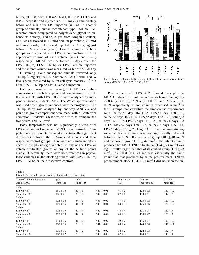
Ž .Fig. 1. Infarct volumes: LPS 0.9 mgrkg or saline i.v. at several timesbefore MCAO. ) P -0.05; ) ) P -0.02.
Pre-treatment with LPS at 2, 3 or 4 days prior toMCAO reduced the volume of the ischemic damage by
Ž . Ž . Ž22.8% P-0.05 , 25.9% P-0.02 and 20.5% P-
. 30.02 , respectively. Infarct volumes expressed in mm inthe 5 groups that constitute the time-course experimentswere: saliner1 day 162"22, LPSr1 day 138"30,saliner2 days 163"35, LPSr2 days 122"23, saliner3days 162"37, LPSr3 days 116"26, saliner4 days 163"12, LPSr4 days 128"27, saliner7 days 165"11,
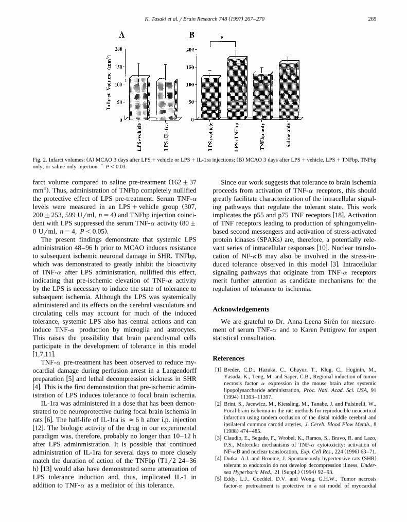
Ž .LPSr7 days 163"25 Fig. 1 . In the blocking studies,ischemic lesion volume was not significantly different
Ž 3.between the LPSq IL-1ra-treated group 109"48 mmŽ 3.and the control group 118"42 mm . The infarct volume
Ž 3.produced by LPSqTNFbp treatment 174"24 mm wasŽsignificantly larger than that of its control group 119"23
3 . Ž .mm , P-0.03 Fig. 2 and was essentially the samevolume as that produced by saline pre-treatment. TNFbp
Ž 3.pre-treatment alone 131"19 mm did not increase in-
Table 1Physiologic variables at occlusion of the middle cerebral artery
Time of LPS administration pO pCO pH Hematocrit Glucose MABP2 2Ž . Ž . Ž . Ž . Ž . Ž .pre-MCAO mm Hg mm Hg % mgr100 ml mm Hg
1 dayŽ .LPS ns8 155"10 39"3 7.38"0.01 41"2 123"12 130"12Ž .Saline ns8 136"21 39"2 7.42"0.02 42"1 130"11 142"7
2 daysŽ .LPS ns8 128"38 44"2 7.38"0.02 47"3 123"12 129"12Ž .Saline ns8 120"16 41"4 7.40"0.01 43"3 126"16 136"12
3 daysŽ .LPS ns8 122"19 40"4 7.40"0.01 44"4 121"17 132"9Ž .Saline ns8 120"10 42"4 7.40"0.02 46"3 130"27 138"8
4 daysŽ .LPS ns8 142"15 41"5 7.40"0.02 39"2 146"17 129"10Ž .Saline ns8 133"15 38"2 7.41"0.02 40"4 144"10 141"11
7 daysŽ .LPS ns8 136"15 40"2 7.40"0.02 38"2 122"13 142"7Ž .Saline ns8 132"22 38"5 7.40"0.02 42"3 124"11 148"9
( )K. Tasaki et al.rBrain Research 748 1997 267–270 269
Ž . Ž .Fig. 2. Infarct volumes: A MCAO 3 days after LPSqvehicle or LPSq IL-1ra injections; B MCAO 3 days after LPSqvehicle, LPSqTNFbp, TNFbponly, or saline only injection. ) P-0.03.
Žfarct volume compared to saline pre-treatment 162"373.mm . Thus, administration of TNFbp completely nullified
the protective effect of LPS pre-treatment. Serum TNF-aŽlevels were measured in an LPSqvehicle group 307,
.200"253, 599 Urml, ns4 and TNFbp injection coinci-Ždent with LPS suppressed the serum TNF-a activity 80"
.0 Urml, ns4, P-0.05 .The present findings demonstrate that systemic LPS
administration 48–96 h prior to MCAO induces resistanceto subsequent ischemic neuronal damage in SHR. TNFbp,which was demonstrated to greatly inhibit the bioactivityof TNF-a after LPS administration, nullified this effect,indicating that pre-ischemic elevation of TNF-a activityby the LPS is necessary to induce the state of tolerance tosubsequent ischemia. Although the LPS was systemicallyadministered and its effects on the cerebral vasculature andcirculating cells may account for much of the inducedtolerance, systemic LPS also has central actions and caninduce TNF-a production by microglia and astrocytes.This raises the possibility that brain parenchymal cellsparticipate in the development of tolerance in this modelw x1,7,11 .
TNF-a pre-treatment has been observed to reduce my-ocardial damage during perfusion arrest in a Langendorff
w xpreparation 5 and lethal decompression sickness in SHRw x4 . This is the first demonstration that pre-ischemic admin-istration of LPS induces tolerance to focal brain ischemia.
IL-1ra was administered in a dose that has been demon-strated to be neuroprotective during focal brain ischemia in
w xrats 6 . The half-life of IL-1ra is f6 h after i.p. injectionw x12 . The biologic activity of the drug in our experimentalparadigm was, therefore, probably no longer than 10–12 hafter LPS adminmistration. It is possible that continuedadministration of IL-1ra for several days to more closely
Žmatch the duration of action of the TNFbp T1r2 24–36. w xh 13 would also have demonstrated some attenuation of
LPS tolerance induction and, thus, implicated IL-1 inaddition to TNF-a as a mediator of this tolerance.
Since our work suggests that tolerance to brain ischemiaproceeds from activation of TNF-a receptors, this shouldgreatly facilitate characterization of the intracellular signal-ing pathways that regulate the tolerant state. This work
w ximplicates the p55 and p75 TNF receptors 18 . Activationof TNF receptors leading to production of sphingomyelin-based second messengers and activation of stress-activated
Ž .protein kinases SPAKs are, therefore, a potentially rele-w xvant series of intracellular responses 10 . Nuclear translo-
cation of NF-k B may also be involved in the stress-in-w xduced tolerance observed in this model 3 . Intracellular
signaling pathways that originate from TNF-a receptorsmerit further attention as candidate mechanisms for theregulation of tolerance to ischemia.
Acknowledgements
We are grateful to Dr. Anna-Leena Siren for measure-´ment of serum TNF-a and to Karen Pettigrew for expertstatistical consultation.
References
w x1 Breder, C.D., Hazuka, C., Ghayur, T., Klug, C., Huginin, M.,Yasuda, K., Teng, M. and Saper, C.B., Regional induction of tumornecrosis factor a expression in the mouse brain after systemiclipopolysaccharide administration, Proc. Natl. Acad. Sci. USA, 91Ž .1994 11393–11397.
w x2 Brint, S., Jacewicz, M., Kiessling, M., Tanabe, J. and Pulsinelli, W.,Focal brain ischemia in the rat: methods for reproducible neocorticalinfarction using tandem occlusion of the distal middle cerebral andipsilateral common carotid arteries, J. Cereb. Blood Flow Metab., 8Ž .1988 474–485.
w x3 Claudio, E., Segade, F., Wrobel, K., Ramos, S., Bravo, R. and Lazo,P.S., Molecular mechanisms of TNF-a cytotoxicity: activation of
Ž .NF-k B and nuclear translocation, Exp. Cell Res., 224 1996 63–71.w x Ž .4 Dutka, A.J. and Broome, J. Spontaneously hypertensive rats SHR
tolerant to endotoxin do not develop decompression illness, Under-Ž . Ž .sea Hyperbaric Med., 21 Suppl. 1994 92–93.
w x5 Eddy, L.J., Goeddel, D.V. and Wong, G.H.W., Tumor necrosisfactor-a pretreatment is protective in a rat model of myocardial
( )K. Tasaki et al.rBrain Research 748 1997 267–270270
ischemia-reperfusion injury, Biochem. Biophys. Res. Commun., 184Ž .1992 1056–1059.
w x6 Garcia, J.H., Liu, K.-F. and Relton, J.K., Interleukin-1 receptorantagonist decreases the number of necrotic neurons in rats with
Ž .middle cerebral artery occlusion, Am. J. Pathol., 147 1995 1477–1486.
w x7 Gutierrez, E.G., Banks, W.A. and Kastin, A.J., Murine tumor necro-sis factor alpha is transported from blood to brain in the mouse, J.
Ž .Neuroimmunol., 47 1993 169–176.w x8 Hogan, M.M. and Vogel, S.N., Production of tumor necrosis factor
Ž .by rIFN-g-primed C3HrHeJ LPSd macrophages requires the pres-Ž .ence of lipid A-associated proteins, J. Immunol., 141 1988 4196–
4202.w x9 Isayama, K., Pitts, L.H. and Nishimura, M.C., Evaluation of 2,3,5-
triphenyltetrazolium chloride staining to delineate rat brain infarcts,Ž .Stroke, 22 1991 1394–1398.
w x10 Kyriakis, J.M., Banerjee, P., Nikolakaki, E., Dai, T., Rubie, E.A.,Ahmad, M.F., Avruch, J. and Woodgett, J.R., The stress-activated
Ž .protein kinase subfamily of c-jun kinases, Nature, 369 1994156–160.
w x11 Laye, S., Parnet, P., Goujon, E. and Dantzer, R., Peripheral adminis-´tration of lipopolysaccharide induces the expression of cytokinetranscripts in the brain and pituitary of mice, Mol. Brain. Res., 27Ž .1994 157–162.
w x12 Martin, D., Chinookoswong, N. and Millfer, G., The interleukin-1Ž .antagonist rhIL-1ra protects against cerebral infarction in a rat
Ž .model of hypoxia-ischemia, Exp. Neurol., 130 1994 362–367.w x13 Martin, D., Near, S.L., Bendele, A. and Russell, D.A., Inhibition of
tumor necrosis factor is protective against neurologic dysfunctionafter active immunization of Lewis rats with myelin basic protein,
Ž .Exp. Neurol., 131 1995 221–228.w x14 Pugin, J., Ulevitch, R.J. and Tobias, P.S., Tumor necrosis factor-a
and interleukin-1b mediate human endothelial cell activation inŽ .blood at low endotoxin concentrations, J. Inflammat., 45 1995
49–55.w x15 Sakaki, T., Yamada, K., Otsuki, H., Yuguchi, T., Kohmura, E. and
Hayakawa, T., Brief exposure to hypoxia induces bFGF mRNA andprotein and protects rat cortical neurons from prolonged hypoxic
Ž .stress, Neurosci. Res., 23 1995 289–296.w x16 Schobitz, B., De Kloet, E.R. and Holsboer, F., Gene expression and¨
function of Interleukin 1, interleukin 6 and tumor necrosis factor inŽ .the brain, Prog. Neurobiol., 44 1994 397–432.
w x17 Simon, R.P., Niiro, M. and Gwinn, R., Prior ischemic stress protectsŽ .against experimental stroke, Neurosci. Lett., 163 1993 135–137.
w x18 Vilcek, J. and Lee, T.H., Tumor necrosis factor. New insights intothe molecular mechanisms of its multiple actions, J. Biol. Chem.,
Ž .266 1991 7313–7316.


![Tail suspension is useful as a sarcopenia model in rats...muscle weakness [9, 10]. Spontaneously hypertensive rats (SHR) are widely used in hypertension and insulin-resistance models](https://img.pdfslide.us/doc/110x75/60fd29b9db06e05a8002d7af/tail-suspension-is-useful-as-a-sarcopenia-model-in-rats-muscle-weakness-9.jpg)