Embed Size (px)
Citation preview
Lipopolysaccharide Induces Pro-Inflammatory Cytokinesand MMP Production via TLR4 in Nasal Polyp-DerivedFibroblast and Organ CultureJung-Sun Cho1,2, Ju-Hyung Kang1, Ji-Young Um1, In-Hye Han1, Il-Ho Park2,3, Heung-Man Lee1,2,3*
1 Brain Korea 21 Plus for Biomedical Science, College of Medicine, Korea University, Seoul, South Korea, 2 Institute for Medical Devices Clinical Trial Center, Korea
University, Guro Hospital, Seoul, South Korea, 3 Department of Otorhinolaryngology-Head and Neck Surgery, Korea University, Guro Hospital, Seoul, South Korea
Abstract
Nasal polyposis is characterized by persistent inflammation and remodeling in sinonasal mucosa. Toll-like receptors (TLRs)play a role in the innate immune response to microbes in the sinonasal cavity. The aim of this study was to evaluate whethernasal polyp-derived fibroblasts (NPDFs) and organ-cultured nasal polyps can synthesize pro-inflammatory cytokines andmatrix metalloproteinases (MMPs) after exposure to lipopolysaccharide (LPS), a TLR4 agonist. NPDFs and organ-culturednasal polyps were isolated from nasal polyps of 8 patients and exposed to LPS. The mRNA and protein expression levels ofTLRs, cytokines, and MMPs were determined using a gene expression microarray, real-time RT-PCR, western blot analysis,enzyme-linked immunosorbent assay, and immunofluorescence staining. The enzymatic activities of MMPs were analyzedusing collagen or gelatin zymography. The protein expression level of MMP-1 increased in nasal polyp tissues compared toinferior turbinate tissues. LPS induced mRNA expression of TLR4, IL-6, IL-8, and MMP-1 and activated MAPK signaling inNPDFs. LPS promoted the release of interleukin (IL)-6 through extracellular signal-related kinase (ERK) and IL-8 through ERKand c-Jun N-terminal kinases (JNK). Production of IL-6 and IL-8 was induced by PI3K/Akt signaling in LPS-stimulated NPDFs.LPS increased the transcript and protein expression levels of MMP-1 and induced collagenase activity of MMP-1 via ERK andp38, but did not induce gelatinase activity of MMP-2 and MMP-9. LPS from Rhodobacter sphaeroides (LPS-RS) inhibited thestimulatory effects of LPS in NPDFs as well as in organ culture of nasal polyp. LPS triggers immune response via TLR 4 andactivates MAPK and PI3K/Akt signaling pathway, which is involved in remodeling of nasal polyps.
Citation: Cho J-S, Kang J-H, Um J-Y, Han I-H, Park I-H, et al. (2014) Lipopolysaccharide Induces Pro-Inflammatory Cytokines and MMP Production via TLR4 in NasalPolyp-Derived Fibroblast and Organ Culture. PLoS ONE 9(11): e90683. doi:10.1371/journal.pone.0090683
Editor: Shrikant Anant, University of Kansas School of Medicine, United States of America
Received August 26, 2013; Accepted February 3, 2014; Published November 12, 2014
Copyright: � 2014 Cho et al. This is an open-access article distributed under the terms of the Creative Commons Attribution License, which permits unrestricteduse, distribution, and reproduction in any medium, provided the original author and source are credited.
Funding: This work was supported by the National Research Foundation of Korea (NRF) grant funded by the Korea Government (MEST; no. 2012-0005455) and aKorea University Grant. The funders had no role in study design, data collection and analysis, decision to publish, or preparation of the manuscript.
Competing Interests: The authors have declared that no competing interests exist.
* Email: [email protected]
Introduction
Chronic rhinosinusitis with nasal polyps (CRSwNP) is a form of
sinonasal inflammatory disease characterized by persistent eosin-
ophilic inflammation, edematous mucosa with nasal polyps, and
thickened sinonasal secretions [1,2]. Although it has been
proposed that CRSwNP is fundamentally an inflammatory disease
rather than an infection, it also has been hypothesized that
microbes often present in the sinonasal cavity play a role in
initiating or perpetuating mucosal inflammation [3,4]. As the first
site of contact between the host and outside environment, the
sinonasal cavity plays a critical role in immunity.
Fibroblasts are major structural components of tissues, where
they confer mechanical strength by providing a supporting
framework for the extracellular matrix (ECM) and are also
thought to be responsible for local recruitment of inflammatory
cells owing to their ability to produce a variety of chemokines
[5,6]. Although fibroblasts play an important role as a source of
biological mediators in initiating and amplifying inflammation,
overproduction of these factors by fibroblasts may prevent
resolution of this condition, leading to chronic inflammation [7].
Whether bacterial products, such as lipopolysaccharide (LPS), can
directly elicit cytokine responses in fibroblasts remains controver-
sial [8,9].
Toll-like receptors (TLRs) are transmembrane receptors with an
extracellular domain that interacts with a pathogen ligand and an
intracellular domain that is involved in signaling [10]. Mammals
express at least 10 TLRs that recognize specific pathogen
molecules. Each of these TLRs is thought to play a role in the
innate immune response to innocuous microbes in the sinonasal
cavity as well as airborne bacterial, fungal, or viral pathogens; for
example, TLR4 recognizes LPS from gram-negative bacteria. The
mRNA for all 10 TLRs is expressed in the sinonasal mucosa, both
in health and in sinus disease [11]. Although the mechanisms
underlying persistent inflammation in CRS are unknown, innate
immune processes may play a role.
Matrix metalloproteinases (MMPs) comprise a large family of
proteolytic enzymes containing a zinc-binding catalytic domain
and are involved in the degradation of ECM components. Their
extracellular activities are regulated by tissue inhibitors of MMP
(TIMP) [12]. Fibroblasts also secrete MMPs that may contribute
to tissue destruction [13]. Expression levels of MMPs have been
found to be elevated in nasal polyp tissues compared to control
tissues and play important roles in the formation of nasal polyposis
PLOS ONE | www.plosone.org 1 November 2014 | Volume 9 | Issue 11 | e90683
[14,15]. However, the role of LPS-induced pro-inflammatory
cytokines and MMPs in nasal polyp-derived fibroblasts (NPDFs)
has not been reported.
We hypothesized that LPS exposure up-regulates not only pro-
inflammatory cytokines, but also tissue remodeling via MMPs in
patients with nasal polyposis. In this study, we evaluated whether
NPDFs and organ-cultured nasal polyps can synthesize pro-
inflammatory cytokines and MMPs following exposure to LPS.
Materials and Methods
MaterialsLPS from Pseudomonas aeruginosa was obtained from Sigma
(St. Louis, MO). InvivoGen (San Diego, CA) provided LPS
isolated from the photosynthetic bacterium Rhodobacter sphaer-oides (LPS-RS). Inhibitors of extracellular related kinase (ERK)
(U0126), p38 (SB203580), c-Jun N-terminal kinase (JNK)
(SP600125) and PI3K/Akt inhibitor (LY294002) were purchased
from Calbiochem (Billerica, MA). The inhibitors were dissolved in
dimethyl sulfoxide. Antibodies against phospho-ERK, phospho-
p38, and phospho-JNK were purchased from Cell Signaling
Technology (Danvers, MA). Antibodies against TLR4 (sc-10741),
MMP-1 (sc-21731), MMP-2 (sc-10736), MMP-9 (sc-21733),
GAPDH (sc-47724), and b –actin (sc-4778) were obtained from
Santa Cruz Biotechnology (Santa Cruz, CA).
Nasal polyp tissues and NPDF cultureNasal polyp tissues were collected from 8 patients with chronic
rhinosinusitis (CRS) and nasal polyp who were recruited from the
Department of Otorhinolaryngology at the Korea University
Medical Center. No patients had a history of allergy, asthma, or
aspirin sensitivity, and they had not been treated with oral anti-
allergic agents for at least 2 months. Written informed consent was
obtained from each patient, and the study was approved by the
Korea University Medical Center Institutional Review Board
(KUGH12041). NPDFs were isolated from surgical tissues and
purified according to our previous study [16]. Cells used for
experiments were obtained from the fourth cell passage.
Microarray analysisNPDFs (56106 cells/mL) were exposed to LPS (10 mg/mL) for
12 h. Total RNA was isolated using Trizol reagent (Invitrogen,
Carlsbad, CA). For control and test RNAs, synthesis of target
cRNA probes and hybridization were performed using the Low
RNA Input Linear Amplification kit (Agilent Technology, Santa
Clara, CA). Hybridized images were scanned using a DNA
microarray scanner and quantified using Feature Extraction
Software (Agilent). All data normalization and selection of fold-
change of the genes were performed using GeneSpringGX 7.3
(Agilent) in Table 2. The accession number of our microarray data
in GEO (Gene Expression Omnibus) is GSE52505.
Real-time RT-PCRTotal tissue RNA was extracted using NucleoSpin RNA II
(Macherey-Nagel, Duren, Germany) according to the manufac-
turer’s instructions. The RNA concentration was determined using
the NanoDrop ND-2000 Spectrophotometer (Thermo Scientific,
Wilmington, DE). Quality and integrity of total RNA was assessed
on 1% formaldehyde-agarose gels. For real-time RT-PCR, 2 mg of
total RNA in a 50 ml reverse transcriptase reaction mixture was
reverse transcribed to cDNA by using the ReverTra Ace qPCR
RT Kit (Toyobo, Osaka, Japan) following the manufacturer’s
protocols. Quantitative PCR was then carried out in a 7300 Real-
Time PCR System (Applied Biosystems, Foster City, CA) using
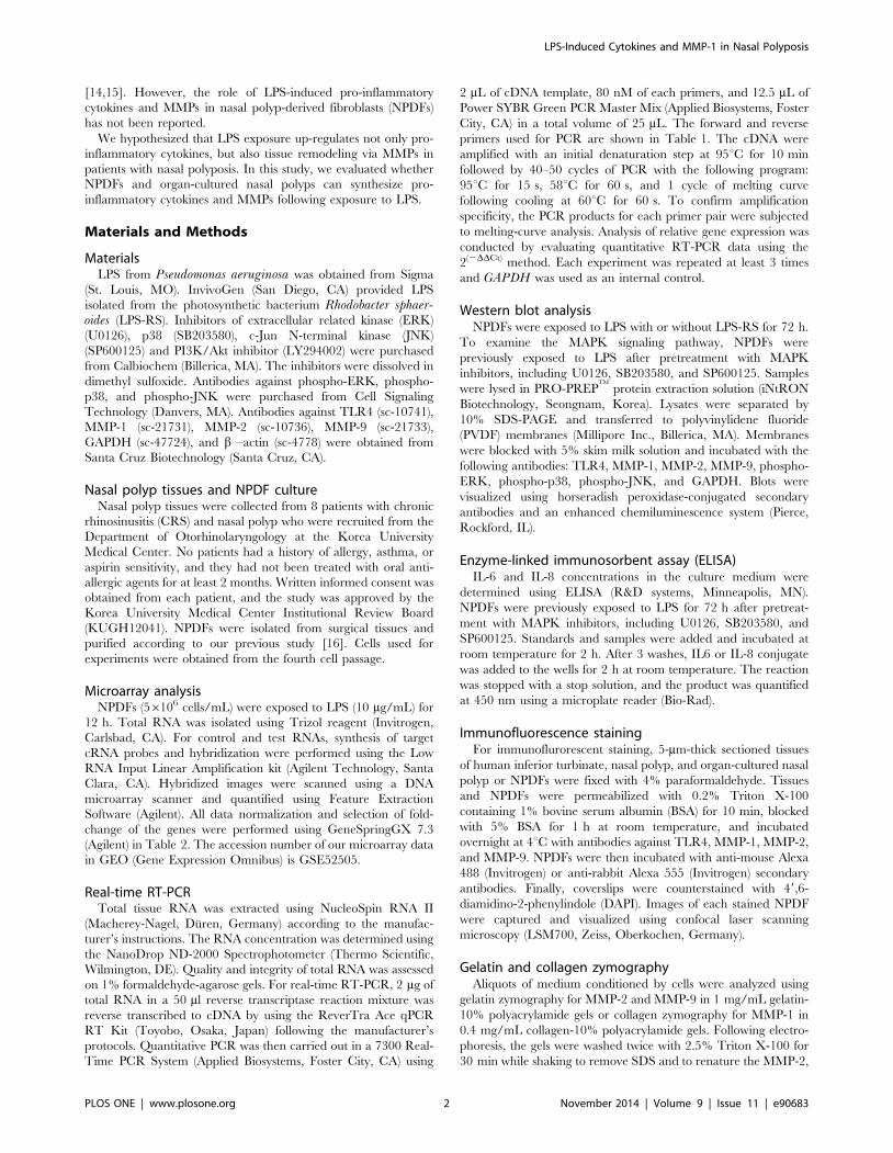
2 mL of cDNA template, 80 nM of each primers, and 12.5 mL of
Power SYBR Green PCR Master Mix (Applied Biosystems, Foster
City, CA) in a total volume of 25 mL. The forward and reverse
primers used for PCR are shown in Table 1. The cDNA were
amplified with an initial denaturation step at 95uC for 10 min
followed by 40–50 cycles of PCR with the following program:
95uC for 15 s, 58uC for 60 s, and 1 cycle of melting curve
following cooling at 60uC for 60 s. To confirm amplification
specificity, the PCR products for each primer pair were subjected
to melting-curve analysis. Analysis of relative gene expression was
conducted by evaluating quantitative RT-PCR data using the
2(2DDCt) method. Each experiment was repeated at least 3 times
and GAPDH was used as an internal control.
Western blot analysisNPDFs were exposed to LPS with or without LPS-RS for 72 h.
To examine the MAPK signaling pathway, NPDFs were
previously exposed to LPS after pretreatment with MAPK
inhibitors, including U0126, SB203580, and SP600125. Samples
were lysed in PRO-PREPTM
protein extraction solution (iNtRON
Biotechnology, Seongnam, Korea). Lysates were separated by
10% SDS-PAGE and transferred to polyvinylidene fluoride
(PVDF) membranes (Millipore Inc., Billerica, MA). Membranes
were blocked with 5% skim milk solution and incubated with the
following antibodies: TLR4, MMP-1, MMP-2, MMP-9, phospho-
ERK, phospho-p38, phospho-JNK, and GAPDH. Blots were
visualized using horseradish peroxidase-conjugated secondary
antibodies and an enhanced chemiluminescence system (Pierce,
Rockford, IL).
Enzyme-linked immunosorbent assay (ELISA)IL-6 and IL-8 concentrations in the culture medium were
determined using ELISA (R&D systems, Minneapolis, MN).
NPDFs were previously exposed to LPS for 72 h after pretreat-
ment with MAPK inhibitors, including U0126, SB203580, and
SP600125. Standards and samples were added and incubated at
room temperature for 2 h. After 3 washes, IL6 or IL-8 conjugate
was added to the wells for 2 h at room temperature. The reaction
was stopped with a stop solution, and the product was quantified
at 450 nm using a microplate reader (Bio-Rad).
Immunofluorescence stainingFor immunoflurorescent staining, 5-mm-thick sectioned tissues
of human inferior turbinate, nasal polyp, and organ-cultured nasal
polyp or NPDFs were fixed with 4% paraformaldehyde. Tissues
and NPDFs were permeabilized with 0.2% Triton X-100
containing 1% bovine serum albumin (BSA) for 10 min, blocked
with 5% BSA for 1 h at room temperature, and incubated
overnight at 4uC with antibodies against TLR4, MMP-1, MMP-2,
and MMP-9. NPDFs were then incubated with anti-mouse Alexa
488 (Invitrogen) or anti-rabbit Alexa 555 (Invitrogen) secondary
antibodies. Finally, coverslips were counterstained with 49,6-
diamidino-2-phenylindole (DAPI). Images of each stained NPDF
were captured and visualized using confocal laser scanning
microscopy (LSM700, Zeiss, Oberkochen, Germany).
Gelatin and collagen zymographyAliquots of medium conditioned by cells were analyzed using
gelatin zymography for MMP-2 and MMP-9 in 1 mg/mL gelatin-
10% polyacrylamide gels or collagen zymography for MMP-1 in
0.4 mg/mL collagen-10% polyacrylamide gels. Following electro-
phoresis, the gels were washed twice with 2.5% Triton X-100 for
30 min while shaking to remove SDS and to renature the MMP-2,
LPS-Induced Cytokines and MMP-1 in Nasal Polyposis
PLOS ONE | www.plosone.org 2 November 2014 | Volume 9 | Issue 11 | e90683
MMP-9, or MMP-1 in the gels. Renaturated gels were incubated
in developing buffer containing 100 mM Tris–HCl, 5 mM CaCl2,
0.005% Brij-35, and 0.001% NaN3 (pH 8.0), overnight at 37uC.
Gels were stained with 0.25% Coomassie brilliant blue G-250
(50% methanol, 10% acetic acid) and destained using destaining
solution (50% methanol, 10% acetic acid). Proteinase activity was
observed as cleared (unstained) regions. Finally, the gels were dried
for 2 h using a gel dryer (Bio-Rad Labs).
Ex vivo organ cultureNasal polyp tissues were cut into 2 to 3 mm3 pieces under sterile
conditions. Tissue fragments were washed 3 times with phosphate-
buffered saline and then rinsed with culture medium. The culture
medium was composed of 98% Dulbecco’s minimum essential
medium (Invitrogen), 2% heat-inactivated fetal bovine serum
(Invitrogen), 100 U/mL penicillin (Invitrogen), 100 mg/mL strep-
tomycin (Invitrogen), and 0.25 mg/mL fungizone. The rinsed
tissue fragments were placed on pre-hydrated 1061061 mm
gelatin sponge (Spongostan, Johnson & Johnson, San Angelo, TX)
with the mucosa side facing up and the submucosa side facing
down. Tissue fragments were placed into 6-well plates and filled
with 1.5 mL of culture medium per well such that the mucosa was
above the liquid phase. Nasal polyps were stimulated with LPS
(20 mg/mL) with or without LPS-RS (20 mg/mL) for 72 h. The
Table 1. Sequences of RT-PCR oligonucleotide primers.
Primer Direction Sequence Size (bp)
TLR4 forward 59- TGA GCA GTC GTG CTG GTA TC -39 167
reverse 59- CAG GGC TTT TCT GAG TCG TC -39
IL-6 forward 59- GGT ACA TCC TCG ACG GCA TCT -39 81
reverse 59- GTG CCT CTT TGC TGC TTT CAC -39
IL-8 forward 59- ATG ACT TCC AAG CTG GCC -39 282
reverse 59- TCT TCA AAA ACT TCT CCA CAA CCC -39
MMP-1 forward 59- CAG AGA TGA AGT CCG GTT TTT C -39 75
reverse 59- GGG GTA TCC GTG TAG CAC AT -39
MMP-2 forward 59- AGA TCT TCT TCT TCA AGG AAC CGT T -39 224
reverse 59- GGC TGG TCA GTG GCT TGG GGT A -39
MMP-9 forward 59- GCG GAG ATT GGG AAC CAG CTG TA -39 208
reverse 59- GAC GCG CCT GTG TAC ACC CAC A -39
GAPDH forward 59- GTG GAT ATT GTT GCC ATC AAT GAC C -39 271
reverse 59- GCC CCA GCC TTC TTC ATG GTG GT -39
doi:10.1371/journal.pone.0090683.t001
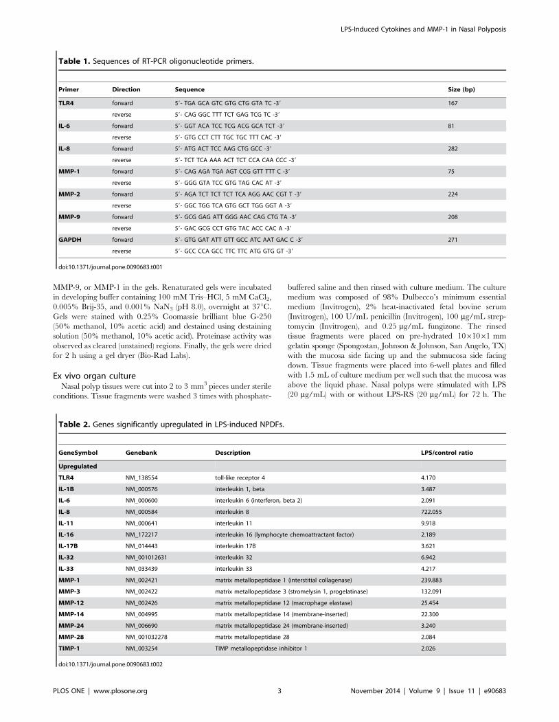
Table 2. Genes significantly upregulated in LPS-induced NPDFs.
GeneSymbol Genebank Description LPS/control ratio
Upregulated
TLR4 NM_138554 toll-like receptor 4 4.170
IL-1B NM_000576 interleukin 1, beta 3.487
IL-6 NM_000600 interleukin 6 (interferon, beta 2) 2.091
IL-8 NM_000584 interleukin 8 722.055
IL-11 NM_000641 interleukin 11 9.918
IL-16 NM_172217 interleukin 16 (lymphocyte chemoattractant factor) 2.189
IL-17B NM_014443 interleukin 17B 3.621
IL-32 NM_001012631 interleukin 32 6.942
IL-33 NM_033439 interleukin 33 4.217
MMP-1 NM_002421 matrix metallopeptidase 1 (interstitial collagenase) 239.883
MMP-3 NM_002422 matrix metallopeptidase 3 (stromelysin 1, progelatinase) 132.091
MMP-12 NM_002426 matrix metallopeptidase 12 (macrophage elastase) 25.454
MMP-14 NM_004995 matrix metallopeptidase 14 (membrane-inserted) 22.300
MMP-24 NM_006690 matrix metallopeptidase 24 (membrane-inserted) 3.240
MMP-28 NM_001032278 matrix metallopeptidase 28 2.084
TIMP-1 NM_003254 TIMP metallopeptidase inhibitor 1 2.026
doi:10.1371/journal.pone.0090683.t002
LPS-Induced Cytokines and MMP-1 in Nasal Polyposis
PLOS ONE | www.plosone.org 3 November 2014 | Volume 9 | Issue 11 | e90683
cultured nasal polyps were examined to detect protein expression
levels of IL-6, IL-8, and MMP-1. The plates were placed in a
humidified incubator and maintained at 37uC in 5% CO2.
Statistical analysisThe results were obtained from at least 3 independent
experiments. The statistical significance of the differences between
control and experimental data was analyzed using the unpaired t-test or one-way analysis of variance (ANOVA) followed by Tukey’s
test (GraphPad Prism, version 5, GraphPad Software, San Diego,
CA). Significance was established at the 95% confidence level. P-
values ,0.05 were accepted as statistically significant.
Results
MMP-1 protein expression levels are increased in nasalpolyp tissues
In a recent study, MMP-1 expression was shown to be
significantly increased in nasal polyps and CRS compared with
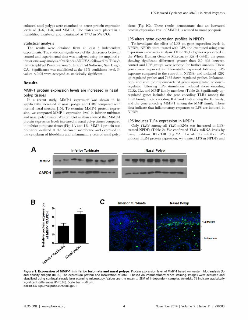
normal nasal mucosa [15]. To examine MMP-1 protein expres-
sion, we compared MMP-1 expression level in inferior turbinate
and nasal polyp tissues. Western blot analysis showed that MMP-1
protein expression levels increased in nasal polyp tissues compared
to inferior turbinate tissues (Fig. 1A and 1B). MMP-1 protein was
primarily localized at the basement membrane and expressed in
the cytoplasm of fibroblasts and inflammatory cells of nasal polyp
tissue (Fig. 1C). These results demonstrate that an increased
protein expression level of MMP-1 is related to nasal polyposis.
LPS alters gene expression profiles in NPDFsTo investigate the effect of LPS on gene expression levels in
NPDFs, NPDFs were treated with LPS and examined using gene
expression microarray analysis. Of the 34,127 genes represented in
the Whole Human Genome Microarray Kit (4644K), the genes
showing significant differences greater than 2.0 fold between
control and LPS groups were selected for further analysis. These
genes were regarded as differentially expressed following LPS
exposure compared to the control in NPDFs, and included 1297
up-regulated probes and 7462 down-regulated probes. Inflamma-
tion- and immune response-related genes up-regulated or down-
regulated following LPS stimulation included those encoding
TLRs, ILs, and MMP family members (Table 2). Significantly up-
regulated genes included the gene encoding TLR4 among the
TLR family, those encoding IL-6 and IL-8 among the IL family,
and the gene encoding MMP-1 among the MMP family. These
data indicate that inflammatory responses to LPS are induced in
NPDFs.
LPS induces TLR4 expression in NPDFsOnly TLR4 among all TLR mRNA was increased in LPS-
treated NPDFs (Table 2). We confirmed TLR4 mRNA levels by
using real-time RT-PCR (Fig. 2A). To identify whether LPS
induces TLR4 protein expression, we treated LPS in NPDFs and
Figure 1. Expression of MMP-1 in inferior turbinate and nasal polyps. Protein expression level of MMP-1 based on western blot analysis (A)and density analysis (B). (C) The expression pattern and localization of MMP-1 based on immunofluorescence staining. Images were acquired andvisualized using confocal z-stack laser scanning microscopy. Values are the mean 6 SEM of independent samples. Asterisks (*) indicate statisticallysignificant differences (P,0.05). Scale bar = 50 mm.doi:10.1371/journal.pone.0090683.g001
LPS-Induced Cytokines and MMP-1 in Nasal Polyposis
PLOS ONE | www.plosone.org 4 November 2014 | Volume 9 | Issue 11 | e90683
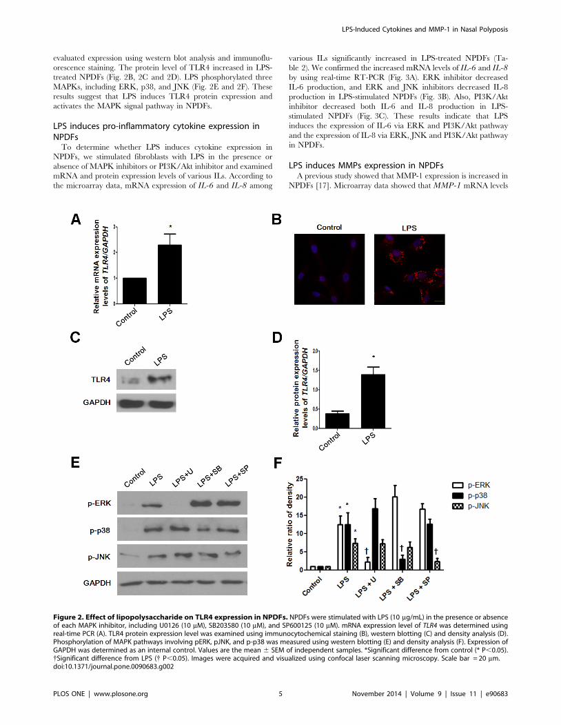
evaluated expression using western blot analysis and immunoflu-
orescence staining. The protein level of TLR4 increased in LPS-
treated NPDFs (Fig. 2B, 2C and 2D). LPS phosphorylated three
MAPKs, including ERK, p38, and JNK (Fig. 2E and 2F). These
results suggest that LPS induces TLR4 protein expression and
activates the MAPK signal pathway in NPDFs.
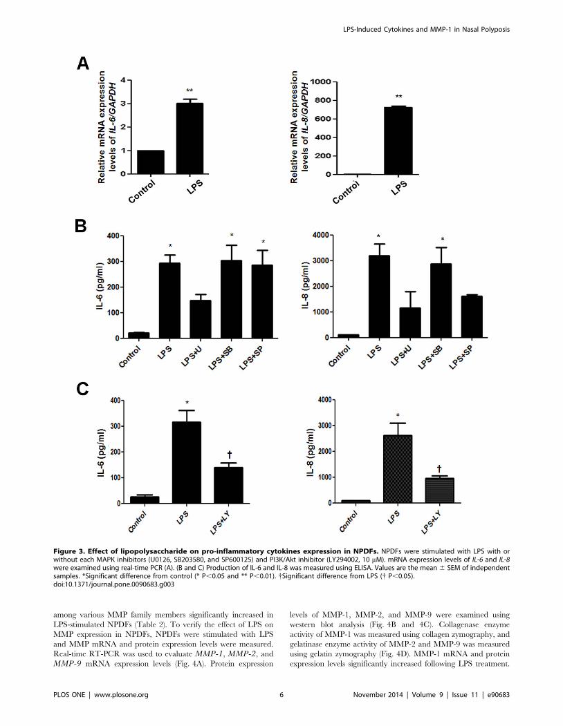
LPS induces pro-inflammatory cytokine expression inNPDFs
To determine whether LPS induces cytokine expression in
NPDFs, we stimulated fibroblasts with LPS in the presence or
absence of MAPK inhibitors or PI3K/Akt inhibitor and examined
mRNA and protein expression levels of various ILs. According to
the microarray data, mRNA expression of IL-6 and IL-8 among
various ILs significantly increased in LPS-treated NPDFs (Ta-
ble 2). We confirmed the increased mRNA levels of IL-6 and IL-8by using real-time RT-PCR (Fig. 3A). ERK inhibitor decreased
IL-6 production, and ERK and JNK inhibitors decreased IL-8
production in LPS-stimulated NPDFs (Fig. 3B). Also, PI3K/Akt
inhibitor decreased both IL-6 and IL-8 production in LPS-
stimulated NPDFs (Fig. 3C). These results indicate that LPS
induces the expression of IL-6 via ERK and PI3K/Akt pathway
and the expression of IL-8 via ERK, JNK and PI3K/Akt pathway
in NPDFs.
LPS induces MMPs expression in NPDFsA previous study showed that MMP-1 expression is increased in
NPDFs [17]. Microarray data showed that MMP-1 mRNA levels
Figure 2. Effect of lipopolysaccharide on TLR4 expression in NPDFs. NPDFs were stimulated with LPS (10 mg/mL) in the presence or absenceof each MAPK inhibitor, including U0126 (10 mM), SB203580 (10 mM), and SP600125 (10 mM). mRNA expression level of TLR4 was determined usingreal-time PCR (A). TLR4 protein expression level was examined using immunocytochemical staining (B), western blotting (C) and density analysis (D).Phosphorylation of MAPK pathways involving pERK, pJNK, and p-p38 was measured using western blotting (E) and density analysis (F). Expression ofGAPDH was determined as an internal control. Values are the mean 6 SEM of independent samples. *Significant difference from control (* P,0.05).{Significant difference from LPS ({ P,0.05). Images were acquired and visualized using confocal laser scanning microscopy. Scale bar = 20 mm.doi:10.1371/journal.pone.0090683.g002
LPS-Induced Cytokines and MMP-1 in Nasal Polyposis
PLOS ONE | www.plosone.org 5 November 2014 | Volume 9 | Issue 11 | e90683
among various MMP family members significantly increased in
LPS-stimulated NPDFs (Table 2). To verify the effect of LPS on
MMP expression in NPDFs, NPDFs were stimulated with LPS
and MMP mRNA and protein expression levels were measured.
Real-time RT-PCR was used to evaluate MMP-1, MMP-2, and
MMP-9 mRNA expression levels (Fig. 4A). Protein expression
levels of MMP-1, MMP-2, and MMP-9 were examined using
western blot analysis (Fig. 4B and 4C). Collagenase enzyme
activity of MMP-1 was measured using collagen zymography, and
gelatinase enzyme activity of MMP-2 and MMP-9 was measured
using gelatin zymography (Fig. 4D). MMP-1 mRNA and protein
expression levels significantly increased following LPS treatment.
Figure 3. Effect of lipopolysaccharide on pro-inflammatory cytokines expression in NPDFs. NPDFs were stimulated with LPS with orwithout each MAPK inhibitors (U0126, SB203580, and SP600125) and PI3K/Akt inhibitor (LY294002, 10 mM). mRNA expression levels of IL-6 and IL-8were examined using real-time PCR (A). (B and C) Production of IL-6 and IL-8 was measured using ELISA. Values are the mean 6 SEM of independentsamples. *Significant difference from control (* P,0.05 and ** P,0.01). {Significant difference from LPS ({ P,0.05).doi:10.1371/journal.pone.0090683.g003
LPS-Induced Cytokines and MMP-1 in Nasal Polyposis
PLOS ONE | www.plosone.org 6 November 2014 | Volume 9 | Issue 11 | e90683
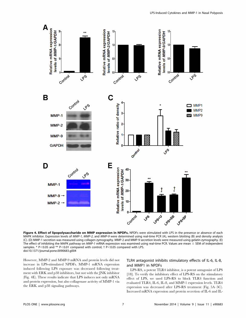
However, MMP-2 and MMP-9 mRNA and protein levels did not
increase in LPS-stimulated NPDFs. MMP-1 mRNA expression
induced following LPS exposure was decreased following treat-
ment with ERK and p38 inhibitors, but not with the JNK inhibitor
(Fig. 4E). These results indicate that LPS induces not only mRNA
and protein expression, but also collagenase activity of MMP-1 via
the ERK and p38 signaling pathways.
TLR4 antagonist inhibits stimulatory effects of IL-6, IL-8,and MMP1 in NPDFs
LPS-RS, a potent TLR4 inhibitor, is a potent antagonist of LPS
[18]. To verify the inhibitory effect of LPS-RS on the stimulatory
effect of LPS, we used LPS-RS to block TLR4 function and
evaluated TLR4, IL-6, IL-8, and MMP-1 expression levels. TLR4
expression was decreased after LPS-RS treatment (Fig. 5A–5C).
Increased mRNA expression and protein secretion of IL-6 and IL-
Figure 4. Effect of lipopolysaccharide on MMP expression in NPDFs. NPDFs were stimulated with LPS in the presence or absence of eachMAPK inhibitor. Expression levels of MMP-1, MMP-2, and MMP-9 were determined using real-time PCR (A), western blotting (B) and density analysis(C). (D) MMP-1 secretion was measured using collagen zymography. MMP-2 and MMP-9 secretion levels were measured using gelatin zymography. (E)The effect of inhibiting the MAPK pathway on MMP-1 mRNA expression was examined using real-time PCR. Values are mean 6 SEM of independentsamples. * P,0.05 and ** P,0.01 compared with control; { P,0.05 compared with LPS.doi:10.1371/journal.pone.0090683.g004
LPS-Induced Cytokines and MMP-1 in Nasal Polyposis
PLOS ONE | www.plosone.org 7 November 2014 | Volume 9 | Issue 11 | e90683
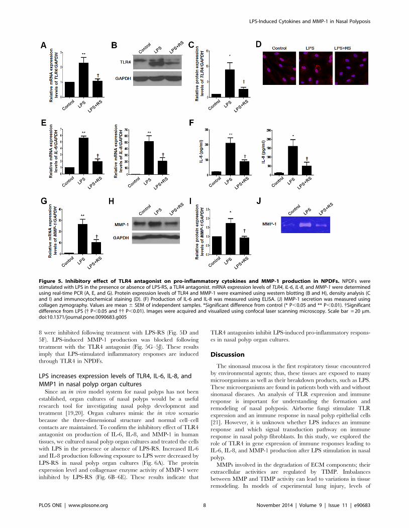
8 were inhibited following treatment with LPS-RS (Fig. 5D and
5F). LPS-induced MMP-1 production was blocked following
treatment with the TLR4 antagonist (Fig. 5G–5J). These results
imply that LPS-stimulated inflammatory responses are induced
through TLR4 in NPDFs.
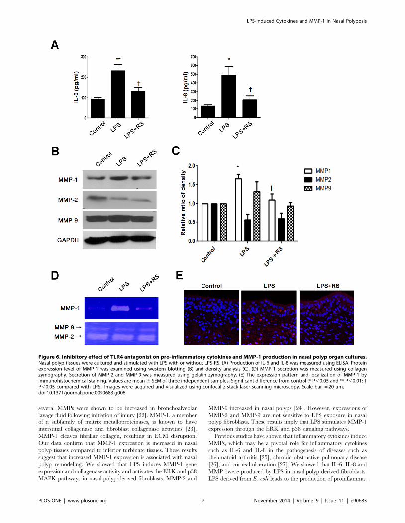
LPS increases expression levels of TLR4, IL-6, IL-8, andMMP1 in nasal polyp organ cultures
Since an in vivo model system for nasal polyps has not been
established, organ cultures of nasal polyps would be a useful
research tool for investigating nasal polyp development and
treatment [19,20]. Organ cultures mimic the in vivo scenario
because the three-dimensional structure and normal cell–cell
contacts are maintained. To confirm the inhibitory effect of TLR4
antagonist on production of IL-6, IL-8, and MMP-1 in human
tissues, we cultured nasal polyp organ cultures and treated the cells
with LPS in the presence or absence of LPS-RS. Increased IL-6
and IL-8 production following exposure to LPS were decreased by
LPS-RS in nasal polyp organ cultures (Fig. 6A). The protein
expression level and collagenase enzyme activity of MMP-1 were
inhibited by LPS-RS (Fig. 6B–6E). These results indicate that
TLR4 antagonists inhibit LPS-induced pro-inflammatory respons-
es in nasal polyp organ cultures.
Discussion
The sinonasal mucosa is the first respiratory tissue encountered
by environmental agents; thus, these tissues are exposed to many
microorganisms as well as their breakdown products, such as LPS.
These microorganisms are found in patients both with and without
sinonasal diseases. An analysis of TLR expression and immune
response is important for understanding the formation and
remodeling of nasal polyposis. Airborne fungi stimulate TLR
expression and an immune response in nasal polyp epithelial cells
[21]. However, it is unknown whether LPS induces an immune
response and which signal transduction pathway on immune
response in nasal polyp fibroblasts. In this study, we explored the
role of TLR4 in gene expression of immune responses leading to
IL-6, IL-8, and MMP-1 production after LPS stimulation in nasal
polyp.
MMPs involved in the degradation of ECM components; their
extracellular activities are regulated by TIMP. Imbalances
between MMP and TIMP activity can lead to variations in tissue
remodeling. In models of experimental lung injury, levels of
Figure 5. Inhibitory effect of TLR4 antagonist on pro-inflammatory cytokines and MMP-1 production in NPDFs. NPDFs werestimulated with LPS in the presence or absence of LPS-RS, a TLR4 antagonist. mRNA expression levels of TLR4, IL-6, IL-8, and MMP-1 were determinedusing real-time PCR (A, E, and G). Protein expression levels of TLR4 and MMP-1 were examined using western blotting (B and H), density analysis (Cand I) and immunocytochemical staining (D). (F) Production of IL-6 and IL-8 was measured using ELISA. (J) MMP-1 secretion was measured usingcollagen zymography. Values are mean 6 SEM of independent samples. *Significant difference from control (* P,0.05 and ** P,0.01). {Significantdifference from LPS ({ P,0.05 and {{ P,0.01). Images were acquired and visualized using confocal laser scanning microscopy. Scale bar = 20 mm.doi:10.1371/journal.pone.0090683.g005
LPS-Induced Cytokines and MMP-1 in Nasal Polyposis
PLOS ONE | www.plosone.org 8 November 2014 | Volume 9 | Issue 11 | e90683
several MMPs were shown to be increased in bronchoalveolar
lavage fluid following initiation of injury [22]. MMP-1, a member
of a subfamily of matrix metalloproteinases, is known to have
interstitial collagenase and fibroblast collagenase activities [23].
MMP-1 cleaves fibrillar collagen, resulting in ECM disruption.
Our data confirm that MMP-1 expression is increased in nasal
polyp tissues compared to inferior turbinate tissues. These results
suggest that increased MMP-1 expression is associated with nasal
polyp remodeling. We showed that LPS induces MMP-1 gene
expression and collagenase activity and activates the ERK and p38
MAPK pathways in nasal polyp-derived fibroblasts. MMP-2 and
MMP-9 increased in nasal polyps [24]. However, expressions of
MMP-2 and MMP-9 are not sensitive to LPS exposure in nasal
polyp fibroblasts. These results imply that LPS stimulates MMP-1
expression through the ERK and p38 signaling pathways.
Previous studies have shown that inflammatory cytokines induce
MMPs, which may be a pivotal role for inflammatory cytokines
such as IL-6 and IL-8 in the pathogenesis of diseases such as
rheumatoid arthritis [25], chronic obstructive pulmonary disease
[26], and corneal ulceration [27]. We showed that IL-6, IL-8 and
MMP-1were produced by LPS in nasal polyp-derived fibroblasts.
LPS derived from E. coli leads to the production of proinflamma-
Figure 6. Inhibitory effect of TLR4 antagonist on pro-inflammatory cytokines and MMP-1 production in nasal polyp organ cultures.Nasal polyp tissues were cultured and stimulated with LPS with or without LPS-RS. (A) Production of IL-6 and IL-8 was measured using ELISA. Proteinexpression level of MMP-1 was examined using western blotting (B) and density analysis (C). (D) MMP-1 secretion was measured using collagenzymography. Secretion of MMP-2 and MMP-9 was measured using gelatin zymography. (E) The expression pattern and localization of MMP-1 byimmunohistochemical staining. Values are mean 6 SEM of three independent samples. Significant difference from control (* P,0.05 and ** P,0.01; {P,0.05 compared with LPS). Images were acquired and visualized using confocal z-stack laser scanning microscopy. Scale bar = 20 mm.doi:10.1371/journal.pone.0090683.g006
LPS-Induced Cytokines and MMP-1 in Nasal Polyposis
PLOS ONE | www.plosone.org 9 November 2014 | Volume 9 | Issue 11 | e90683
tory cytokines such as IL-1b, IL-6, and TNFa as well as
chemokines such as IL-8 [28]. LPS derived from Pseudomonasaeruginosa induces IL-1a, IL-6, MCP-1, IL-1b and TNFa in
airway inflammation and goblet cell hyperplasia [29]. In the
previous study, LPS activated phosphorylation of the downstream
mediators ERK and p38 and initiated an inflammatory response
in granulosa cells through accumulation of IL-6 and IL-8 in rat
synovial membranes [30]. Also, LPS induced inflammatory
responses via PI3K/Akt signaling in human peripheral blood
mononuclear cells [31]. In the present study, LPS activated ERK
phosphorylation for IL-6 induction and induced activation of
ERK and JNK, but not p38, for IL-8 induction in NPDFs.
Additionally, PI3K/Akt activation increased LPS-induced IL-6
and IL-8 production in NPDFs. These data indicate that LPS
stimulates IL-6 and IL-8 production through the ERK, JNK and
PI3K/Akt signaling pathways in NPDFs.
LPS from the photosynthetic bacterium Rhodobacter sphaer-oides (LPS-RS) is a potent inhibitor of TLR4 and antagonist of
LPS [18]. In the present study, we found that LPS-RS inhibits the
expression of LPS-stimulated TLR4, IL-6, IL-8, and MMP-1. Our
data suggest that LPS induces expression of IL-6, IL-8, and
MMP1 via TLR4.
A limitation of this study is that an in vivo model system for
nasal polyposis was not established. To mimic the in vivo model
system of nasal polyposis, we used organ cultures of nasal polyps to
investigate nasal polyp development and treatment [19,20]. The
organ culture systems provide accessible tissue in an artificial
environment and maintain the three-dimensional structure as well
as standard cell–cell contacts. Organ cultures allow experimenta-
tion under more controlled conditions than is possible in in vivoexperiments with a minimal alteration of natural conditions. Our
study revealed that TLR4 is important for the production of pro-
inflammatory cytokines and MMP-1 in nasal polyp organ cultures.
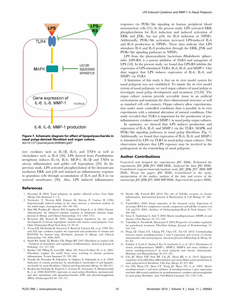
In summary, we showed that LPS induces production and
expression of IL-6, IL-8, and MMP-1 via the TLR4, MAPK and
PI3K/Akt signaling pathways in nasal polyp fibroblasts (Fig. 7).
Additionally, we found that expression of IL-6, IL-8, and MMP-1
is stimulated by LPS via TLR4 in nasal polyp organ cultures. This
observation indicates that LPS exposure may be involved in the
pathogenesis in the remodeling of nasal polyposis.
Author Contributions
Conceived and designed the experiments: JSC HML. Performed the
experiments: JSC JHK JYU IHH HML. Analyzed the data: JSC HML.
Contributed reagents/materials/analysis tools: JSC JHK JYU IHH IHP
HML. Wrote the paper: JSC HML. Contributed to the study,
interpretation of the studies, analysis of the data and review of the
manuscript: JSC JHK JYU IHH IHP HML. Supervised the project: HML.
References
1. Pawankar R (2003) Nasal polyposis: an update: editorial review. Curr Opin
Allergy Clin Immunol 3: 1–6.
2. Norlander T, Westrin KM, Fukami M, Stierna P, Carlsoo B (1996)
Experimentally induced polyps in the sinus mucosa: a structural analysis of
the initial stages. Laryngoscope 106: 196–203.
3. Shin SH, Ponikau JU, Sherris DA, Congdon D, Frigas E, et al. (2004) Chronic
rhinosinusitis: An enhanced immune response to ubiquitous airborne fungi.
Journal of Allergy and Clinical Immunology 114: 1369–1375.
4. Bernstein JM, Kansal R (2005) Superantigen hypothesis for the early
development of chronic hyperplastic sinusitis with massive nasal polyposis. Curr
Opin Otolaryngol Head Neck Surg 13: 39–44.
5. Teran LM, Mochizuki M, Navarrete F, Bartels J, Valencia EL, et al. (1999) Th1
and Th2 type cytokines regulate the expression and production of eotaxin and
RANTES by human lung fibroblasts. Journal of Allergy and Clinical
Immunology 103: S206–S207.
6. Smith RS, Smith TJ, Blieden TM, Phipps RP (1997) Fibroblasts as sentinel cells
- Synthesis of chemokines and regulation of inflammation. American Journal of
Pathology 151: 317–322.
7. Buckley CD, Pilling D, Lord JM, Akbar AN, Scheel-Toellner D, et al. (2001)
Fibroblasts regulate the switch from acute resolving to chronic persistent
inflammation. Trends Immunol 22: 199–204.
8. Nonaka M, Pawankar R, Fukumoto A, Ogihara N, Sakanushi A, et al. (2004)
Induction of eotaxin production by interleukin-4, interleukin-13 and lipopoly-
saccharide by nasal fibroblasts. Clinical and Experimental Allergy 34: 804–811.
9. Rostkowska-Nadolska B, Pospiech L, Fortuna W, Szymaniec S, Miedzybrodzki
R, et al. (2006) RANTES expression in nasal polyps fibroblasts; spontaneously
and after stimulation with lipopolisaccharides (LPS) and phytohemagglutinin
(PHA). Otolaryngol Pol 60: 143–147.
10. Drexler SK, Foxwell BM (2010) The role of Toll-like receptors in chronic
inflammation. International Journal of Biochemistry & Cell Biology 42: 506–
518.
11. VanderMeer (2005) Innate immunity of the sinonasal cavity: Expression of
messenger RNA for complement cascade components and toll-like receptors (vol
130, pg1374, 2004). Archives of Otolaryngology-Head & Neck Surgery 131:
406–406.
12. Sorsa T, Tjaderhane L, Salo T (2004) Matrix metalloproteinases (MMPs) in oral
diseases. Oral Diseases 10: 311–318.
13. Takashiba S, Naruishi K, Murayama Y (2003) Perspective of cytokine regulation
for periodontal treatment: Fibroblast biology. Journal of Periodontology 74:
103–110.
14. Wang LF, Chien CY, Chiang FY, Chai CY, Tai CF (2012) Corelationship
between matrix metalloproteinase 2 and 9 expression and severity of chronic
rhinosinusitis with nasal polyposis. American Journal of Rhinology & Allergy 26:
E1–E4.
15. Eyibilen A, Cayli S, Aladag I, Koc S, Gurbuzler L, et al. (2011) Distribution of
matrix metalloproteinases MMP-1, MMP-2, MMP-8 and tissue inhibitor of
matrix metalloproteinases-2 in nasal polyposis and chronic rhinosinusitis.
Histology and Histopathology 26: 615–621.
16. Cho JS, Moon YM, Park IH, Um JY, Moon JH, et al. (2012) Epigenetic
regulation of myofibroblast differentiation and extracellular matrix production in
nasal polyp-derived fibroblasts. Clin Exp Allergy 42: 872–882.
17. Liu CM, Hong CY, Shun CT, Wang JS, Hsiao TY, et al. (2001) Matrix
metalloproteinase-1 and tissue inhibitor of metalloproteinase-1 gene expressions
and their differential regulation by proinflammatory cytokines and prostaglandin
in nasal polyp fibroblasts. Ann Otol Rhinol Laryngol 110: 1129–1136.
Figure 7. Schematic diagram for effect of lipopolysaccharide innasal polyp-derived fibroblast and organ culture.doi:10.1371/journal.pone.0090683.g007
LPS-Induced Cytokines and MMP-1 in Nasal Polyposis
PLOS ONE | www.plosone.org 10 November 2014 | Volume 9 | Issue 11 | e90683
18. Coats SR, Pham TT, Bainbridge BW, Reife RA, Darveau RP (2005) MD-2
mediates the ability of tetra-acylated and penta-acylated lipopolysaccharides toantagonize Escherichia coli lipopolysaccharide at the TLR4 signaling complex.
Journal of Immunology 175: 4490–4498.
19. Park SK, Jang WH, Yang YI (2009) Expression of pro-angiogenic cytokines andtheir inhibition by dexamethasone in an ex vivo model of nasal polyps. Biochem
Biophys Res Commun 379: 255–260.20. Cho JS, Moon YM, Park IH, Um JY, Kang JH, et al. (2013) Effects of histone
deacetylase inhibitor on extracellular matrix production in human nasal polyp
organ cultures. American Journal of Rhinology & Allergy 27: 18–23.21. Shin SH, Lee YH (2010) Airborne fungi induce nasal polyp epithelial cell
activation and Toll-like receptor expression. International archives of allergy andimmunology 153: 46–52.
22. Gibbs DF, Shanley TP, Warner RL, Murphy HS, Varani J, et al. (1999) Role ofmatrix metalloproteinases in models of macrophage-dependent acute lung
injury. Evidence for alveolar macrophage as source of proteinases. Am J Respir
Cell Mol Biol 20: 1145–1154.23. Vincenti MP, Brinckerhoff CE (2002) Transcriptional regulation of collagenase
(MMP-1, MMP-13) genes in arthritis: integration of complex signaling pathwaysfor the recruitment of gene-specific transcription factors. Arthritis Res 4: 157–
164.
24. Lechapt-Zalcman E, Coste A, d’Ortho MP, Frisdal E, Harf A, et al. (2001)Increased expression of matrix metalloproteinase-9 in nasal polyps. J Pathol 193:
233–241.
25. Hyc A, Osiecka-Iwan A, Niderla-Bielinska J, Moskalewski S (2011) Influence of
LPS, TNF, TGF-beta 1 and IL-4 on the expression of MMPs, TIMPs and
selected cytokines in rat synovial membranes incubated in vitro. International
Journal of Molecular Medicine 27: 127–137.
26. Chung KF (2001) Cytokines in chronic obstructive pulmonary disease.
European Respiratory Journal 18: 50s–59s.
27. Wong Y, Sethu C, Louafi F, Hossain P (2011) Lipopolysaccharide Regulation of
Toll-Like Receptor-4 and Matrix Metalloprotease-9 in Human Primary Corneal
Fibroblasts. Investigative Ophthalmology & Visual Science 52: 2796–2803.
28. Segura M, Vadeboncoeur N, Gottschalk M (2002) CD14-dependent and -
independent cytokine and chemokine production by human THP-1 monocytes
stimulated by Streptococcus suis capsular type 2. Clin Exp Immunol 127: 243–
254.
29. Harris JF, Aden J, Lyons CR, Tesfaigzi Y (2007) Resolution of LPS-induced
airway inflammation and goblet cell hyperplasia is independent of IL-18. Respir
Res 8: 24.
30. Bromfield JJ, Sheldon IM (2011) Lipopolysaccharide Initiates Inflammation in
Bovine Granulosa Cells via the TLR4 Pathway and Perturbs Oocyte Meiotic
Progression in Vitro. Endocrinology 152: 5029–5040.
31. Ngkelo A, Meja K, Yeadon M, Adcock I, Kirkham PA (2012) LPS induced
inflammatory responses in human peripheral blood mononuclear cells is
mediated through NOX4 and G(i)alpha dependent PI-3kinase signalling.
Journal of Inflammation-London 9.
LPS-Induced Cytokines and MMP-1 in Nasal Polyposis
PLOS ONE | www.plosone.org 11 November 2014 | Volume 9 | Issue 11 | e90683